a case of late infantile 7 amaurotic idiocy, · counterstain, by mallory's phosphotungstic...
TRANSCRIPT
A CASE OF LATE INFANTILE 7AMAUROTIC IDIOCY,WITH PATHOLOGICAL REPORT
R
BY I
BERNARD SCHLESINGER, M.D., M.R.C.P.,_J. G. GREENFIELD, M.D., F.R.C.P., and R. 0. STERN, M.D.
(From the Royal Northern Hospital, and the Pathological Laboratory ofthe National Hospital for Nervous Diseases, London.)
Few degenerative diseases of the nervous system have acquired a widerexpansion of their original definition than amaurotic idiocy. Believed inthe first place to be limited to Jewish infants (Waren Tay', Sachs2), it wasnot long before cases were discovered among Gentiles, in whom also theocular changes and age of onset were not typical (Batten3). During the lastfifty years these variations of the disease have appeared under various titles;'late infantile form ' (Jansky4, Bielschowsky5); ' juvenile form ' (Vogt6,Spielmeyer7, Greenfield and Holmes8); and more recently ' late form(Kufs9, Meyer"0). Clinically there may be great differences in these varioustypes; in fact, but for the characteristic histo-pathological changes in allof them, it would be difficult to establish a common identity. The clinicalpicture of Tay-Sachs' disease in infants, for instance, is quite unrecognizablein Kufs' ex-service man of 42. The marked differences in the appearanceof the disease depend on variations in the intensity, extent and speciallocalization of the cell degeneration in the brain. The rate at which thedegenerative process proceeds also plays an important part, and in thejuvenile and late forms the illness may be prolonged for over ten years(Sch6nfeld'", Kufs9). Not uncommonly the case remains undiagnosed untila study of the morbid anatomy becomes possible.
Even the name amaurotic family idiocy may finally need modification,since sporadic cases are not uncommon at any age, and amaurosis may beabsent or play a secondary part in the juvenile and late forms. Idiocyappears to be the only constant feature. The greatest changes in the clinicalpicture of the varieties of this disorder make their appearance somewherebetween the infantile and juvenile forms. Ihence the rare ' late infantile '
cases become especially important as a link between the two, for already thehall-mark has become blurred. Racial preponderance is no longer obvious,macular changes are often absent and the course of the disease ismore protracted.
These considerations prompt us to publish this ' late infantile ' case,particularly as it is the earliest in a Gentile which has been pathologically
A
on Decem
ber 29, 2019 by guest. Protected by copyright.
http://adc.bmj.com
/A
rch Dis C
hild: first published as 10.1136/adc.9.49.1 on 1 February 1934. D
ownloaded from
ARCHIVES OF DISEASE IN CHILDHOOD
recorded. The degree of swellings on the axons in the cortex is probablyalso unique.
Case report.Pauline M. first came under observation at the age of 1 year and 10 months. She
was the first child of English parents with no history of Jewish intermarriage in thefamily, and there had been no previous miscarriage. The birth had been normal,with an easy confinement lasting twenty-four hours. Up to the age of 6 monthsthe child appeared to be developing normally. She seemed intelligent, smiled andcried like an ordinary infant, and had already become well trained in the use of thechamber. At 10 months the parents first noticed something amiss when no attemptwas made to sit up, and it was not until 14 months that the infant began to lifther head from the pillow. The two other most obvious signs of backwardness werean inability to take solids and very indefinite grasping at objects. Meanwhile theprevious cleanliness in habits gra(lually gave way to incontinence, the child no longersmiled or uttered a cry, but lay still and apathetic in her cot. Short convulsionslasting about a minute commenced at the age of 15 months, two or three daily forthree or four days every few weeks. At the same time the child was noticed to jumpat any sudden noise.
When first seen spasticity of the arms and legs was already most obvious andthe ' toes were pointed.' All the tendon reflexes were much increased, those of theupper extremity less markedly so than the knee and ankle jerks. The abdominalreflexes were absent and the Babinski plantar responses extensor. A loud noiseproduced a sudden straightening of all the limbs with increased spasticity as in decere-brate rigidity. Gradually the child's general condition deteriorated, the appetitediminished, wasting was rapid, and the fits, which throughout were uninfluenced byluminal and bromide, eventually gave way to complete drowsiness with periodictwitching of the face. The arms were held straight and pronated with the wristsand fingers tightly flexed, while the legs were fixed in a position of moderate talipesequino-varus. The eyes fixed on no object and appeared to be blind; the pupils werehalf dilated, reacted sluggishly to light, and a varying left external strabismus waspresent. Both discs showed primary optic atrophy, but there were no macularchanges.
The Wassermann reaction of both blood and spinal fluid was negative, and an-intradermal Mantoux test provoked no reaction. Further examination showed thecerebro-spinal fluid to be clear and colourless, with less than one cell per c.mm.Sugar was present, globulin absent, and the protein 30 mgm. per c.mm.
The final course of the disease was rapid and the chil(l succumbed to broncho-pneuimonia at the age of 1 year and 10 months.
Pathological examinationThe brain, spinal cord an(l optic nerve with small nortion of retina were remove(d
within twelve hours of death an(l immediately nlaced in formol saline.Dr. Ellison who performed the rest of the post-mortem examination reported
patches of broncho-pneumonia. The liver and soleen were carefully examined butshowed no changes such as described by Neimann and Pick. Other organs in thebody showed no morbid changes of any particular interest.
Histological examination.-Frozen sections of the brain and sDinal cord werestained with Scharlach R. and an acid haematoxylin counterstain to demonstratelipoid deposits in the nerve cells; and histo-chemical tests were nerformed on frozensections of the basal ganglia in order to ascertain the nature of these deposits.Anderson's Victoria blue method for neuroglia, and Da Fano's modification ofBielschowsky's method for neurofibrils were also used on frozen sections. Celloidin
* A grant from the Medical Research Council was received in connexion with thispart of the investigation,
2
on Decem
ber 29, 2019 by guest. Protected by copyright.
http://adc.bmj.com
/A
rch Dis C
hild: first published as 10.1136/adc.9.49.1 on 1 February 1934. D
ownloaded from
A CASE OF LATE INFANTILE AMAUROTIC IDIOCY
sections were made from various parts of the, brain, spinal cord and optic nerve.These were stained by the Nissi method, by iron haematoxylin with van Gieson'scounterstain, by Mallory's phosphotungstic acid method for neuroglia, and by theWeigert-Pal method. The fragment of retina was embedded in paraffin andsubsequently cut in serial section.
The essential features of the microscopical examination are convenientlydiscussed under the headings of the nerve cells, the neuroglia, and the myelin.
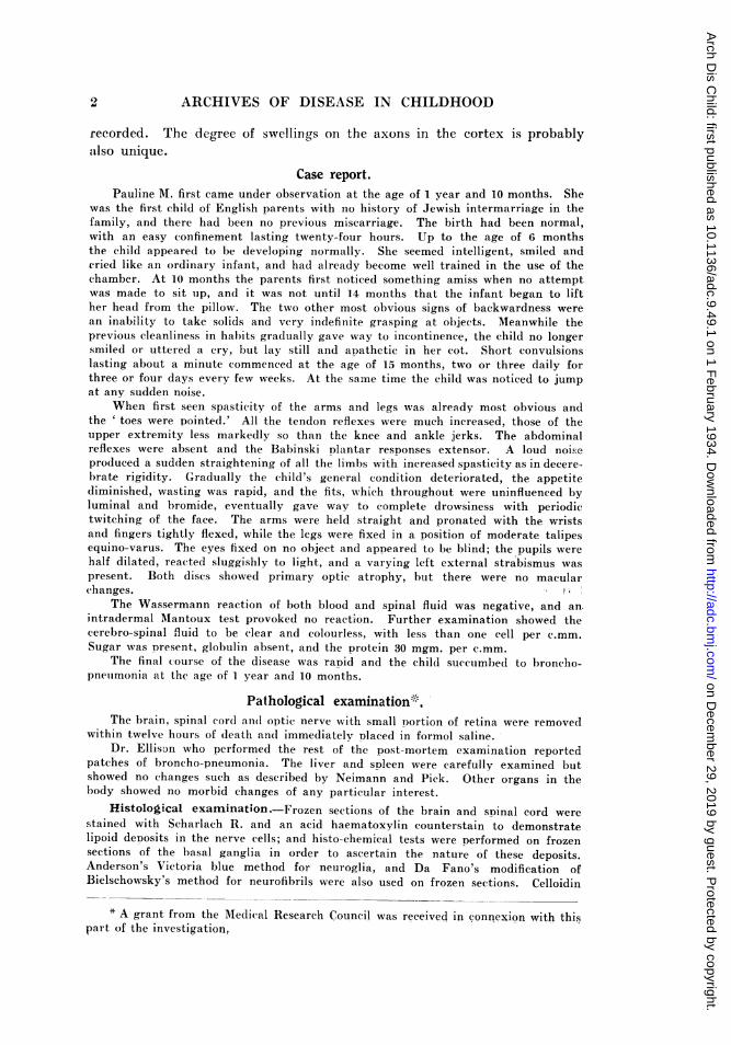
THE NERVE CELLS.-The changes in the nerve cells in this case conformed generallyto those which have been described previously in the infantile form of amauroticfamily idiocy. The nerve cells of the frontal, temporal and occipital cortex, and ofthe basal ganglia, midbrain, pons, medulla and sDinal cord, all presented the rounded,ballooned appearance characteristic of this disease. The cell bodies were swollen anddistended by lipoid deposits, the nature of which will be considered later. In the
Fi(;. 1. Two Dyrami(lal nerve cePsfrom the occipital cortex withballoon-like swellings on the axons
(Bielschowsky stain).
Fic;. '2. Two pyramidal nerve cellswith balloon-like swelling on theiraxons; in one the cell bodv is alsodistende(d with lipoid(Bielschowsky
stain).
cortex cerebri many cells showed fusiform or globular swellings on the proximalpart of their axon. The most bizarre forms were seen in the occipital cortex (Fig. Iand 2). Here many of the pyramidal nerve cells an)peared normal in size, whilst theswellings on their axons were larger than the cells themselves. The shape of theswellings was variable; globular, ellipitical and fusiform examples were common, andoccasionally in the occipital cortex curious kidney-shaped swellings were found.
Among the nerve cells of the frontal cortex there were some smaller cells with aglobular, sharply outlined cell body, and a small irregular nucleus resembling thatof microglial cells. With Scharlach R. these cells stained rather more brightly thanthe nerve cells, and with high powers could be seen to be filled with clear yellowishor pale pink globules. Darker staining fatty granules could also be seen in someof them.
The cerebellum was not greatly atrophied, nor was there much shrinkage ofthe foliae, although they had a more rounded contour and felt firmer than normal.
A\ '
3
on Decem
ber 29, 2019 by guest. Protected by copyright.
http://adc.bmj.com
/A
rch Dis C
hild: first published as 10.1136/adc.9.49.1 on 1 February 1934. D
ownloaded from

ARCHIVES OF DISEASE IN CHILDHOOD
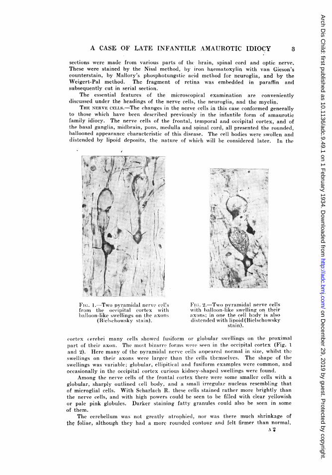
Histologically the changes here were diffuse rather than intense. The Purkinje cellswere not noticeably decreased in number and were surrounded by normal basketfibres. Their cell bodies were considerably swollen with lipoid, and lipoid swellingswere seen on their dendrites, usually at the points of branching (Fig. 3). The granulecell layer was considerably atrophied. At least half the granule cells had disappeared,and between those that remained were seen numerous small globular cells filled withyellowish clear pigment, similar to those found in the frontal cortex. There wasalso, external to the thinned zone of granules and in line with the Purkinje cells, a fairlycompact layer, one or two cells thick, of clear, oval nuclei which appeared to give originto firm neuroglial fibres (Fig. 4). Similar nuclei were seen among the granules and
FIGT. 3. Purkinje cells from cerebellarcortex, showing antler-like swellings ontheir dendrites. In one the axonal end ofthe cell is also distended with lipoid
(Bielschowsky stain).
in the white centre of the foliae. No lipoid could be seen round any of these neuroglialnuclei.
Although the contour of the nerve cells throughout the central nervous systemwas altered by the deposition of lipoid substances within their cytoplasm, theneurofibrils were singularly unaffected. Bielschowsky preparations showed that theneurofibrils were displaced to the perinhery of the body of the affected cell, but thatthey passed normally into the dendrites and through the swellings.
Histo-chemical investigation of the intracellular liDoid deposits gave thefollowing results -with Scharlach R, the lipoid gave a bright pink coloratiorn
4
on Decem
ber 29, 2019 by guest. Protected by copyright.
http://adc.bmj.com
/A
rch Dis C
hild: first published as 10.1136/adc.9.49.1 on 1 February 1934. D
ownloaded from
A CASE OF LATE INFANTILE AMAUROTIC IDIOCY
distine-t from that given by neutral fats. Heidenhain's haematoxylin stained theintracellular lipoid deeply. It was not dissolved out by immersing sections fortwenty-four hours in absolute alcohol, acetone, chloroform or acid alcohol,but thereafter no coloration,> or only a faint pink tinge, was obtained withScharlach R. It was not doubly refractile.
A comparison between the chemical nature of the intracellular lipoid peculiarto the disease process in amaurotic family idiocy, normal myelin and the neutralfat products of degeneration resulting from disintegration of myelin, was possible by
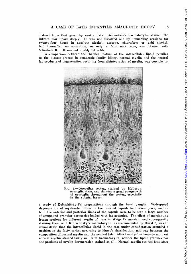
FIG. 4.-Cerebeilar cortex, stained by Mallory'sneuroglia stain, and showing a great overgrowthof neuroglia throughout the cortex, especiallyin the subpial layer;
a study of Kultschitsky-Pal preparations through the basal ganglia. Widespreaddegeneration of myeliinated fibres in the internal capsule had taken place, and inboth the anterior and posterior limbs of the capsule were to be seen a large numberof compound granular corpuscles loaded with fat granules. The effect of mordantingfrozen sections for different lengths of time in Weigert's mordant and subsequentlystaining them with Kultschitsky's haematoxylin, as recommended by Hurst'2, was todemonstrate that the intracellular lipoid in the case under consideration occupied aposition in the fatty series, according to Hurst's classification, mid-way between thecomposition of normal myelin and the neutral fats. After twenty-four hours in mordantnormal myelin stained fairly well with haematoxylin; neither the lipoid granules northe products of myelin degeneration stained at all. Normal myelin stained best after
on Decem
ber 29, 2019 by guest. Protected by copyright.
http://adc.bmj.com
/A
rch Dis C
hild: first published as 10.1136/adc.9.49.1 on 1 February 1934. D
ownloaded from

ARCHIVES OF DISEASE IN CHILDHOOD)
forty-eight hours in mordant, at a time when the degeneration products did not stainat all, though the intracellular lipoid stained well. Optimum staining of theintracellular lipoid was obtained by mordanting for four days. The degenerationproducts of myelin also stained well when mordanted for the same lenlgth of time,whereas normal myelin gave only a faint blue coloration. The intracellular lipoidwas recognizable at the end of a week in mordant, when no appreciable staining ofnormal myelin could be obtained. The neutral fat in the compound granularcorpuscles and in the (legenerated fibres stained well after this period of mordanting.These results indicated that the intracellular lipoid consisted of a compound ofphosphatides and cerebrosides, which, in its reactions, was intermediate betweennormal myelin an(1 neutral fat.
FIG. 5. Frontal cortex stained by Weigert-Palmethod. The lipoid swellings stain alongwith the radial fibres.
NEUTROGLIA.-The sub-pial neuroglia in the cerebral cortex ha(l greatlyproliferated, producing a dense gliosis of the superficial layers of the cortex. Thiswas especially well seen in the occipital region, where there was a thick down-growthof neuroglia into the third layer of the cortex. leri-ventricular gliosis was alsostriking, especially in the fourth ventricle. Other regions of the brain showed aremarkable gliosis; in the cerebellum there was considerable gliosis of the molecularlayer, especially just under the surface, where a thick feltwork of neuroglial fibreswas to be seen in preparations stained with Victoria bluie or phosphotungstic acidhaematoxylin (Fig. 4). In the thalamus, which had felt unduly firm macroscopically,a very severe gliosis was found, perhaps more intense in the medial than in thelateral part. The lenticular and caudate nuclei were relatively unaffected, thoughsome proliferation of neuroglia had occurred in them also.
MYELIN.-The fibres in the cerebral cortex were, in general, well myelinated,though in the frontal region some of the more superficial tangential fibres stained
6
on Decem
ber 29, 2019 by guest. Protected by copyright.
http://adc.bmj.com
/A
rch Dis C
hild: first published as 10.1136/adc.9.49.1 on 1 February 1934. D
ownloaded from

A CASE OF LATE INFANTILE AMAUROTIC IDIOCY
poorly. In the internal capsule, the fibres in the anterior two-thirds of the anteriorlimb of the capsule had undergone almost complete degeneration. In the genuthere were many finely myelinated fibres, but in the posterior limb, with theexception of a few finely myelinated fibres in its most anterior part, all the fibres
.. .. .. .......
FIG. 6 & 7.-Transverse sections of medulla and cervical cord, stainedby the Weigert-Pal method to show the degeneration of thepyramidal tracts.
had degenerated. The outer parts of the crura were unaffected, but the medial andfronto-pontine fibres were degenerated. In the medulla there was almost completedestruction of the pyramids, and the pyramidal tracts were dlegenerated throughoutthe spinal cord.
7
on Decem
ber 29, 2019 by guest. Protected by copyright.
http://adc.bmj.com
/A
rch Dis C
hild: first published as 10.1136/adc.9.49.1 on 1 February 1934. D
ownloaded from

8 ARCHIVES OF DISEAS'E IN CIIILDHOOD0I-ric NERVE AND RETINA.-In longitudinal sections of the optic nerve many
poorly myelinated fibres were seen, and in addition there were many fibres in anearly stage of degeneration. There was no neuroglial reaction to this degeneration.
It was not possible to decide whether the appearance of the retina was patho-logical or not, owing to the conditions of its fixation. The only definite opinion thatcould be given on the fragment sectioned was regarding the number of ganglion cells;these were considerably fewer in number than those seen in sections of a normalretina. Those present appeared healthy.
Summary.-1. The nerve cells throughout the central nervous system presentedthe swollen, ballooned appearance characteristic of amaurotic family idiocy.
2. This swelling of the nerve cells was due to distension by deposition in themof a lipoid substance. Histo-chemical tests showed this to be a compound ofcerebrosides and phosphatides, occupying an intermediate position chemicallybetween normal myelin and neutral fat.
3. Similar swellings were found on many axons of nerve cells; in some situations,notably in the frontal and occipital cortex, these axonal swellings were larger thanthe cells themselves.
4. Intense gliosis was present in the superficial layers of the cerebral cortex andof the cerebellum; also in the optic thalamus and round the ventricles.
5. There was degeneration of the fronto-pontine and of the pyramidal fibres,and of some fibres of the optic nerve.
6. There was a loss of ganglion cells in the retina.
Discussion.The view of both Pick and Bielschowsky" is that all forms of familial
amaurotic idiocy are due to a disturbance of lipoid metabolism, andMarinesco's14 work goes a long way towards establishing this. The sameprimary disturbance of lipoid metabolism is present in certain cases ofNiemann-Pick disease, and it is now generally held that the two conditionsare related by more than a chance association, and that, in fact, they aredifferent manifestations of the same disease process. That cases ofamaurotic idiocy do occur with no lipoid changes in the liver and spleen,and vice versa, is no reason to doubt this.
The alteration in the cells of the liver and spleen in Niemann-Pickdisease is mainly due to a deposition of phosphatid molecules. In amauroticidiocy we have shown, as others have done, that the lipoid deposit causingdistension of the cells of the brain is also largely composed of phosphatid.The fundamental cause of this disturbance of lipoid metabolism, however,still remains unknown.
REFERENCES.1. Tay, W., Trans. Ophthalm. Soc., Lond., 1881, I, 55.2. Sachs, B., J. Nerv. and Ment. Dis., Richmond, Va., 1887, XIV, 541.3. Batten, F. E., Trans. Opthalm. Soc., Lond., 1903, XXIII, 386.4. Jansky, J., Rev. de med. tcheque., Prague, 1908, I, 58.5. Bielschowsky, M., Deutsch. Ztschr. f. Nervenheilk., Berlin, 1914, L, 7.6. Vogt, H., Monatschr. f. Psychiatr. u. Neurol., Berlin, 1906, XVIII, 161 & 310.7. Spielmeyer, W., Neurol. Centralb., Leipzig, 1906, XXV, 51.8. Greenfield, J. G., & Holmes, G., Brain, Lond., 1925, XLVIII, 183.9. Kufs, H., Ztschr. f. d. ges. Neurol. u. Psychiat., Berlin, 1929, CXXII, 395.
10. Meyer, A., Arch. f. Psych., Berlin, 1930, XCIII, 155.11. Schorfeld, A., Ztschr. f. Psychiat., Berlin, 1930, XCIII, 155.12. Hurst, E. W., Brain, Lond., 1925, XLVIII, 1.-13. Bielschowsky, M., J. Psychol. u. Neur., Leipzig, 1928, XXXVI, 103.14. Marinesco, G., L'Encephale, Paris, 1927, XXII, 605.
on Decem
ber 29, 2019 by guest. Protected by copyright.
http://adc.bmj.com
/A
rch Dis C
hild: first published as 10.1136/adc.9.49.1 on 1 February 1934. D
ownloaded from