a bryophyte from kolli hills, eastern ghats of tamilnadu ... · pdf filea bryophyte from kolli...
TRANSCRIPT

Antioxidant and Antimicrobial Studies on Biosynthesized Silver Nanoparticles using Bryum medianum mitt.
A Bryophyte From Kolli Hills, Eastern Ghats Of Tamilnadu, India S.Sahaya Sathish1*, A.Vimala1, A.Kanaga1 , M.Murugan2
1Centre for Cryptogamic Studies, Department of Botany, St. Joseph’s College (Autonomous), Tiruchirappalli - 620 002, Tamilnadu, India
2Centre for Biological Science, Noorul Islam Centre for Higher Education, Kumaracoil -629 180, Tamil Nadu, India
Abstract The present study deals with the synthesis of silver nanoparticles using a moss plant Bryum medianum. The plant extract were mixed with AgNo3 solution and incubated in light condition for 24 h. The solution turned into yellowish to dark brown colour due to excitation of Surface Plasmon Resonance. The synthesized silver nanoparticles characterized by UV-Vis spectrophotometer, FTIR, FESEM, EDX and XRD analysis. UV-Vis spectroscopy exhibited the maximum absorbance peak at 431nm which indicates the formation of silver nanoparticles. The FTIR peak showed the alkanes and ether compounds attached with silver nanoparticles. The FESEM image revealed the size of the AgNo3 in the range of 85nm and XRD indicated the face centred cubic structure. The antimicrobial activity was studied for the synthesized nanoparticles against Proteus mirabilis, Escherichia coli, Klebsiella pneumonia, Aspergillus fumigatus, Candida albicans and Trichophyton mentagrophytes. Except Proteus mirabilis and Trichophyton mentagrophytes all the organisms showed the higher degree of inhibition. The antioxidant activity of synthesized AgNps determined by the DPPH assay showed the higher percentage of inhibition. Thus the Bryum medianum moss plant has the potential to scavenge the free radicals and good source for the antioxidant activity.
Keywords: Bryum medianum, Silver Nanoparticles, FESEM, antioxidant activity and antimicrobial activity.
INTRODUCTION Nano science is one of the fastest developing sciences over the last few years. Nanoscience embraces many diverse fields of biology, chemistry, physics, engineering, medicine etc. Silver nanoparticles have extensive application in the development of new technologies in the area of electronics, material sciences and medicine at the nanoscale level therefore; silver nano metal has drawn attention of researchers. Synthesis of metal nanoparticles and there characterization has been an emerging field of nanotechnology. Silver particles have found tremendous applications in the field of medicinal, pharmaceuticals, agricultural industry water purification and in bio molecular detection and diagnostics [1], antimicrobials and therapeutic [2] and micro-electronics [3]. The nanoparticles are commonly synthesised by the conventional physical and chemical methods. Biological synthesis of nanomaterial has been shown to have several advantages over chemical synthesis including high productivity and low cost. These biological methods are consider safe and ecologically sound for the nanomaterial fabrication and it is a best alternative to conventional methods. Using plants for nanoparticles synthesis can be advantageous over other biological process because which have the rich bioactive compound and it eliminates the elaborate process of maintaining cell cultures and can also be stately scaled up for large - scale nanoparticle synthesis [4]. Although, the synthetic procedures result in good control over shape, size and crystallinity, they often involve in the use of hazardous reagents, volatile solvents and intense physico-chemical conditions. Silver nanoparticles are used
as antimicrobial agents in most of the public places such as elevators and railway stations in China. Besides, they are used as antimicrobial agents in surgically implanted catheters in order to reduce the infections caused during surgery and are proposed to possess anti-fungal, anti-inflammatory and anti-angiogenic. Almost all the researchers are worked in the highly evolved group of plants, i.e. in the angiosperms to synthesise nanoparticles. But very little work has been done in the lower groups. Bryophytes, nonvascular cryptogams, are primitive land plants with highest assemblage next to angiosperm. As compared to angiosperms plants; bryophytes are advantageous as the lower down to their simple primitive organization of body. Among bryophytes mosses are highly developed group occupying unique position between lower cryptogams and vascular cryptogams. Therefore the present attempt was carried on a moss plant. On the basis of abundance Bryum medianum was selected for the silver nanoparticle fabrication. It is a pleurocarpic moss plant. It belongs to the family Bryaceae, tufted with green stem and found in evergreen forests.
MATERIAL AND METHODS Plant sample The moss plant Bryum medianum Mitt. were collected from Kolli Hills of Eastern Ghats at the place of Kuzhivalavu (1450 m), Namakkal District, Tamilnadu. India. The collected mosses were shade dried and made it into powder. 10 g of powder taken in 250 ml Erlenmeyer flask along with 100 ml of distilled water and then boiling the mixture for 30 min. The extract was filtered through the whatmann No.1 filter paper and stored in refrigerator for further use.
S.Sahaya Sathish et al /J. Pharm. Sci. & Res. Vol. 8(8), 2016, 704-709
704

Synthesis of silver nanoparticles An aliquot of plant extract treated with aqueous 1Mm silver nitrate solution. The mixture was incubated at room temperature for 24 h and light yellow to reddish brown colour formation was observed. The colour change indicating the formation of silver nanoparticle.
Characterization of synthesized silver nanoparticles UV-Visible spectral analysis The bio reduction of Ag ion solution was monitored by UV-Vis spectra. After dilution a small aliquot of the sample is added into the distilled water. UV-Vis spectral analysis was carried out by using UV-Vis spectrophotometer (Perkin-Elmer Lamda-35). Fourier Transform Infrared Spectroscopy (FTIR) analysis The analysis of Fourier Transform Infrared was possible to detect functional group responsible for the synthesis of silver nanoparticles. The small aliquot of the reaction mixture was measured in the transmittance made at the 400 to 4000 cm-1. The 10 µl of the sample of the formed silver nanoparticle from the moss Bryum medianum powder extract was subjected to FTIR analysis using Perkin Elmer – RX1 spectrophotometer in the diffuse reflectance mode ata resolution of 4 cm-1. Field Emission Scanning Electron Microscopy (FESEM) analysis FESEM was used to characterize the mean particle size, morphology of the AgNps. FESEM produces clear images with spatial resolution down to 1 1/2 nm that are electro-statically less distorted, i.e. 3 to 6 times better than conventional SEM. The powder sample and freeze dried sample of the AgNps solution was sonicated with distilled water; small drop of this sample was placed on glass slide allowed to dry. A thin layer of platinum was coated to make the samples conductive Jeol JSM-6480 LV FESEM machine was operated at a vacuum of the order of 10-5 torr. The accelerating voltage of the microscope was kept in the range 10-20 kV. Energy Dispersive X-ray Spectroscopy (EDX) analysis To confirm the elemental composition of synthesized materials, the synthesized material is subjected to EDS microanalysis system which automatically identifies and labels the elements responsible for the peaks in this energy distribution. X-ray diffraction (XRD) analysis The structure and composition were analyzed by XRD. The silver nanoparticles was collected for the determination of the formation of AgNps by an X1 pert Pro X – ray diffract meter operated at a voltage of 40kv and a current of 30mA with CuKα reduction in a θ - 2θ configurations. The crystallite domain size was calculated from the width of the XRD peaks, assuring that they are face from nonuniform strains using the scherrer formula (D= 0.94λ / β Cos θ → 1). Antioxidant activity by DPPH assay The total antioxidant potential of sample was determined by DPPH assay by the method described by Kitts et al. [5] with slight modifications. Briefly, 1mL of DPPH (1, 1-diphenyl-2-picrylhydrazyl) working solution was mixed
with 1mL of diluted plant extract. The mixture was kept for 30 min incubation in dark at ambient temperature. The absorbance of the remaining DPPH radicals was measured at 520 nm. The free radical scavenging property of DPPH was calculated according to the following equation [(Abs control – Abs sample) / Abs of control] x 100.
Antimicrobial activity Antibacterial activity Antibacterial activities of the moss extract reduced AgNPs were studied by disc diffusion method. The nutrient agar was used for the bacterial culture. The bacterial strains Proteus mirabilis, Escherichia coli and Klebsiella pneumoniae were selected as test bacteria. Sterile discs were dipped in silver nanoparticles solution and placed in the nutrient media plate and Chloromphenical is used as a positive control. The plates were kept for incubation at 37°C for 24 h and the zone of inhibition were measured. Antifungal activity Potato dextrose agar plates were prepared, sterilized and solidified. After solidification, three fungal cultures (Aspergillus fumigates, Candida albicans and Trichophyton mentagrophytes) were swabbed on the potato dextrose agar. Sterile discs were dipped in silver nanoparticles solution, placed in the media and Nystatin used as a positive control. The plates were kept for incubation for 2 d and the zone of inhibition were measured.
RESULT AND DISCUSSION The attempt was to synthesise silver nanoparticles using Bryum medianum extract employed with silver nitrate solution. Within 2 hours the silver ions get reduced and it is indicated by the colour change from pale yellow to dark brown. The Surface Plasmon Resonance vibration is responsible for the occurrence of the colour change. These colour changes indicates the formation of silver nanoparticles. Green synthesized particles were further confirmed through the characterization studies such as UV – Vis, FTIR, SEM and XRD.UV- Visible spectroscopy The reductions of Ag ions were measured by UV – Visible spectrophotometer. Characteristic Surface Plasma Resonance (SPR) of colloidal silver nanoparticles ranges between 340 nm to around 450 nm. Bryum medianum mediated synthesized silver particle shows the maximum absorbance at 431nm (Figure 1). Sundarajan and Ranjitha Kumari [6] synthesised of AgNPs by using the aqueous extract of Lagerstroemia speciosa and obtained maximum absorbance at 435nm and 432nm respectively. FTIR analysis Fourier transform infrared was carried out to possible functional groups responsible for the reduction of Ag+ ions. The strong band 2950 Cm-1 corresponds to the–CH stretching the alkanes. Likewise, 1228 Cm-1 corresponds to the -C-O-C stretching Ether compound (Figure 2). These revealed the compounds responsible for the reduction of silver ions. Mallikarjuna et al. [7] reported the C–O stretching alcohols, esters, ether compounds encapsulated on the synthesized silver nanoparticles from Piper bettle.
S.Sahaya Sathish et al /J. Pharm. Sci. & Res. Vol. 8(8), 2016, 704-709
705

Figure 1: UV- Visible spectrum for Bryum medianum mediated synthesized silver nanoparticles
Figure 2: FTIR peaks for Bryum medianum mediated synthesized silver nanoparticles
Figure 3: FESEM micrograph of AgNps synthesized from Bryum medianum extract
4000.0 3600 3200 2800 2400 2000 1800 1600 1400 1200 1000 800 600 400.0
0.0
5
10
15
20
25
30
35
40
45
50
55
60
65
70
75
80
85
90
95
100.0
cm-1
%T
3774.40
3441.14
2950.53
2357.72 2074.59
1636.33
1440.131318.46
1227.29
671.09
S.Sahaya Sathish et al /J. Pharm. Sci. & Res. Vol. 8(8), 2016, 704-709
706
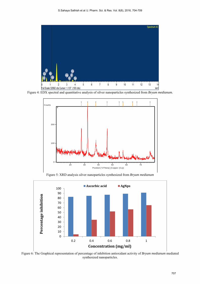
Figure 4: EDX spectral and quantitative analysis of silver nanoparticles synthesized from Bryum medianum.
Figure 5: XRD analysis silver nanoparticles synthesized from Bryum medianum
Figure 6: The Graphical representation of percentage of inhibition antioxidant activity of Bryum medianum mediated synthesized nanoparticles.
Position [°2Theta] (Copper (Cu))
20 30 40 50 60 70
Counts
0
100
200
6
0
10
20
30
40
50
60
70
80
90
100
0.2 0.4 0.6 0.8 1
Percentageinhibition
Concentration(mg/ml)
Ascorbicacid AgNps
S.Sahaya Sathish et al /J. Pharm. Sci. & Res. Vol. 8(8), 2016, 704-709
707
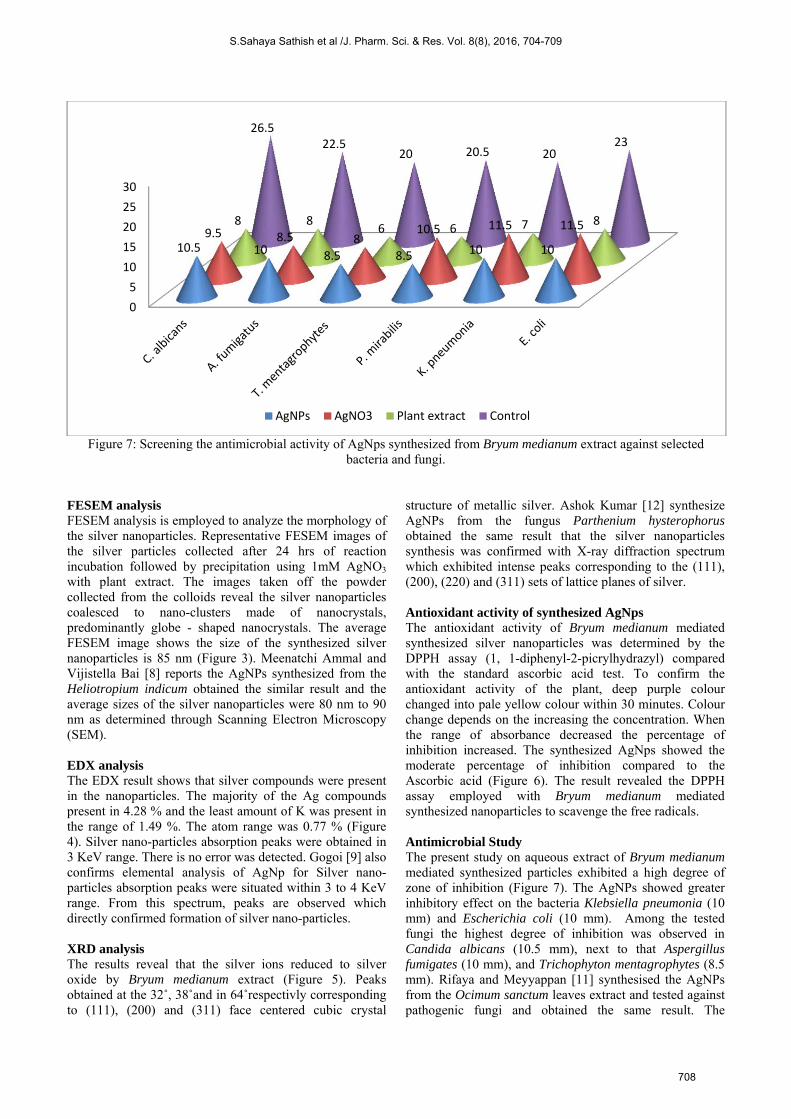
Figure 7: Screening the antimicrobial activity of AgNps synthesized from Bryum medianum extract against selected bacteria and fungi.
FESEM analysis FESEM analysis is employed to analyze the morphology of the silver nanoparticles. Representative FESEM images of the silver particles collected after 24 hrs of reaction incubation followed by precipitation using 1mM AgNO3 with plant extract. The images taken off the powder collected from the colloids reveal the silver nanoparticles coalesced to nano-clusters made of nanocrystals, predominantly globe - shaped nanocrystals. The average FESEM image shows the size of the synthesized silver nanoparticles is 85 nm (Figure 3). Meenatchi Ammal and Vijistella Bai [8] reports the AgNPs synthesized from the Heliotropium indicum obtained the similar result and the average sizes of the silver nanoparticles were 80 nm to 90 nm as determined through Scanning Electron Microscopy (SEM).
EDX analysis The EDX result shows that silver compounds were present in the nanoparticles. The majority of the Ag compounds present in 4.28 % and the least amount of K was present in the range of 1.49 %. The atom range was 0.77 % (Figure 4). Silver nano-particles absorption peaks were obtained in 3 KeV range. There is no error was detected. Gogoi [9] also confirms elemental analysis of AgNp for Silver nano-particles absorption peaks were situated within 3 to 4 KeV range. From this spectrum, peaks are observed which directly confirmed formation of silver nano-particles.
XRD analysis The results reveal that the silver ions reduced to silver oxide by Bryum medianum extract (Figure 5). Peaks obtained at the 32˚, 38˚and in 64˚respectivly corresponding to (111), (200) and (311) face centered cubic crystal
structure of metallic silver. Ashok Kumar [12] synthesize AgNPs from the fungus Parthenium hysterophorus obtained the same result that the silver nanoparticles synthesis was confirmed with X-ray diffraction spectrum which exhibited intense peaks corresponding to the (111), (200), (220) and (311) sets of lattice planes of silver.
Antioxidant activity of synthesized AgNps The antioxidant activity of Bryum medianum mediated synthesized silver nanoparticles was determined by the DPPH assay (1, 1-diphenyl-2-picrylhydrazyl) compared with the standard ascorbic acid test. To confirm the antioxidant activity of the plant, deep purple colour changed into pale yellow colour within 30 minutes. Colour change depends on the increasing the concentration. When the range of absorbance decreased the percentage of inhibition increased. The synthesized AgNps showed the moderate percentage of inhibition compared to the Ascorbic acid (Figure 6). The result revealed the DPPH assay employed with Bryum medianum mediated synthesized nanoparticles to scavenge the free radicals.
Antimicrobial Study The present study on aqueous extract of Bryum medianum mediated synthesized particles exhibited a high degree of zone of inhibition (Figure 7). The AgNPs showed greater inhibitory effect on the bacteria Klebsiella pneumonia (10 mm) and Escherichia coli (10 mm). Among the tested fungi the highest degree of inhibition was observed in Candida albicans (10.5 mm), next to that Aspergillus fumigates (10 mm), and Trichophyton mentagrophytes (8.5 mm). Rifaya and Meyyappan [11] synthesised the AgNPs from the Ocimum sanctum leaves extract and tested against pathogenic fungi and obtained the same result. The
0
5
10
15
20
25
30
10.5 10 8.5 8.5 10 109.5 8.5 8
10.5 11.5 11.58 86 6 7 8
26.522.5
20 20.5 2023
AgNPs AgNO3 Plant extract Control
S.Sahaya Sathish et al /J. Pharm. Sci. & Res. Vol. 8(8), 2016, 704-709
708

antibacterial activity of phyto mediated silver nanoparticles was assessed by the paper disc method against K. pneumonia and 12 mm clear zone was observed. Asmita et al. [12] also reported AgNPs from Azadirachta indica obtained the similar result were noted for gentamicin and piperacillin resistant Salmonella typhi, Fluconazole resistant C. albicans were found of multiple drug resistant E. coli was inhibited by synergistic action of gentamicin and silver nanoparticles synthesized by aqueous extract of Neem and Triphala.
CONCLUSION In the field of nanotechnology the green synthesis is an ecofriendly and more reliable process. The reductions of metal ions through the moss extract leading to the formation of silver nanoparticles. The synthesis of silver nanoparticle using aqueous extract of Bryum medianum provides a natural, simple, cost effective and efficient route for begin of nanoparticle. FESEM image exhibited the globe shaped nanoparticles in the range of 85nm. The FTIR confirmed that alkanes and ether bio molecules responsible for the reduction of silver ions. XRD spectra of the AgNPs confirmed the formation of metallic crystalline silver. The moss plant mediated synthesized particles showed the higher percentage of scavenging the free radicals; it is a good source to produce the antioxidant property. The antimicrobial screening demonstrated that the synthesized AgNps had a high inhibitory effect on bacteria and fungi. So the Bryum medianum (moss plant) have the ability to produce the silver nanoparticles and also these moss plant mediated synthesized particle inhibited the pathogenic
organisms moreover it has a high amount of antioxidant property. The mosses has a good antimicrobial action hence it is preferred for the pharmaceutical field to produce the drugs.
ACKNOWLEDGEMENT We would like to thankful to Archbishop Casimir Instrumentation Centre (ACIC), St. Joseph’s college, Tiruchirappalli for the UV – VIS and FTIR analysis and also grateful to Centre for Nano Science and Nanotechnology, Sathya Bama University, Chennai for the support to carry over FESEM, EDX and XRD analysis.
REFERENCES [1] Leela, A., Vivenkanandan, M., Afr. J. Biotechnol. 2008; 7(17), 3162-
3165. [2] Elechiguerra, J.L., Burt, J.L., Morones, J.R., Camacho-Bragado, A.,
Gao, X., Lara, H.H., Yacaman, M.J., J. Nanobiol. 2005; 3(6), 1-10. [3] Gittins, D., J. Matt Chem. 2000; 10, 79-83. [4] Garder Toressgay, J.L., Gomez, E., Peralta-Videa, J., Parsons, J.G.,
Troiani, H.E., Jose – Yacaman, M., Langmir. 2003; 13, 13-57. [5] Kitts, Mol. Cell. Biochem. 2000; 203, 1-10. [6] Sundarajan, Ranjithakumari, Int. J.Pharm. Pharm. Sci. 2014; 6(3),
30- 34. [7] Mallikarjuna, Dillip, G.R., Narasimha, G., John Sushma, N., Deva
Prasad Raju, B., Res. J. Nanosci. Nanotech. 2012; 2(1), 17- 23. [8] Meenatchi Ammal, Vijistella Bai, Int. J. Pharm. Chem. Biol. Sci.
2014; 4(1), 101- 111. [9] Gogoi, Int. J. Pharm. Res. Biosci. 2014; 3(1), 321-327. [10] Ashok Kumar, Int. Res. J. Pharm. 2012; 3(2), 169-173. [11] Rifaya, A., Meyyappan, Afr. J. Microbiol. Res. 2014; 8(1), 118-128. [12] Asmita, J., Gavhane, P., Padmanabhan, Suresh, P., Kamble, Suresh,
N., Int. J. Pharm. Biosci. 2012; 3(3), 88-100.
S.Sahaya Sathish et al /J. Pharm. Sci. & Res. Vol. 8(8), 2016, 704-709
709