a brief, hands-on lab manual with frequent …... hands-on lab manual with frequent opportunities to...
TRANSCRIPT
A Brief, Hands-On Lab Manual with Frequent Opportunities to Practice
Visual Anatomy & Physiology Lab Manual uses stunning images from Ric Martini’s Visual Anatomy & Physiology textbook and active-learning exercises to get students practicing in the lab.
377
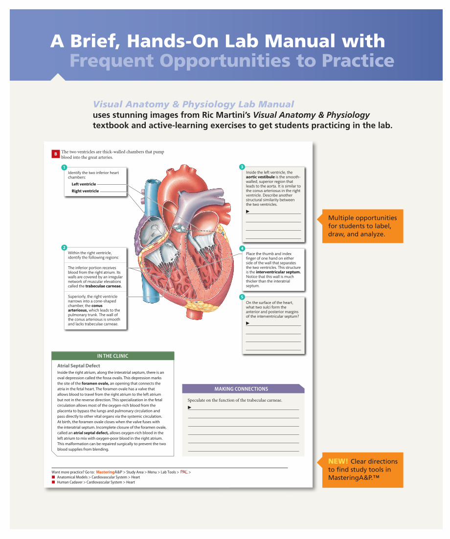
B The two ventricles are thick-walled chambers that pump blood into the great arteries.
1
5
42
▶
3
▶
Identify the two inferior heart chambers:
Left ventricle
Right ventricle
Within the right ventricle, identify the following regions:
Place the thumb and index finger of one hand on either side of the wall that separates the two ventricles. This structure is the interventricular septum. Notice that this wall is much thicker than the interatrial septum.
On the surface of the heart, what two sulci form the anterior and posterior margins of the interventricular septum?
Inside the left ventricle, the aortic vestibule is the smooth-walled, superior region that leads to the aorta. It is similar to the conus arteriosus in the right ventricle. Describe another structural similarity between the two ventricles.
The inferior portion receives blood from the right atrium. Its walls are covered by an irregular network of muscular elevations called the trabeculae carneae.
Superiorly, the right ventricle narrows into a cone-shaped chamber, the conus arteriosus, which leads to the pulmonary trunk. The wall of the conus arteriosus is smooth and lacks trabeculae carneae.
in tHE clinic
Atrial Septal DefectInsidetherightatrium,alongtheinteratrialseptum,thereisanovaldepressioncalledthefossaovalis.Thisdepressionmarksthesiteoftheforamen ovale,anopeningthatconnectstheatriainthefetalheart.Theforamenovalehasavalvethatallowsbloodtotravelfromtherightatriumtotheleftatriumbutnotinthereversedirection.Thisspecializationinthefetalcirculationallowsmostoftheoxygen-richbloodfromtheplacentatobypassthelungsandpulmonarycirculationandpassdirectlytoothervitalorgansviathesystemiccirculation.Atbirth,theforamenovalecloseswhenthevalvefuseswiththeinteratrialseptum.Incompleteclosureoftheforamenovale,calledanatrial septal defect,allowsoxygen-richbloodintheleftatriumtomixwithoxygen-poorbloodintherightatrium.Thismalformationcanberepairedsurgicallytopreventthetwobloodsuppliesfromblending.
Speculate on the function of the trabeculae carneae.
▶
MAkinG connEctions
Want more practice? Go to: masteringa&p > Study Area > Menu > Lab Tools > ™
> ■ Anatomical Models > Cardiovascular System > Heart ■ Human Cadaver > Cardiovascular System > Heart
M21_SARI3854_02_SE_C21.indd 377 18/10/16 12:16 PM
Multiple opportunities for students to label, draw, and analyze.
NEW! Clear directions to find study tools in MasteringA&P.™
A01_SARI3854_02_SE_VWT.indd 1 26/10/16 12:45 PMA01_SARI3854_02_SE_FM_IRC.indd 1 28/10/16 4:12 PM

15
Care and Use of the Compound Light Microscope
Learning OutcOmesThese Learning Outcomes correspond by number to the laboratory activities in this exercise. When you complete the activities, you should be able to:
activity 2.1 Identify the parts of a compound light microscope and explain their functions.
activity 2.2 Demonstrate the proper method for viewing a specimen with the compound microscope.
activity 2.3 Describe the principle of inversion of image.
activity 2.4 Understand the concept of depth of field.
activity 2.5 Measure the diameter of the field of view and estimate the size of structures in a tissue section.
LabOratOry suppLies• Compoundlightmicroscopes• Preparedmicroscopeslidesofvarious
tissues• Preparedmicroscopeslidesofthe
lettere• Preparedmicroscopeslidesof
intersectingcoloredthreads• Clearmillimeterrulers• Lenspaper
pre-Lab QuizBefore you begin, read all the activities in Exercise 2 and the required reading in your textbook that is assigned by your instructor.
1. Whencarryingamicroscope,youshouldholditsecurelywithbothhands.Onehandshouldbeonthe___________,andtheotherhandshouldbeunderthe___________. a. arm...base b. head...stage c. arm...stage d. stage...base
2. Onacompoundmicroscope,whereisthelightsourcelocated? a. ontheoculars b. onthebase c. onthestage d. onthecord
3. TrueorFalse:Thespacebetweentheobjectivelensandthemicroscopestageiscalledtheworkingdistance.___________
4. TrueorFalse:Whenanimageisapproximatelyinfocus,youcanusethecoarseadjustmentknobtobringittoexactfocus.___________
5. Theilluminatedareathatyouviewwithamicroscopeiscalledthe___________.
6. Thenosepieceonamicroscopeisarevolvingstructurethatholdsthe___________.
7. The___________concentratesthelightbeforeittravelsthroughthetissueontheslide.
8. Duringthislaboratoryexercise,youwilluseapreparedslidewiththeletteretodemonstrate a. depthoffield.b. therelationshipbetweentotalmagnificationandfielddiameter. c. theworkingdistance.d. inversionofimage.
9. Thethicknessofthetissuelayerthatisinfocusiscalled a. resolvingpower.b. imageinversion. c. depthoffield.d. fielddiameter.
10. Duringthislaboratoryexercise,youwilluseaclearmillimeterrulerto a. estimatethediameterofthefieldofview.b. measuretheworkingdistance. c. estimatetheresolvingpowerofthemicroscope.d. estimatethesizeofastructureonatissuesection.
2ExErCisE
M02_SARI3854_02_SE_C02.indd 15 19/10/16 12:54 PM
49
C Stratified Squamous, Keratinized Epithelium
1 View a microscope slide of thick skin from the sole under low power and identify the epithelium. This epithelium, known as the epidermis of the skin, is a stratified squamous type, with a thick layer of dead cells at the surface filled with keratin fibers. The layer of dead cells, known as the keratinized layer, serves as a protective barrier that prevents dehydration and infection from airborne pathogens.2 Deep to the epithelium, identify a layer of connective
tissue, known as the dermis.
B Stratified Squamous Epithelium
1 View a microscope slide of the esophagus under low power. Observe that the epithelium lining the lumen (internal space) of the esophagus has many layers of cells.2 Switch to high magnification and observe that the deeper
layers contain cube-shaped cells but the superficial layers consist of flattened cells. That is an example of a stratified squamous epithelium. This type of epithelium provides protection from abrasive and frictional forces, such as when food rubs against the wall of the esophagus.
Lumen ofesophagus
Stratifiedsquamous
epithelium
Connectivetissue
Esophagus LM * 400
Keratinized layerof the epithelium
Stratifiedsquamous
epithelium
Connectivetissue
Epidermis of thick skin LM * 470
D In the spaces provided, sketch what you see under the microscope.
1 In box (a), draw your observations of the esophagus.2 In box (b), draw
your observations of thick skin.3 On both drawings,
label the stratified squamous epithelium and connective tissue.4 On the appropriate
drawing, label the keratinized layer.
(a) ▶
(b) ▶
During this activity, you observed that the lumen of a blood vessel, a passageway for blood, is lined by a simple squamous epithelium, which is a very thin cell layer. You also observed that the lumen of the esophagus, a passageway for food to the stomach, is lined by a stratified squamous epithelium, which is much thicker. Why do you think these two structures have epithelia that are so strikingly different?
▶
MAKiNG CONNECTiONs
■ Want more practice? Go to: masteringa&p > Study Area > Menu > Lab Tools > ™
> Histology > Epithelial Tissue
M05_SARI3854_02_SE_C05.indd 49 18/10/16 10:32 AM
NEW! Pre-Lab Quizzes open each exercise by asking students questions that will help ensure they come to lab prepared. These quizzes are also assignable in MasteringA&P.™
Making Connectionsgive students an opportunity to pause, internalize information, and apply their understanding.
Prepare, Practice, and Put It All
A01_SARI3854_02_SE_VWT.indd 2 26/10/16 12:45 PMA01_SARI3854_02_SE_FM_IRC.indd 2 28/10/16 4:12 PM

Together in the Lab
NEW! Before You Move On feature wraps up each exercise by asking students to think critically about the lab they just completed, and then connect that information to next lab.
In the Clinic boxes throughout the lab manual help students connect what they learn in lab to the real world.
76 ExErcisE 6 The integumentary system
Activity 6.3
A Examining a model of the integumentary system allows you to study the spatial arrangement of the accessory structures and sensory receptors in the cutaneous membrane and to compare your observations with previous microscope work.
Examining an Anatomical Model of the Integumentary System
Hair follicle
Arrectorpili muscle
Observe a model of the integumentary system and identify the following tissue layers:
Epidermis (stratified squamous, keratinized epithelium)
Dermis• Papillary layer• Reticular layer
1
Locate the hypodermis (superficial fascia) deep to the dermis.
2
Notice that arteries, veins, and nerves travel through the hypodermis and give rise to smaller branches that ascend through the dermis.
3
Identify sebaceous glands, located within triangular regions formed bythe epidermis, a hair follicle, and an arrector pili muscle.
4Notice that merocrine sweat glands give rise to ducts that travel through the dermis and epidermis and empty onto the skin surface.
5
In ThE clInIc
Malignant MelanomaMalignantmelanomaisatypeofskincancerthatcausesunregulatedreproductionofmelanocytes.Becausethesecellsproducethepigmentmelanin,melanomasareofteneasilyspottedbecauseoftheirdarkcoloration.Althoughthesemalignanciesmaygrowslowlyatfirst,theybecomeaggressiveand,ifnotcaughtearly,haveahighmortalityrate.Normally,melaninprotectsusfromgeneticdamagecausedbyultravioletradiation,whichcanleadtomelanomaandotherskincancers.Peoplewhohaveskinwithahighlevelofmelaninhavemoreprotectionagainstthisdamage.Theriskofmelanomaisabout20timeshigherforpeopleofEuropeandescentthanforpeopleofAfricandescent.BecausetheoccurrenceofmelanomainpeopleofAfricandescentisrelativelyrare,itsdiagnosisisoftendelayed.Asaresult,whenthediseaseisdiscovered,itisusuallyatamoreadvancedstage,andthechancesofsurvivalarereduced.Earlydetectionisthekey,andthatrequiresfrequentself-examinationofyourskin,includingareasthatarenotexposedtosunlight.
M06_SARI3854_02_SE_C06.indd 76 18/10/16 11:26 AM
BEFoRE YoU MovE on . . .
124
Looking Forward ❯❯
Be aware that the skeletal system not only includes all the bones of the body, but also the cartilage, tendons, and ligaments associated with the articulations (joints). Tendons and ligaments are both composed of dense regular connective tissue. At a joint, a tendon connects a muscle to a bone; a ligament connects one bone to another bone. In the next laboratory exercise (Laboratory Exercise 9), you will study articulations. Think of articulations as the functional junctions between bones. They bind various parts of the skeletal system together, are locations on the body where movement occurs, allow bone growth and development, and permit parts of the skeleton to change shape.
2Clavicle4Pectoral
girdle
60Upperlimbs
2Pelvicgirdle
60Lowerlimbs
2Scapula
2Humerus
2Radius
2Ulna
2Hip bone(coxal bone)
16Carpal bones
10Metacarpal bones
28Phalanges(proximal,
middle, distal)
2Femur
2Patella
2Tibia
2Fibula
14Tarsal bones
10Metatarsal bones
28Phalanges
❮❮ Looking Back
The appendicular skeleton comprises the bones of the upper and lower limbs (the appendages). In this laboratory exercise, you learned that each upper limb includes a clavicle and scapula (1/2 pectoral girdle) and the bones of the arm, forearm, wrist, and hand. Each lower limb includes a coxal (hip) bone (1/2 pelvic girdle) and the bones of the thigh, leg, and foot. You also observed that the organization of the bones in the upper limb is comparable to those in the lower limb. For example, the arm and thigh each contain one large long bone; the forearm and leg each contain two smaller long bones that are roughly parallel.
Despite these similarities, there are important structural and functional differences between the upper and lower limbs.
Consider these questions: ▶
1. Why are the bones of the lower limb larger than those of the upper limb?
2. What is the fundamental difference in function between the foot and the hand?
M08_SARI3854_02_SE_C08.indd 124 24/10/16 11:49 AM
A01_SARI3854_02_SE_VWT.indd 3 26/10/16 12:45 PMA01_SARI3854_02_SE_FM_IRC.indd 3 28/10/16 4:12 PM
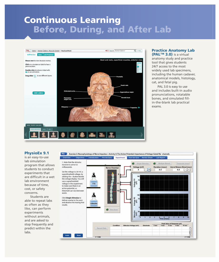
Continuous Learning Before, During, and After Lab
Practice Anatomy Lab (PAL™ 3.0) is a virtual anatomy study and practice tool that gives students 24/7 access to the most widely used lab specimens, including the human cadaver, anatomical models, histology, cat, and fetal pig.
PAL 3.0 is easy to use and includes built-in audio pronunciations, rotatable bones, and simulated fill- in-the-blank lab practical exams.
PhysioEx 9.1 is an easy-to-use lab simulation program that allows students to conduct experiments that are difficult in a wet lab environment because of time, cost, or safety concerns.
Students are able to repeat labs as often as they like, can perform experiments without animals, and are asked to stop frequently and predict within the labs.
A01_SARI3854_02_SE_VWT.indd 4 26/10/16 12:45 PMA01_SARI3854_02_SE_FM_IRC.indd 4 28/10/16 4:12 PM
with MasteringA&P™
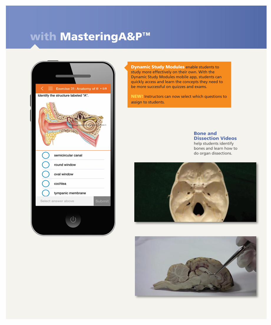
Dynamic Study Modules enable students to study more effectively on their own. With the Dynamic Study Modules mobile app, students can quickly access and learn the concepts they need to be more successful on quizzes and exams.
NEW! Instructors can now select which questions to assign to students.
Bone and Dissection Videos help students identify bones and learn how to do organ dissections.
A01_SARI3854_02_SE_VWT.indd 5 26/10/16 12:45 PMA01_SARI3854_02_SE_FM_IRC.indd 5 28/10/16 4:12 PM
MasteringA&P™
Additional assignable MasteringA&P activities include:• Bone & Dissection Video Coaching
Activities• A&P Flix™ for Anatomy Topics• PAL™ Assessments• PhysioEx™ Assessments• Pre-Lab and Post-Lab Quizzes• And More!
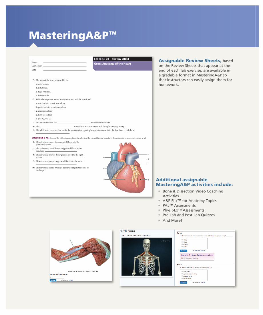
ExErcisE 21 Gross Anatomy of the Heart 389
Name
LabSection
Date
Gross Anatomy of the Heart
ExErCISE 21 REvIEw SHEEt
1. The apex of the heart is formed by the
a. right atrium.
b. left atrium.
c. right ventricle.
d. left ventricle.
2. Which heart groove travels between the atria and the ventricles?
a. anterior interventricular sulcus
b. posterior interventricular sulcus
c. coronary sulcus
d. both (a) and (b)
e. (a), (b), and (c)
3. The epicardium and the are the same structure.
4. The artery forms an anastomosis with the right coronary artery.
5. The adult heart structure that marks the location of an opening between the two atria in the fetal heart is called the .
QUEStIONS 6–10: Answer the following questions by selecting the correct labeled structure. Answers may be used once or not at all.
a
g
f
e
d
b
c
6. This structure pumps deoxygenated blood into the pulmonary trunk.
7. The pulmonary veins deliver oxygenated blood to this structure.
8. This structure delivers deoxygenated blood to the right atrium.
9. This structure pumps oxygenated blood into the aorta.
10. This structure and its branches deliver deoxygenated blood to the lungs.
M21_SARI3854_02_SE_C21.indd 389 18/10/16 12:17 PM
Assignable Review Sheets, based on the Review Sheets that appear at the end of each lab exercise, are available in a gradable format in MasteringA&P so that instructors can easily assign them for homework.
A01_SARI3854_02_SE_VWT.indd 6 26/10/16 12:45 PMA01_SARI3854_02_SE_FM_IRC.indd 6 28/10/16 4:12 PM

Access the Complete Lab Manual On or Offline with eText 2.0
NEW! The Second Edition is available in Pearson’s fully-accessible eText 2.0 platform.*
*The eText 2.0 edition will be live for Fall 2017 classes.
NEW! The eText 2.0 mobile app offers offline access and can be downloaded for most iOS and Android phones and tablets from the iTunes or Google Play stores.
Powerful interactive and customization functions include instructor and student note-taking, highlighting, bookmarking, search, and links to glossary terms.
A01_SARI3854_02_SE_VWT.indd 7 26/10/16 12:45 PMA01_SARI3854_02_SE_FM_IRC.indd 7 28/10/16 4:12 PM
Instructor and Student Support
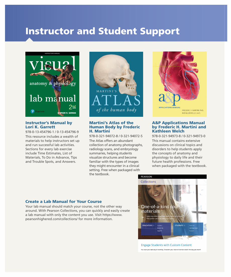
Instructor’s Manual by Lori K. Garrett 978-0-13-454796-1 / 0-13-454796-9This resource includes a wealth of materials to help instructors set up and run successful lab activities. Sections for every lab exercise include Time Estimates, List of Materials, To Do in Advance, Tips and Trouble Spots, and Answers.
INSTRUCTOR’S MANUAL
Martini’s Atlas of the Human Body by Frederic H. Martini978-0-321-94072-8 / 0-321-94072-5The Atlas offers an abundant collection of anatomy photographs, radiology scans, and embryology summaries, helping students visualize structures and become familiar with the types of images they might encounter in a clinical setting. Free when packaged with the textbook.
A&P Applications Manual by Frederic H. Martini and Kathleen Welch978-0-321-94973-8 / 0-321-94973-0This manual contains extensive discussions on clinical topics and disorders to help students apply the concepts of anatomy and physiology to daily life and their future health professions. Free when packaged with the textbook.
Create a Lab Manual for Your CourseYour lab manual should match your course, not the other way around. With Pearson Collections, you can quickly and easily create a lab manual with only the content you use. Visit https://www.pearsonhighered.com/collections/ for more information.
A01_SARI3854_02_SE_VWT.indd 8 26/10/16 12:45 PMA01_SARI3854_02_SE_FM_IRC.indd 8 28/10/16 4:12 PM