a atr t a y u a r r ary ar t try - hscbusiness.hscni.net ophthalmology_guidance...herpes simplex...
TRANSCRIPT
PAEDIATRIC OPHTHALMOLOGY
GUIDANCE FOR PRIMARY CARE OPTOMETRY
Belfast Health and Social Care Trust (v2 revised January 2018)
2
Guidelines for Paediatric Ophthalmology Referrals
Referral Pathways
The following guidance outlines the actions advised and required when presented with the
conditions listed below. This list is not exclusive and exhaustive of the conditions which may
present. Annex 1 provides ‘summary’ information on paediatric ophthalmic conditions and
the recommended referral pathways/options.
To note in the Belfast Trust area there are 3 pathways which a paediatric ophthalmology
referral may follow:
1. Paediatric Ophthalmology - Urgent and Routine Referrals. These can be accessed via
CCG under the RVH ‘OPHTHALMOLOGY – PAEDIATRIC OPHTHALMOLOGY’. Please
read the guidance in order to assist in your decision making as to whether the
referral should be noted as ‘Urgent’ or ‘Routine’ and the doctors will subsequently
triage and assign an appropriate appointment. In particular please note the advice in
regard to the management of urgent referrals on page 10 of this guidance document.
2. Eye Casualty (Regional Acute Eye Service – RAES)
3. A&E at the Royal Belfast Hospital for Sick Children (RBHSC)
Paediatric Ophthalmic conditions
Blepharitis & Staphylococcal Hypersensitivity Disease (see Annex 4)
- Advice re lid cleaning
- Course of topical chloramphenicol ointment to the lids at bedtime one month
- Issue advice leaflet
- If very photosensitive or chronic corneal changes, refer to Paediatric Ophthalmology
as an Urgent Referral for follow up
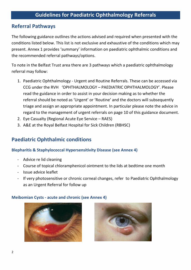
Meibomian Cysts - acute and chronic (see Annex 4)
3
- Advice re lid cleaning and hot bathing long term
- Oral antibiotic not indicated; course of chloramphenicol ointment
- Issue advice leaflet as they tend to occur in crops
- Chronic cysts will settle over time; refer for opinion to outpatients if causing
mechanical problems
Other Lid Lesions
- Molluscum contagiosum of lid with secondary red eye, refer to Paediatric
Ophthalmology as an Urgent Referral
Ptosis
- If obscuring the visual axis in neonate refer to Paediatric Ophthalmology as an Urgent
Referral
- If part of suspected new Horner’s syndrome refer to Paediatric Ophthalmology as an
Urgent Referral
- Otherwise, refer for routine paediatric outpatient appointment
Viral Conjunctivitis
- Endemic in children, highly infectious and self-limiting.
- Often associated with respiratory infection and pre-auricular lymph nodes
- DO NOT refer routinely to hospital, give infection control advice
Acute Bacterial Conjunctivitis
- Rarely seen. Purulent green discharge.
- Treat with topical Gt Chloramphenicol four times daily 1 week.
Epiphora
- Chronic watering and discharge since birth suggests blocked nasolacrimal duct
(usually clear or mucoid discharge)
- Oral antibiotics not indicated
- Gt Chloramphenicol only if red eye or discharge becomes green
- Lacrimal sac massage by parent advocated as per advice sheet
4
- Refer to paediatric ophthalmology outpatients if persisting beyond 12 months,
parents are keen for probing and child is fit for GA
- Look for epiblepharon in babies, in turning eyelashes; refer to Paediatric
Ophthalmology as an Urgent Referral
Periorbital Cellulitis
- Treat any local infection. Refer to GP for oral antibiotic if involving whole lid
- If not responding to oral antibiotic, or with systemic upset, needs referral to RBHSC
A&E to consider admission for IV antibiotic
Dacryocystitis
- Lacrimal sac abscess (swelling medially between eye and nose)
- Refer to GP to commence oral antibiotic and to refer to Paediatric Ophthalmology as
an Urgent Referral
- Emergency referral to RBHSC for IV antibiotic if systemically unwell
Chronic Blinking (see also Annex 2)
- Suggests corneal epithelial disturbance
- May be secondary to allergic conjunctivitis (history of allergy/allergic rhinitis)
- May be secondary to lid inflammation (red lids, styes and meibomian cysts)
- Try simple lubricant first; Treat any lid inflammation; refer to Paediatric
Ophthalmology as an Urgent Referral if chronic photophobia, has chronic red eye or
doesn’t respond to simple treatment
Allergic Eye Disease
- Trial of antihistamine drops
- Advise oral antihistamine if eyes are involved in acute periorbital allergic reaction
- Refer to Paediatric Ophthalmology as an Urgent Referral for advice if symptoms
causing chronic upset in daily life and not responding to treatment

5
Herpes Simplex Blepharitis
- Refer to GP to start oral aciclovir and refer to Paediatric Ophthalmology as an Urgent
Referral.
- May have more generalised rash associated with eczema (eczema herpeticum) and
require dermatology advice via GP
Herpes Simplex Keratitis
- Refer to GP to commence oral aciclovir and refer to Paediatric Ophthalmology as an
Urgent Referral.
Herpes Zoster Ophthalmicus (Shingles)
- Refer to GP to start oral acyclovir and refer to Paediatric Ophthalmology as an
Urgent Referral
- If on immunosuppressant, refer Royal Belfast Hospital for Sick Children (RBHSC)
urgently for admission for IV aciclovir
Chickenpox Rash and Eye Involvement
- To GP to commence oral aciclovir if keratitis, iritis, optic neuritis, retinitis or other
cranial nerve involvement
- Refer to Paediatric Ophthalmology as an Urgent Referral
Chemical Injury
- Irrigation and oral analgesia prior to referral to hospital
- Often pupil will need dilated with cyclopentolate (0.5% under 1 year old, 1% over 1
year old) to allow any assessment of corneal epithelial defect. Refer on to Eye
Casualty for advice and topical treatment or seek advice re treatment by phone.
Corneal Abrasion
- Relieve pain with topical anaesthetic and dilating drops (amethocaine and
cyclopentolate – 0.5% under 1 year old, 1% over 1 year old )
- If minor send home on chloramphenicol eye ointment
- If extensive, or unable to examine, refer acutely for ophthalmology opinion.
Remember to dilate pupil for pain relief
6
Corneal Foreign Body
- Relieve pain with topical anaesthetic (amethocaine) to facilitate exam
- Attempt removal with cotton tipped applicator if FB accessible
- Cover with topical chloramphenicol ointment until heals
- Refer acutely to Eye Casualty if child distressed
- Refer to Paediatric Ophthalmology as an Urgent Referral if problem persists; dilate
pupil if photophobic
Corneal Ulcer
- Relieve any pain with topical anaesthetic and dilating drops (amethocaine and
cyclopentolate – 0.5% under 1 year old, 1% over 1 year old)
- Refer to Eye Casualty if child distressed possibility of perforation or sight threatening
infection. Otherwise, commence gt chloramphenicol and refer to Paediatric
Ophthalmology as an Urgent Referral.
Hyphaema & blunt trauma
- Refer to Eye Casualty for acute assessment and treatment; may require admission
Iritis/Uveitis
- Refer to Eye Casualty for assessment and treatment
Acute Onset Squint Unwell child:
- Systemic upset, gaze palsy, swollen discs, infection; if some / all present, refer to RBHSC for urgent neuroimaging
- Check red reflex - retinoblastoma can present with squint If child is well with normal red reflexes:
- Refer to Paediatric Ophthalmology as an Urgent Referral for follow up; enquire if history of previous shunt surgery for raised intracranial pressure.
7
Swollen Discs query (see also Annex 3)
- classical papilloedema with disc haemorrhages, - systemically unwell child - gaze palsy - neurosurgical history - refer to RBHSC A& E for urgent neuroimaging
- If disc swelling detected in well child on routine optometric assessment with normal
visual functions, refer to Paediatric Ophthalmology as an Urgent Referral and note the query as optic disc drusen
- Please note that the provision of OCT scanning for Optic Discs in children is not advised as inconsistencies in scanning methods can arise and the paediatric ophthalmology service in BHSCT do not wish to receive OCT scans where optic disc drusen are queried.
Unequal Pupils
- If child systemically well, and this is new finding: refer to paediatric ophthalmology outpatients.
- If pupil size is associated with other signs, eg gaze palsy, ptosis, refer routinely to Paediatric Ophthalmology as an Urgent Referral
Assessment of the baby who does not appear to see
- Ascertain child’s corrected age before making referral (premature babies will not begin to fix and follow until term + 6 weeks)
- Check red reflexes are present - Record if there is nystagmus or roving eye movements - Refer to Paediatric Ophthalmology as an Urgent Referral for assessment
Proptosis
- If child systemically unwell, refer to Eye Casualty - If child systemically well, and this is acute finding: refer to Paediatric Ophthalmology
as an Urgent Referral - If child systemically well, and proptosis is longstanding: refer for routine Paediatric
Ophthalmology appointment
8
Headache (see also Annex 3)
- Headache is a common problem in children, and is managed by the GP - Normal eye exam, refer on to GP for advice if interfering with activities of daily living
e.g. migraine - Severe headache with unwell child and symptoms of raised intracranial pressure,
rare but important not to miss worse on wakening, lying down, daily vomiting and hearing whooshing sounds, examine discs , swelling with haemorrhages; refer urgently to Paediatric Ophthalmology as an Urgent Referral
Absent Red Reflex
- Refer without delay to Paediatric Ophthalmology as an Urgent Referral, record if unilateral or bilateral
- Neonates with darkly pigmented eyes are often a challenge for the non- specialist to assess as pupils are small and the reflexes appear paler. Examine in a darkened room.
Leucocoria
- Check red reflex; if absent refer to Paediatric Ophthalmology as an Urgent Referral without delay
- If white reflex present in one pupil in every photo, refer to Paediatric Ophthalmology as an Urgent Referral without delay
- If red reflex present on your exam and white reflex present as isolated finding on one photo, reassure parents that a serious problem is unlikely and refer to Paediatric Ophthalmology to arrange Outpatient appointment
Screening for Congenital Glaucoma
- If hazy cornea, enlarged globe with epiphora, refer to eye casualty - If all normal, send accurate family history of childhood glaucoma and refer as routine
paediatric ophthalmology referral (NB family history of adult onset glaucoma not significant in this condition)
Screening babies with family history of Retinoblastoma
- Send referral to Paediatric Ophthalmology as an Urgent Referral with details of family history documented.
9
Incidental fundal findings at routine optometric assessment
- Incidental findings in otherwise normal eye exam (e.g. flat naevi and CHRPE) identified as new finding because of digital imaging, do not need ophthalmology assessment. These innocent lesions can be safely photographed and observed.
Reduced vision at optometric assessment with otherwise normal exam
- Record distance and near vision using different tests to check consistency of responses
- Check pupil responses and colour vision; formal VF test not useful in children - Observe if visual behaviour is consistent with level of tested vision - Document test results and refer to Paediatric Ophthalmology as an Urgent Referral if
there are major concerns
10
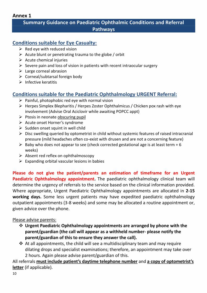
Annex 1 Summary Guidance on Paediatric Ophthalmic Conditions and Referral
Pathways Conditions suitable for Eye Casualty: Red eye with reduced vision Acute blunt or penetrating trauma to the globe / orbit Acute chemical injuries Severe pain and loss of vision in patients with recent intraocular surgery Large corneal abrasion Corneal/subtarsal foreign body Infective keratitis
Conditions suitable for the Paediatric Ophthalmology URGENT Referral: Painful, photophobic red eye with normal vision Herpes Simplex Blepharitis / Herpes Zoster Ophthalmicus / Chicken pox rash with eye
involvement (Advise Oral Aciclovir while awaiting POPCC appt) Ptosis in neonate obscuring pupil Acute onset Horner’s syndrome Sudden onset squint in well child Disc swelling queried by optometrist in child without systemic features of raised intracranial
pressure (mild headaches often co-exist with drusen and are not a concerning feature) Baby who does not appear to see (check corrected gestational age is at least term + 6
weeks) Absent red reflex on ophthalmoscopy Expanding orbital vascular lesions in babies
Please do not give the patient/parents an estimation of timeframe for an Urgent Paediatric Ophthalmology appointment. The paediatric ophthalmology clinical team will determine the urgency of referrals to the service based on the clinical information provided. Where appropriate, Urgent Paediatric Ophthalmology appointments are allocated in 2-15 working days. Some less urgent patients may have expedited paediatric ophthalmology outpatient appointments (3-8 weeks) and some may be allocated a routine appointment or, given advice over the phone.
Please advise parents: Urgent Paediatric Ophthalmology appointments are arranged by phone with the
parent/guardian (the call will appear as a withheld number- please notify the parent/guardian of this to ensure they answer the call).
At all appointments, the child will see a multidisciplinary team and may require dilating drops and specialist examinations; therefore, an appointment may take over 2 hours. Again please advise parent/guardian of this.
All referrals must include patient’s daytime telephone number and a copy of optometrist’s letter (if applicable).

11
Conditions suitable for referral to a Paediatric Ophthalmology ROUTINE Referral: Blepharitis and cysts Other lid lesions e.g. dermoid Watering eyes Chronic surface problems secondary to allergy Misdirected lashes Simple Ptosis (not covering the pupil) Unequal pupils (longstanding/no systemic problems) Nystagmus Asymptomatic anterior segment/fundus findings queried by optometrist on routine
assessment (may be sent back to referring optometrist if felt not to merit further investigation by ophthalmologist)
Vision problems detected by optometrist not related to squint Screening for congenital glaucoma in asymptomatic baby – send accurate family history,
including age of onset, with referral Screening for congenital/childhood cataract – check and record that good red reflexes
present; Send accurate family history with referral.
BHSCT – Paediatric Ophthalmology – revised January 2018
2018
PLEASE NOTE: Paediatric
Ophthalmology referrals
can be made via CCG.
Please select the
appropriate box ‘Urgent’ on
the referral template
If referring via GP please
ask GP to refer via CCG
using the urgent referral
option under
‘ROYAL VICTORIA
HOSPITAL
/OPHTHALMOLOGY/
OPHTHALMOLOGY –
PAEDIATRIC
OPHTHALMOLOGY’
Annex 2
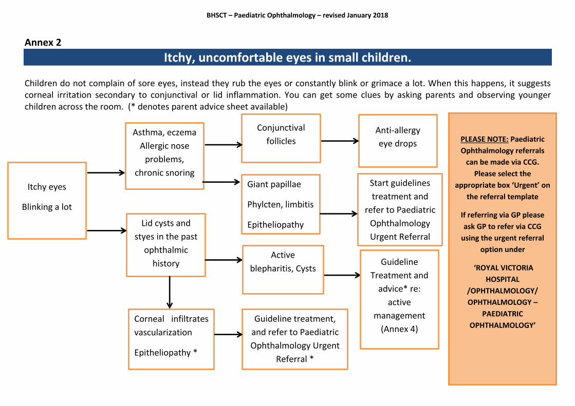
Itchy, uncomfortable eyes in small children. Children do not complain of sore eyes, instead they rub the eyes or constantly blink or grimace a lot. When this happens, it suggests corneal irritation secondary to conjunctival or lid inflammation. You can get some clues by asking parents and observing younger children across the room. (* denotes parent advice sheet available)
Itchy eyes
Blinking a lot
Lid cysts and
styes in the past
ophthalmic
history
Conjunctival
follicles Asthma, eczema
Allergic nose
problems,
chronic snoring
Anti-allergy
eye drops
Start guidelines
treatment and
refer to Paediatric
Ophthalmology
Urgent Referral
Giant papillae
Phylcten, limbitis
Epitheliopathy
Corneal infiltrates
vascularization
Epitheliopathy *
Active
blepharitis, Cysts
Guideline
Treatment and
advice* re:
active
management
(Annex 4) Guideline treatment,
and refer to Paediatric
Ophthalmology Urgent
Referral *

BHSCT – Paediatric Ophthalmology – revised January 2018
2018
Annex 3
Headache and Difficult Discs in Children
Headache is a common condition, rarely due to focusing problems in this age group.
However, GP’s often suggest an eye test when a child complains of headache, mainly to
rule out papilloedema. Children can suffer from migraine and older children can have sinus-
related problems and chronic daily headache related to posture and stress. All these
conditions should be diagnosed and managed by the GP.
Papilloedema is a rare diagnosis, but important not to miss. It is a sign of raised intracranial
pressure. Pressure is raised by the mass effect of expanding intracranial tumours, blocked
shunts (inserted in children with raised intracranial pressure often as small babies) and can
be idiopathic.
Children who have raised intracranial pressure have severe headaches increasing in
intensity, worst on wakening in the morning and lying down. They usually begin to look
unwell and cannot maintain everyday activities. As intracranial pressure increases, children
can become irritable, behave badly and begin to vomit.
Those with large tumours have other presenting signs and all patients need a careful
evaluation to look for signs of direct optic nerve/pathway compression, lateral rectus palsy
and other cranial nerve involvement. Formal visual field testing is of no value. NB a child
with chronic headache and chronic papilloedema may have small grey atrophic discs and
reduced vision due to optic atrophy. This is a very late presentation.
Record the results of the following;
Age appropriate vision test - often normal or slightly reduced with early presentation
of papilloedema
Pupil responses - direct and indirect
Colour vision
Range of ocular movements and cover test
Visual fields to confrontation
Appearance of optic nerve heads and whether spontaneous venous pulsation (SVP) is
present or can be induced by gently pressing on the globe through the lid while
observing the veins as they branch at the centre of the optic disc
BHSCT – Paediatric Ophthalmology – revised January 2018
2018
Papilloedema Vs Drusen
NB papilloedema is a cause of binocular symmetrical disc swelling with vessel tortuosity,
often accompanied by haemorrhages at optic disc margin.
Optic disc drusen cause lumpy discs, most often bi-nasally and can be quite asymmetric in
appearance. Drusen are present in about 1% of the general population. They tend to be
buried in children but can be seen as white shiny lumps at the disc surface in older children.
SVP is present or can be induced.
Identifying papillodema in children
A well child presenting for routine assessment who is identified as having prominent lumpy
nerve heads but normal visual functions has drusen. Refer for ophthalmology assessment if
SVP is not present or if there is uncertainty about diagnosis.
If optometric imaging shows unchanged lumpy nerve head appearance over serial tests,
onward referral is not necessary.
In a child with chronic headache, disc swelling, no SVP, vessel tortuosity and disc
haemorrhages, refer urgently via CCG to Paediatric Ophthalmology as an Urgent Referral or,
via the GP requesting that they annotate the referral as Urgent.
A child presenting with acute papilloedema and other signs such as lateral rectus palsy or
who is unwell should be sent to RBHSC A&E for admission
Referral and implications for parents and children
A diagnosis of swollen discs in an unwell child can aid the speedy diagnosis of a potentially
life-threatening condition.
Thankfully this is a rare occurrence. Drusen discs are common. Benign headaches in
children are very common.
It is important to identify the rare diagnosis; however it is equally important to objectively
evaluate the more common drusen discs. If a referral onward is felt to be clinically
necessary, refer to Paediatric Ophthalmology as an Urgent Referral. Well children do not
need emergency referral with unnecessary neuroimaging and all the anxiety that this
referral route brings.

BHSCT – Paediatric Ophthalmology – revised January 2018
2018
Annex 4
Patient Advice
The following patient advice leaflets may help you in practice when presented with a child
who has one or, more of the following conditions:
Inflammatory Lid Problems in Children
Blepharitis & Staphylococcal hypersensitivity disease
Meibomian cysts, acute and chronic
Watering Eyes in Babies
PLEASE NOTE: These leaflets are available in PDF format and are hosted on the
BSO website at: http://www.hscbusiness.hscni.net/services/2376.htm

BHSCT – Paediatric Ophthalmology – revised January 2018
2018
Inflammatory Lid Problems in Children
Advice for Parents
Blepharitis & Staphylococcal hypersensitivity disease
The skin at the base of the eyelashes can become inflamed and lead to a lot of problems on the
surface of the eye. You may notice scaly deposits and debris that look like dandruff along the lid
margins. These allow normal friendly skin bacteria to overgrow and produce a toxin in the tears.
This causes the eyes to become very irritated. You may notice your child rubbing the eyes and
blinking a lot. If untreated, this can progress to an allergic reaction with redness and inflammation
of the surface of the eyes. It can progress to constant blinking and intolerance of bright lights,
interfering with daily activities.
Your GP will prescribe antibiotic eye ointment. This should be used 4 times a day for 5-7 days. If
the lids are very inflamed, it may need to be continued at night only for another 3 weeks.
The lid inflammation is a skin problem. It will flare up and down as your child grows. It is
important to carry out simple measures to control this condition, as part of your child’s routine.
Daily hot bathing and lid cleaning will help. This is done with a clean facecloth and wash hand
basin full of hand hot water (temperature suitable for skin contact). Wring out face cloth and fold
to make hot towel. Show your child how to close eyes and apply hot cloth to closed eye lids. The
aim is to heat the oil glands in the eyelids and allow them to open. The cloth can be reheated again
as soon as it begins to cool. Ideally this should be done every day for up to 2 minutes at a time.
Finally, wipe the cloth gently along the base of the lashes to help dislodge any debris.
Lid cleaning can be incorporated as a game into bath time routine in very small children.
A daily omega 3 oil supplement can be useful. It is now available to buy as chewable fruit flavoured
capsules for smaller children. Milled linseeds can also be added to breakfast cereals as an omega 3
source.
BHSCT – Paediatric Ophthalmology – revised January 2018
2018
Inflammatory Lid Problems in Children
Advice for Parents
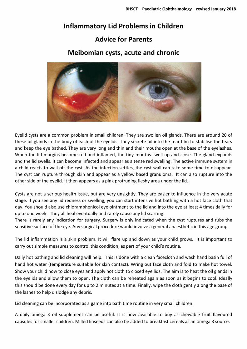
Meibomian cysts, acute and chronic
Eyelid cysts are a common problem in small children. They are swollen oil glands. There are around 20 of
these oil glands in the body of each of the eyelids. They secrete oil into the tear film to stabilise the tears
and keep the eye bathed. They are very long and thin and their mouths open at the base of the eyelashes.
When the lid margins become red and inflamed, the tiny mouths swell up and close. The gland expands
and the lid swells. It can become infected and appear as a tense red swelling. The active immune system in
a child reacts to wall off the cyst. As the infection settles, the cyst wall can take some time to disappear.
The cyst can rupture through skin and appear as a yellow based granuloma. It can also rupture into the
other side of the eyelid. It then appears as a pink protruding fleshy area under the lid.
Cysts are not a serious health issue, but are very unsightly. They are easier to influence in the very acute
stage. If you see any lid redness or swelling, you can start intensive hot bathing with a hot face cloth that
day. You should also use chloramphenicol eye ointment to the lid and into the eye at least 4 times daily for
up to one week. They all heal eventually and rarely cause any lid scarring.
There is rarely any indication for surgery. Surgery is only indicated when the cyst ruptures and rubs the
sensitive surface of the eye. Any surgical procedure would involve a general anaesthetic in this age group.
The lid inflammation is a skin problem. It will flare up and down as your child grows. It is important to
carry out simple measures to control this condition, as part of your child’s routine.
Daily hot bathing and lid cleaning will help. This is done with a clean facecloth and wash hand basin full of
hand hot water (temperature suitable for skin contact). Wring out face cloth and fold to make hot towel.
Show your child how to close eyes and apply hot cloth to closed eye lids. The aim is to heat the oil glands in
the eyelids and allow them to open. The cloth can be reheated again as soon as it begins to cool. Ideally
this should be done every day for up to 2 minutes at a time. Finally, wipe the cloth gently along the base of
the lashes to help dislodge any debris.
Lid cleaning can be incorporated as a game into bath time routine in very small children.
A daily omega 3 oil supplement can be useful. It is now available to buy as chewable fruit flavoured
capsules for smaller children. Milled linseeds can also be added to breakfast cereals as an omega 3 source.
BHSCT – Paediatric Ophthalmology – revised January 2018
2018
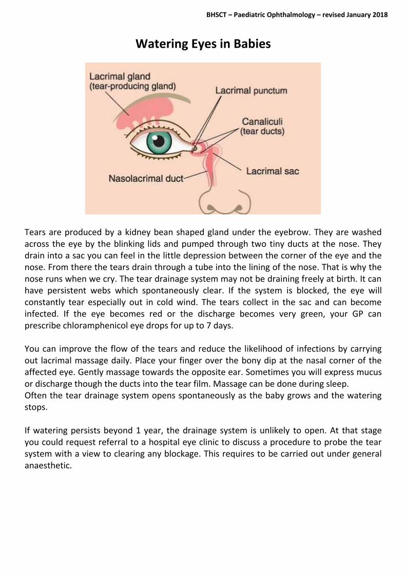
Watering Eyes in Babies
Tears are produced by a kidney bean shaped gland under the eyebrow. They are washed across the eye by the blinking lids and pumped through two tiny ducts at the nose. They drain into a sac you can feel in the little depression between the corner of the eye and the nose. From there the tears drain through a tube into the lining of the nose. That is why the nose runs when we cry. The tear drainage system may not be draining freely at birth. It can have persistent webs which spontaneously clear. If the system is blocked, the eye will constantly tear especially out in cold wind. The tears collect in the sac and can become infected. If the eye becomes red or the discharge becomes very green, your GP can prescribe chloramphenicol eye drops for up to 7 days. You can improve the flow of the tears and reduce the likelihood of infections by carrying out lacrimal massage daily. Place your finger over the bony dip at the nasal corner of the affected eye. Gently massage towards the opposite ear. Sometimes you will express mucus or discharge though the ducts into the tear film. Massage can be done during sleep. Often the tear drainage system opens spontaneously as the baby grows and the watering stops. If watering persists beyond 1 year, the drainage system is unlikely to open. At that stage you could request referral to a hospital eye clinic to discuss a procedure to probe the tear system with a view to clearing any blockage. This requires to be carried out under general anaesthetic.