a 5669-fr burn test
TRANSCRIPT

8/6/2019 A 5669-FR burn test
http://slidepdf.com/reader/full/a-5669-fr-burn-test 1/13
Final Report KGL #5669
TABLE OF CONTENTS
I. OBJECTIVE.........................................................................................................2 II. EXPERIMENTAL DESIGN.................................................................................2
A. General Considerations................................................................................2
B. Panelist Selection...........................................................................................3Light Source (Solar Simulator)............................................................................3D. MED Determination ......................................................................................4E. Test Products and Treatment Procedures ...................................................4F. Expert Grader’s Evaluations..........................................................................5 G. Minolta Chromameter L* a* b* ......................................................................6 H. Cortex Technology DermaLab Water Loss Meter.........................................7 I. Data Analysis...................................................................................................8
III. RESULTS..........................................................................................................8 A. Panelist Accountability...................................................................................8 B. Expert Grader Scores....................................................................................9
C. Minolta Chromameter ............................................................................10 D. DermaLab® Water Loss Probe..................................................................12
IV. CONCLUSIONS..............................................................................................13V. RECORD RETENTION....................................................................................13
Appendix A: Calendar of Events Appendix B: Weather Information Appendix C: Demographic Data Appendix D: Expert Grader Data Appendix E: Chromameter Data Appendix F: Water Loss Data
Page 1
8/6/2019 A 5669-FR burn test
http://slidepdf.com/reader/full/a-5669-fr-burn-test 2/13
Final Report KGL #5669
I. OBJECTIVE
To determine if the use of a topically applied test formulation can diminish
the sunburn response which is induced when the skin is photo-insulted with 2 MED's of UV-B.
II. EXPERIMENTAL DESIGN
A. General Considerations
This study was conducted under the supervision of Kays Kaidbey, M.D.and Gary Grove, Ph.D., at the Skin Study Center in Broomall, Pennsylvania.Copies of Dr. Kaidbey and Dr. Grove’s curriculum vitae are on file with theSponsor.
In conducting this study, we followed the general guidelinesrecommended in Good Clinical Practices (GCP) and Good Laboratory Practices(GLP) as well as the COLIPA Efficacy Testing Guidelines.
This study was conducted from August 19, 2004 to August 27, 2004. Acalendar of events outlining the schedule of treatments and evaluative
procedures that were followed is attached as Appendix A. The daily weather records covering this time as extracted from newspaper reports were recorded and are attached as Appendix B. A more detailed account issued by the US
Weather Bureau can be provided upon the Sponsor’s request.
Briefly, this study was a single-blind, controlled, randomized study whichdetermined the effects of a test material on skin photo-insulted with 2 MED's of UV-B. . The panelists reported first for MED determination and then returned tothe laboratory the following week for Baseline assessments and to receive a 2 MED dose on 4 of the 6 sites. They returned the following day for additional evaluations and to commence twice daily product treatments. The panelists thenreturned each day for the remainder of the week for additional assessments and treatments (no treatment on Friday).
Page 2

8/6/2019 A 5669-FR burn test
http://slidepdf.com/reader/full/a-5669-fr-burn-test 3/13
Final Report KGL #5669
B. Panelist Selection
The volunteers for this study were selected from a pool of healthy women
who were between 18-55 years of age and must have been willing to comply with the requirements of this experimental design. Each candidate wasinterviewed to make certain that they had no medical problems and were not using concomitant medications that might interfere with the study results.Women who were either pregnant or breast-feeding were also excluded from
participating in this study.
Each volunteer signed a consent form after being informed as to her obligations and risks that might be encountered as a participant in this study.The selected panelists were advised of the general nature of this study and wereinstructed not to "tamper" with the sites in any way.
Prior to testing, all candidates were assessed by Ms. Angelit Barnes, for suitability to be included on the panel. Any individuals with cuts, scratches or any clinical signs of erythema on the lower back were excluded at that time.Qualified panelists were assigned a panelist number in the order of their admittance to the study panel.
Light Source (Solar Simulator)
This is a 150-watt compact xenon arc source equipped with a UV-reflecting dichroic mirror and a 1mm thick Schott WG-320 filter to producesimulation of the solar spectrum. A 1mm thick UG5 filter is added to removereflected heat and remaining visible radiation (Berger, D.S.: Specification and design of solar ultraviolet simulators. J. Invest. Dermatol. 53:192-199, 1969).Warm up time of the lamp before use is 20-25 minutes. Total irradiance at skinlevel is measured with a calibrated Eppley Thermopile and the UVB component is monitored with a Robertson-Berger sunburn meter (R-B meter). The size of the irradiated field is approximately a 1 cm diameter circle.
Page 3

8/6/2019 A 5669-FR burn test
http://slidepdf.com/reader/full/a-5669-fr-burn-test 4/13
Final Report KGL #5669
D. MED Determination
One test area serves for determining each panelist’s Minimal Erythema
Dose (MED). The MED is determined by exposing several unprotected skinsites (1cm in diameter) over the mid back to a series of exposures from the solar simulator that were done in 25% increments. The MED is defined as the time of exposure required to produce minimally perceptible erythema 22 + 4 hours after exposure. The MED of the panelist’s unprotected skin was determined the week
prior to the actual testing. Visual grading of the MED is done under standardized lighting conditions when the panelists returned to the testing facility approximately 24 hours after irradiation.
E. Test Products and Treatment Procedures
The test material utilized in this study which was a biocatalyst that had been filtered through 5 micron and 1 micron filters, then irradiated with UV and filtered through activated charcoal was supplied by the Sponsor in a bottle and labeled as follows:
Sample A
Three sites (approximately 1” x 1”) were marked on both the left and right lower back (6 sites total) by Ms. Barnes. After completing the visual grades and instrument measurements on Day 1, two times each individual’s MED dose wasadministered to 4 of the 6 test sites.
On Day 2, panelists reported to the Skin Study Center for additional visual assessment by the Expert Grader and for instrumental measurements using theDermaLab Water Loss Probe and Minolta Chromameter. After theseassessment/measurements were completed, twice daily treatmentscommenced. Treatment was done in a randomized fashion for the following:
1 site on each side of the back: 2 MED + Sample A1 site on each side of the back: 2 MED + no treatment 1 site on each side of the back: Control (no 2 MED + no treatment)
Page 4
8/6/2019 A 5669-FR burn test
http://slidepdf.com/reader/full/a-5669-fr-burn-test 5/13
Final Report KGL #5669
The test product was applied to the designated sites at the lab in themorning and in the afternoon by Mrs. Marie Windle. Approximately 0.1cc of thetest material was applied to the assigned sites and rubbed into the skin using a
cotted finger. Additional product (approximately 0.1cc) was also applied to aBand-Aid Brand bandage which was placed over the site. During the afternoonvisit, the morning bandages were removed and product was again applied directly to the skin and covered with additional product on a fresh bandage.Care was taken to prevent cross-contamination of the treatment sites during application and throughout the treatment/measurement phase of the study.
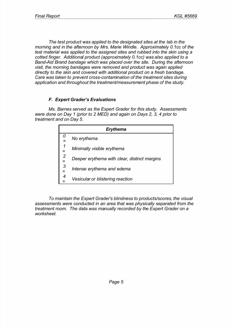
F. Expert Grader’s Evaluations
Ms. Barnes served as the Expert Grader for this study. Assessments
were done on Day 1 (prior to 2 MED) and again on Days 2, 3, 4 prior totreatment and on Day 5.
Erythema
0 =
No erythema
1=
Minimally visible erythema
2 =
Deeper erythema with clear, distinct margins
3
=
Intense erythema and edema
4=
Vesicular or blistering reaction
To maintain the Expert Grader's blindness to products/scores, the visual assessments were conducted in an area that was physically separated from thetreatment room. The data was manually recorded by the Expert Grader on aworksheet.
Page 5

8/6/2019 A 5669-FR burn test
http://slidepdf.com/reader/full/a-5669-fr-burn-test 6/13
Final Report KGL #5669
G. Minolta Chromameter L* a* b*
Skin surface color was measured instrumentally using reflectancetechniques based on the standardized tristimulus system recommended by CIE.
The specific model employed for such measurements was the Minolta CR-200 Chromameter that has an 8mm measuring area using the illuminant conditionsof D65 which most closely approximates normal daylight conditions. This is ahand held device that is gently placed against the surface to be color characterized. When triggered, a pulsed xenon light source flashes and thislight is reflected off the surface and measured back into the device. Within thedevice, there are 6 silicon photocells that are filtered to detect primary stimulusvalues for red, green and blue wavelengths of light. For color readings, thevalues are translated into the L*a*b* coordinates whose spacing correlatesclosely with color changes perceived by the human eye. This is aninternationally recognized convention for numerically expressing color
differences established by the C.I.E. (Commission International de L’Eclairage).The L* value represents the density value from black to white. The a* and b* values represent the color axes ranging from green to red and from blue toyellow, respectively. Higher a* values along the red-green axis are an indicationthat a site is more irritated. [Babulak, S.W., Rhein, L.D., Scala, D.D., Simion,
A.F. and Grove, G.L., Quantitation of Erythema in a Soap Chamber Test Using the Minolta Chroma (Reflectance) Meter: Comparison of Instrumental Resultswith Visual Assessments, J. Soc. Cosmet. Chem. 37:475-479, 1986.]
On Day 1 prior to 2 MED exposure, Days 2, 3 and 4 prior to treatment and again on Day 5, three sets of L*, a* and b* readings from each of the test sites were taken by Mrs. Trish Alfano with the assistance of Mrs. Nancy Bates
and the average value was computed for each site.
Page 6

8/6/2019 A 5669-FR burn test
http://slidepdf.com/reader/full/a-5669-fr-burn-test 7/13
Final Report KGL #5669
H. Cortex Technology DermaLab Water Loss Meter
All water loss measurements were taken following a 15-30 minute
acclimation period in a controlled environment with the relative humidity maintained at less than 50% and temperature maintained at 70 + 2 oF.
This instrument is based on the vapor pressure gradient estimationmethod as designed by Nilsson and initially utilized by the Servo Med Evaporimeter. There are slight dimensional differences and the sensor technology is greatly improved in the DermaLab® TEWL probe but theunderlying principles of the measurement remain the same. Both probescontain two sensors which measure the temperature and relative humidity at twofixed points along the axis normal to the skin surface. This arrangement is suchthat the device can electronically derive a value that corresponds to evaporative
water loss expressed in gm/m2
hr. The DermaLab®
Modular System with TEWLProbe is more fully described in:
Grove, G.L., M.J. Grove, C. Zerweck and E. Pierce: Comparative metrology of the evaporimeter and the DermaLab® TEWL probe. Skin Res. & Tech. 5:1-8, 1999.
Grove, G.L., M.J. Grove, C. Zerweck and E. Pierce: Computerized evaporimetry using the DermaLab® TEWL probe. Skin Res. & Tech. 5:9-13, 1999.
The guidelines established for using the Servo Med Evaporimeter asdescribed by Pinnagoda [Pinnagoda, J., R.A. Tupker, T. Anger and J. Serup.Guidelines for transepidermal water loss (TEWL) measurement. In: Contact Dermatitis 1990: 22:164-178] are quite appropriate for the DermaLab® TEWLProbe as well.
The data from the DermaLab® Modular System is completely computerized and continuously communicates with its PC through a serial port using an RS-232C cable and associated cyberDERM, inc. software for theEvaporimeters. We use the application program entitled C_BASIX_ whichcaptures the water loss data from the attached evaporimeter at a sampling rateof 5 inputs/second. These inputs are graphed as a real time display on thecomputer monitor. The extracted value refers to the average evaporative water
loss rate collected over a twenty second interval once steady state conditionshave been achieved. These are directly transferred to an Excel spread sheet fileusing a DDE link.
Page 7

8/6/2019 A 5669-FR burn test
http://slidepdf.com/reader/full/a-5669-fr-burn-test 8/13
Final Report KGL #5669
At each session, duplicate water loss readings were taken from each test site and electronically recorded using a spreadsheet format based on Excel 7.0 software which computed the average value for each test site. These values arealso manually recorded on a worksheet that serves as a back-up in case there
are problems with the computerized records.
On Day 1, prior to 2 MED exposure, Days 2, 3 and 4 prior to treatment and again on Day 5, evaporative water loss measurements were obtained fromeach of the test sites by Ms. Barnes and electronically recorded using aspreadsheet format based on Excel software that computes the average valuefor each test site. These values were also manually recorded on a worksheet that serves as a back-up in case there were problems with the computerized records.
Such measures provide a noninvasive method for determining the barrier
function of the stratum corneum. Damage leads to a disruption of the barrier that is accompanied by elevated water loss rates.
I. Data Analysis
Dr. Grove was responsible for devising a sorting template based on Excel 2000 spreadsheet software and implemented on the IBM clone desktopcomputer. The sorted data was tabulated and arranged in order of panelist number for each point of evaluation. In creating these tables, column averageswere computed, but only to give a preliminary look at the findings.
Due to the small sample size in this study, a full statistical analysis of thefindings cannot be performed. The means were calculated to ascertain any trends in the data which would suggest that further study is warranted.
III. RESULTS
A. Panelist Accountability
Six panelists were recruited for this study, all of whom were accepted onto the study panel. Appendix C contains a listing of the selected panelistsalong with their age and sex. This table also provides a listing of the actual UV-B doses (2 MED’s) used to challenge the skin.
All of the panelists were able to successfully complete the study. Therewere no missed visits and we have no reason to believe that the panelists werenot fully compliant with the requirements of the study.
Page 8

8/6/2019 A 5669-FR burn test
http://slidepdf.com/reader/full/a-5669-fr-burn-test 9/13
Final Report KGL #5669
B. Expert Grader Scores
Appendix D contains the tabulated and sorted erythema scores fromBaseline and Days 2, 3, 4 and 5. These results are graphically summarized inthe figure shown below:
As expected the mean erythema scores increased due to the skinbecoming redder which is the classic response of the skin to a dose of UV known to induce a sunburn. Over time, the degree of erythema did decreaseand there are trends in the data that strongly suggest that this is happening to agreater extent with Sample A than the control.
Page 9

8/6/2019 A 5669-FR burn test
http://slidepdf.com/reader/full/a-5669-fr-burn-test 10/13
Final Report KGL #5669
C. Minolta Chromameter
The tabulated and sorted data for the L*, a* and b* measurements
obtained with the Minolta Chromameter on Days 1, 2, 3, 4 and 5 are enclosed as Appendix E . These results are graphically summarized in the figures shownbelow:
For the a* readings which are a measure of redness, we see patterns that are consistent with the Expert Grader’s visual scores. First, there is an dramatic
increase in redness at all of the skin sites exposed to 2 MED’s of UV followed by a gradual decline. Although the differences are modest, this does seem to behappening at a greater rate in those irradiated sites that are being treated withSample A.
Page 10

8/6/2019 A 5669-FR burn test
http://slidepdf.com/reader/full/a-5669-fr-burn-test 11/13
Final Report KGL #5669
A similar pattern is seen in the L* readings which is most likely a reflectionthat the sites are a darker due to increased redness and perhaps someimmediate tanning. Again over time, the values are returning to a more normal range with the rate seeming to be somewhat quicker in those sites treated with
Sample A as shown below:
There are no readily apparent changes in the b* readings which are measuring the blue to yellow color components of the skin as shown below:
Page 11
8/6/2019 A 5669-FR burn test
http://slidepdf.com/reader/full/a-5669-fr-burn-test 12/13
Final Report KGL #5669
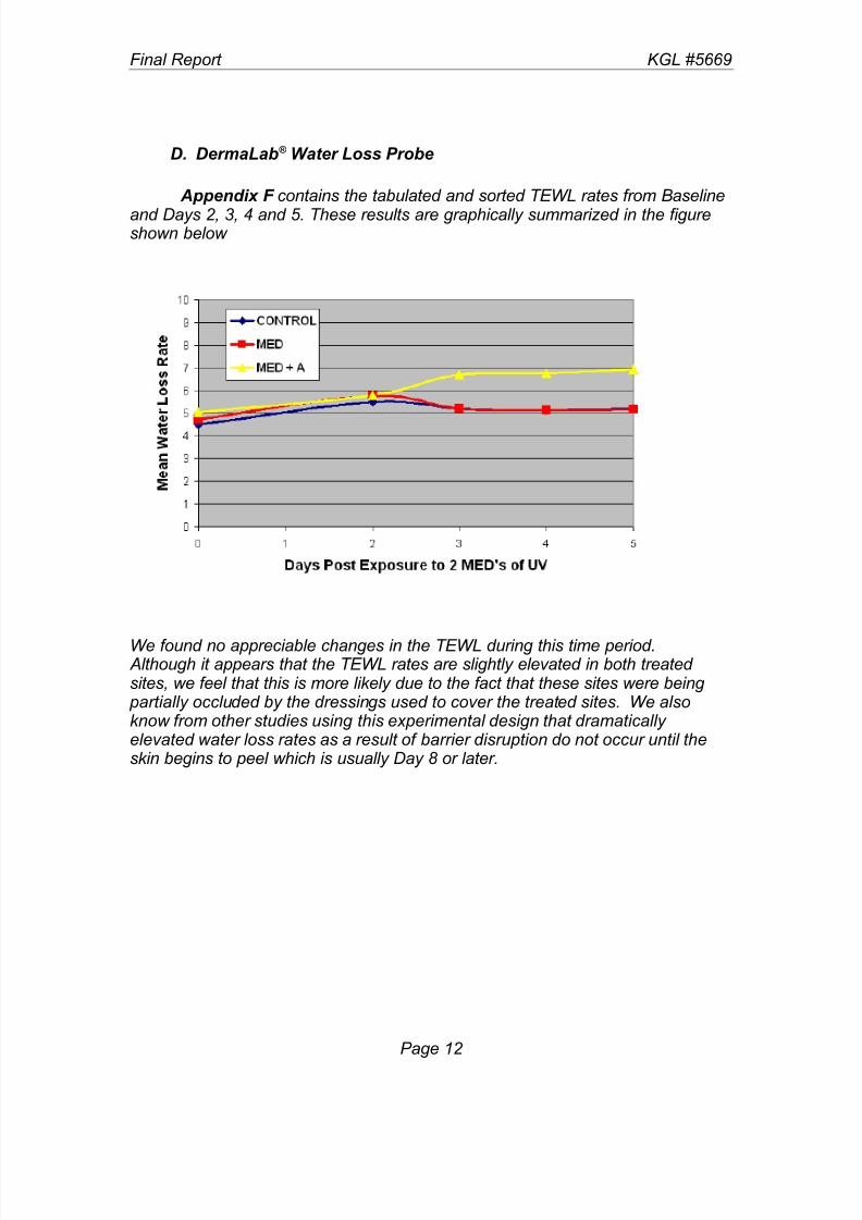
D. DermaLab® Water Loss Probe
Appendix F contains the tabulated and sorted TEWL rates from Baseline
and Days 2, 3, 4 and 5. These results are graphically summarized in the figureshown below
We found no appreciable changes in the TEWL during this time period. Although it appears that the TEWL rates are slightly elevated in both treated sites, we feel that this is more likely due to the fact that these sites were being
partially occluded by the dressings used to cover the treated sites. We alsoknow from other studies using this experimental design that dramatically elevated water loss rates as a result of barrier disruption do not occur until theskin begins to peel which is usually Day 8 or later.
Page 12

8/6/2019 A 5669-FR burn test
http://slidepdf.com/reader/full/a-5669-fr-burn-test 13/13
Final Report KGL #5669
IV. CONCLUSIONS
On the basis of the information collected during the course of this study,
we feel that it is reasonable to conclude that Sample A can measurably reducethe appearance of redness of sunburned skin. That this occurred wassuggested by both the Expert Grader’s ratings of visual erythema and theinstrumental measures provided by the a* readings of the Minolta Chromameter.Given the small panel size, it is impossible to evaluate the significance of thesefindings but these data do suggest that treating with Sample A can be clinically effective.
V. RECORD RETENTION
Please be advised that the records for this study will remain on file at KGL, Inc. (or a remote storage site) for a period of 1 year from the issue date of the final report and then destroyed unless we are notified otherwise by theSponsor using the form accompanying this report.
Page 13