a 12 year-old-boy with proximal muscle weakness and
TRANSCRIPT

A 12 year-old-boy with proximal muscle weakness and
cardiomyopathy
Samra Vazirian, M.D.*, Kara Jones, M.D. Ph.D.#, Steven Moore, M.D. Ph.D.#, Perry B. Shieh, M.D. Ph.D.*
*University of California, Los Angeles#University of Iowa Health Care

Case presentation12-year-old boy with eight years of progressive leg weakness.
Birth:
Normal spontaneous vaginal delivery at 41 weeks
Normal prenatal labs and ultrasounds.
Development:
Cognition is apparently normal. In seventh grade- regular classes.
Language: Bilingual- fluent in Spanish and English.
Normal fine and gross motor development.
Social: apparently normal.

Initial Presentation and clinical courseAt four years of age, he would occasionally fall while playing soccer. He was keeping up with his peers, however, and remained very active.
At four years of age, routine labs revealed elevated transminases. Subsequent testing revealed consistently elevated CKs ~1500.
He underwent a muscle biopsy, which reportedly showed mild type II fiber hypertrophy.

Clinical courseDespite this evaluation, he did not develop any obvious progressive weakness or other muscle symptoms over the next several years
At 10 years of age, the patient did recall experienced muscle pain in his legs while playing soccer at school.
He denies having any noticeable symptoms or complain of weakness.
No fatigue or overt exercise intolerance.
No shortness of breath. No swallowing problems.

Case presentationPast Medical History
Hypertrophic cardiomyopathy, discovered at age 9 on periodic screening echocardiograms that were being performed because the mother had died of heart failure.
Past Surgical History Muscle biopsy at four years of age.
Social History: Lives with his father and two sisters. Currently in seventh grade – regular classes.

Case presentationFamily History:
Born to a non-consanguineous parents of Mexican ancestry.
Mother was diagnosed with a heart disease at 31 years of age. She had dilated
cardiomyopathy with EF of 15% requiring ICD placement. She passed away at
35 years of age. The mother did not have weakness. She had one miscarriage.
Two sisters – age 8 and 6, both healthy.
Maternal aunt died of hypertrophic cardiomyopathy. Aunt has three children
who are apparently healthy

Neurological ExamMental status: Age appropriate
Cranial Nerves: Mild nasal sounding speech, but palate elevation and tongue was normal and the remainder of cranial nerves were normal.
Sensory: Intact to all modalities.
Reflexes: Absent throughout.
Coordination: Intact FNF, HTS, RAM.
Gait: Slow to rise from the floor, but has apparently normal gait.

Motor Exam (left/right)
Shoulder abduction 4/4Elbow flexion 5/5Elbow extension 5/5Wrist flexion 5/5Wrist extension 5/5Finger extension 5/5Finger flexion 5/5Finger abduction 5/5Finger adduction 5/5Thumb abduction 5/5
Hip flexion 4/4Hip Extension 5/5Hip abduction 5/5Hip adduction 5/5Knee extension 5/5Knee flexion 5/5Foot dorsiflexion 5/5Foot plantar flexion 5/5Foot inversion 5/5Foot eversion 5/5
Neck Flexion/Extension 4/5

Neurological Exam

Diagnostic work-upLabs
AST 491; ALT 465
CK: 1776
CK-MB fraction of 0, CK-MM fraction 100.
LDH 1287
Brain Natriuretic Peptide > 1000
Hepatic ultrasound:
Mild fatty hepatomegaly.

Cardiac evaluationEKG: • Sinus bradycardia. HR 48 bpm• LVH with repolarization abnormalities and strain pattern.
Holter monitor:• Sinus rhythm with junctional rhythm at lower heart rates.• Rare isolated monomorphic PVCs. Rare isolated PACs. • No SVT or VT.
Stress test:• No dysrhythmias or evidence of airway reactivity• Reduced BP at peak exertion with prompt normalization during recovery

EchocardiogramSerial studies until 8 years of age was normal.
Echo May 2016 (at 9 years of age)• Hyperdynamic left ventricular systolic function.• Mild left ventricular intra-cavitary obstruction.• Severe asymmetric left ventricular hypertrophy.• Interventricular septum measured 2.9 cm. LV free wall measures 2.2 cm.
Echo – most recent• Hyperdynamic left ventricular systolic shortening.• Mild dynamic left ventricular outflow tract obstruction• Severe hypertrophic cardiomyopathy.

Mild type-II fiber hypotrophy; there were no dystrophic changes, evidence of myofiber degeneration, mitochondrial changes, or evidence of storage disease
MUSCLE BIOPSY- 2011


Thoughts ?

Next work-up
Because of the history of hypertrophic cardiomyopathy and possible muscle disease, we had some concern that he may have Danon disease or some other neuromuscular disease associated with hypertrophic cardiomyopathy…

Genetic testing
Cardiac Sequencing panel showed hemizygous variant in the LAMP2 gene: c.815T>C (p.Leu272Pro)
The muscle biopsy specimen was sent for LAMP2 staining.
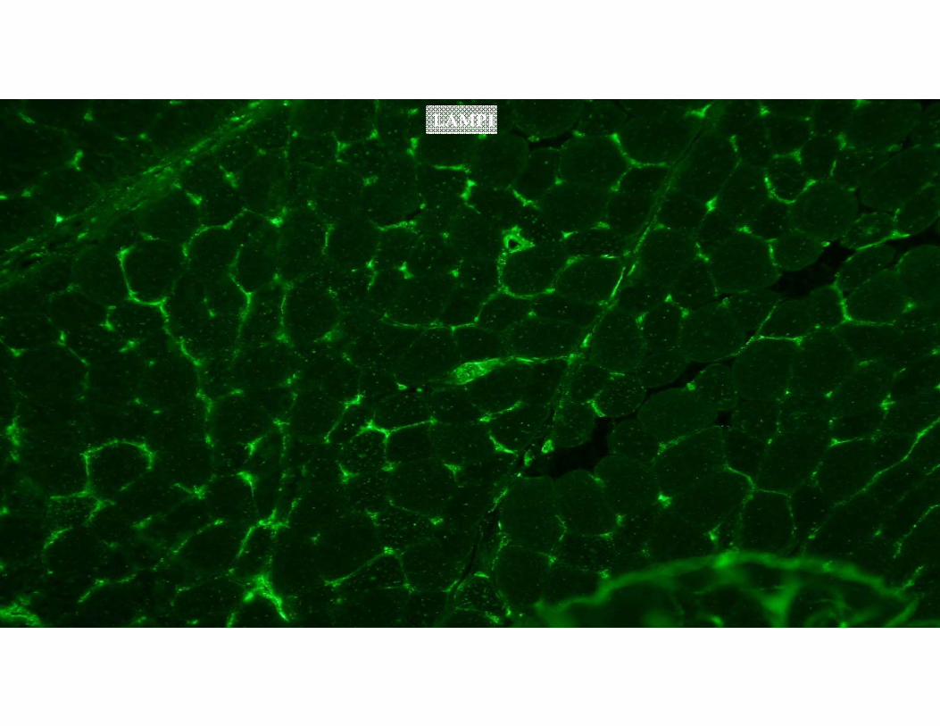
LAMP1

Control LAMP2
Patient’s LAMP2
DAPI(nuclei)

GREEN: LAMP2BLUE: DAPI (nuclei)

Acetylcholinesterase

Acetylcholinesterase

Acetylcholinesterase

Dystrophin
Spectrin

Dystrophin
Spectrin

Danon diseaseDanon disease (DD) is a rare X-linked dominant genetic disorder caused by mutations in the LAMP2 gene (Xq24).
In 1980, Moris Danon first described the disease.
Two frozen muscle specimens, from two patients were reviewed, which were remarkably similar both clinically and pathologically but normal activity of acid malatase.
“Lysosomal glycogen storage disease with normal acid maltase”

History of Danon disease
In 1993, Di Mauro, Servidei, and Tsujino redefined this disorder as “Cardiomyopathy, mental retardation, and autophagic vacuolar myopathy”
In 2000, Nishino and coworkers sequenced a candidate gene on chromosome Xq24, LAMP-2, in ten unrelated patients with Danondisease, including one of the two boys.
They found pathogenic mutations in all 10 patients and documented lack of LAMP-2 (lysosome-associated membrane protein 2) both by Western blot analysis and by immunohistochemistry.

Danon DiseaseA classic triad of cardiomyopathy, skeletal myopathy, and intellectual disability.
Males usually manifest at an earlier age of onset (average 12.1 years old) with more severe symptoms than females (average 27.9 years old)
Males invariably require heart transplantation.
The exact prevalence of DD is unknown and the majority of published summary data on Danon disease comes from two major case series.

The features of 20 affected men and 18 affected women in 13 families with genetically confirmed Danon disease were reviewed.
All patients had cardiomyopathy.
18 of 20 male patients (90%) and 6 of 18 female patients (33%) had skeletal myopathy.
14 of 20 male patients (70%) and one of 18 female patients (6%) had intellectual disability.
Men were affected before age 20 years whereas most affected women developed cardiomyopathy in adulthood.
Muscle histology revealed basophilic vacuoles that contain acid phosphatase-positive material within membranes that lack lysosome-associated membrane protein-2.
Neurology 2002

In 2011- reported 82 patients with Danon disease from 36 families, the largest series to date.
Men are hemizygous:severely affected with cognitive disabilities (100%), hypertrophic cardiomyopathy (88%)muscle weakness (80%).
high morbidity and were unlikely to reach the age of 25 years without a cardiac transplantation.
Genet Med 2011

Clinical Manifestation in Men vs. Women
Women are heterozygous:
Women less severely affected.
Women reported higher than expected levels of cognitive disabilities (47%)
Skeletal muscle complaints in women (50%)
Equal prevalence of dilated cardiomyopathy and hypertrophic cardiomyopathy.

Age specific manifestation in Men vs. Women
First symptom:
12.1 years in men
27.9 years in women.
Cardiac transplantation
17.9 years in men
33.7years in women.
Death
19.0 years in men,
34.6 years in women.

Neurological and ophthalmologic manifestations
Male
Proximal muscle weakness and neck muscle weakness.
Most have intellectual disability or learning disorder.
Decrease or loss of central and visual acuity.
Peripheral pigmentaryretinopathy.
Female
Mild or no proximal muscle weakness.
Typically no intellectual disability.
Peripheral pigmentaryretinopathy.
Lamellar opacities in the lens.

Cardiac manifestationsPalpitations or documented arrhythmias, syncope, chest pain or cardiac arrest.
Electrical conduction abnormalities are common. WPW pattern is the most common ECG abnormality.
Atrial and ventricular arrhythmias are typically present.

LabsSerum CK levels are elevated two-three fold, even if clinical myopathy is mild.
Liver enzymes may be persistently elevated in absence of liver dysfunction.
AST, ALT, LDH, Aldolase tend to be elevated in one half of the patients.

ImagingMRI brain: but is often normal.
Echocardiogram and EKGs – signs of hypertrophic cardiomyopathy
Cardiac MRI can be performed to assess hypertrophy

Genetic DiagnosisX-linked dominant trait.
pathogenic variants in the LAMP2 gene (“lysosome-associated membrane protein-2”).
Patients with VUS can be further evaluated based on LAMP2 staining on muscle biopsy

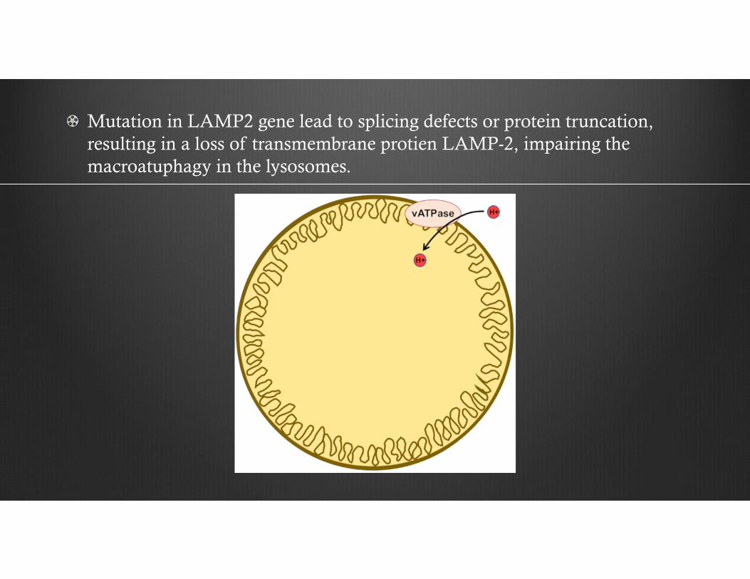
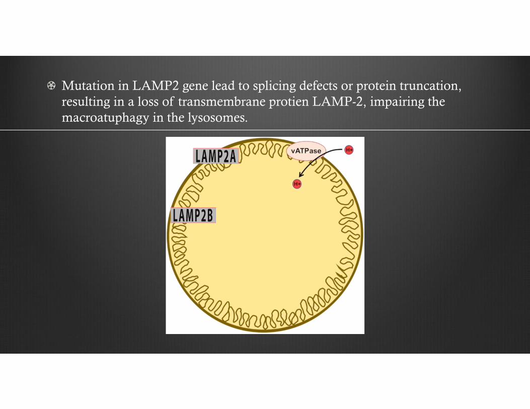
Mutation in LAMP2 gene lead to splicing defects or protein truncation, resulting in a loss of transmembrane protien LAMP-2, impairing the macroatuphagy in the lysosomes.

Mutation in LAMP2 gene lead to splicing defects or protein truncation, resulting in a loss of transmembrane protien LAMP-2, impairing the macroatuphagy in the lysosomes.
Mutation in LAMP2 gene lead to splicing defects or protein truncation, resulting in a loss of transmembrane protien LAMP-2, impairing the macroatuphagy in the lysosomes.

Mutation in LAMP2 gene lead to splicing defects or protein truncation, resulting in a loss of transmembrane protien LAMP-2, impairing the macroatuphagy in the lysosomes.
Mutation in LAMP2 gene lead to splicing defects or protein truncation, resulting in a loss of transmembrane protien LAMP-2, impairing the macroatuphagy in the lysosomes.

Mutation in LAMP2 gene lead to splicing defects or protein truncation, resulting in a loss of transmembrane protien LAMP-2, impairing the macroatuphagy in the lysosomes.


Muscle Biopsy

Acetylcholinesterase

Electron Microscopy

Immunoreaction with antibodies to LIMP-1 (left panel) immunoreaction with antibodies to LAMP-2 (right panel) is completely lacking in the patient’s muscle.

Prognosis
The prognosis for male patients is poor. The severity of cardiomyopathy is the major prognostic factor.
Most males require a heart transplant during the second to third decade of life.
In contrast, females may develop either DCM (28%) or HCM (33%) and cardiac transplantation in females typically occurs much later in life.
SCD, likely due to a ventricular arrhythmia, is the major cause of death.

ManagementEducation
Avoid strenuous exercise.
Limit caffeine in case of tachyarrhythmia.
Genetic counseling.
The timely identification of de-novo LAMP2 mutated family members, many of whom are heterozygous females, remains critical for their treatment and family counseling.

Treatment options
Implantable Cardioverter Defibrillator (ICD)
Heart transplantation.
Medications: Diuretics, beta-blockers, ACE inhibitors

“Nature is nowhere accustomed more openly to display her secret mysteries than in cases where she shows tracings of her workings apart from the beaten paths; nor is
there any better way to advance the proper practice of medicine than to give our minds to the discovery of the usual law of nature, by careful investigation of cases of rarer
forms of disease”.
~ William Harvey, M.D.

References1. Danon MJ, Oh SJ, DiMauro S, Manaligod JR, Eastwood A, Naidu S, Schliselfeld LH
Neurology. 1981 Jan; 31(1):51-7. Lysosomal glycogen storage disease with normal acid maltase.
2. Sugie K, Yamamoto A, Murayama K, et al. Clinicopathological features of genetically confirmed Danon disease. Neurology. 2002;58(12):1773–1778.
3. Boucek D, Jirikowic J, Taylor M. Natural history of Danon disease. Genet Med. 2011;13(6):563–568.
4. Nishino I, Fu J, Tanji K, et al. Primary LAMP-2 deficiency causes X-linked vacuolar cardiomyopathy and myopathy (Danon disease). Nature. 2000;406(6798):906–910.
5. D’souza RS, Levandowski C, Slavov D, et al. Danon disease: clinical features, evaluation, and management. Circ Heart Fail. 2014;7(5):843–849
6. Ryan S. D’souza, Luisa Mestroni, and Mathew R. G. Taylor. Danon disease for the cardiologist: case report and review of the literature.
7. Maron BJ, Roberts WC, Arad M, Haas TS, Spirito P, Wright GB, Almquist AK, Baffa JM, Saul JP, Ho CY, Seidman J, Seidman CE. Clinical outcome and phenotypic expression in LAMP2 cardiomyopathy.JAMA. 2009 Mar 25; 301(12):1253-9.