9.13 the human brain
TRANSCRIPT

9.13 The Human Brain Lecture 2
Outline for Today
I. Motion Demo: What do we need visual motion information for?
II. Basic Neuroanatomy Refresher (prep for dissection Wednesday)
III. Cortex. criteria for a visual area case study: visual motion area MT(V5)
1

(Why) Do We Need to be Able to See Motion?
1. How do we use visual motion information?
2. Might this ability be important enough that our brains would allocatespecial machinery to seeing motion?
3. If you had to write an algorithm to take video input and figure out if an object is moving or in what direction, what would that code look like?
The Marr reading assigned for today points out that:we cannot understand perception without thinking about
what each perceptual inference is necessary for ecologically, the computational challenges involved in making that inference.
More on that next week. 2

-
* = jeff bezos’ net worthSome Bare Basics about the Brain *no you don’t have to
remember this
• human brain contains ~ 100 billion (1011) neurons*~~ thousand of synapses per neuron
• human brain runs on 20 wattsvs: IBM’s Watson: 20,000 watts
• primary focus of this course: the cortexfolded outer surface approx. area of a large pizza
• But there are lots of other important bits
Diagram courtesy of USNCI/SEER via Wikimedia. License: CC BY SA. This content is excluded from our Creative Commons license. See https://ocw.mit.edu/fairuse for more information.
Brain image © source unknown. This content is excluded from our Creative Commons license. See https://ocw.mit.edu/fairuse for more information. 3

3. White Matter
Four Major Components of the Brain 2. Limbic system1. Brain stem & cerebellum
(subcortical regions)
This course will focus mostly on the cortex. 4. Cerebral cortex (outer sheet)But the other parts are
easier to see in a dissection, so we will
briefly review them here. Brain image © sources unknown. This content is excluded from our Creative Commons license. See https://ocw.mit.edu/fairuse for more information.
slide adapted from Michael Cohen 4
Who Cares about White Matter? we will discuss in more detail on May 1, but just to foreshadow….
I. White matter makes up 45% of the human brain.
2. We cannot understand cortex w/out knowing the connectionsbetween regions.3. The specific connections of each region may serve as a“fingerprint” of that region across species, enabling us todiscover interspecies homologies.4. The specific connections of each region may play acausal in its development.5. Disruptions of white matter may be key to clinical disorders6. Structural connections provide a major constraint in circuitdesign and likely too in brain design.
5

9.13 The Human Brain Lecture 2
Outline for Today
I. Motion Demo: What do we need visual motion information for?
II. Basic Neuroanatomy Refresher (prep for dissection Wednesday)
III. Cortex.criteria for a visual area case study: visual motion area MT(V5)
let’s start with the easy parts, which you have already seen... 6

Refresher: What is a Receptive Field?
Place an electrode next to a cell in monkey visual cortex Train the monkey to stare at a fixation spot w/out moving its eyes Stimulate various regions of visual space A cell will respond to stimulation in one part of space more than
any others The region of visual space that drives a particular cell forms its
receptive field (RF)
Different cells have different RFs Some cells’ responses are tuned not only to the location of the
stimulus but also other properties (shape, color, direction of motion)
Nearby cells in the cortex have nearby receptive fields, producing retinotopic maps in visual cortex…..
Figure from Gazzaniga, M., Ivry, R. & Mangun, G. (2002). Cognitive Neuroscience: The Biology of Mind. All rights reserved. This content is excluded from our Creative Sources: Gazzaniga, Ivry & Mangun, 2002 Commons license. See https://ocw.mit.edu/fairuse for more information. Jody Culham 7

Retinotopic Maps
Retinotopy in Macaque V1 Retinotopy in Human V1 Tootell et al., 1982 Polimeni et al (2009) fMRI at 7T deoxyglucose method
• Retinotopy: Adjacent parts of thevisual scene are mapped to adjacent parts of the cortex
• Terminology: V1 = primary visualcortex = striate cortex
Figures above © sources unknown. All rights reserved. This content is excluded from our Creative Commons license. See https://ocw.mit.edu/fairuse for more information. 8

What exactly is a cortical area? Criteria: A region of cortex distinct from its neighbors in
• Function• Connectivity to other areas• Distinctive layer structure/cell types (“cytoarchitecture”)
» (sometimes)Let’s look at a classic example: Visual Motion area MT Meets all the criteria for a visual area. How do we know this?
lots of ways…
9Figures above © sources unknown. All rights reserved. This content is excluded from our Creative Commons license. See https://ocw.mit.edu/fairuse for more information.

MT: Function Single unit recording
– Single neurons in MT are tuned to the direction of motion– Nearby neurons within MT have similar directional selectivity
(sound familiar?)
What about humans? Can we record from single neurons in
Figures above © sources unknown. This content is excluded from our Creative Commons license. See https://ocw.mit.edu/fairuse for more information. humans? 10

see
11
Visual motion area MT
Figures on this page © sources unknown. This content is excluded from our Creative Commons license. For more information,
https://ocw.mit.edu/fairuse.
Does this tell us that MT represents the direction of motion, or just the presence of motion? Do we have neurons tuned to the direction of motion?

Visual motion area MT How might you use the motion aftereffect to test for direction selectivity in M
Figure above © source unknown. This content is excluded from our Creative Commons license. See https://ocw.mit.edu/fairuse for more information.
Cool, BUT:Does this tell us that MT is necessary for motion perception? 12

More MT Function
• Microstimulation– stimulation affects the perception of motion
• Lesions– lesions to MT lead to deficits in perceiving motion
A patient with bilateral lesions to MT can no longer perceive motion (Zihl et al., 1983)
“Akinetopsia”
13Figure on right © BU Brain & Vision Lab; bottom figure © source unknown. All rights reserved. This content is excluded from our Creative Commons license. See https://ocw.mit.edu/fairuse for more information.

What exactly is a cortical area? Example: Visual Motion Area MT
Criteria: A region of cortex distinct from its neighbors in • Function, e.g. selectivity/processing a specific dimension, e.g.
• MT selectively engaged in processing motion– single neurons in monkeys– fMRI in humans– psychophysics (aftereffects)– microstimulation in monkeys– lesions in humans
• Specific Connectivity– To other areas
a distinct “connectivity fingerprint” a signature of that region
• What about physical/cellular diffs?= ”cytoarchitecture”an old idea….
Figure from Felleman DJ, Van Essen DC., J Cerebral Cortex, Vol. 1 No. 1 Jan/Feb (1991) 47. © Oxford Academic Journals. All rights reserved. This content is excluded from our Creative Commons license. See https://ocw.mit.edu/fairuse 14

Brodmann Areas
• Korbinian Brodmann (1868 –1918)
Identified 52 distinct “areas’ based on cytoarchitecture Thought of them as like “organs” Photo of K. Brodmann is in the public
domain. Source: Wikimedia Commons.
“The specific histologicaldifferentiation of the cortical areas proves irrefutably their specificfunctional differentiation--for it rests as we have seen on the division of labor.”
Very clear for primary cortical regions (visual, auditory, ss, motor).Less clear for most others, except...
Colored brain © source unknown. This content is excluded from our Creative Commons license. See https://ocw.mit.edu/fairuse.
15

MT is also distinctive in Cytoarchitecture
• MT is stained with cytochromeoxidase (which indicates highmetabolic activity)
Figures above © sources unknown. All rights reserved. This content is excluded from our Creative Commons license. See https://ocw.mit.edu/fairuse for more information. 16
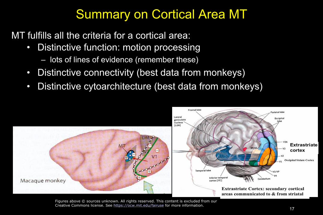
Summary on Cortical Area MT MT fulfills all the criteria for a cortical area:
• Distinctive function: motion processing – lots of lines of evidence (remember these)
• Distinctive connectivity (best data from monkeys) • Distinctive cytoarchitecture (best data from monkeys)
Figures above © sources unknown. All rights reserved. This content is excluded from our Creative Commons license. See https://ocw.mit.edu/fairuse for more information.
17

Concepts you Should be Comfortable withfrom this Lecture
• cerebellum, thalamus, amygdala, hippocampus, grey vs white matter • retina, LGN, primary visual cortex • retinotopy, receptive fields, cortical maps, cytoarchitcture • what is a “map” in cortex? • Criteria for a cortical area • What does MT do and what methods have told us that? • What is akinetopsia?
Questions?Please do not arrive late for next class.
18

MIT OpenCourseWare https://ocw.mit.edu/
9.13 The Human Brain Spring 2019
For information about citing these materials or our Terms of Use, visit: https://ocw.mit.edu/terms.
19