9125d93 i5)3b55>9>7&b?f945b - announcements - … · a-:ei:b7:g a 9125d93 i5 ?eb>1<...
TRANSCRIPT

26 l September 201 6 l DiabeticEyeJournal
Advertorial Spotlight on DESP
Diabetic Eye Screening Provider
Although the name EMIS Care is a relatively new one – we rebranded in Apri l 201 6 – you wil l have
come across us before. Formerly working under the name of Medical Imaging (UK) Ltd, we have
been at the forefront of innovating how patients with diabetes are cared for since we began diabetic
eye screening over 25 years ago.
From our days as a pioneer in the mobile retinal screening model – back when digital photography
was sti l l in its infancy – we have grown to become who we are today, providing services to over
500,000 patients across England and the Republic of Ireland.
Diabetic eye screening is just one of several ophthalmic and health screening services that we offer.
Our DESP programmes and locations
From rural areas to urban centres, we are based all across England and are able to provide screening services to patients in a variety of different ways.
• Arden, Herefordshire & Worcestershire – our longest standing programme where
we started off over 30 years ago. This programme was recently merged and now supports
over 94,000 patients.
• Bradford & Airedale – a Quality in Care award-winning programme and the DESP area
where we gather the most patient feedback, sampling 1 in 1 5 patients we screened last year.
• Central Mersey – a mixed screening model programme, where we work with both local
optometrists and the local NHS trust.
• Kent & Medway – a head-to-head competitor for our largest programme – with another
94,000 patients cared for – this area uses a combination of fixed screening and self-contained
mobile screening vans to reach patients.
“It is a fantastic service. It is important for
me to know if something is wrong it will be
picked up during screening and can be dealt
with. It is peace of mind knowing this
service exists. Once it is done I can forget
about it til l next appointment. Thanks to all
people involved.”
Patient quote from Bradford and Airedale
All of these areas feed into our Administration Centre based at our Worcester office, which acts as
the heart of EMIS Care. Here our team provide administration to several diabetic eye screening
programmes. Our Worcester office is also home to our Quality Management Team, who provide
support and business intel l igence to everyone involved in service delivery.
Our administration team boast some impressive statistics, booking over 31 8,000 appointments
so far in 201 6, whilst taking 330,000 incoming calls since 201 5.
• North of Tyne & Gateshead – winner of a Healthwatch award, this is our most rural-based programme that is run through mobile screening
sessions.
• South West London – we were recently commissioned to run this densely-populated programme. We also head-up a state of the art grading
office in New Malden.
• Birmingham, Solihull & Black Country – where we run administration services for the DESP programme.
As a part of the EMIS Group – a collection of healthcare-focused companies that include EMIS Health, Patient. info and Egton – we are working to help
cl inicians and patients through a variety of different services.

DiabeticEyeJournal l September 201 6 l 27
Spotlight on DESP
Patient care
Patient care is at the core of the service that we provide. Across our DESP areas, we
have implemented several initiatives in order to ensure that patients have access to the
best care and are satisfied with the attention that they are given.
“Appointment right on time, no waiting,
cheerful staff, whole experience painless.”
Patient quote from North of Tyne and
Gateshead
We consistently l isten to patients to understand what they think about their care and to see if there are any ways to improve their experience. In Kent &
Medway this takes the form of a Patient Champion Programme, where patient representatives attend meetings to give us views on service
improvements. Through forums like these, we can gain important face time with patients outside of their screening appointments.
Surveys are also a key part of gaining feedback. For our Programme Managers l ike Denise Young at
North of Tyne & Gateshead, this information is key to creating programme frameworks: “This quarter
we will be working towards analysing the survey results and forming an action plan on outcomes and
quotes, so that we can address any patient issues. You said, we did!”
Such initiatives have allowed us to gain key insight into making patients as happy as possible whilst in
our care.
After surveying over 1 8,000 service users, over 99% thought the service was excellent or
good, and over 99% would recommend the service to a friend or family member with diabetes.
Reaching as many patients as possible
I t is key to us that patients are given an appointment in a timely manner and that they also
attend these appointments. This way we can ensure that patients receive the care that they
deserve, and that the effects of diabetes on a patient’s eyesight is kept to a minimum. One
of the ways in which we are achieving these goals is through CQUINS. Our Bradford and
Airedale programme is leading the way under Programme Manager Suzanne Beshara, who
is targeting those who are most at risk: “We are working on our CQUIN – improving access
and service provision for people with learning disabil ities and mental health issues.”
Analysing and improving
North of Tyne & Gateshead are also currently completing a CQUIN in an attempt to engage patients
and drive uptake of appointments. This project runs alongside their impressive service of offering
1 00% of newly registered patients an appointment within 1 2 weeks.
“Excellent customer care skills
from lovely people.” Patient quote
from South West London
We are also looking into when and why patients may not come to DESP sessions. In Central Mersey, Programme Manager Kimberly Gall ienne notes
that, “We have looked into a group of patients who have not responded to DESP invitations or attended a booked appointment. Our experienced
graders have been contacting these patients to encourage attendance for diabetic eye screening, establ ishing the reasons for non-attendance and
answer any queries the patient may have.”
These projects run alongside mobile screening sessions and out of hours/ weekend appointments – like our work with local optometry stores in North of
Tyne and Gateshead – to ensure that DNA rates are low and that as many patients as possible are reached.
28 l September 201 6 l DiabeticEyeJournal
Spotlight on DESP
Education
One way that we help our patients is to educate them about their condition and what they can do to best care for themselves. As a standard, al l of our
cl inicians and screeners inform patients of ways to prevent diabetic retinopathy and deal with diabetes.
We are also working with Diabetes Essential in our Central Mersey programme in order to ensure
that referrals are created and appointments are kept.
And it’s not just patients that we are helping to inform. In Bradford & Airedale, we have begun
trial l ing our apprenticeship scheme, where we are training administrators to become ful ly qualified.
The future
With the success of our projects across our current DESP areas, we are looking at
how we can help even more patients in the future. This means setting our sites on
our next DESP area: Lancashire. The process is set to begin in October 201 6 and
wil l end in Apri l 201 7, when we complete the service transition for the several areas
that wil l form the programme.
Once accomplished, we wil l be looking after over 78,000 patients in the area. With
initiatives already in place - such as working with independent local optometrists and
implementing community based screening services - we are looking forward to
ensuring that even more patients with diabetes recieve the best care possible.
For more information about EMIS Care, visit www.emiscare.com
You can fol low us on twitter@emiscare or l ike us on facebook.com/emiscare
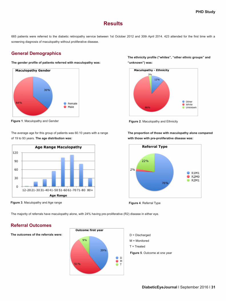
“You said we did” campaign case study
Our “you said we did” campaign gains
important feedback from patients in order
to support service improvements. We gather
data through a variety of different ways, from
recommendations by clinics and patients, to
social media comments and feedback
survey results. Through these different
formats, we have been able to compile
feedback from 6,000 responses, helping us
to create our aims for this year:
Alexander Keep
Communications Manager & Marketing Business
Partner, Contracts & Quality Management Team,
EMIS Care

DiabeticEyeJournal l September 201 6 l 29
• Improving accessibility to make sure everyone can provide feedback
When reviewing data from our in-cl inic ‘Screening Survey, ’ we identified a lack of engagement from certain demographics. After further research, we
discovered this was due to a translation/l iteracy issue.
We Did: We have ensured Google Translate available throughout the EMIS Care website and have purchased a translation module that integrates with
our survey feedback software. We are also working with a translation firm to convert our surveys and feedback forms to suit our patients’ needs.
• Helping patients know more about their data
Patients have informed us that they want to know more about how their information is stored.
We Did: We have made the letters we send to new patients widely available from clinics and have shared the letters with local stakeholders. Patients
can also visit a designated webpage which - since its deployment last month - has helped over 40 users. Patient letters now all include a footer
message informing them of how to find out more information about their data, as well providing them with the new website l ink:
www.emiscare.com/fair-processing-notice.
• Improving peace of mind
In a multiple choice question, we asked patients: “What was the best thing
about your visit?” 38.7% said peace of mind.
We did: To increase this statistic, we have started offering more support to
patients outside of their appointments. On every dedicated DESP webpage,
we offer l ive chat functionality through Facebook. This means that any
patient at any time can speak to a clinical ly knowledgeable member of the
team - 24/7. We have also started a pilot partnership with Diabetes
Essentials - a diabetes education programme in our Central Mersey DESP.
We want to empower patients - and by using this partnership as a template,
we are looking to implement similar schemes with local services across all
our sites in England.
• Helping patients to feel comfortable
We gathered feedback from one clinic where patients had highl ighted that
water facil ities were not available.
We did: In this particular cl inic, we agreed a contract to instal l water coolers.
After this initiative, we decided to implement further operational changes in al l
our programmes, ensuring that al l patients have access to water in their
waiting areas.
We did: We want patients to know we value their feedback. Their comments shape the service,
which is why we launched this campaign.
To help collect their responses, we have also launched a feedback tablet pi lot scheme in our
Bradford & Airedale DESP. This al lows us to gather feedback effectively, quickly and in a format
that can be easily translated. Using just one tablet in a single month, the team gathered 539
responses. From those surveyed, 92.39% of patients were very happy with the service and 7.61%
were happy with the service - that means 1 00% of patients were happy with their diabetic eye
screening appointment. As a final note, 99.63% of those patients would recommend our service to
a friend or family member with diabetes.
• Keep up the good work!
Over 99% of patients thought our service was excellent or good. In our surveys alone, we have received over 1 200 compliments. The word “good” was
used 487 times, “excellent” was used 297 times and “helpful” was used 1 39 times.
Spotlight on DESP
30 l September 201 6 l DiabeticEyeJournal
Characteristics and outcome of referable diabetic maculopathy
Dr Andrew Brown Clinical Lead
Staffordshire Diabetic Eye Screening Programme
Introduction
Diabetes causes 1 3.8% of blindness in the working population 1 , with 11 .9% being due to retinopathy, and is one of the leading causes of severe visual
loss in the same population 2.
Sight threatening retinopathy wil l triple by 2050 when compared to 2000, with the amount of sight threatening disease quadrupling in the over 65s.
This is partly due to longer survival rates due to more effective treatments and partly to increased prevalence and incidence of type 2 diabetes 3 .
A national programme screening for sight threatening diabetic retinopathy began in England in 2007, and was offered to al l patients with diabetes
twelve years of age and over, 4 with digital surveil lance pathways being introduced in 201 2 5 to provide a pathway for monitoring stable patients with a
referable grade in a community setting.
Since the burden of diabetic retinopathy on hospital eye services wil l rise significantly, the effectiveness of screening, referral and monitoring needs
continual re-evaluation to ensure maximal efficiency. The purpose of this retrospective study is to review the characteristics of diabetic maculopathy
referred to secondary care from an English Diabetic Eye Screening Programme in order to inform the use of the digital surveil lance model to ease the
growing burden on secondary care.
Method
Data was collected retrospectively for referrals made to a secondary care provider from October 201 2 to Apri l 201 4. Those that have had previous eye
clinic management were excluded. This period was selected because the national screening definition of maculopathy changed in September 201 2, so
referrals before October 201 2 could potential ly have different characteristics. Since the examination interval in national diabetic eye screening
programme is at present twelve months, analysis of the outcome of patients with at least one year’s fol low–up was chosen.
The outcome of the first year’s fol low-up, the presence of pre-prol iferative disease (R2 grade), whether the maculopathy was bilateral or unilateral and
if uni lateral, the degree of retinopathy in the fel low eye was obtained. The type of maculopathy was determined by re-examining the screening images
obtaining the fol lowing categories:
I f two or more types of maculopathy were present in the same patient, the type closest to the top of this l ist was selected. Where available, patient’s
HbA1C and serum cholesterol was recorded.
Outcomes were defined as:
I t was recorded if any of the patients progressed to prol iferative disease allowing an analysis of the l ikel ihood of such progression during the first year
of referral.
Inferential analysis of this data using one-sample and two sample t-tests for parametric data, Whitney-Mann U tests for non paramateric data, together
with Chi squared and Pearson Correlation analysis al lows an examination of any relationship between medical risk factors, demographic factors and
type of maculopathy, and outcomes.
• Discharged at first visit to screening
• Discharged at first visit to digital surveil lance
• Discharged during the first year to screening
• Discharged in the first year to digital surveil lance
• Monitored in the hospital eye service
• Treatment (laser or intra-vitreal therapy) in the first year
• Maculopathy in association with R2 disease
• Maculopathy due to hard exudate within one disc diameter of the centre of the fovea
• Maculopathy due to the presence of a group of hard exudates in the macula region
• Vision of 6/1 2 or less with haemorrhage or micro-aneurysms within one disc diameter of the centre of the fovea
PHD Study
• Maculopathy in association with R2 disease
• Maculopathy due to hard exudate within one disc diameter of the centre of the fovea
• Maculopathy due to the presence of a group of hard exudates in the macula region
• Vision of 6/1 2 or less with haemorrhage or micro-aneurysms within one disc diameter of the centre of the fovea
DiabeticEyeJournal l September 201 6 l 31
Results
665 patients were referred to the diabetic retinopathy service between 1 st October 201 2 and 30th Apri l 201 4. 423 attended for the first time with a
screening diagnosis of maculopathy without prol iferative disease.
General Demographics
The gender profile of patients referred with maculopathy was:
Figure 1 . Maculopathy and Gender
The ethnicity profile (“whites”, “other ethnic groups” and
“unknown”) was:
Figure 2. Maculopathy and Ethnicity
The average age for this group of patients was 60.1 0 years with a range
of 1 9 to 93 years. The age distribution was:
Figure 3. Maculopathy and Age range
The proportion of those with maculopathy alone compared
with those with pre-proliferative disease was:
Figure 4. Referral Type
The majority of referrals have maculopathy alone, with 24% having pre-prol iferative (R2) disease in either eye.
Referral Outcomes
The outcomes of the referrals were:
Figure 5. Outcome at one year
D = Discharged
M = Monitored
T = Treated
PHD Study

32 l September 201 6 l DiabeticEyeJournal
Of the patients discharged, the manner of discharge were:
Figure 6.
Discharge Modality
D1 = Discharged to screening first visit
DS1 = Discharged to digital surveil lance first visit
D = Discharged to screening after a period of monitoring in hospital cl inic
DS = Discharged to digital surveil lance after a period of monitoring in hospital cl inic
Patients discharged at first visit to screening may be regarded as a false positive
referral. They were thought to have referable maculopathy at screening but this
was not confirmed when further examination was performed and were discharged
with a non-referable grade.
Co-existing pre-proliferative (R2) retinopathy
Of the 422 patients referred with maculopathy who attended their appointment, 1 02 (24.2%) had significant pre-prol iferative disease (R2) in one or both
eyes.
Of these, 1 5 (1 4.7%) were managed in the community compared with 1 51 (47.2%) of the patients without prol iferative disease.
Table 1 . Maculopathy, R2 and management
Patients with significant pre-prol iferative disease in either eye were
significantly more likely to require hospital management than those without
X2(1 )=32.850, p<0.05.
40 (9.5%) patients in this group required interventional treatment. 1 9 of
these had significant pre-prol iferative retinopathy (R2) whilst 21 did not.
Table 2. Maculopathy, R2 and treatment
Patients with significant pre-prol iferative disease in either eye were
significantly more likely to require interventional treatment than those without
X2(1 )=11 .753, p<0.05.
Of the 422 patients referred with maculopathy, 4 (0.9%) progressed to
prol iferative retinopathy. 3 of these patients had co-existing pre-prol iferative
disease at the time of referral.
Table 3. Progression to R3
Patients with co-existent pre-prol iferative disease were not statistical ly more
likely to progress to prol iferative disease X2(1 )=3.237 p=0.072, though a p
value of 0.07 suggests a possible relationship, which could be investigated
using a larger number of patients.
PHD Study
The positive predictive value (PPV) represents the proportion of subjects with a positive test who actual ly have the disease, and is a measure of a test’s
efficiency.
For the 422 patients referred with maculopathy (including pre-prol iferative disease), 61 where discharged at first visit:
PPV=True Positive/(True positive + False Positive)=361 /422=0.86=86%
If the patients with maculopathy alone are considered (a worst grade of R1M1 ) 59 patients out of 320 referred with this grade were discharged at first
visit giving a PPV of:
PPV=True Positive/(True positive + False Positive) =261 /320= 0.82=82%
This demonstrates a moderate PPV for the screening test for both groups of patients.
DiabeticEyeJournal l September 201 6 l 33
Bilateral Maculopathy
Of the 422 patients referred with maculopathy, 31 5 (75%) had maculopathy alone with no other significant retinopathy (i.e pre-prol iferative retinopathy
(R2) in either eye. 74 (23.5%) of these patients had bilateral disease (i.e. R1 M1 grade in both eyes). In this group of patients 1 50 were discharged to
community monitoring at some point, whilst 1 65 were managed in hospital (monitoring and/or treatment).
Considering whether bilateralism had an association
with mode of management:
Table 4. Bi lateral Disease and management
Patients with bilateral disease were significantly more likely to require
hospital based management X2(1 )=22.280, p<0.05.
Considering whether patients with bilateral disease were more
likely to require treatment:
Table 5. Bi lateral Disease and treatment
Patients with bilateral disease were not significantly more likely to
require treatment than those with unilateral disease X2(1 ) =3.611
p=0.057. A p value of 0.057 suggests a possible association that
requires further investigation.
Diabetic Control
The average HbA1C of the patients who attended with maculopathy
was 70.80 mmol/mol.
The distribution of HbA1C was:
The proportion of patients at or below the HbA1C of target
(locally set) value of 53 mmol/mol was:
A one-sample t test shows the average HbA1C level of the patients referred with diabetic maculopathy is significantly different to the national and local
target for HbA1C t(537)=21 .727, p<0.005. The average serum cholesterol in patients with R2 was 4.00 mmol/L compared with 4.1 5 mmol/L in those
without. A Whitney-Mann U test (the data is not normally distributed) shows there was no significant difference in serum cholesterol levels in patients
with referable maculopathy between those with and those without pre-prol iferative disease (R2) U=8804.00, z=0.899, p=0.369. The mean HbA1C was
68.35 mmol/mol in the unilateral group and 69.82 mmol/mol in those with bilateral disease. A two sample test showed there was no significant
difference between the HbA1C levels between patients with unilateral and bilateral disease: t(264)=-0.748, p=0.455.
Serum cholesterol measurements were available in 236 patients in this group and the average value is 4.1 6 mmol/L . Those with bilateral disease had
average cholesterol levels of 4.269 mmol/L, compared with 4.1 25 mmol/L in those with unilateral disease. A Whitney-Mann U test showed there was no
significant difference in serum cholesterol levels between patients with bilateral and unilateral disease U=4882.50, z=-0.072, p=0.943.
Figure 8. Maculopathy and Target HbA1CFigure 7. Maculopathy and HbA1C
PHD Study

PHD Study
Discussion
Patients with maculopathy tend to have poor control with higher HbA1C levels than target. Patients with co-existing pre-prol iferative (R2) disease
tended to have worse control sti l l , although those with maculopathy without R2 tended to have similar control regardless of whether the disease was
unilateral or bi lateral. Sub-optimal cholesterol levels did not seem to be an issue within this cohort of patient, and was not associated with the presence
or absence of pre-prol iferative disease. Some of these patients may be monitored in a community setting in the digital surveil lance module, especial ly
by programme deploying OCT scanning. I t would appear that these patients do require further medical intervention, so enhanced pathways for GP
referral to facil itate this should be considered.
Only a small number (1 0%) of patients referred required interventional treatment in the first year and a large number of patients were discharged
without treatment (39%), with 49% of those discharged being discharged at first visit. When considering the requirement for hospital management the
positive predictive value for maculopathy is between 86% and 82%, depending upon whether maculopathy and R2 or maculopathy alone is considered.
A significant number of patients presently monitored in hospital could potential ly therefore be monitored in a community setting, with greater use if the
digital surveil lance module, possibly with the use of OCT.
Patients with co-existing prol iferative disease in either eye are significantly more likely to require interventional treatment, have poorer control (they
have significantly higher HbA1C) and were almost significantly more likely to progress to prol iferative disease. I f this type of patient is to be monitored in
a community setting then more frequent review intervals might be advisable. Alternatively, the development of R2 disease in a patient being monitored
in the community with maculopathy could prompt consideration of onward referral to secondary care.
23.5% of the patients with maculopathy alone had bilateral maculopathy. Although this type of patient was more likely to be monitored in hospital cl inics
the need for treatment was no more likely when compared with the group as a whole. I t would appear that there is therefore potential for safely
monitoring patients with bilateral maculopathy in the absence of pre-prol iferative disease (R2) in a digital surveil lance setting.
I t is hoped that these findings stimulate further discussion and consideration of the most cost effective way of managing patients with referable diabetic
retinopathy detected at eye screening.
References
1 EVANS J (1 995) Causes of bl indness and partial sight in England and Wales 1 990-1 991 . London OPCS 1 995:1 -29
2 KOCUR I, RESNIKOFF S. (2002) Visual impairment and blindness in Europe and their prevention. Br J Ophthalmol 86 (7): 71 6-22
3 SAADINE JB, HONEYCUTT AA, NARAYAN KM, et al. (2008) Projection of diabetic retinopathy and other major eye diseases among people with
diabetes mell itus: United States 2005-2050. Arch Ophthalmol. 1 26: 1 740-1 747
4 SCANLON P, (2008) The English national screening programme for sight-threatening diabetic retinopathy J Med Screening 1 5: 1 -4
5 DIABETIC EYE SCREENING SURVEILLANCE PATHWAYS (version 1 .3 24 October 201 2) available from http://www.diabeticeye.screening.nhs.uk
[Accessed 28th May 201 6]
SHARING GOOD
PRACTICE
PRESENTING
CASE
STUDIES
IMPROVING
PATIENTS CARE
Are you working in London DESP?
Then this could be for you!
Management of Early Maculopathy
1 0th of November
Location to be confirmed, London
To register your interest email:

DiabeticEyeJournal l September 201 6 l 35
Screeners in Diabetic Eye CareersOver the next several issues, we hope to bring to you a range of interviews of people who have developed a career in the Diabetic Eye
Screening Programme. What challenges have they faced? What do they enjoy about their work? How did they start off and where are they
now? What inspires them to share their story and continue in this field of work?
Q: What made you choose to go into an eye
screening career Rahila?
A: I was a 27 year old KS1 primary school teacher and decided to
move from the education sector into eye medicine which had become
a developing interest of mine in the medical sector. But a lot of the jobs
had entry requirements of high standard experience. Then, I came
across a job advertisement for a medical administrator in a private
ophthalmic company which was giving unexperienced applicants an
opportunity for work with ful l training provisions and so it was a perfect
opportunity at the right time for me. After nine months, I was promoted
to a screener/grader and with continuing support was given ful l training
and began to seek further opportunities.
Q: How different is it to other jobs you have done?
A: Very different. As a KS1 teacher, I also had other responsibi l ities and
roles such as primary curriculum advisor, parental consultant, acting
deputy head, child protection officer and course co-ordinator. But with
screening and grading, I found it fairly repetitive yet each day is a new day.
You see new patients, a new set of fundus image sets, amazing pathology,
histology, lesions in the anterior and posterior segments and learn about
many retinal eye diseases. There is so much to see and learn. As an
independent eye screening cl incian and retinal image grader for almost 8
ful l years, I would see up to 42 patients a day at satel l ite cl inics. Taking
pre-history detai ls, measuring visual acuties, insti l l ing drops, screening
patients, assessing and triaging their retina status, managing an entire
waiting area all by myself, it was all great!
Q: Can you tell us about some of your acheivements so far?
A: I was awarded with the Peter Hansell Scholarship in 201 5. I received a nomination medal by NELFT for patient care in the excellence award
category. I was also nominated for Diabetes UK inspire awards. I have been awarded by the local cl inical commisioning chairman for receiving high
volumes of patient compliments in cl inical eye healthcare.
Q: Does your work allow you to explore other channels of interest?
A: Yes I bel ieve it does. I know from personal experience that I have been able to look for other avenues and been successful due to my work in the
eye screening profession. For example, I was the first guest speaker at the local Diabetes UK event. I am also a foundation trust member for Great
Ormond Street Hospital for Children. I ’ve worked with Alzheimer UK and Dementia UK charity events and have also completed training courses in Slit
Lamp Bio Microscopy to help charities who run eye camps in countries affected by natural disaster and in third world countries. Designing leaflets on
eye care and producing posters for global education exhibitions has also been an interest through work.
DEC Interview
We start this segment presenting an interview with
Rahila Bashir, who is a Senior Grader based at the
Reading Centre at Moorfields Eye Hospital, City Road
in London. She is also a research advisory panel
member (RAP) for the National Institute for Health
Research - Collaboration for Leadership in Applied
Health Research and Care (NIHR CLAHRC) North
Thames.

36 l September 201 6 l DiabeticEyeJournal
Q: You said you were a teacher before, any skills you carried over?
A: I think it would be my writing skil ls. In 2007, I was appointed as the UK Education Ambassador for a reading firm in Calgary, Canada. My primary
education articles were later published in the middle east and as far as Hong Kong. Immediately in my new job, I began to write articles for the company
and pharmaceutical newsletters. Very soon these were recognised by other journals who offered to print them and since 2011 , I ’ve been an official
writer for various eye journals or magazines. I ’m passionate about learning and sharing knowledge with others.
Q: You have mentioned a lot of positive acheivements, any failing moments?
A: Oh yes! And I find these are a learning curve. Everyday is a new day. You pick yourself up and move forward to better things. I ’ve applied for jobs
where I ’ve not met the candidate criteria or I ’ve not got the job because I don’t have a photography degree. I remember when I applied for a particular
scholarship. My application was unsuccessful one year but the fol lowing year I tried again and got it! I t can be nerve racking for junior staff if EQA
scores are low or City and Guild modules are failed. That is why due to my high score levels in both, I joined the grading college and also became a
City and Guilds assessor to help others.
Q: Do you think screening allows opportunities for career progression?
A: I certainly think it does. I have seen many people come into the eye screening profession who are now certified cl inical photographers, operation
managers, fai lsafe officers, working for Glaucoma services or RNIB or even as business managers in large organisations which support diabetic eye
screening programmes. There are plenty of opportunities for everyone and you just have to keep working hard and you’l l achieve your dreams.
Q: Tell us a bit more about what motivates you to get up and come into work every morning?
A: I thoroughly enjoy my work. I am based in a department of exceptional ly kind, friendly and highly qualified team members and it’s a good feeling
coming into work where you feel safe, appreciated, have a laugh and work together because you enjoy what you do and are achieving targets and aims
which help patient’s stay safe from losing their vision. I ts very rewarding and like I said before, each day is a new day and it brings new ideas and
contributions.
Q: You said your work was fairly repetitive so how important is continuity of education in your job role?
A: Very important. Job tasks being repetitive helps you improve yourself and become capable of passing on skil ls to others. I have noticed when
training junior staff how relevant it is to educate yourself al l the time. That is why I joined IMI , RPS, RCOP, RSM, BUOS, OIA & BARS just to name
afew. After completing the City and Guilds Diploma in Retinopathy Screening, I then looked for an appealing prospectus and went to Warwick Medical
School to complete the Msc in Health Science – Retinal Screening core module, Diabetic Retinopathy. I later returned to complete a new Master level
paper at Medical School titled Public Health Screening. Furthermore, I also studied a course paper on Eye anatomy Dissection at the university of
Sussex Medical School. Attending regular training and conferences has always been important to me. You educate yourself, your patients and share
learning with your colleagues and this, I bel ieve, is a valuable ongoing process towards being a better healthcare professional.
Q: Where do you see yourself in 5 years from now?
A: I am a family person but I also enjoy what I do so much that I think I ’ l l sti l l have close links to Diabetic Eye Screening Services. Even today feels
l ike the first day! The same excitement and joy of achieving pinnacles of success. The same interest and commitment in learning about the retina and
disease activites. The same dedication and concentration of meeting targets and deadlines. I think it's an amazing profession and I hope to continue
growing my interest in ophthalmology. Many moments of small , positive interactions build an extroadinary career. Often people think that you have to
fight your way to the top but I ’ve seen positive working relationships in the workplace mount to great moments in the work environment. In the new
year I ’ l l commence my 1 0th year in diabetic eye screening services. The past eight to nine years were a mixture of cl inics and grading and now I solely
grade at different levels which is a huge task, but one which I continue to enjoy very much everyday. When you see patients go through the pathway
and return to routine digital screening, you’ve been a part of that journey in helping save their eyesight. I t’s a rewarding feeling.
If you have a story to share, we would love to hear from you!
Please email us on [email protected] or directly to [email protected] where this interview took place.
DEC Interview

DiabeticEyeJournal l September 201 6 l 37
Case Study
Case Study of type 2 Diabetes Patientby Rahila Bashir Senior Grader at Reading Centre MEH London
In November 2008, a 46 year old gentleman attended for a routine digital screening (RDS) (figure 1 ). His visual acuity was 6/6 for the right eye and only
perception of l ight (PL) in the left eye. His left eye sudden vision loss was caused by an occular occlusion in August 2008. He had been seen once and
was awaiting a second appointment at the hospital eye service (HES), but at some point was flagged as DNA and so was discharged from hospital in
Apri l 2011 .
Figure 1 a. Right eye macula view Figure 1 b. Right eye nasal view
Figure 1 c. Left eye macula view Figure 1 d . Left eye nasal view
The patient returned for a routine digital screening in November 2011 (figure 2). His visual acuity was 6/5 in the right eye and 6/24 in the left eye. His in
care of ophthalmology (ICO) notes stated that he had been treated at his local hospital with a course of six injections at intervals to the left eye and had
been reinstated after his initial DNA, so was re-attending for reviews once every 6 months. In January 201 3, he was final ly discharged from the HES
back to RDS as he had completed the course of treatment and all the required fol low-ups to monitor and review his eyes.

38 l September 201 6 l DiabeticEyeJournal
Case Study
Figure 2a.
Right eye macula view
Figure 2b.
Right eye nasal view
Figure 2c.
Left eye macula view
Figure 2d .
Left eye nasal view

