8 the citric acid cycle - minificciones · in a series of reactions variously called the citric...
TRANSCRIPT

Learning Objectives✓ Why is the reaction catalyzed by the pyruvate dehydrogenase complex a
crucial juncture in metabolism?
✓ How is the pyruvate dehydrogenase complex regulated?
✓ What is the advantage of oxidizing acetyl CoA in the citric acid cycle?
✓ How is the citric acid cycle regulated?
You learned in Chapter 15 that glucose can be metabolized in glycolysis topyruvate, yielding some ATP. However, the process of glycolysis is inefficient,
capturing only a fraction of the energy inherent in a glucose molecule as ATP.More of the energy can be accessed if the pyruvate is completely oxidized to car-bon dioxide and water. The combustion of fuels to carbon dioxide and water togenerate ATP is called cellular respiration and is the source of more than 90% ofthe ATP required by human beings. Cellular respiration, unlike glycolysis, is an aer-obic process, requiring molecular oxygen—O2. In eukaryotes, cellular respirationtakes place inside the double-membrane bounded mitochondria, whereas glycol-ysis is cytoplasmic.
Cellular respiration can be divided into two parts. First, carbon fuels are com-pletely oxidized with a concomitant generation of high-transfer-potential electrons
SECTION
8The Citric Acid Cycle

in a series of reactions variously called the citric acid cycle (CAC), the tricarboxylicacid (TCA) cycle, or the Krebs cycle. In the second part of cellular respiration,referred to as oxidative phosphorylation, the high-transfer-potential electrons aretransferred to oxygen to form water in a series of oxidation–reduction reactions.This transfer is highly exergonic, and the released energy is used to synthesize ATP.We will focus on the citric acid cycle in this section, leaving oxidative phosphory-lation until Section 9.
The citric acid cycle is the central metabolic hub of the cell. It is the gatewayto the aerobic metabolism of all fuel molecules. The cycle is also an importantsource of precursors for the building blocks of many other molecules such asamino acids, nucleotide bases, and porphyrin (the organic component of heme).The citric acid cycle component oxaloacetate also is an important precursor toglucose (p. 254).
We begin this section by examining one of the most important reactions inliving systems: the conversion of glucose-derived pyruvate into acetyl CoA, an acti-vated acetyl unit and the actual substrate for the citric acid cycle. This reactionlinks glycolysis and cellular respiration, thus allowing for the complete combus-tion of glucose, a fundamental fuel in all living systems. We will then study thecitric acid cycle itself, the final common pathway for the oxidation of all fuel mol-ecules, carbohydrates, fats, and amino acids.
Chapter 18: HarvestingElectrons from the Cycle
Chapter 17: Preparation for the Cycle

268
CHAPTER
17 Preparation for the Cycle
17.1 Pyruvate Dehydrogenase FormsAcetyl Coenzyme A fromPyruvate
17.2 The Pyruvate DehydrogenaseComplex Is Regulated by TwoMechanisms
17.3 The Disruption of PyruvateMetabolism Is the Cause ofBeriberi
A s you learned in Chapter 15, the pyruvate produced by glycolysis can havemany fates. In the absence of oxygen (anaerobic conditions), the pyruvate is
converted into lactic acid or ethanol, depending on the organism. In the presenceof oxygen (aerobic conditions), it is converted into a molecule, called acetyl coen-zyme A (acetyl CoA; Figure 17.1), that is able to enter the citric acid cycle. Thepath that pyruvate takes depends on the energy needs of the cell and the oxygenavailability. In most tissues, pyruvate is processed aerobically because oxygen isreadily available. For instance, in resting human muscle, most pyruvate isprocessed aerobically by first being converted into acetyl CoA. In very activemuscle, however, much of the pyruvate is processed to lactate because the oxy-gen supply cannot meet the oxygen demand.
The majestic Brooklyn Bridge links Brooklyn with Manhattan in New York City. Pyruvatedehydrogenase links glycolysis with cellular respiration by converting pyruvate into acetylCoA. Unlike the Brooklyn Bridge, however, molecular traffic flows in only one direction. [RonChapple Stock/Alamy.]

26917.1 Pyruvate Dehydrogenase
GTPorATP
High-transfer-potential electrons
2 CO2
H3CAcetyl unit
O
C
Six-carbonmolecule
Four-carbonacceptor
Figure 17.2 An overview of the citric acidcycle. The citric acid cycle oxidizes two-carbon units, producing two molecules ofCO2, one molecule of GTP or ATP, andhigh-transfer-potential electrons.
Figure 17.3 Mitochondrion. The double membrane of the mitochondrionis evident in this electron micrograph. The oxidative decarboxylation ofpyruvate and the sequence of reactions in the citric acid cycle take placewithin the matrix. [(Left) Omikron/Photo Researchers.]
Matrix
Innermitochondrialmembrane
Outermitochondrialmembrane
A schematic portrayal of the citric acid cycle is shown in Figure 17.2. Thecitric acid cycle accepts two-carbon acetyl units in the form of acetyl CoA. Thesetwo-carbon acetyl units are introduced into the cycle by binding to a four-carbonacceptor molecule. The two-carbon units are oxidized to CO2, and the resultinghigh-transfer-potential electrons are captured. The acceptor molecule is regener-ated, capable of processing another two-carbon unit. The cyclic nature of thesereactions enhances their efficiency.
In this chapter, we examine the enzyme complex that catalyzes the formationof acetyl CoA from pyruvate, how this enzyme is regulated, and some pathologiesthat result if the function of the enzyme complex is impaired.
17.1 Pyruvate Dehydrogenase Forms Acetyl Coenzyme Afrom Pyruvate
Glycolysis takes place in the cytoplasm of the cell, but the citric acid cycle takesplace in the mitochondria (Figure 17.3). Pyruvate must therefore be trans-ported into the mitochondria to be aerobically metabolized. This transport isfacilitated by a special transporter (p. 324). In the mitochondrial matrix, pyru-vate is oxidatively decarboxylated by the pyruvate dehydrogenase complex toform acetyl CoA.
Pyruvate + CoA + NAD+ ¡ acetyl CoA + CO2 + NADH + H+
HN
C O
CH3
CH3
H
HO
HN
O
S
O
CH3
O PO
PO
O O OO
O
O OHP
O
OO
N
N
N
N
NH2
– –
2–
Acetyl coenzyme A (Acetyl CoA)
CFigure 17.1 Coenzyme A. Coenzyme A isthe activated carrier of acyl groups. AcetylCoA, the fuel for the citric acid cycle, isformed by the pyruvate dehydrogenasecomplex.
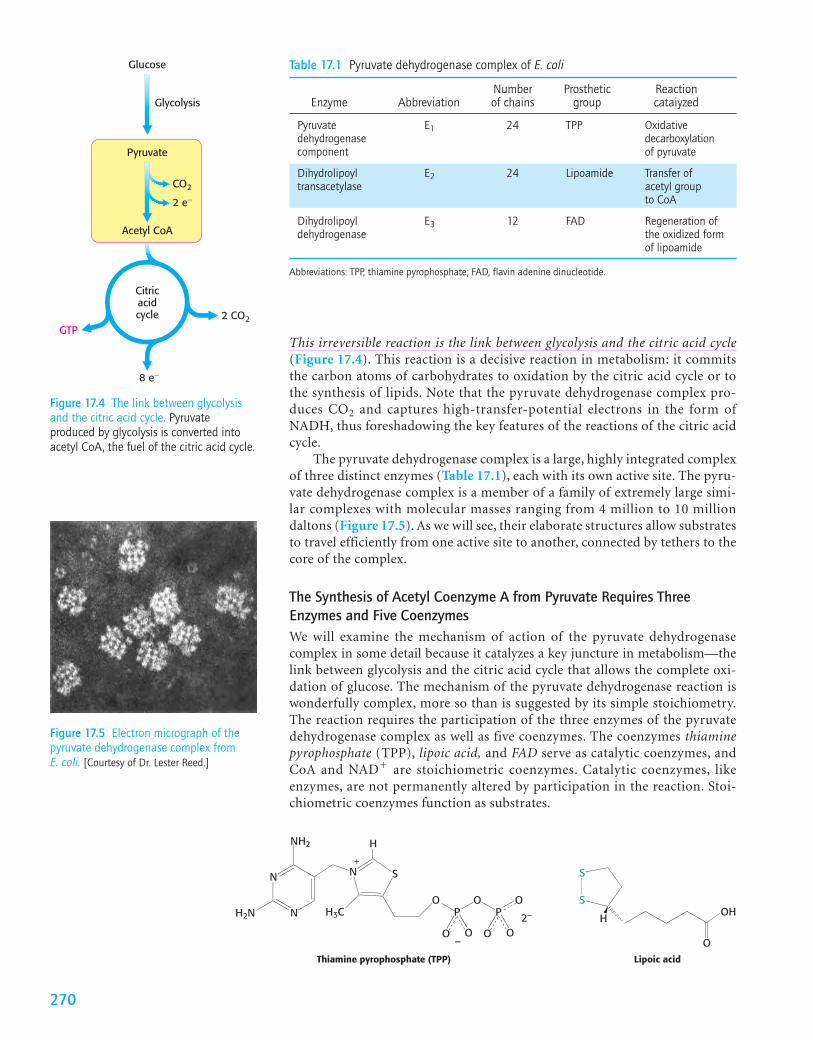
This irreversible reaction is the link between glycolysis and the citric acid cycle(Figure 17.4). This reaction is a decisive reaction in metabolism: it commitsthe carbon atoms of carbohydrates to oxidation by the citric acid cycle or tothe synthesis of lipids. Note that the pyruvate dehydrogenase complex pro-duces CO2 and captures high-transfer-potential electrons in the form ofNADH, thus foreshadowing the key features of the reactions of the citric acidcycle.
The pyruvate dehydrogenase complex is a large, highly integrated complexof three distinct enzymes (Table 17.1), each with its own active site. The pyru-vate dehydrogenase complex is a member of a family of extremely large simi-lar complexes with molecular masses ranging from 4 million to 10 milliondaltons (Figure 17.5). As we will see, their elaborate structures allow substratesto travel efficiently from one active site to another, connected by tethers to thecore of the complex.
The Synthesis of Acetyl Coenzyme A from Pyruvate Requires ThreeEnzymes and Five CoenzymesWe will examine the mechanism of action of the pyruvate dehydrogenase complex in some detail because it catalyzes a key juncture in metabolism—thelink between glycolysis and the citric acid cycle that allows the complete oxi-dation of glucose. The mechanism of the pyruvate dehydrogenase reaction iswonderfully complex, more so than is suggested by its simple stoichiometry.The reaction requires the participation of the three enzymes of the pyruvatedehydrogenase complex as well as five coenzymes. The coenzymes thiaminepyrophosphate (TPP), lipoic acid, and FAD serve as catalytic coenzymes, andCoA and NAD� are stoichiometric coenzymes. Catalytic coenzymes, likeenzymes, are not permanently altered by participation in the reaction. Stoi-chiometric coenzymes function as substrates.
Acetyl CoA
Pyruvate
Glucose
Glycolysis
GTP2 CO2
CO2
8 e−
2 e−
Citricacidcycle
Figure 17.4 The link between glycolysisand the citric acid cycle. Pyruvateproduced by glycolysis is converted intoacetyl CoA, the fuel of the citric acid cycle.
Figure 17.5 Electron micrograph of thepyruvate dehydrogenase complex fromE. coli. [Courtesy of Dr. Lester Reed.]
Table 17.1 Pyruvate dehydrogenase complex of E. coli
Number Prosthetic Reaction Enzyme Abbreviation of chains group cataiyzed
Pyruvate E1 24 TPP Oxidativedehydrogenase decarboxylationcomponent of pyruvate
Dihydrolipoyl E2 24 Lipoamide Transfer of transacetylase acetyl group
to CoA
Dihydrolipoyl E3 12 FAD Regeneration ofdehydrogenase the oxidized form
of lipoamide
Abbreviations: TPP, thiamine pyrophosphate; FAD, flavin adenine dinucleotide.
S
S
O
OHH
Lipoic acid
N SN
N
NH2
H2N
H
H3CO
PO
PO
O OO O
Thiamine pyrophosphate (TPP)
+
–
2–
270
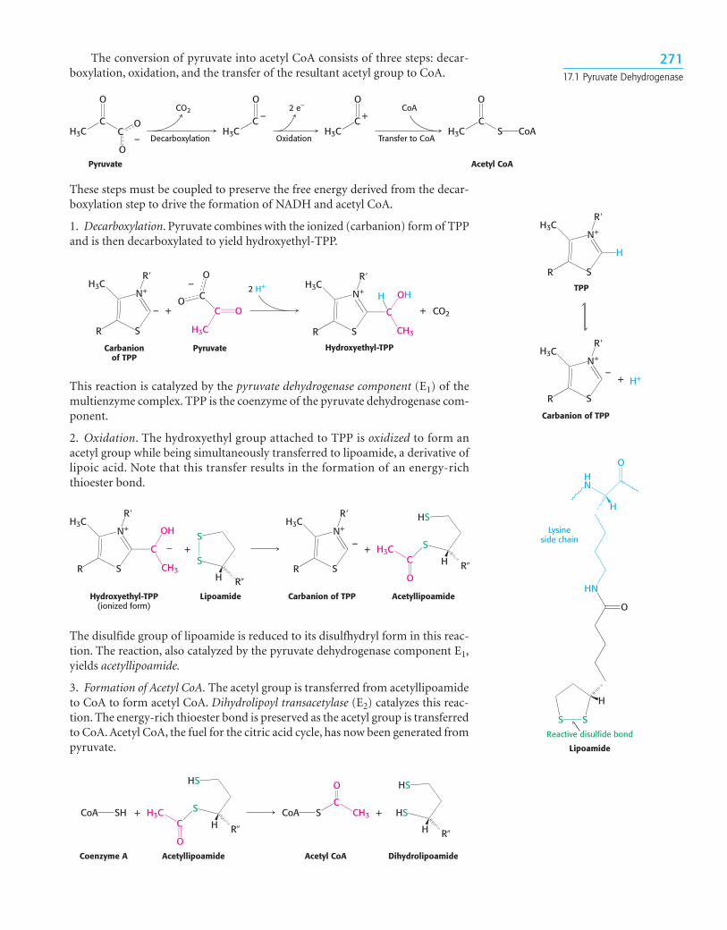
These steps must be coupled to preserve the free energy derived from the decar-boxylation step to drive the formation of NADH and acetyl CoA.
1. Decarboxylation. Pyruvate combines with the ionized (carbanion) form of TPPand is then decarboxylated to yield hydroxyethyl-TPP.
H3C
O
OPyruvate Acetyl CoA
OH3C
O
H3C
O
S CoA–
CO2
Decarboxylation
–
H3C
O
+CoA
Transfer to CoA
2 e–
Oxidation
CC
C C C
C
C
H3C
OO
O
N+
S
R�H3C
R
+ +
–
–
PyruvateCarbanionof TPP
N+
S
R�H3C
R
C
OH
CH3
CO2
H
Hydroxyethyl-TPP
2 H+
N+
S
R�H3C
R
–
Carbanion of TPP
TPP
N+
S
R�H3C
+
R
H
H+
N+
S
R�H3C
R
C
OH
CH3
S
S
R�H
N+
S
R�H3C
R
HS
S
R�H
O
H3C+ +
Hydroxyethyl-TPP(ionized form)
Lipoamide
––
Carbanion of TPP Acetyllipoamide
C
S S
O
H
HN
HN
O
H
Lipoamide
Lysineside chain
Reactive disulfide bond
HS
S
R�H
O
H3CSHCoA
HS
HS
R�H
SCoA CH3
O
AcetyllipoamideCoenzyme A Acetyl CoA Dihydrolipoamide
+ +C
C
27117.1 Pyruvate Dehydrogenase
The conversion of pyruvate into acetyl CoA consists of three steps: decar-boxylation, oxidation, and the transfer of the resultant acetyl group to CoA.
This reaction is catalyzed by the pyruvate dehydrogenase component (E1) of themultienzyme complex. TPP is the coenzyme of the pyruvate dehydrogenase com-ponent.
2. Oxidation. The hydroxyethyl group attached to TPP is oxidized to form anacetyl group while being simultaneously transferred to lipoamide, a derivative oflipoic acid. Note that this transfer results in the formation of an energy-richthioester bond.
The disulfide group of lipoamide is reduced to its disulfhydryl form in this reac-tion. The reaction, also catalyzed by the pyruvate dehydrogenase component E1,yields acetyllipoamide.
3. Formation of Acetyl CoA. The acetyl group is transferred from acetyllipoamideto CoA to form acetyl CoA. Dihydrolipoyl transacetylase (E2) catalyzes this reac-tion. The energy-rich thioester bond is preserved as the acetyl group is transferredto CoA. Acetyl CoA, the fuel for the citric acid cycle, has now been generated frompyruvate.

This electron transfer from FAD to NAD� is unusual, because the commonrole for FAD is to receive electrons from NADH. The electron-transfer poten-tial of FAD is increased by its association with the enzyme, enabling it to trans-fer electrons to NAD�. Proteins tightly associated with FAD are calledflavoproteins.
Flexible Linkages Allow Lipoamide to Move Between Different Active SitesThe structures of all of the component enzymes of the pyruvate dehydrogenasecomplex are known, albeit from different complexes and species. Thus, it is nowpossible to construct an atomic model of the complex to understand its activity(Figure 17.6).
The core of the complex is formed by the transacetylase component E2.Transacetylase consists of eight catalytic trimers assembled to form a hollowcube. Each of the three subunits forming a trimer has three major domains(Figure 17.7). At the amino terminus is a small domain that contains a flexiblelipoamide cofactor. The lipoamide domain is followed by a small domain that
27217 Preparation for the Cycle
E1(�2�2)
E3(��)
E2(�3)
Figure 17.6 A schematic representation ofthe pyruvate dehydrogenase complex. Thetransacetylase core (E2) is shown in red,the pyruvate dehydrogenase component(E1) in yellow, and the dihydrolipoyldehydrogenase (E3) in green. The numberand type of subunits of each enzyme isgiven parenthetically.
Atrimer
Lipoamide
Lipoamidedomain
Domaininteracting withE3 component
Transacetylasedomain
Figure 17.7 Structure of the transacetylase(E2) core. Each red ball represents a trimerof three E2 subunits. Notice that eachsubunit consists of three domains: alipoamide-binding domain, a small domainfor interaction with E3, and a largetransacetylase catalytic domain. Thetransacetylase domain has three subunits,with one subunit depicted in red and theother two in white in the ribbonrepresentation.
The pyruvate dehydrogenase complex must “reset” lipoamide so that thecomplex can catalyze another set of reactions. The complex cannot completeanother catalytic cycle until the dihydrolipoamide is oxidized to lipoamide. In afourth step, the oxidized form of lipoamide is regenerated by dihydrolipoyl dehydro-genase (E3). Two electrons are transferred to an FAD prosthetic group of theenzyme and then to NAD�.
HS
HS
R�H
Dihydrolipoamide
S
S
R�H
Lipoamide
+ FAD+
NAD+
FAD FADH2 + NADH + H+

interacts with E3 within the complex. A larger transacetylase domain completesan E2 trimer. The eight E2 trimers are surrounded by twenty-four copies of E1(an �2�2 tetramer ) and 12 copies of E3 (an �� dimer) surround the E2 core.How do the three distinct active sites work in concert? The key is the long, flex-ible lipoamide arm of the E2 subunit, which carries substrate from active siteto active site (Figure 17.8).
1. Pyruvate is decarboxylated at the active site of E1, forming the hydroxyethyl-TPP intermediate, and CO2 leaves as the first product. This active site lies deepwithin the E1 complex, connected to the enzyme surface by a 20-Å-longhydrophobic channel.
2. E2 inserts the lipoamide arm of the lipoamide domain into the deep channelin E1 leading to the active site.
3. E1 catalyzes the transfer of the acetyl group to the lipoamide. The acetylatedarm then leaves E1 and enters the E2 cube to visit the active site of E2, locateddeep in the cube at the subunit interface.
4. The acetyl moiety is then transferred to CoA, and the second product, acetylCoA, leaves the cube. The reduced lipoamide arm then swings to the active siteof the E3 flavoprotein.
CoAAcetyl CoA
Pyruvate CO2NAD+ NADH + H+
TPP FADH2
E1 E3
E2
TPP FAD
TPP
FAD
C
OH
CH3
H
TPP C
OH
CH3
O
CH3
H
S S
S SS SS S
SH HS
S
SH
5
6 1
2
34
FADTPP TPP FAD FAD
Figure 17.8 Reactions of the pyruvate dehydrogenase complex. At the top (center), theenzyme (represented by a yellow, a green, and two red spheres) is unmodified and ready fora catalytic cycle. (1) Pyruvate is decarboxylated to form hydroxyethyl-TPP. (2) The lipoamidearm of E2 moves into the active site of E1. (3) E1 catalyzes the transfer of the two-carbongroup to the lipoamide group to form the acetyl–lipoamide complex. (4) E2 catalyzes thetransfer of the acetyl moiety to CoA to form the product acetyl CoA. The dihydrolipoamidearm then swings to the active site of E3. E3 catalyzes (5) the oxidation of thedihydrolipoamide acid and (6) the transfer of the protons and electrons to NAD� tocomplete the reaction cycle.
273

5. At the E3 active site, the lipoamide is oxidized by coenzyme FAD. The reacti-vated lipoamide is ready to begin another reaction cycle.
6. The final product, NADH, is produced with the reoxidation of FADH2 to FAD.
The structural integration of three kinds of enzymes and the long flexiblelipoamide arm make the coordinated catalysis of a complex reaction possible.The proximity of one enzyme to another increases the overall reaction rate andminimizes side reactions. All the intermediates in the oxidative decarboxyla-tion of pyruvate remain bound to the complex throughout the reactionsequence and are readily transferred as the flexible arm of E2 calls on eachactive site in turn.
17.2 The Pyruvate Dehydrogenase Complex Is Regulated by Two Mechanisms
The pyruvate dehydrogenase complex is stringently regulated by multipleallosteric interactions and covalent modifications. As stated earlier, glucose can beformed from pyruvate through the gluconeogenic pathway (p. 251). However, theformation of acetyl CoA from pyruvate is an irreversible step in animals and thusthey are unable to convert acetyl CoA back into glucose. The oxidative decarboxyla-tion of pyruvate to acetyl CoA commits the carbon atoms of glucose to either oftwo principal fates: (1) oxidation to CO2 by the citric acid cycle with the concomi-tant generation of energy or (2) incorporation into lipid, inasmuch as acetyl CoAis a key precursor for lipid synthesis (Chapter 28 and Figure 17.9). High concen-trations of reaction products inhibit the reaction: acetyl CoA inhibits thetransacetylase component (E2) by directly binding to it, whereas NADH inhibitsthe dihydrolipoyl dehydrogenase (E3). High concentrations of NADH and acetylCoA inform the enzyme that the energy needs of the cell have been met or thatenough acetyl CoA and NADH have been produced from fatty acid degradation(p. 407). In either case, there is no need to metabolize pyruvate to acetyl CoA. Thisinhibition has the effect of sparing glucose, because most pyruvate is derived fromglucose by glycolysis.
The key means of regulation of the complex in eukaryotes is covalentmodification—in this case, phosphorylation (Figure 17.10). Phosphorylation ofthe pyruvate dehydrogenase component (E1) by a specific kinase switches off theactivity of the complex. Deactivation is reversed by the action of a specific phos-phatase. Both the kinase and the phosphatase are physically associated with thetransacetylase component (E2), again highlighting the structural and mechanis-tic importance of this core. Moreover, both the kinase and the phosphatase arethemselves regulated.
To see how this regulation works under biological conditions, consider mus-cle that is becoming active after a period of rest (Figure 17.11). At rest, the mus-cle will not have significant energy demands. Consequently, the NADH/NAD�,
27417 Preparation for the Cycle
Glucose
Pyruvate
Pyruvatedehydrogenasecomplex
Acetyl CoA
LipidsCO2
Figure 17.9 From glucose to acetyl CoA.The synthesis of acetyl CoA by thepyruvate dehydrogenase complex is a keyirreversible step in the metabolism ofglucose.
ActivePDH
InactivePDH
ATP ADP
Kinase
Phosphatase
Pi H2O
PFigure 17.10 The regulation of thepyruvate dehydrogenase complex. Aspecific kinase phosphorylates andinactivates pyruvate dehydrogenase (PDH),and a phosphatase activates thedehydrogenase by removing thephosphoryl group. The kinase and thephosphatase also are highly regulatedenzymes.

acetyl CoA/CoA, and ATP/ADP ratios will be high. These high ratios stimulate thekinase, promoting phosphorylation and, hence, deactivation of the pyruvatedehydrogenase complex. In other words, high concentrations of immediate(acetyl CoA and NADH) and ultimate (ATP) products of the pyruvate dehydro-genase complex inhibit its activity. Thus, pyruvate dehydrogenase is switched offwhen the energy charge is high.
As exercise begins, the concentrations of ADP and pyruvate will increase asmuscle contraction consumes ATP and glucose is converted into pyruvate to meetthe energy demands. Both ADP and pyruvate activate the dehydrogenase by inhibit-ing the kinase. Moreover, the phosphatase is stimulated by Ca2�, a signal that alsoinitiates muscle contraction. A rise in the cytoplasmic Ca2� level to stimulate mus-cle contraction elevates the mitochondrial Ca2� level. The rise in mitochondrialCa2� activates the phosphatase, enhancing pyruvate dehydrogenase activity.
In some tissues, the phosphatase is regulated by hormones. In liver,epinephrine binds to the �-adrenergic receptor to initiate the phosphatidylinos-itol pathway (p. 180), causing an increase in Ca2� concentration that activatesthe phosphatase. In tissues capable of fatty acid synthesis (such as the liver andadipose tissue), insulin (the hormone that signifies the fed state) stimulates thephosphatase, increasing the conversion of pyruvate into acetyl CoA. In these tis-sues, the pyruvate dehydrogenase complex is activated to funnel glucose topyruvate and then to acetyl CoA and ultimately to fatty acids.
Clinical Insight
Defective Regulation of Pyruvate Dehydrogenase Results in aPathological ConditionIn people with a phosphatase deficiency, pyruvate dehydrogenase is always phos-phorylated and thus inactive. Consequently, glucose always has to take the anaero-bic path and is processed to lactate rather than acetyl CoA. This condition results inunremitting lactic acidosis—high blood levels of lactic acid. In such an acidic envi-ronment, many tissues malfunction, most notably the central nervous system. ■
17.3 The Disruption of Pyruvate Metabolism Is the Cause of Beriberi
The importance of the coordinated activity of the pyruvate dehydrogenasecomplex is illustrated by disorders that result from the absence of a key coen-zyme. Recall that thiamine pyrophosphate is a coenzyme for the pyruvate dehy-drogenase activity of the pyruvate dehydrogenase complex. Beriberi, aneurological and cardiovascular disorder, is caused by a dietary deficiency of
Pyruvate
Acetyl CoA
(A) HIGH ENERGY CHARGE (B) LOW ENERGY CHARGE
+
+NAD+
ADP
e−CAC
PDH
NADH
Pyruvate
Acetyl CoA
−−−
NAD+
ADP
e−
ATP ATPCAC
PDH
NADH
Figure 17.11 Response of the pyruvatedehydrogenase complex to the energycharge. The pyruvate dehydrogenasecomplex is regulated to respond to theenergy charge of the cell. (A) The complexis inhibited by its immediate products,NADH and acetyl CoA, as well as by theultimate product of cellular respiration,ATP. (B) The complex is activated bypyruvate and ADP, which inhibit the kinasethat phosphorylates PDH.
QUICK QUIZ List some of theadvantages of organizing the
enzymes that catalyze the formation ofacetyl CoA from pyruvate into a singlelarge complex.
27517.3 Disruption of Pyruvate Metabolism

thiamine (also called vitamin B1). Thiamine deficiency results in insufficientpyruvate dehydrogenase activity because thiamine pyrophosphate cannot beformed. The disease has been and continues to be a serious health problem inthe Far East because rice, the major food, has a rather low content of thiamine.This deficiency is partly ameliorated if the whole rice grain is soaked in waterbefore milling; some of the thiamine in the husk then leaches into the rice ker-nel (Figure 17.12). The problem is exacerbated if the rice is polished, becauseonly the outer layer contains significant amounts of thiamine. Beriberi is alsooccasionally seen in alcoholics who are severely malnourished and thus thi-amine deficient. The disease is characterized by neurological and cardiac symp-toms. Damage to the peripheral nervous system is expressed as pain in thelimbs, weakness of the musculature, and distorted skin sensation. The heartmay be enlarged and the cardiac output inadequate.
Thiamine pyrophosphate is not just crucial to the conversion of pyruvateto acetyl CoA. In fact, this coenzyme is the prosthetic group of three importantenzymes: pyruvate dehydrogenase, a-ketoglutarate dehydrogenase (a citric acidcycle enzyme, p. 283), and transketolase. Transketolase functions in the pentosephosphate pathway, which will be considered in Chapter 25. The common fea-ture of enzymatic reactions utilizing TPP is the transfer of an activated aldehydeunit. As expected in a body in which TPP is deficient, the levels of pyruvate and�-ketoglutarate in the blood of patients with beriberi are higher than normal.The increase in the level of pyruvate in the blood is especially pronounced afterthe ingestion of glucose. A related finding is that the activities of the pyruvatedehydrogenase complex and the �-ketoglutarate dehydrogenase complex invivo are abnormally low. The low transketolase activity of red blood cells inberiberi is an easily measured and reliable diagnostic indicator of the disease.
Why does TPP deficiency lead primarily to neurological disorders? The ner-vous system relies essentially on glucose as its only fuel. The product of glycolysis—pyruvate—can enter the citric acid cycle only through the pyruvate dehydrogenasecomplex. With that enzyme deactivated, the nervous system has no source of fuel.In contrast, most other tissues can use fats as a source of fuel for the citric acid cycle.
Symptoms similar to those of beriberi appear in organisms exposed to mer-cury or arsenite . Both substances have a high affinity for sulfhydryls inclose proximity to one another, such as those in the reduced dihydrolipoyl groupsof the E3 component of the pyruvate dehydrogenase complex (Figure 17.13).The binding of mercury or arsenite to the dihydrolipoyl groups inhibits the com-plex and leads to central nervous system pathologies. The proverbial phrase “mad
(AsO33-)
SH
SH
R H
As O–
HO
HO
S
S
R H
As O–
HO
HS
HS
S
S
As–O
HO
Dihydrolipoamidefrom pyruvate
dehydrogenasecomponent E3
Arsenite
+
Arsenite chelateon enzyme
2,3-Dimercaptopropanol(BAL)
2 H2O SH
SH
R H
Excreted
Restored enzyme
Figure 17.13 Arsenite poisoning. Arsenite inhibits the pyruvate dehydrogenase complex byinactivating the dihydrolipoamide component of the transacetylase. Some sulfhydrylreagents, such as 2,3-dimercaptoethanol, relieve the inhibition by forming a complex withthe arsenite that can be excreted.
“A certain very troublesome affliction,which attacks men, is called by theinhabitants Beriberi (which meanssheep). I believe those, whom this samedisease attacks, with their kneesshaking and the legs raised up, walklike sheep. It is a kind of paralysis, orrather Tremor: for it penetrates themotion and sensation of the hands andfeet indeed sometimes of the wholebody.”
—Jacob Bonitus, a physician working in Java in 1630
Figure 17.12 Milled and polished rice.Brown rice is milled to remove only theouter husk. Further milling (polishing)removes the inner husk also, resulting inwhite rice. [Image Source/Age Fotostock.]
27617 Preparation for the Cycle
as a hatter” refers to the strange behavior of poisoned hat makers who used mer-cury nitrate to soften and shape animal furs (Figure 17.14). This form of mercuryis absorbed through the skin. Similar symptoms afflicted the early photographers,who used vaporized mercury to create daguerreotypes.
Treatment for these poisons is the administration of sulfhydryl reagents withadjacent sulfhydryl groups to compete with the dihydrolipoyl residues for bind-ing with the metal ion. The reagent–metal complex is then excreted in the urine.Indeed, 2,3-dimercaptopropanol (see Figure 17.13) was developed after WorldWar I as an antidote to lewisite, an arsenic-based chemical weapon. This com-pound was initially called BAL, for British anti-lewisite.
SUMMARY
17.1 Pyruvate Dehydrogenase Forms Acetyl Coenzyme A from PyruvateMost fuel molecules enter the citric acid cycle as acetyl CoA. The linkbetween glycolysis and the citric acid cycle is the oxidative decarboxylationof pyruvate to form acetyl CoA. In eukaryotes, this reaction and those ofthe cycle take place inside mitochondria, in contrast with glycolysis, whichtakes place in the cytoplasm. The enzyme complex catalyzing this reaction,the pyruvate dehydrogenase complex, consists of three distinct enzymeactivities. Pyruvate dehydrogenase catalyzes the decarboxylation of pyru-vate and the formation of acetyllipoamide. Dihydrolipoyl transacetylaseforms acetyl CoA, and dihydrolipoyl dehydrogenase regenerates the activetransacetylase. The complex requires five cofactors: thiamine pyrophos-phate, lipoic acid, coenzyme A, NAD�, and FAD.
17.2 The Pyruvate Dehydrogenase Complex Is Regulated by Two MechanismsThe irreversible formation of acetyl CoA from pyruvate is an importantregulatory point for the entry of glucose-derived pyruvate into the citricacid cycle. The pyruvate dehydrogenase complex is regulated by feedbackinhibition by acetyl CoA and NADH. The activity of the pyruvate dehydro-genase complex is stringently controlled by reversible phosphorylation byan associated kinase and phosphatase. High concentrations of ATP andNADH stimulate the kinase, which phosphorylates and inactivates the com-plex. ADP and pyruvate inhibit the kinase, whereas Ca2� stimulates thephosphatase, which dephosphorylates and thereby activates the complex.
17.3 The Disruption of Pyruvate Metabolism Is the Cause of BeriberiThe importance of the pyruvate dehydrogenase complex to metabolism,especially to catabolism in the central nervous system, is illustrated byberiberi. Beriberi is a neurological condition that results from a deficiencyof thiamine, the vitamin precursor of thiamine pyrophosphate. The lack ofTPP impairs the activity of the pyruvate dehydrogenase component of thepyruvate dehydrogenase complex. Arsenite and mercury are toxic because oftheir effects on the complex. These chemicals bind to the lipoic acid coen-zyme of dihydrolipoyl dehydrogenase, inhibiting the activity of this enzyme.
Figure 17.14 Mad Hatter. The Mad Hatteris one of the characters that Alice meets ata tea party in her journey throughWonderland. Real hatters worked withmercury, which inhibited an enzymeresponsible for providing the brain withenergy. The lack of energy would lead topeculiar behavior, often described as “mad.”[The Granger Collection.]
277Summary

6. Alternative fuels. As we will see (Chapter 26), fatty acidbreakdown generates a large amount of acetyl CoA. Whatwill be the effect of fatty acid breakdown on pyruvate dehy-drogenase complex activity? On glycolysis?
7. Alternative fates. Compare the regulation of the pyru-vate dehydrogenase complex in muscle and liver.
8. Mutations. (a) Predict the effect of a mutation thatenhances the activity of the kinase associated with the PDHcomplex. (b) Predict the effect of a mutation that reducesthe activity of the phosphatase associated with the PDHcomplex.
9. Flaking wallpaper. Claire Boothe Luce, Ambassador toItaly in the 1950s (and Connecticut congressperson, play-wright, editor of Vanity Fair, and the wife of Henry Luce,founder of Time magazine and Sports Illustrated) became illwhen she was staying at the ambassadorial residence in Italy.The wallpaper of her bedroom in the ambassadorial resi-dence was colored a mellow green owing to the presence ofcupric arsenite. Suggest a possible cause of AmbassadorLuce’s illness.
10. Energy rich. What are the thioesters in the reaction catalyzed by PDH complex?
Problems
N+
S
R�
H
H3C
R
N
S
R�H3C
R
O
TPP Thiazolone analogof TPP
Answer to QUICK QUIZ
The advantages are as follows:
1. The reaction is facilitated by having the active sites inproximity.
2. The reactants do not leave the enzyme until the finalproduct is formed. Constraining the reactants minimizesloss due to diffusion and minimizes side reactions.
3. All of the enzymes are present in the correct amounts.
4. Regulation is more efficient because the regulatoryenzymes—the kinase and phosphatase—are part of thecomplex.
278 17 Preparation for the Cycle
1. Naming names. What are the five enzymes (includingregulatory enzymes) that constitute the pyruvate dehydro-genase complex? Which reactions do they catalyze?
2. Coenzymes. What coenzymes are required by the pyru-vate dehydrogenase complex and what are their roles?
3. More coenzymes. Distinguish between catalytic coen-zymes and stoichiometric coenzymes in the pyruvate dehy-drogenase complex.
4. A potent inhibitor. Thiamine thiazolone pyrophosphatebinds to pyruvate dehydrogenase about 20,000 times asstrongly as does thiamine pyrophosphate, and it competi-tively inhibits the enzyme. Why?
5. Lactic acidosis. Patients in shock often suffer from lac-tic acidosis owing to a deficiency of O2. Why does a lack ofO2 lead to lactic acid accumulation? One treatment forshock is to administer dichloroacetate, which inhibits thekinase associated with the pyruvate dehydrogenase com-plex. What is the biochemical rationale for this treatment?
Selected readings for this chapter can be found online at www.whfreeman.com/Tymoczko
Key Terms
acetyl CoA (p. 268)citric acid cycle (p. 268)pyruvate dehydrogenase complex
(p. 269)thiamine pyrophosphate (TPP)
(p. 270)
lipoic acid (p. 270)pyruvate dehydrogenase (E1) (p. 271)acetyllipoamide (p. 271)dihydrolipoyl transacetylase (E2)
(p. 271)
dihydrolipoyl dehydrogenase (E3)(p. 272)
flavoproteins (p. 272)beriberi (p. 275)