8-oxoguanine dna damage: at the crossroad of alternative repair pathways
TRANSCRIPT

Mutation Research 531 (2003) 127–139
8-Oxoguanine DNA damage: at the crossroad ofalternative repair pathways
P. Fortinia, B. Pascuccia,b, E. Parlantia, M. D’Errico a, V. Simonellia, E. Dogliottia,∗a Laboratory of Comparative Toxicology and Ecotoxicology, Istituto Superiore di Sanità, Viale Regina Elena 299, Rome 00161, Italy
b Istituto di Cristallografia, CNR, Sezione di Roma, P.O. Box 10, I-00016 Monterotondo Stazione, Roma, Italy
Received 11 April 2003; received in revised form 20 June 2003; accepted 18 July 2003
Abstract
Radical oxygen species (ROS) generate various modified DNA bases. Among them 8-oxo-7,8-dihydroguanine (8oxoG)is the most abundant and seems to play a major role in mutagenesis and in carcinogenesis. 8oxoG is removed from DNAby the specific glycosylase OGG1. An additional post-replication repair is needed to correct the 8oxoG/A mismatches thatare produced by persistent 8oxoG residues. This review is focused on the mechanisms of base excision repair (BER) of thisoxidized base. It is shown that, in vitro, efficient and complete repair of 8oxoG/C pairs requires a core of four proteins, namelyOGG1, APE1, DNA polymerase (Pol)�, and DNA ligase I. Repair occurs predominantly by one nucleotide replacementreactions (short-patch BER) and Pol� is the polymerase of election for the resynthesis step. However, alternative mechanismscan act on 8oxoG residues since Pol�-null cells are able to repair these lesions. 8oxoG/A mismatches are repaired by humancell extracts via two BER events which occur sequentially on the two strands. The removal of the mismatched adenineis followed by preferential insertion of a cytosine leading to the formation of 8oxoG/C pairs which are then corrected byOGG1-mediated BER. Both repair events are inhibited by aphidicolin, suggesting that a replicative DNA polymerase isinvolved in the repair synthesis step. We propose that Pol�/ε-mediated BER (long-patch BER) is the mode of repair whenlesions persist or are formed at replication. Finally, we address the issues of the relative contribution of the two BER pathwaysto oxidative damage repair in vivo and the possible role of BER gene variants as cancer susceptibility genes.© 2003 Elsevier B.V. All rights reserved.
Keywords:DNA; BER; 8oxoG
1. Introduction
Since DNA is prone to oxidative attack cells haveevolved multiple protective strategies to prevent thedeleterious effects of DNA oxidation. An exam-ple is given by the multiple layers of control of8-oxo-7,8-dihydroguanine (8oxoG) which is the moststable product of oxidative DNA damage. A specificDNA glycosylase, OGG1, corrects 8oxoG/C pairs,
∗ Corresponding author. Tel.:+39-06-49902580.E-mail address:[email protected] (E. Dogliotti).
MYH glycosylase (Escherichia coli MutY homo-logue) excises A residues misincorporated oppositeunrepaired 8oxoG during replication, and MTH (E.coli MutT homologue) hydrolyzes 8oxodGTP fromnucleotide pools thus preventing the incorporation of8oxodGMP into nascent DNA. The importance ofthis third level of protection is clearly shown by theincrease (hundreds- to thousand-fold) in spontaneousmutation rate of theE. coli MutT-deficient strain com-pared to the wild-type[1] and by the cancer suscepti-bility of MTH1-deficient mice[2]. The base excisionrepair (BER) pathway is thought to be the main repair
0027-5107/$ – see front matter © 2003 Elsevier B.V. All rights reserved.doi:10.1016/j.mrfmmm.2003.07.004
128 P. Fortini et al. / Mutation Research 531 (2003) 127–139
mechanism for either 8oxoG/C pairs in resting DNA(reviewed in[3,4]) or 8oxoG/A mismatches producedduring replication[5]. The mismatch repair (MMR)system plays also an important role by contributingto the excision of 8oxodGMP incorporated duringreplication. A large part of the mutator phenotypeof MMR defective cells is indeed a consequence ofdefective processing of oxidative DNA damage[6].In this paper, we will review the current knowledgeon the involvement of BER in the repair of 8oxoGand we will speculate on the possible role of defectsin this repair pathway in cancer susceptibility.
1.1. The multiple choices of BER after removal of adamaged base
The oxidized bases are repaired predominantly bythe BER pathway, which until recently was believedto be the simplest and most defined of all repairprocesses. This repair mechanism is initiated by aspecific DNA glycosylase that recognizes and re-moves the damaged base throughN-glycosylic bondhydrolysis. The generated AP site can be repairedin human cells by two alternative pathways whichinvolve either the replacement of one (short-patchBER) or more nucleotides (long-patch BER) at thelesion site. Short-patch BER requires three proteins:the major mammalian AP endonuclease APE1, DNApolymerase (Pol)�, and the DNA ligase III/XRCC1heterodimer or DNA ligase I[7,8]. Pol� has an intrin-sic dRPase activity[9] as well as a DNA polymeraseactivity. The polymerization step, which precedes the
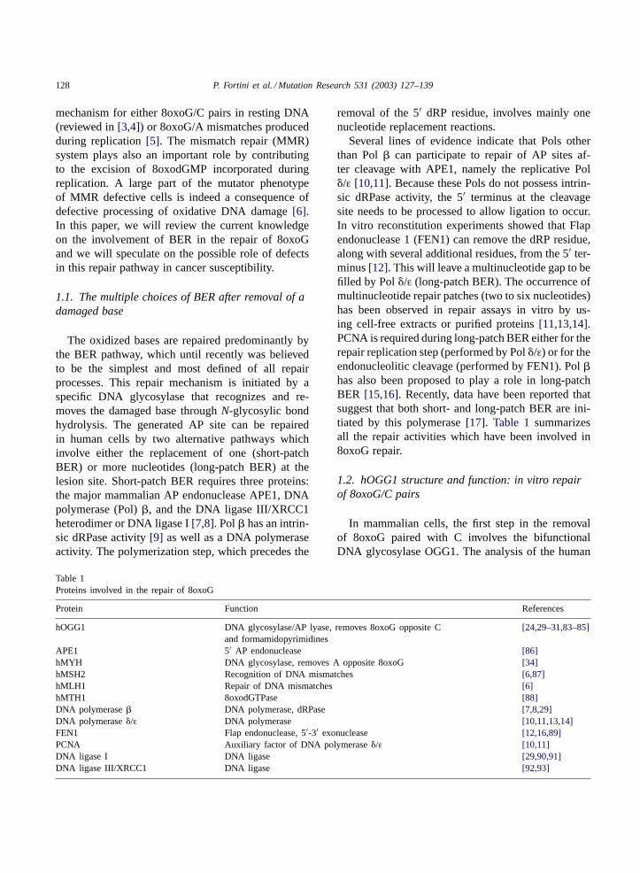
Table 1Proteins involved in the repair of 8oxoG
Protein Function References
hOGG1 DNA glycosylase/AP lyase, removes 8oxoG opposite Cand formamidopyrimidines
[24,29–31,83–85]
APE1 5′ AP endonuclease [86]hMYH DNA glycosylase, removes A opposite 8oxoG [34]hMSH2 Recognition of DNA mismatches [6,87]hMLH1 Repair of DNA mismatches [6]hMTH1 8oxodGTPase [88]DNA polymerase� DNA polymerase, dRPase [7,8,29]DNA polymerase�/ε DNA polymerase [10,11,13,14]FEN1 Flap endonuclease, 5′-3′ exonuclease [12,16,89]PCNA Auxiliary factor of DNA polymerase�/ε [10,11]DNA ligase I DNA ligase [29,90,91]DNA ligase III/XRCC1 DNA ligase [92,93]
removal of the 5′ dRP residue, involves mainly onenucleotide replacement reactions.
Several lines of evidence indicate that Pols otherthan Pol� can participate to repair of AP sites af-ter cleavage with APE1, namely the replicative Pol�/ε [10,11]. Because these Pols do not possess intrin-sic dRPase activity, the 5′ terminus at the cleavagesite needs to be processed to allow ligation to occur.In vitro reconstitution experiments showed that Flapendonuclease 1 (FEN1) can remove the dRP residue,along with several additional residues, from the 5′ ter-minus[12]. This will leave a multinucleotide gap to befilled by Pol�/ε (long-patch BER). The occurrence ofmultinucleotide repair patches (two to six nucleotides)has been observed in repair assays in vitro by us-ing cell-free extracts or purified proteins[11,13,14].PCNA is required during long-patch BER either for therepair replication step (performed by Pol�/ε) or for theendonucleolitic cleavage (performed by FEN1). Pol�has also been proposed to play a role in long-patchBER [15,16]. Recently, data have been reported thatsuggest that both short- and long-patch BER are ini-tiated by this polymerase[17]. Table 1summarizesall the repair activities which have been involved in8oxoG repair.
1.2. hOGG1 structure and function: in vitro repairof 8oxoG/C pairs
In mammalian cells, the first step in the removalof 8oxoG paired with C involves the bifunctionalDNA glycosylase OGG1. The analysis of the human

P. Fortini et al. / Mutation Research 531 (2003) 127–139 129
cDNA sequences revealed two kinds of messengerRNAs coding for�-hOGG1 and�-hOGG1, respec-tively (reviewed in [18]). By immunocytochemicalexperiments, it was shown that the� form of hOGG1is targeted to the nucleus while the� form is local-ized to the mitochondria[19]. The OGG1 expressionis not modulated during the cell cycle[20,21] and,even under severe oxidative stresses, neither OGG1mRNA nor OGG1 protein are induced[22]. hOGG1is dynamically regulated with respect to its nuclearlocalization, such that the DNase-sensitive chromatinas well as the nuclear matrix appear to be the prefer-ential sites of association during interphase, while theprotein re-localizes to condensed chromatin duringmitosis [23]. These results suggest that OGG1 maybe involved in several aspects of DNA metabolismand repair and that its functional role is mainly playedduring the pre-replicative stages of the cell cycle.
The human enzyme OGG1 is a bifunctional DNAglycosylase endowed with an AP lyase activity withspecificity for cleavage at 8oxoG opposite C[24].The crystallographic X-ray analysis of both the freehOGG1 enzyme as well as the catalytically inactivemutant bound to an 8oxoG/C containing DNA haverecently been solved[25,26]. Structural analyses haveshown that upon DNA binding, the enzyme under-goes extensive local conformational changes associ-ated with its strong preference for C in the oppositestrand. Moreover, the structure of a trapped catalyticintermediate in DNA repair by hOGG1 and biochem-ical data have shown that the enzyme sequesters theexcised 8oxoG base and employs it as a cofactor inthe subsequent hOGG1�-lyase cascade[27].
The in vitro reconstitution of the first step of8oxoG repair has shown the uncoupling between theDNA glycosylase and AP lyase activities of hOGG1[24,28,29]and the stimulation by APE1 of its catalyticactivity [30,31]. It has been hypothesized that APE1exerts its function by preventing hOGG1 reassociationto the AP site. In fact, substrate concentration ver-sus velocity plots for the DNA glycosylases (as wellas for APE1) show non-Michaelis-Menten kineticsand are indicative of product inhibition[30]. In thismodel, APE1 would preclude the AP lyase reactionof hOGG1[31]. Moreover, it was observed that APE1can stimulate the AP lyase activity of hOGG1. Anincision assay conducted either on a circular plasmidmolecule [29] or on a duplex oligonucleotide (our
unpublished data) containing a single 8oxoG residue,showed that when hOGG1 is stoichiometrically inexcess, APE1 might promote a second distinct inter-action of hOGG1 with the AP site, thus giving a newchance for AP lyase cleavage of the DNA.
Several studies indicate that the AP sites generatedby the catalytic action of hOGG1 are repaired predom-inantly by short-patch BER and that DNA Pol� isresponsible for the resynthesis step[32,33]. The pref-erential repair of 8oxoG/C pairs by short-patch BERwas confirmed by the low efficiency of repair of this le-sion by Pol�-deficient mouse cells as compared withtheir wild-type counterpart[32]. The in vitro reconsti-tution of BER of 8oxoG with human purified proteinsshowed that a core of four proteins is required, namelyhOGG1, APE1, Pol�, and DNA ligase I[29]. The ad-dition of FEN1 to the repair mixture or the use of highconcentration of Pol� were responsible of strand dis-placement DNA synthesis at incised 8oxoG in the ab-sence of DNA ligase. The recruitment of DNA ligaseI at the 5′-phosphate terminal moiety (created by thecombined activities of hOGG1 and APE1) abolishedthe Pol �/FEN1-induced strand displacement DNAsynthesis and confined the repair patches to one nu-cleotide. These findings suggest that cells are able tooperate a tight control of the repair patches at 8oxoG(as well as at AP sites). In fact, the same authors haveshown the critical importance of DNA ligase I as apatch size determinator in Pol�-mediated BER of APsites[13]. Additionally, it was shown that the induc-tion by FEN1 of Pol� strand displacement dependson the nature of the gap ahead of the polymerase. Thestimulation of strand displacement is stronger whenthe downstream nucleotide is represented by a 5′-dRPas compared with an ordinary 5′-P and the profileof the repair intermediates appears to be gap-specific[29].
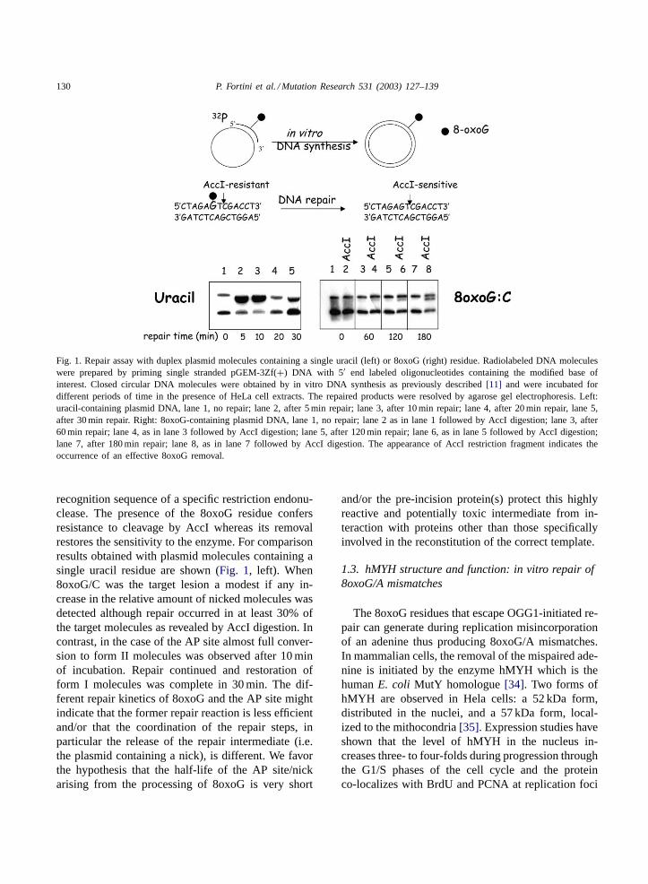
Altogether these data suggest that there are multi-ple interactions between BER enzymes both at earlyand late steps during repair of 8oxoG and that theseinteractions are probably responsible for the highlycoordinated repair of 8oxoG paired with C. The co-ordination of the repair steps is shown inFig. 1.Radiolabeled form I duplex molecules containing asingle 8oxoG were incubated with cell extracts inthe presence of dNTPs for different times and therelative yield of forms I and II molecules was deter-mined (Fig. 1, right). The lesion was located in the
130 P. Fortini et al. / Mutation Research 531 (2003) 127–139
Fig. 1. Repair assay with duplex plasmid molecules containing a single uracil (left) or 8oxoG (right) residue. Radiolabeled DNA moleculeswere prepared by priming single stranded pGEM-3Zf(+) DNA with 5′ end labeled oligonucleotides containing the modified base ofinterest. Closed circular DNA molecules were obtained by in vitro DNA synthesis as previously described[11] and were incubated fordifferent periods of time in the presence of HeLa cell extracts. The repaired products were resolved by agarose gel electrophoresis. Left:uracil-containing plasmid DNA, lane 1, no repair; lane 2, after 5 min repair; lane 3, after 10 min repair; lane 4, after 20 min repair, lane 5,after 30 min repair. Right: 8oxoG-containing plasmid DNA, lane 1, no repair; lane 2 as in lane 1 followed by AccI digestion; lane 3, after60 min repair; lane 4, as in lane 3 followed by AccI digestion; lane 5, after 120 min repair; lane 6, as in lane 5 followed by AccI digestion;lane 7, after 180 min repair; lane 8, as in lane 7 followed by AccI digestion. The appearance of AccI restriction fragment indicates theoccurrence of an effective 8oxoG removal.
recognition sequence of a specific restriction endonu-clease. The presence of the 8oxoG residue confersresistance to cleavage by AccI whereas its removalrestores the sensitivity to the enzyme. For comparisonresults obtained with plasmid molecules containing asingle uracil residue are shown (Fig. 1, left). When8oxoG/C was the target lesion a modest if any in-crease in the relative amount of nicked molecules wasdetected although repair occurred in at least 30% ofthe target molecules as revealed by AccI digestion. Incontrast, in the case of the AP site almost full conver-sion to form II molecules was observed after 10 minof incubation. Repair continued and restoration ofform I molecules was complete in 30 min. The dif-ferent repair kinetics of 8oxoG and the AP site mightindicate that the former repair reaction is less efficientand/or that the coordination of the repair steps, inparticular the release of the repair intermediate (i.e.the plasmid containing a nick), is different. We favorthe hypothesis that the half-life of the AP site/nickarising from the processing of 8oxoG is very short
and/or the pre-incision protein(s) protect this highlyreactive and potentially toxic intermediate from in-teraction with proteins other than those specificallyinvolved in the reconstitution of the correct template.
1.3. hMYH structure and function: in vitro repair of8oxoG/A mismatches
The 8oxoG residues that escape OGG1-initiated re-pair can generate during replication misincorporationof an adenine thus producing 8oxoG/A mismatches.In mammalian cells, the removal of the mispaired ade-nine is initiated by the enzyme hMYH which is thehumanE. coli MutY homologue[34]. Two forms ofhMYH are observed in Hela cells: a 52 kDa form,distributed in the nuclei, and a 57 kDa form, local-ized to the mithocondria[35]. Expression studies haveshown that the level of hMYH in the nucleus in-creases three- to four-folds during progression throughthe G1/S phases of the cell cycle and the proteinco-localizes with BrdU and PCNA at replication foci
P. Fortini et al. / Mutation Research 531 (2003) 127–139 131
[36]. The expression profile and the localization datasuggest that hMYH is involved in a repair processwhich occurs at replication.
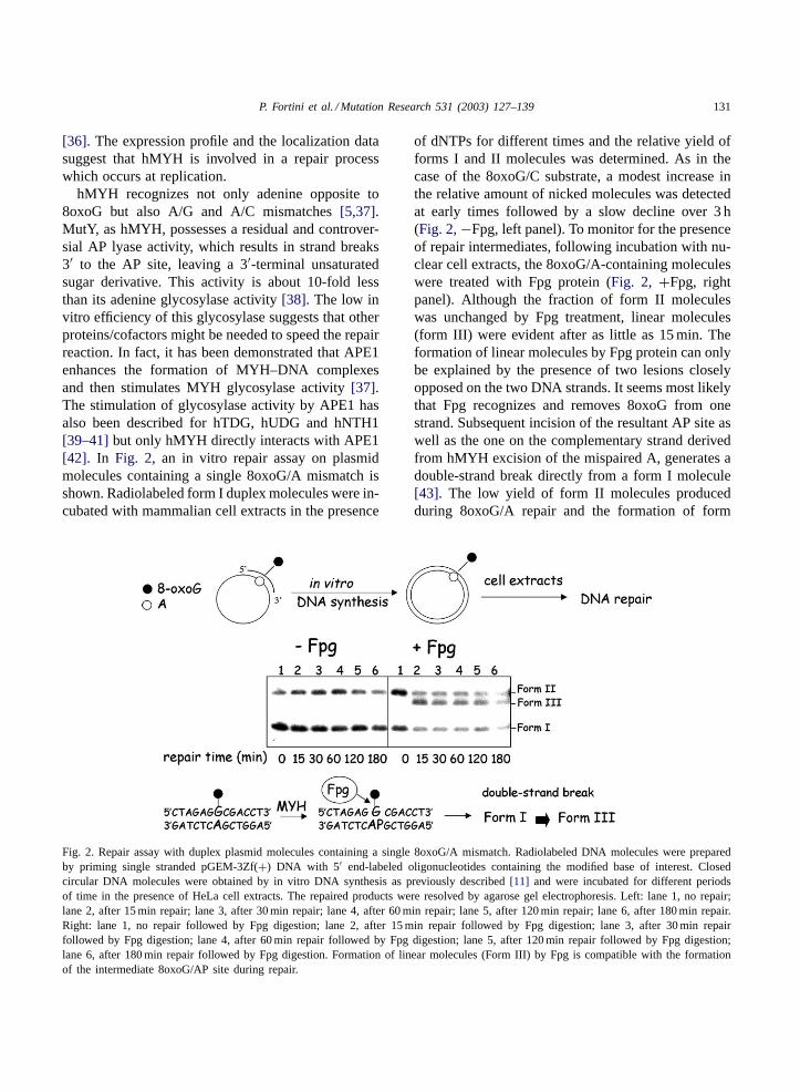
hMYH recognizes not only adenine opposite to8oxoG but also A/G and A/C mismatches[5,37].MutY, as hMYH, possesses a residual and controver-sial AP lyase activity, which results in strand breaks3′ to the AP site, leaving a 3′-terminal unsaturatedsugar derivative. This activity is about 10-fold lessthan its adenine glycosylase activity[38]. The low invitro efficiency of this glycosylase suggests that otherproteins/cofactors might be needed to speed the repairreaction. In fact, it has been demonstrated that APE1enhances the formation of MYH–DNA complexesand then stimulates MYH glycosylase activity[37].The stimulation of glycosylase activity by APE1 hasalso been described for hTDG, hUDG and hNTH1[39–41]but only hMYH directly interacts with APE1[42]. In Fig. 2, an in vitro repair assay on plasmidmolecules containing a single 8oxoG/A mismatch isshown. Radiolabeled form I duplex molecules were in-cubated with mammalian cell extracts in the presence
Fig. 2. Repair assay with duplex plasmid molecules containing a single 8oxoG/A mismatch. Radiolabeled DNA molecules were preparedby priming single stranded pGEM-3Zf(+) DNA with 5′ end-labeled oligonucleotides containing the modified base of interest. Closedcircular DNA molecules were obtained by in vitro DNA synthesis as previously described[11] and were incubated for different periodsof time in the presence of HeLa cell extracts. The repaired products were resolved by agarose gel electrophoresis. Left: lane 1, no repair;lane 2, after 15 min repair; lane 3, after 30 min repair; lane 4, after 60 min repair; lane 5, after 120 min repair; lane 6, after 180 min repair.Right: lane 1, no repair followed by Fpg digestion; lane 2, after 15 min repair followed by Fpg digestion; lane 3, after 30 min repairfollowed by Fpg digestion; lane 4, after 60 min repair followed by Fpg digestion; lane 5, after 120 min repair followed by Fpg digestion;lane 6, after 180 min repair followed by Fpg digestion. Formation of linear molecules (Form III) by Fpg is compatible with the formationof the intermediate 8oxoG/AP site during repair.
of dNTPs for different times and the relative yield offorms I and II molecules was determined. As in thecase of the 8oxoG/C substrate, a modest increase inthe relative amount of nicked molecules was detectedat early times followed by a slow decline over 3 h(Fig. 2, −Fpg, left panel). To monitor for the presenceof repair intermediates, following incubation with nu-clear cell extracts, the 8oxoG/A-containing moleculeswere treated with Fpg protein (Fig. 2, +Fpg, rightpanel). Although the fraction of form II moleculeswas unchanged by Fpg treatment, linear molecules(form III) were evident after as little as 15 min. Theformation of linear molecules by Fpg protein can onlybe explained by the presence of two lesions closelyopposed on the two DNA strands. It seems most likelythat Fpg recognizes and removes 8oxoG from onestrand. Subsequent incision of the resultant AP site aswell as the one on the complementary strand derivedfrom hMYH excision of the mispaired A, generates adouble-strand break directly from a form I molecule[43]. The low yield of form II molecules producedduring 8oxoG/A repair and the formation of form

132 P. Fortini et al. / Mutation Research 531 (2003) 127–139
III molecules by Fpg digestion are compatible withthe binding of hMYH to the intermediate 8oxoG/APsite during repair. Binding to the AP site appearsto prevent its cleavage by APE1 in the extract. Thepersistence of uncleaved AP sites is consistent withthe acknowledged high affinity of purified mMYHfor the 8oxoG/AP site that it produces and the poorturnover of the enzyme[37]. TheE. coli MutY has asimilar behavior: it remains bound to its product anddoes not turn over[44,45]. This behavior could be ex-plained by the need of “shielding” the highly reactiveAP site, till the correct enzyme, namely APE1, entersthe repair pathway. According to this model, the Fpgactivity is inhibited by the tight binding of MutY to8oxoG/AP, as well to 8oxoG/G and 8oxoG/T[46].
When the A misincorporated opposite 8oxoG hasbeen removed and the AP site incised, a DNA poly-merase has to fill in the gap created at the lesion site.Although both replicative and repair polymeraseshave been hypothesized to play a role in this reac-tion (e.g. Pol�, �, ε, �, �) Pol � is the most likelycandidate. In fact, when the repair of a plasmid con-taining a single 8oxoG/A by human cell extracts wasinvestigated, at early repair times (1 h repair) a pref-erential incorporation of a C opposite 8oxoG wasdetected and this reaction was aphidicolin-sensitive[5]. Both features are consistent with Pol� playinga role in the resynthesis step: Pol� preferentiallyincorporates C opposite 8oxoG[47] and is sensitiveto aphidicolin[48]. The involvement of a replicative(and then PCNA-dependent) polymerase, suggeststhat the 8oxoG/A repair occurs by a long-patch,PCNA-dependent BER pathway. According to thishypothesis, the interaction between hMYH and PCNAhas been shown both in vitro[42] and in vivo, asco-localization at replication foci[36]. PCNA is oneof the most important players in DNA replication,acting as the processivity factor for Pol�/ε. In thelong-patch BER, it may act as a scaffold for the repairmachinery, interacting with other factors involved inthis pathway, namely Pol�/ε, RFC, FEN1 and DNALigase I. Moreover, hMYH interacts also with RPA[42] that is known to stimulate FEN1 activity[49]. In-terestingly, another DNA repair glycosylase, UNG2,has been reported to interact and co-localize at repli-cation foci with PCNA and RPA. The levels of thisglycosylase, as that of hMYH, increase in S phase[50]. It is tempting to speculate that both DNA gly-
cosylases initiate a replication-associated BER whichis Pol �-mediated.
After the removal of the mispaired adenine andits substitution by a C, an 8oxoG/C pair is formed.As expected this is the substrate for another BERevent which involves the replacement of the oxidizedguanine with a normal G but, surprisingly, also thisreaction was aphidicolin-sensitive[5]. It is likelythat the repair of 8oxoG/C, when it is formed dur-ing the repair of the 8oxoG/A mismatch, involvesthe replication-associated repair (RAR) machinery,that includes Pol�/ε, PCNA, and DNA ligase I[51].This pathway will repair any BER substrate resultingfrom misincorporation opposite modified bases (asA/8oxoG and A/U) or persisting/formed at replica-tion. The presence of the lesion at or near the replica-tion fork will be the signal that will select this repairmode. It will act even on lesion like 8oxoG/C that, innon-replicating DNA, is repaired by Pol�-dependentshort-patch BER[32,33] (Fig. 3).
Although it is not the subject of this review, it shouldbe mentioned that MMR might also be involved in8oxoG/A repair. Yeast Msh2/Msh6 heterodimer hasbeen shown to bind 8oxoG/A mismatches and to beinvolved in repair of 8oxoG in DNA[52]. More re-cently, it has been shown that hMYH interacts with theheterodimer hMSH2/hMSH6 that is also able to stim-ulate the DNA binding and glycosylase activities ofhMYH [53]. Therefore, hMYH-dependent BER andMMR might both contribute to the removal of theA misincorporated opposite 8oxoG by two differentreplication-associated pathways.
1.4. Repair of oxidative DNA damage inmammalian cells
The experiments carried out either with purifiedproteins or with cell extracts indicate that 8oxoGis preferentially repaired via short-patch BER whenpaired with a C whereas the long-patch pathway isthe main route of repair when 8oxoG/A mismatchesare formed. Are the in vitro repair systems a goodmodel for repair in mammalian cells? A useful cellsystem which is defective in the short-patch BER hasbeen developed by Sobol et al.[54] who succeededto establish Pol�-null mouse embryonic fibroblastcell lines. These cells were showed to be hypersensi-tive to the cytotoxic effects of both monofunctional

P. Fortini et al. / Mutation Research 531 (2003) 127–139 133
Fig. 3. Model for the switching between short- and long-patch BER at replication. When 8oxoG/C pairs are present in non-replicatingDNA the Pol �-dependent short-patch BER will be the mechanism of election for repair. Conversely, when a BER substrate is formed at,or near, a replication fork (as in the case of the adenine mismatched with 8oxoG or the 8oxoG/C pairs arising during repair of 8oxoG/Amismatches) a replication-associated BER pathway that involves Pol�/ε, PCNA, FEN1 will be the preferential repair mode.
alkylating agents (e.g. methyl methanesulfonate,MMS) [54] and oxidative agents (i.e. hydrogen perox-ide, H2O2) [55,56]. O6-Alkylguanine,N-alkylpurinesand AP sites are the major base modifications inducedby alkylating agents whereas, oxidized purines (e.g.8oxoG, 8oxoA) and pyrimidines (e.g. thymine gly-col), regular and oxidized AP sites (OAS) and franksingle strand breaks (ssb) are specifically inducedby H2O2. All these lesions, with the exception ofO6-alkylguanine, are BER-substrates. The revertionof the hypersensitive phenotype by expression of Pol� in defective cells testifies the essential role of Pol� in the protection against the cytotoxicity inducedby these lesions[54,56]. It should be noted that thesensitivity to the cytotoxic effect of MMS was in-creased five-fold (by comparing the D37 values) in Pol�-null fibroblasts as compared to the wild-type cellswhereas after H2O2 treatment, the sensitivity was in-creased 2.7-fold in the defective cell line[55]. Thesefindings suggest that the Pol�-mediated short-patchBER is the pathway of election for the processingof the regular AP sites which arise after removal ofN-alkylpurines. These sites represent only 10% of theabasic sites induced by oxidative agents which aremainly OAS. When DNA ssb induced by MMS andH2O2 treatment were measured in wild-type and Pol�-null fibroblasts by single cell gel electrophoresis[55], a decrease in the rate of DNA ssb rejoining wasobserved after MMS exposure in the defective cell
line, confirming that the regular AP sites are predom-inantly repaired by Pol�-mediated short-patch BER.In contrast, after H2O2 treatment, no difference inthe repair kinetics of wild-type and Pol�-null cellswas observed. The apparent discrepancy between thecytotoxic response and the DNA ssb repair data inthe case of H2O2 can be explained considering thatdifferent types of lesions are likely to be responsiblefor lethal events and ssb formation. We favor thehypothesis that H2O2-induced DNA ssb arise mainlyfrom OAS which are efficiently repaired also by Pol�-independent long-patch BER[57].
A role for Pol � in the response to oxidative stresshas been confirmed by experiments in cells in culturesand, more recently, in in vivo models. Mouse mono-cytes and fibroblasts pre-treated with lipopolysaccha-ride [58], which induces production of nitric oxide,were more resistant to the cytotoxic effects induced byMMS than untreated cells and showed increased Pol� expression and in vitro BER capacity. Conversely,Pol �-null cells failed to show this adaptive response.Similarly [59], intraperitoneal injection in mice of 2 ni-tropropane (2-NP), a known in vivo hepatocarcinogenable to induce high levels of 8oxoG, induced a signif-icant increase in Pol� levels. When 2-NP was admin-istered to heterozygous Pol� knock out mice (whichexhibit a 50% reduction in BER capacity), the levelsof 8oxoG were similar to those detected in wild-typemice but a significant increase in the level of DNA ssb

134 P. Fortini et al. / Mutation Research 531 (2003) 127–139
was observed. This effect can be ascribed to a delayin the repair replication reaction subsequent to strandincision at 8oxoG residues by OGG1/APE1. If thereis clear evidence that Pol� is induced by oxidativestress and enters as a major player in the repair pro-cess, no inducibility of OGG1 has been reported inmice subjected to oxidative stress[59,60].
Is there any evidence that long-patch BER mightoccur during oxidative damage repair ? FEN1, astructure-specific endonuclease involved in DNAreplication, has also a crucial role in the removal of thelesion-containing 5′ flap nucleotides displaced duringlong-patch BER. Homozygous mutant chicken cells(FEN1−/−) have been generated[61] indicating thatthis is not an essential gene. The mutant cells werecharacterized for the sensitivity to various damagingagents and the cells were showed to be hypersensitiveto monofunctional methylating agents (e.g. MMS)but not to UV light, X-rays and etoposide supportingthe specific role of FEN1 in BER. A revertion to thewild-type phenotype was obtained by ectopic FEN1expression. Interestingly, the cytotoxic effect of H2O2in FEN1-null cells was more pronounced than that ofthe methylating agents indicating that long-patch BERhas an important role in the processing of cytotoxicoxidative damage-induced lesions.
1.5. Base excision repair of oxidative DNA damageand human cancer
Excessive production of reactive oxygen species(ROS) beyond the antioxidant capacity of the bodycan cause oxidative stress. Oxidative stress has beenimplicated in the etiology of a variety of pathologiesincluding cancer. The interplay between exposure toenvironmental mutagens and defects in DNA repaircan lead to genomic instability. What is the currentevidence that the malfunctioning of BER is associatedwith cancer risk?
Frequent allele variants have been described forOGG1, XRCC1, Pol�, APE1, and MYH. Althoughonly a small proportion of studies are population-basedand are sufficiently large, evidence starts to accumu-late on the possible role of these polymorphisms incancer risk. Mutations in theOGG1gene have beenidentified in human lung and kidney tumours[62] andthe S326C polymorphism appears to be associatedwith an increased risk in several case–control stud-
ies of esophageal[63], lung [64], and prostate can-cers[65]. It should be noted, however, that one studyon gastric cancer and another one recently publishedon breast cancer found no association between thispolymorphism and cancer risk[66,67]. The relation-ship between two polymorphic variants of XRCC1(Arg194Trp and Arg399Gln) and different types ofcancer shows some controversial aspects (reviewed in[68]). Although both these polymorphisms reside inimportant functional domains (Arg194Trp in the linkerregion between Pol� and PARP domains; Arg399Glnin the BRCT domain) a consistent reduced cancerrisk of breast, lung, bladder, stomach, and squamouscell carcinoma of head and neck (SCCHN) has beenfound only for the XRCC1 Arg194Trp. On the con-trary, Arg399Gln was associated with an increasedrisk of breast and stomach cancer while seems pro-tective against non melanoma skin cancer and bladdercancer.
Pol �, the key polymerase in BER of oxidativedamage, seems to be involved directly and indi-rectly in the development of several cancers that arestrongly linked to environmental exposure, althoughonly small numbers of tumors have been analyzed. Ahigh frequency of splice variants has been reportedin colorectal, breast, brain, lung, and bladder cancer[69–72]. Splice variants are also observed in normalcells although their frequency in tumor cells seemsto be higher. The only two variants, which have beencharacterized, are the exon� insertion and the exonXI deletion. Insertion of exon� does not affect thepolymerase activity of Pol�, while the exon XIsplice variant generates a protein with a dominantnegative activity[69,70]. Pol � polymorphic vari-ants have also been described in bladder cancer[72]and cancer-specific missense mutations and deletionshave been found at high frequency in gastric cancers[73]. One of these mutations (Glu295Lys) led to areduction of BER activity and the mutant protein wasreported to exert a dominant negative activity. Theassociation between APE1 variants and cancer risk isnot described in the epidemiological literature, eventhough three out of seven variants identified (L104R,E126D, and R237A) exhibit 40–60% reduction inthe AP-endonuclease function[74]. Similarly, the as-sociation of MYH with cancer risk has never beenanalyzed in large studies. The only available resultsconcern two missense mutations (Tyr165Cys and
P. Fortini et al. / Mutation Research 531 (2003) 127–139 135
Gly382Asp) found in patients affected by multipleadenomas and carcinomas[75–77]. Functional stud-ies revealed a decrease in DNA glycosylase activityof these two variant forms[75,78].
2. Conclusions
Although BER is the main mechanism for 8oxoG/Cand 8oxoG/A repair, several lines of evidence indicatethat the repair of oxidative damage in mammaliancell nuclei is more complex. For instance, the pref-erential repair of 8oxoG located in the transcribedsequences involves DNA glycosylases/mechanismsother than the OGG1-initiated BER[79,80] and thereplication-associated repair of oxidized bases incor-porated into the nascent strand from the deoxynu-cleotide pool awaits to be clarified (reviewed in[81]).We believe that the DNA polymerase�/ε-mediatedBER, which shares several components with DNAreplication, is a good candidate for RAR of oxidativeDNA damage[5]. If distinct complexes are involvedin repair of transcriptionally active regions and in re-pair of quiescent versus dividing cells, the challengefor the future is to identify and characterize thesecomplexes. Up to now the mouse models have pro-vided limited clues as to the function of the genes in-volved in repair of oxidative damage. OGG1-deficientmice, similarly to other DNA glycosylase-defectivemice, do not exhibit an excess of tumors[82]. How-ever, if the role of these repair genes is to protect thelong-term integrity of the genome, a knock out animalmight be a poor model for screening for reductionin fitness. Interestingly, MTH1-deficient mice displaygreater numbers of tumors in the lungs, liver, andstomach compared to their wild-type littermates[2].Until recently, inherited deficiencies in the repair ofoxidative DNA damage had not been casually linkedwith any human genetic disorder. Several lines ofevidence indicate an association between polymor-phisms in BER genes and cancer risk. Recently, ithas been demonstrated that inherited defects of MYHpredispose to multiple colorectal adenomas and car-cinomas[75–77]. Molecular epidemiological studieson multiple polymorphisms within genes and on mul-tiple genes within DNA repair pathways may leadto a better understanding of the gene–environmentinteractions in cancer aetiology. At the same time,
experimental studies should be particularly addressedto analyze whether or how DNA repair gene poly-morphisms may affect DNA repair function.
Acknowledgements
This work was partially supported by the Associ-azione Italiana Ricerca sul Cancro (A.I.R.C.) and byCompagnia di S. Paolo, Turin, Italy (Project coordi-nator: Dr. G. Frosina).
References
[1] T. Tajiri, H. Maki, M. Sekiguchi, Functional cooperation ofMutT, MutM and MutY proteins in preventing mutationscaused by spontaneous oxidation of guanine nucleotide inEscherichia coli, Mutat. Res. 336 (1995) 257–267.
[2] T. Tsuzuki, A. Egashira, H. Igarashi, T. Iwakuma, Y.Nakatsuru, Y. Tominaga, H. Kawate, K. Nakao, K. Nakamura,F. Ide, S. Kura, Y. Nakabeppu, M. Katsuki, T. Ishikawa, M.Sekiguchi, Spontaneous tumorigenesis in mice defective inthe MTH1 gene encoding 8-oxo-dGTPase, Proc. Natl. Acad.Sci. U.S.A. 98 (2001) 11456–11461.
[3] S. Boiteux, J.P. Radicella, Base excision repair of 8-hydro-xyguanine protects DNA from endogenous oxidative stress,Biochimie 81 (1999) 59–67.
[4] L. Gros, M. Saparbaev, J. Laval, Enzymology of the repairof free radicals-induced DNA damage, Oncogene 21 (2002)8905–8925.
[5] E. Parlanti, P. Fortini, P. Macpherson, J. Laval, E. Dogliotti,Base excision repair of adenine/8-oxoguanine mispairs by anaphidicolin-sensitive DNA polymerase in human cell extracts,Oncogene 21 (2002) 5204–5212.
[6] C. Colussi, E. Parlanti, P. Degan, G. Aquilina, D. Barnes,P. Macpherson, P. Karran, M. Crescenzi, E. Dogliotti, M.Bignami, The mammalian mismatch repair pathway removesDNA 8-oxodGMP incorporated from the oxidized dNTP pool,Curr. Biol. 12 (2002) 912–918.
[7] Y. Kubota, R. Nash, A. Klungland, P. Schar, D. Barnes, T.Lindahl, Reconstitution repair of DNA base excision-repairwith purified human proteins: interaction between DNApolymerase B and the XRCC1 protein, EMBO J. 15 (1996)6662–6670.
[8] D.K. Srivastava, B.J. Vande, R. Prasad, J.T. Molina, W.A.Beard, A.E. Tomkinson, S.H. Wilson, Mammalian baseexcision repair. Identification of the reaction sequence andrate-determining steps, J. Biol. Chem. 273 (1998) 21203–21209.
[9] Y. Matsumoto, K. Kim, Excision of deoxyribose phosphateresidues by DNA polymerase� during DNA repair, Science269 (1995) 699–702.
[10] Y. Matsumoto, K. Kim, D.F. Bogenhagen, Proliferating cellnuclear antigen-dependent abasic site repair inXenopus laevis

136 P. Fortini et al. / Mutation Research 531 (2003) 127–139
oocytes: an alternative pathway of base excision DNA repair,Mol. Cell. Biol. 14 (1994) 6187–6197.
[11] G. Frosina, P. Fortini, O. Rossi, F. Carrozzino, G. Raspaglio,L.S. Cox, D.P. Lane, A. Abbondandolo, E. Dogliotti, Twopathways for base excision repair in mammalian cells, J. Biol.Chem. 271 (1996) 9573–9578.
[12] R. Gary, K. Kim, H.L. Cornelius, M.S. Park, Y. Matsumoto,Proliferating cell nuclear antigen facilitates excision inlong-patch base excision repair, J. Biol. Chem. 274 (1999)4354–4363.
[13] B. Pascucci, M. Stucki, Z.O. Jonsson, E. Dogliotti, U.Hubscher, Long-patch base excision repair with purifiedhuman proteins. DNA ligase I as patch size mediator for DNApolymerases delta and epsilon, J. Biol. Chem. 274 (1999)33696–33702.
[14] Y. Matsumoto, K. Kim, J. Hurwitz, R. Gary, D.S. Levin,A.E. Tomkinson, M.S. Park, Reconstitution of proliferatingcell nuclear antigen-dependent repair of apurinic/apyrimidinicsites with purified human proteins, J. Biol. Chem. 274 (1999)33703–33708.
[15] G. Dianov, R. Prasad, S. Wilson, V. Bohr, Role of DNApolymerase� in the excision step of long-patch mammalianbase excision repair, J. Biol. Chem. 274 (1999) 13741–13743.
[16] R. Prasad, G.L. Dianov, V.A. Bohr, S.H. Wilson, FEN1stimulation of DNA polymerase� mediates an excision stepin mammalian long-patch base excision repair, J. Biol. Chem.275 (2000) 4460–4466.
[17] A.J. Podlutsky, I.I. Dianova, V.N. Podust, V.A. Bohr, G.L.Dianov, Human DNA polymerase� initiates DNA synthesisduring long-patch repair of reduced AP sites in DNA, EMBOJ. 20 (2001) 1–6.
[18] S. Boiteux, J.P. Radicella, The human OGG1 gene: structure,functions, and its implication in the process of carcinogenesis,Arch. Biochem. Biophys. 377 (2000) 1–8.
[19] M. Takao, H. Aburatani, K. Kobayashi, A. Yasui, Mito-chondrial targeting of human DNA glycosylases for repairof oxidative DNA damage, Nucleic Acids Res. 26 (1998)2917–2922.
[20] A. Dhenaut, S. Boiteux, J.P. Radicella, Characterization ofthe hOGG1 promoter and its expression during the cell cycle,Mutat. Res. 461 (2000) 109–118.
[21] M. Bouziane, F. Miao, S.E. Bates, L. Somsouk, B.C. Sang,M. Denissenko, T.R. O’Connor, Promoter structure and cellcycle dependent expression of the human methylpurine-DNAglycosylase gene, Mutat. Res. 461 (2000) 15–29.
[22] T. Saitoh, K. Shinmura, S. Yamaguchi, M. Tani, S. Seki,H. Murakami, Y. Nojima, J. Yokota, Enhancement of OGG1protein AP lyase activity by increase of APEX protein, Mutat.Res. 486 (2001) 31–40.
[23] F. Dantzer, L. Luna, M. Bjoras, E. Seeberg, Human OGG1undergoes serine phosphorylation and associates with thenuclear matrix and mitotic chromatin in vivo, Nucleic AcidsRes. 30 (2002) 2349–2357.
[24] M. Bjoras, L. Luna, B. Johnsen, E. Hoff, T. Haug, T.Rognes, E. Seeberg, Opposite base-dependent reactions ofa human base excision repair enzyme on DNA containing7,8-dihydro-8-oxoguanine and abasic sites, EMBO J. 16(1997) 6314–6322.
[25] S.D. Bruner, D.P. Norman, G.L. Verdine, Structural basisfor recognition and repair of the endogenous mutagen8-oxoguanine in DNA, Nature 403 (2000) 859–866.
[26] M. Bjoras, E. Seeberg, L. Luna, L.H. Pearl, T.E. Barrett,Reciprocal “flipping” underlies substrate recognition andcatalytic activation by the human 8-oxo-guanine DNAglycosylase, J. Mol. Biol. 317 (2002) 171–177.
[27] J.C. Fromme, S.D. Bruner, W. Yang, M. Karplus, G.L.Verdine, Product-assisted catalysis in base-excision DNArepair, Nat. Struct. Biol. 10 (2003) 204–211.
[28] D.O. Zharkov, T.A. Rosenquist, S.E. Gerchman, A.P.Grollman, Substrate specificity and reaction mechanism ofmurine 8-oxoguanine-DNA glycosylase, J. Biol. Chem. 275(2000) 28607–28617.
[29] B. Pascucci, G. Maga, U. Hubscher, M. Bjoras, E. Seeberg,I.D. Hickson, G. Villani, C. Giordano, L. Cellai, E.Dogliotti, Reconstitution of the base excision repair pathwayfor 7,8-dihydro-8-oxoguanine with purified human proteins,Nucleic Acids Res. 30 (2002) 2124–2130.
[30] J.W. Hill, T.K. Hazra, T. Izumi, S. Mitra, Stimulation ofhuman 8-oxoguanine-DNA glycosylase by AP-endonuclease:potential coordination of the initial steps in base excisionrepair, Nucleic Acids Res. 29 (2001) 430–438.
[31] A.E. Vidal, I.D. Hickson, S. Boiteux, J.P. Radicella,Mechanism of stimulation of the DNA glycosylase activityof hOGG1 by the major human AP endonuclease: bypassof the AP lyase activity step, Nucleic Acids Res. 29 (2001)1285–1292.
[32] P. Fortini, E. Parlanti, O.M. Sidorkina, J. Laval, E. Dogliotti,The type of DNA glycosylase determines the base excisionrepair pathway in mammalian cells, J. Biol. Chem. 274 (1999)15230–15236.
[33] G. Dianov, C. Bischoff, J. Piotrowski, V.A. Bohr, Repairpathways for processing of 8-oxoguanine in DNA bymammalian cell extracts, J. Biol. Chem. 278 (1998) 33511–33516.
[34] M.M. Slupska, C. Baikalov, W.M. Luther, J.H. Chiang, Y.F.Wei, J.H. Miller, Cloning and sequencing a human homolog(hMYH) of the Escherichia colimutY gene whose function isrequired for the repair of oxidative DNA damage, J. Bacteriol.178 (1996) 3885–3892.
[35] T. Ohtsubo, K. Nishioka, Y. Imaiso, S. Iwai, H. Shimokawa,H. Oda, T. Fujiwara, Y. Nakabeppu, Identification ofhuman MutY homolog (hMYH) as a repair enzyme for2-hydroxyadenine in DNA and detection of multiple formsof hMYH located in nuclei and mitochondria, Nucleic AcidsRes. 28 (2000) 1355–1364.
[36] I. Boldogh, D. Milligan, M.S. Lee, H. Bassett, R.S. Lloyd,A.K. McCullough, hMYH cell cycle-dependent expression,subcellular localization and association with replicationfoci: evidence suggesting replication-coupled repair ofadenine:8-oxoguanine mispairs, Nucleic Acids Res. 29 (2001)2802–2809.
[37] H. Yang, W.M. Clendenin, D. Wong, B. Demple, M.M.Slupska, J.H. Chiang, J.H. Miller, Enhanced activity ofadenine-DNA glycosylase (Myh) by apurinic/apyrimidinicendonuclease (Ape1) in mammalian base excision repair of

P. Fortini et al. / Mutation Research 531 (2003) 127–139 137
an A/GO mismatch, Nucleic Acids Res. 29 (2001) 743–752.
[38] S.D. Williams, S.S. David, Evidence that MutY is amonofunctional glycosylase capable of forming a covalentSchiff base intermediate with substrate DNA, Nucleic AcidsRes. 26 (1998) 5123–5133.
[39] S.S. Parikh, C.D. Mol, G. Slupphaug, S. Bharati, H.E.Krokan, J.A. Tainer, Base excision repair initiation revealed bycrystal structures and binding kinetics of human uracil-DNAglycosylase with DNA, EMBO J. 17 (1998) 5214–5226.
[40] T.R. Waters, P. Gallinari, J. Jiricny, P.F. Swann, Humanthymine DNA glycosylase binds to apurinic sites in DNAbut is displaced by human apurinic endonuclease 1, J. Biol.Chem. 274 (1999) 67–74.
[41] D.R. Marenstein, M.K. Chan, A. Altamirano, A.K. Basu,R.J. Boorstein, R.P. Cunningham, G.W. Teebor, Substratespecificity of human endonuclease III (hNTH1). Effect ofhuman APE1 on hNTH1 activity, J. Biol. Chem. 278 (2003)9005–9012.
[42] A. Parker, Y. Gu, W. Mahoney, S.H. Lee, K.K. Singh, A.L.Lu, Human homolog of the MutY repair protein (hMYH)physically interacts with proteins involved in long-patchDNA base excision repair, J. Biol. Chem. 276 (2001) 5547–5555.
[43] J. Tchou, V. Bodepudi, S. Shibutani, I. Antoshechkin, J.Miller, A.P. Grollman, F. Johnson, Substrate specificity of Fpgprotein. Recognition and cleavage of oxidatively damagedDNA, J. Biol. Chem. 269 (1994) 15318–15324.
[44] D.O. Zharkov, A.P. Grollman, MutY DNA glycosylase: baserelease and intermediate complex formation, Biochemistry 37(1998) 12384–12394.
[45] S.L. Porello, A.E. Leyes, S.S. David, Single-turnover andpre-steady-state kinetics of the reaction of the adenineglycosylase MutY with mismatch-containing DNA substrates,Biochemistry 37 (1998) 14756–14764.
[46] X. Li, P.M. Wright, A.L. Lu, The C-terminal domain ofMutY glycosylase determines the 7,8-dihydro-8-oxo-guaninespecificity and is crucial for mutation avoidance, J. Biol.Chem. 275 (2000) 8448–8455.
[47] H.J. Einolf, F.P. Guengerich, Fidelity of nucleotide insertionat 8-oxo-7,8-dihydroguanine by mammalian DNA polymerasedelta. Steady-state and pre-steady-state kinetic analysis, J.Biol. Chem. 276 (2001) 3764–3771.
[48] T. Weiser, M. Gassmann, P. Thommes, E. Ferrari, P.Hafkemeyer, U. Hubscher, Biochemical and functionalcomparison of DNA polymerases alfa, delta and epsilon fromcalf thymus, J. Biol. Chem. 266 (1991) 10420–10428.
[49] E.E. Biswas, F.X. Zhu, S.B. Biswas, Stimulation of RTH1nuclease of the yeastSaccharomyces cerevisiaeby replicationprotein A, Biochemistry 36 (1997) 5955–5962.
[50] M. Otterlei, E. Warbrick, T.A. Nagelhus, T. Haug, G.Slupphaug, M. Akbari, P.A. Aas, K. Steinsbekk, O.Bakke, H.E. Krokan, Post-replicative base excision repair inreplication foci, EMBO J. 18 (1999) 3834–3844.
[51] K.W. Caldecott, Mammalian DNA single-strand break repair:an X-ra(y)ted affair, Bioessays 23 (2001) 447–455.
[52] T.T. Ni, G.T. Marsischky, R.D. Kolodner, MSH2 and MSH6are required for removal of adenine misincorporated opposite8-oxo-guanine inS. cerevisiae, Mol. Cell 4 (1999) 439–444.
[53] Y. Gu, A. Parker, T.M. Wilson, H. Bai, D.Y. Chang, A.L.Lu, Human MutY homolog, a DNA glycosylase involvedin base excision repair, physically and functionally interactswith mismatch repair proteins human MutS homolog 2/humanMutS homolog 6, J. Biol. Chem. 277 (2002) 11135–11142.
[54] R.W. Sobol, J.K. Horton, R. Kuhn, H. Gu, R.K. Singhal,R. Prasad, K. Rajewsky, S.H. Wilson, Requirement ofmammalian DNA polymerase� in base excision repair,Nature 379 (1996) 183–186.
[55] P. Fortini, B. Pascucci, F. Belisario, E. Dogliotti, DNApolymerase� is required for efficient DNA strand break repairinduced by methyl methanesulfonate but not by hydrogenperoxide, Nucleic Acids Res. 28 (2000) 3040–3046.
[56] J.K. Horton, A. Baker, B.J. Berg, R.W. Sobol, S.H. Wilson,Involvement of DNA polymerase� in protection against thecytotoxicity of oxidative DNA damage, DNA Rep. 1 (2002)317–333.
[57] A. Klungland, T. Lindahl, Second pathway for completion ofhuman DNA base excision-repair: reconstitution with purifiedproteins and requirement for DNase IV, EMBO J. 16 (1997)3341–3348.
[58] K.H. Chen, D.K. Srivastava, S.H. Wilson, Relationshipbetween base excision repair capacity and DNA alkylatingagent sensitivity in mouse monocytes, Mutat. Res. 487 (2001)121–126.
[59] D.C. Cabelof, J.J. Raffoul, S. Yanamadala, Z. Guo, A.R.Heydari, Induction of DNA polymerase�-dependent baseexcision repair in response to oxidative stress in vivo,Carcinogenesis 23 (2002) 1419–1425.
[60] T. Arai, V.P. Kelly, O. Minowa, T. Noda, S. Nishimura, Highaccumulation of oxidative DNA damage, 8-hydroxyguanine,in Mmh/Ogg1 deficient mice by chronic oxidative stress,Carcinogenesis 23 (2002) 2005–2010.
[61] Y. Matsuzaki, N. Adachi, H. Koyama, Vertebrate cellslacking FEN-1 endonuclease are viable but hypersensitive tomethylating agents and H2O2, Nucleic Acids Res. 30 (2002)3273–3277.
[62] S. Chevillard, J.P. Radicella, C. Levalois, J. Lebeau, M.F.Poupon, S. Oudard, B. Dutrillaux, S. Boiteux, Mutationsin OGG1, a gene involved in the repair of oxidative DNAdamage, are found in human lung and kidney tumours,Oncogene 16 (1998) 3083–3086.
[63] D.Y. Xing, W. Tan, N. Song, D.X. Lin, Ser326Cyspolymorphism in hOGG1 gene and risk of esophageal cancerin a Chinese population, Int. J. Cancer 95 (2001) 140–143.
[64] H. Sugimura, T. Kohno, K. Wakai, K. Nagura, K. Genka, H.Igarashi, B.J. Morris, S. Baba, Y. Ohno, C. Gao, Z. Li, J.Wang, T. Takezaki, K. Tajima, T. Varga, T. Sawaguchi, J.K.Lum, J.J. Martinson, S. Tsugane, T. Iwamasa, K. Shinmura,J. Yokota, hOGG1 Ser326Cys polymorphism and lung cancersusceptibility, Cancer Epidemiol. Biomarkers Prev. 8 (1999)669–674.

138 P. Fortini et al. / Mutation Research 531 (2003) 127–139
[65] J. Xu, S.L. Zheng, A. Turner, S.D. Isaacs, K.E. Wiley,G.A. Hawkins, B.L. Chang, E.R. Bleecker, P.C. Walsh, D.A.Meyers, W.B. Isaacs, Associations between hOGG1 sequencevariants and prostate cancer susceptibility, Cancer Res. 62(2002) 2253–2257.
[66] T. Hanaoka, H. Sugimura, K. Nagura, M. Ihara, X.J. Li,G.S. Hamada, I. Nishimoto, L.P. Kowalski, J. Yokota, S.Tsugane, hOGG1 exon7 polymorphism and gastric cancer incase–control studies of Japanese, Brazilians and non-Japanese,Cancer Lett. 170 (2001) 53–61.
[67] U. Vogel, B.A. Nexo, A. Olsen, B. Thomsen, N.R. Jacobsen,H. Wallin, K. Overvad, A. Tjonneland, No associationbetween OGG1 Ser326Cys polymorphism and breast cancerrisk, Cancer Epidemiol. Biomarkers Prev. 12 (2003) 170–171.
[68] E.L. Goode, C.M. Ulrich, J.D. Potter, Polymorphisms inDNA repair genes and associations with cancer risk, CancerEpidemiol. Biomarkers Prev. 11 (2002) 1513–1530.
[69] R. Nowak, P. Bieganowski, R. Konopinski, J.A. Siedlecki,Alternative splicing of DNA polymerase beta mRNA is nottumor-specific, Int. J. Cancer 68 (1996) 199–202.
[70] Y.J. Chyan, P.R. Strauss, T.G. Wood, S.H. Wilson,Identification of novel mRNA isoforms for human DNApolymerase�, DNA Cell Biol. 15 (1996) 653–659.
[71] N. Bhattacharyya, H.C. Chen, S. Comhair, S.C. Erzurum, S.Banerjee, Variant forms of DNA polymerase� in primarylung carcinomas, DNA Cell Biol. 18 (1999) 549–554.
[72] T.E. Thompson, P.K. Rogan, J.I. Risinger, J.A. Taylor, Splicevariants but not mutations of DNA polymerase� are commonin bladder cancer, Cancer Res. 62 (2002) 3251–3256.
[73] A. Iwanaga, M. Ouchida, K. Miyazaki, K. Hori, T. Mukai,Functional mutation of DNA polymerase beta found in humangastric cancer-inability of the base excision repair in vitro,Mutat. Res. 435 (1999) 121–128.
[74] M.Z. Hadi, M.A. Coleman, K. Fidelis, H.W. Mohrenweiser,D.M. Wilson III, Functional characterization of Ape1 variantsidentified in the human population, Nucleic Acids Res. 28(2000) 3871–3879.
[75] N. Al-Tassan, N.H. Chmiel, J. Maynard, N. Fleming, A.L.Livingston, G.T. Williams, A.K. Hodges, D.R. Davies, S.S.David, J.R. Sampson, J.P. Cheadle, Inherited variants of MYHassociated with somatic G:C→ T:A mutations in colorectaltumors, Nat. Genet. 30 (2002) 227–232.
[76] S. Jones, P. Emmerson, J. Maynard, J.M. Best, S. Jordan,G.T. Williams, J.R. Sampson, J.P. Cheadle, Biallelic germlinemutations in MYH predispose to multiple colorectal adenomaand somatic G:C→ T:A mutations, Hum. Mol. Genet. 11(2002) 2961–2967.
[77] O.M. Sieber, L. Lipton, M. Crabtree, K. Heinimann, P.Fidalgo, R.K. Phillips, M.L. Bisgaard, T.F. Orntoft, L.A.Aaltonen, S.V. Hodgson, H.J. Thomas, I.P. Tomlinson,Multiple colorectal adenomas, classic adenomatous polyposis,and germ-line mutations in MYH, N. Engl. J. Med. 348(2003) 791–799.
[78] N.H. Chmiel, A.L. Livingston, S.S. David, Insight intothe functional consequences of inherited variants of thehMYH adenine glycosylase associated with colorectalcancer: complementation assays with hMYH variants and
pre-steady-state kinetics of the corresponding mutatedE. colienzymes, J. Mol. Biol. 327 (2003) 431–443.
[79] F. Le Page, E.E. Kwoh, A. Avrutskaya, A. Gentil, S.A.Leadon, A. Sarasin, P.K. Cooper, Transcription-coupled repairof 8-oxoguanine: requirement for XPG, TFIIH, and CSB andimplications for Cockayne syndrome, Cell 101 (2000) 159–171.
[80] F. Le Page, A. Klungland, D. Barnes, A. Sarasin, S. Boiteux,Transcription coupled repair of 8-oxoguanine in murine cells:the ogg1 protein is required for repair in nontranscribedsequences but not in transcribed sequences, Proc. Natl. Acad.Sci. U.S.A. 97 (2000) 8397–8402.
[81] S. Mitra, T. Izumi, I. Boldogh, K.K. Bhakat, J.W. Hill,T.K. Hazra, Choreography of oxidative damage repair inmammalian genomes, Free Rad. Biol. Med. 33 (2002) 15–28.
[82] A. Klungland, I. Rosewell, S. Hollenbach, E. Larsen, G. Daly,B. Epe, E. Seeberg, T. Lindahl, D.E. Barnes, Accumulationof premutagenic DNA lesions in mice defective in removalof oxidative base damage, Proc. Natl. Acad. Sci. U.S.A. 96(1999) 13300–13305.
[83] T.A. Rosenquist, D.O. Zharkov, A.P. Grollman, Cloningand characterization of a mammalian 8-oxoguanine DNAglycosylase, Proc. Natl. Acad. Sci. U.S.A. 94 (1997) 7429–7434.
[84] J.P. Radicella, C. Dherin, C. Desmaze, M.S. Fox, S. Boiteux,Cloning and characterization of hOGG1, a human homologof the OGG1 gene ofSaccharomyces cerevisiae, Proc. Natl.Acad. Sci. U.S.A. 94 (1997) 8010–8015.
[85] T. Roldan-Arjona, Y.-F. Wei, K.C. Carter, A. Klungland, C.Anselmino, R.-P. Wang, M. Augustus, T. Lindahl, Molecularcloning and functional expression of a human cDNA encodingthe antimutator enzyme 8-hydroxyguanine-DNA glycosylase,Proc. Natl. Acad. Sci. U.S.A. 94 (1997) 8014–8020.
[86] R. Aspinwall, D.G. Rothwell, T. Roldan-Arjona, C.Anselmino, C.J. Ward, J.P. Cheadle, J.R. Sampson, T. Lindahl,P.C. Harris, I.D. Hickson, Cloning and characterization of afunctional human homolog ofE. coli endonuclease III, Proc.Natl. Acad. Sci. U.S.A. 94 (1997) 109–114.
[87] T.L. DeWeese, J.M. Shipman, N.A. Larrier, N.M. Buckley,L.R. Kidd, J.D. Groopman, R.G. Cutler, H. te Riele, W.G.Nelson, Mouse embryonic stem cells carrying one or twodefective Msh2 alleles respond abnormally to oxidative stressinflicted by low-level radiation, Proc. Natl. Acad. Sci. U.S.A.95 (1998) 11915–11920.
[88] K. Sakumi, M. Furuichi, T. Tsuzuki, T. Kakuma, S. Kawabata,H. Maki, M. Sekiguchi, Cloning and expression of cDNA fora human enzyme that hydrolyzes 8-oxo-dGTP, a mutagenicsubstrate for DNA synthesis, J. Biol. Chem. 268 (1993)23524–23530.
[89] S. Tom, L.A. Henricksen, R.A. Bambara, Mechanism wherebyproliferating cell nuclear antigen stimulates flap endonuclease1, J. Biol. Chem. 275 (2000) 10498–10505.
[90] D.S. Levin, A.E. McKenna, T.A. Motycka, Y. Matsumoto,A.E. Tomkinson, Interaction between PCNA and DNA ligaseI is critical for joining of Okazaki fragments and long-patchbase-excision repair, Curr. Biol. 10 (2000) 919–922.

P. Fortini et al. / Mutation Research 531 (2003) 127–139 139
[91] T.A. Ranalli, S. Tom, R.A. Bambara, AP endonuclease 1coordinates flap endonuclease 1 and DNA ligase I activity inlong-patch base excision repair, J. Biol. Chem. 277 (2002)41715–41724.
[92] K.W. Caldecott, C.K. McKeown, J.D. Tucker, S. Ljungquist,L.H. Thompson, An interaction between the mammalian DNA
repair protein XRCC1 and DNA ligase III, Mol. Cell. Biol.14 (1994) 68–76.
[93] E. Cappelli, R. Taylor, M. Cevasco, A. Abbondandolo, K.Caldecott, G. Frosina, Involvement of XRCC1 and DNAligase III gene products in DNA base excision repair, J. Biol.Chem. 272 (1997) 23970–23975.