document
TRANSCRIPT

NATURE MEDICINE • VOLUME 5 • NUMBER 10 • OCTOBER 1999 1105
COMMENTARY
Today it seems self-evident that neuronsmake their electrical signals by openinggates of water-filled pores (ion channels)in the plasma membrane. But it was not always so. Remarkably,pore theory in biology is easily traced as far back as the 1840swhen von Brücke and the circle of ‘biophysicists’ Helmholtz,Ludwig, DuBois Reymond, Fick and others invoked them as anew hypothesis to explain osmosis1. The principal property ofpores as conceived then was that the channels (kanäle) wouldpass water and other small particles ranging in size up to therigid pore diameter. The pore hypothesis remained prominentin many textbooks of biology and physiology from then on,but except for osmosis, tools for making biological tests werelacking. In this century, Michaelis was a strong proponent, dis-cussing proposed effects of hydration and of pore charge on“capillary canals” in apple skin and other membranes.
In 1952, Hodgkin and Huxley published their quantitativedescription of action potential propagation in the squid giantaxon2, for which they received a Nobel prize in 1963. Althoughthey described beautifully the changes of membrane Na+ and K+
permeability after changes in membrane potential, they con-cluded that their study did not favor any particular mecha-nisms for the permeability. Subsequently, Hodgkin andKeynes3 made subtle isotopic K+ flux-ratio experiments inCambridge. In a stroke of genius, they said their unexpected re-sults would be explainable if “ions cross the membrane along achain of negative charges or through narrow tubes or chan-nels... in which they are constrained to move in single file[with] several ions in the channel at any moment.”4
In the next decade, Clay Armstrong and I began our indepen-dent research. In our first papers we brought a clear list of ‘mol-ecular’ assumptions to the table (Fig. 1). They included thefollowing ideas: ions are passing through aqueous pores thatwe called channels, ion channels are proteins, the channels forNa+ and K+ are different, they have swinging gates that openand close them, we can study their architecture by using elec-tric currents to measure gating, permeation and block, andchannel blockers are molecules that enter the pores and physi-cally plug them.
Our starting agenda could have been entirely wrong, andthere were plenty of skeptics, but it was essentially what we setout to show. Although this does not sound like open-mindedscientific inquiry, I certainly picked problems to study becauseof their potential to show that these ideas were right! From1965 to 1973, such ideas were debated annually at meetings ofthe Biophysical Society. There, prominent scientists would rou-tinely rise to request that anyone who chose to use the word“channel” avow first that it bears absolutely no mechanisticimplications! It is probably fair to say that most people thoughtthat discussions about molecular mechanisms were premature.
In 1969, when I had drafted a summaryreview of these ideas4, Kenneth Cole, thedean of American biophysics wrote to
me: “I’m ... worried you may be pushing some of your channelarguments pretty far.” In 1978, Wolfgang Schwarz and I wrotean article5 showing that many features of permeation, selectiv-ity, and block of K+ channels can be explained by extending thelong-pore theory of Hodgkin and Keynes. This manuscript hada hard time in review. Even then, there was a widespread pre-sumption that inward rectifier K+ channels could be carriersand not pores. Three of an eventual five reviewers asserted thatit was “uncritical salesmanship” to write a paper on the subject“potassium channels as multi-ion, single-file pores.”
A serious difficulty in formulating the early structural hy-potheses about ion channels as molecular pores was that notone structure was known (until 1998) and therefore our bio-physical methods of inference were untested and uncalibrated.For example, biologists had long accepted that the permeatingparticle had to be the hydrated ion, whereas crystallographersand physical chemists were just beginning to explain the selec-tivity of ionophores like valinomycin on the basis of direct in-teractions with the unhydrated ions. Which was right forchannels? When I had to explain by a pore theory how the Nachannel could be permeable to Na+, K+, amino-ammonium (hy-drazinium), guanidinium, but not methylammonium ions,after months of hard thinking I felt required to invoke directinteractions between channel residues and the ion6. I could notsee how details of the ion inside could be detected through a
Ion channels: From idea to reality
Pores in the early days
BERTIL HILLE
BERTIL HILLE, CLAY M. ARMSTRONG & RODERICK MACKINNON
Basic Medical Research Award
Fig. 1 My 1967 thesis drawing of an excitable membrane showingthree separate types of ion channels Na, K and leak (L), as well as the Na-K ATPase pump, a “carrier,” a serine protease modeled after acetyl-cholinesterase, and lipids.
© 1999 Nature America Inc. • http://medicine.nature.com©
199
9 N
atu
re A
mer
ica
Inc.
• h
ttp
://m
edic
ine.
nat
ure
.co
m
1106 NATURE MEDICINE • VOLUME 5 • NUMBER 10 • OCTOBER 1999
COMMENTARY
full fuzzy coat of water molecules. Clay and I reached similarconclusions about K+ channels7,8. Twenty years later, RodMacKinnon’s crystal structure of the KcsA channel confirmedour guesses in a most pleasing way9.
When did cells evolve this style of pore? Classical electro-physiological experiments had shown regenerative action po-tentials in Paramecium, green algae and higher plants. Thissuggested that there might be homologous voltage-gated chan-nels throughout the eukaryotes, an idea that was well borneout when cloning of the genes began. From their sequences, wenow suppose that K+ channels arose first and then spawnedvoltage-gated Ca2+ channels early in eukaryotic history by tan-dem duplications. The voltage-gated Na+ channels may havearisen only with animal axons, making our nervous systemspossible. Altogether we know so far of about 100 homologousgenes for K+, Na+ and Ca2+ channels in mammals and nearly asmany in Caenorhabditis elegans. Nevertheless, it was a consider-
able surprise when a gene in Escherichia coli was also reported tobe homologous to animal K+ channels10! The possibility thatthis occurred by some kind of reverse gene transfer was dis-pelled when additional K+ channel genes were found by most ofthe archaeal and bacterial genome projects. One of these chan-nels was the subject of the initial MacKinnon K+ channel crystalstructure9. I hope we can learn soon what these proto-channelsdo—are they used for signaling or do they transport K+ ions forosmotic adjustments?
Thus, the pore theory, originally conceived for osmosis andultrafiltration, received its definitive proof in biology by appli-cation to ion channels of excitable membranes. Happily, thetheory has seen full closure during the last 12 years with the ad-ditional discovery of the family of aquaporins11. These are thereal water pores of eukaryotic and prokaryotic cells.Remarkably, they have an aqueous path that passes water with-out being permeable to the common ions.
Early views of channels and gates
Ion channels are involved in everythought, every perception, every move-ment, every heartbeat. They developedearly in evolution, probably in the service of basic cellulartasks like energy production and osmotic stabilization ofcells, and evolved to underlie the elaborate electrical systemthat provides rapid perception and control.
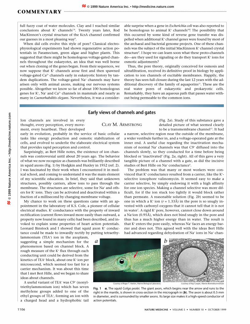
Surprisingly, as Bert Hille notes, the existence of ion chan-nels was controversial until about 20 years ago. The behaviorof what we now recognize as channels was brilliantly describedin squid axons (Fig. 1) by Hodgkin and Huxley in 1952 (ref. 2).I was fascinated by their work when I encountered it in med-ical school, and coming to understand it was the main elementof my scientific education. In brief, they said that unknownstructures, possibly carriers, allow ions to pass through themembrane. The structures are selective, some for Na+ and oth-ers for K+ ions. They can be activated and deactivated within afraction of a millisecond by changing membrane voltage.
My chance to work on these questions came with an ap-pointment in the laboratory of K.S. Cole, a pioneer of cellularelectrical studies. K+ conductance with the property of inwardrectification (current flows inward more easily than outward, aproperty now found in many cells) had been described, and in-voked to explain some properties of heart action potentials.Leonard Binstock and I showed that squid axon K+ conduc-tance could be made to inwardly rectify by putting tetraethy-lammonium (TEA+) ion in the axoplasm,suggesting a simple mechanism for thephenomenon based on channel block. Arough measure of the K+ flux through eachconducting unit could be derived from thekinetics of TEA+ block, about one K+ ion permicrosecond, which seemed too fast for acarrier mechanism. It was about this timethat I met Bert Hille, and we began to shareideas about channels.
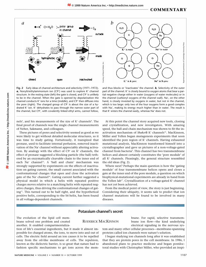
A useful variant of TEA+ was C9+ (nonyl-triethylammonium ion) which has sevenmethylene groups added to one of theethyl groups of TEA+, forming an ion witha charged head and a hydrophobic tail
(Fig. 2a). Study of this substance gave adetailed picture of what seemed clearlyto be a transmembrane channel12. It had
a narrow, selective region near the outside of the membrane,a wider vestibule further in, and a voltage-operated gate at theinner end. A useful clue regarding the inactivation mecha-nism of normal Na+ channels was that C9+ diffused into thechannels slowly, so they conducted for a time before beingblocked or ‘inactivated’ (Fig. 2a, right). All of this gave a verytangible picture of a channel with a gate, as did the incisivestudies of Bert Hille on Na+ conductance6.
The problem was that many or most workers were con-vinced that K+ conductance resulted from a carrier, like the K+-selective ionophore valinomycin. It seemed easy to make acarrier selective, by simply endowing it with a high affinityfor one ion species. Making a channel selective was more dif-ficult, for if the ion stuck too tightly it would block ratherthan permeate. A reasonable solution (Fig. 2b) seemed to beone in which a K+ ion (r = 1.33Å) in the pore is so snugly in-vested with carbonyl oxygens that it cannot tell that it is notin water7. A rigid K+ pore, however, cannot close down arounda Na+ion (0.95Å), which does not bind snugly in the pore andthus has a much higher energy than in water. The result isthat K+ enters the pore easily, whereas Na+ faces an energy bar-rier and does not. This agreed well with the ideas Bert Hillehad advanced regarding dehydration of Na+ ions in Na+ chan-
Fig. 1 a, The squid Goligo pealei. The giant axon, which begins near the arrow and runs to theright in the mantle, is shown in cross-section in the micrograph in (b). The axon is about 0.5 mmin diameter, and is surrounded by smaller axons. Its large size makes it a high-speed conductor ofaction potentials.
CLAY M. ARMSTRONG
a b
Courtesy of Roger T. Hanlon, Marine Biological Laboratory Courtesy of Kay Cooper, Marine Biomedical Institute
© 1999 Nature America Inc. • http://medicine.nature.com©
199
9 N
atu
re A
mer
ica
Inc.
• h
ttp
://m
edic
ine.
nat
ure
.co
m
NATURE MEDICINE • VOLUME 5 • NUMBER 10 • OCTOBER 1999 1107
COMMENTARY
Potassium channel’s secretThe evolution of the lipid cell mem-brane solved one problem and createdanother. It enabled compartmentaliza-tion of life’s essential ingredients, but it made it almost im-possible for charged atoms, the ions, to move into and out ofcells. The electric field around an ion causes it to be repelledaway from the oil-like membrane of cells. The repulsion,known as the dielectric barrier, is so great that nature had tofashion specific mechanisms to get ions across the mem-
brane. For rapid, selective transmem-brane ion flow—the kind underlyingelectrical signaling in the nervous sys-
tem and many other cellular processes—membrane-spanningproteins called ion channels were nature’s solution.
I began studying ion channels long after it was establishedthat they are protein pores in the cell membrane. In 1986, Iabandoned plans to practice medicine and began postdoc-toral studies with Christopher Miller, who provided an inspi-
nels6, and his measurements of the size of K+ channels8. Thefinal proof of channels was the single channel measurementsof Neher, Sakmann, and colleagues.
These pictures of pores and selectivity seemed as good as wewere likely to get without detailed molecular structures, so itwas time to study gating. Fortuitously, it transpired thatpronase, used to facilitate internal perfusion, removed inacti-vation of the Na+ channel without appreciably altering activa-tion. By analogy with the effect of C9+ on K+ channels, theeffect of pronase suggested a blocking particle (the ball) teth-ered by an enzymatically cleavable chain to the inner end ofeach Na+ channel13. A ‘ball and chain’ mechanism wasstrongly supported by experiments on the effects of inactiva-tion on gating current, the small current associated with theconformational changes that open and close the activationgate of the Na+ channel14. Gating current further suggested aphysical model in which a helix with repeated positivecharges moves relative to a matching helix with repeated neg-ative charges, thus driving the conformational changes of gat-ing15. This turned out to be half right, and the hypotheticalpositive helix, corresponding to the S4 helix, has been foundin all voltage-dependent channels.
At this point the channel story acquired new tools, cloningand crystallization, and new investigators. With amazingspeed, the ball and chain mechanism was shown to fit the in-activation mechanism of ShakerB K+ channels16. MacKinnon,Miller and Yellen began mutagenesis experiments that soonidentified the pore region of K+ channels. Having exhaustedmutational analysis, MacKinnon transformed himself into acrystallographer and gave us pictures of a non-voltage-gatedchannel from bacteria9. This channel has two transmembranehelices and almost certainly constitutes the ‘pore module’ ofall K+ channels. Pleasingly, the general structure resembledthe old ideas (Fig. 2).
Where next? Perhaps the main question is how the ‘gatingmodule’ of four transmembrane helices opens and closes agate at the inner end of the pore module, a question on whichbiophysical-mutational experiments are already in hand fromthe Yellen lab17. Crystallization of a voltage-gated K+ channelhas not yet been achieved.
From the medical point of view, the story is just beginning.Considering their ubiquity, it seems safe to predict that ionchannel mutations will be found to be involved in manydiseases.
RODERICK MACKINNON
Fig. 2 Early ideas of channel architecture and selectivity (1971–1972).a, Nonyltriethylammonium ion (C9+) was used to explore K+ channelstructure. In the resting state (left) the gate is closed, and C9+ is unlikelyto be in the channel. When the gate is opened by depolarization thechannel conducts K+ ions for a time (middle), and C9+ then diffuses intothe pore (right). The charged group of C9+ is about the size of a hy-drated K+ ion. K+ dehydrates to pass through the narrow outer part ofthe channel, but C9+, with covalently linked ethyl arms, cannot follow,
and thus blocks or ‘inactivates’ the channel. b, Selectivity of the outerpart of the channel. K+ is closely bound to oxygen atoms that bear a par-tial negative charge either in water (oxygens of water molecules) or inthe channel (carbonyl oxygens of the channel wall). Na+, on the otherhand, is closely invested by oxygens in water, but not in the channel,which is too large: only two of the four oxygens form a good complexwith Na+, making its energy much higher than in water. The result isthat K+ enters the channel easily, whereas Na+ does not.
a b
Rep
rinte
d w
ith t
he p
erm
issi
on o
f Cam
brid
ge
Uni
vers
ity P
ress
Cou
rtes
y of
Mar
cel D
ekke
r, In
c.
© 1999 Nature America Inc. • http://medicine.nature.com©
199
9 N
atu
re A
mer
ica
Inc.
• h
ttp
://m
edic
ine.
nat
ure
.co
m

1108 NATURE MEDICINE • VOLUME 5 • NUMBER 10 • OCTOBER 1999
COMMENTARY
rational beginning in science. The experiments of ClayArmstrong and Bertil Hille describing the gated pores ofsodium and potassium channels were already classics. Genesencoding sodium and calcium channels and the acetylcholineactivated channel had been cloned, and potassium channelclones were on the horizon. The fundamental questions hadreached a new level. What is the structure and chemistry be-hind the operation of an ion channel?
I began by asking how a small scorpion toxin inhibited apotassium channel. Using electrical measurements, I deducedthat the toxin occludes the channel’s ion pathway ratherthan interfering with gating of the channel18. That humbleconclusion set the course that I still follow today—one aimedat understanding how a potassium channel so precisely se-lects the potassium ion over sodium, and conducts it near thediffusion limit.
The first potassium channel gene was cloned in 1987 fromfruit fly: the Shaker gene19. The gene gave us the amino-acidsequence, but little information about the arrangement ofamino acids in the channel. The scorpion toxin allowed me toidentify which of the amino acids form the ion pathway, thefirst step toward understanding the channel’s three-dimen-sional structure. I began this work in Christopher Miller’s lab-oratory and then continued in my own laboratory, in 1989, atHarvard Medical School. Over the next several years, my labo-ratory reached a number of important conclusions about thearchitecture of a potassium channel using site-directed muta-genesis, electrical measurements, and simple reasoning. Weshowed that the channel contains four identical subunitsarranged in a symmetric ring around a central pore20. We alsodefined a special amino acid stretch known as the pore loop.Our experiments revealed that four pore loops, one from eachsubunit, meet near the channel’s central axis to form the nar-rowest point along the ion pathway. Gary Yellen and I ex-ploited the same potassium channel blocker used decadesearlier by Armstrong and Hille, tetraethylammonium, toshow that our pore loop conclusion must be true. Finally, mylaboratory demonstrated that a stretch of eight amino acids
within the pore loop is responsible for potassium selectivity21.We called these amino acids the potassium channel signaturesequence.
The signature sequence has been used as a key to find potas-sium channels even in bacteria. The obvious implication isthat nature used the same structural scaffold to form the se-lectivity filter in all potassium channels. This intriguing con-clusion inspired me to want to directly visualize a potassiumchannel in a way that mutagenesis experiments could not ac-complish. I therefore set out to learn X-ray crystallography,spending long hours purifying and attempting to crystallize afew soluble proteins, learning a new trade while gathering ad-vice from experienced colleagues. A complete change of envi-ronment, from Harvard to Rockefeller University, removedany temptation to fall back on mutagenesis experiments, andhelped me to focus intensely on the new effort. It was fright-ening! From my laboratory at Harvard only one beginningpostdoctoral scientist, Declan Doyle, and my wife Alice (achemist who felt sorry for me) joined the effort to crystallize apotassium channel.
Two very good things happened before despair set in. First,the Streptomyces lividans potassium channel (KcsA) was de-scribed22, and second, my laboratory grew quickly into a stillsmall but very enthusiastic group of talented scientists, dedi-cated to solving ion channel structures. The KcsA channel hasonly two membrane-spanning segments per subunit andclosely resembles the Shaker channel in its amino acid se-quence. The ever-valuable scorpion toxin confirmed that theKcsA channel had to be closely related in structure to its eu-karyotic counterparts23. Through much hard work and deter-mination, crystals were obtained that allowed us to solve the
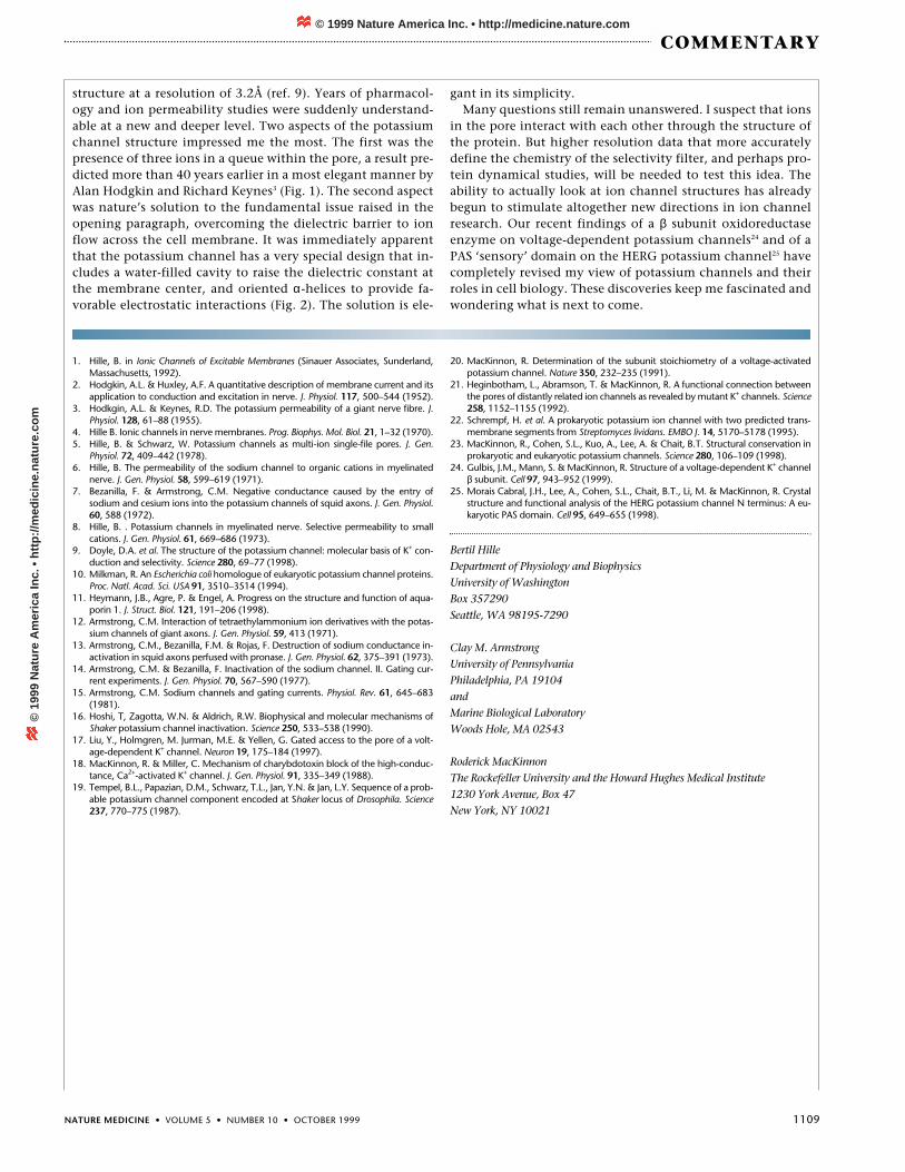
Fig. 2 Potassium channel demonstrates nature’s mechanism of loweringthe dielectric barrier. The channel contains a water-filled cavity (pale blue)in its ion pathway, halfway through the cell membrane (white). Alpha he-lices direct their cation-attractive negative ends (red) toward the cavity,stabilizing the positively charged potassium ion (green sphere). Withoutthe cavity and helices, a potassium ion would be unable to cross the mem-brane center. Reprinted with permission from Science 280, 75 (1998).
Fig. 1 Three ions in the potassium channel. Two subunits of the potas-sium channel are shown as ribbons (blue and gold). Electron density (redmesh) shows three ions within the ion pathway: two are located in the se-lectivity filter (top) and one, in a cavity at the membrane center (bottom).The subunits are oriented with the extracellular solution on top.
© 1999 Nature America Inc. • http://medicine.nature.com©
199
9 N
atu
re A
mer
ica
Inc.
• h
ttp
://m
edic
ine.
nat
ure
.co
m
NATURE MEDICINE • VOLUME 5 • NUMBER 10 • OCTOBER 1999 1109
COMMENTARY
1. Hille, B. in Ionic Channels of Excitable Membranes (Sinauer Associates, Sunderland,Massachusetts, 1992).
2. Hodgkin, A.L. & Huxley, A.F. A quantitative description of membrane current and itsapplication to conduction and excitation in nerve. J. Physiol. 117, 500–544 (1952).
3. Hodkgin, A.L. & Keynes, R.D. The potassium permeability of a giant nerve fibre. J.Physiol. 128, 61–88 (1955).
4. Hille B. Ionic channels in nerve membranes. Prog. Biophys. Mol. Biol. 21, 1–32 (1970).5. Hille, B. & Schwarz, W. Potassium channels as multi-ion single-file pores. J. Gen.
Physiol. 72, 409–442 (1978).6. Hille, B. The permeability of the sodium channel to organic cations in myelinated
nerve. J. Gen. Physiol. 58, 599–619 (1971).7. Bezanilla, F. & Armstrong, C.M. Negative conductance caused by the entry of
sodium and cesium ions into the potassium channels of squid axons. J. Gen. Physiol.60, 588 (1972).
8. Hille, B. . Potassium channels in myelinated nerve. Selective permeability to smallcations. J. Gen. Physiol. 61, 669–686 (1973).
9. Doyle, D.A. et al. The structure of the potassium channel: molecular basis of K+ con-duction and selectivity. Science 280, 69–77 (1998).
10. Milkman, R. An Escherichia coli homologue of eukaryotic potassium channel proteins.Proc. Natl. Acad. Sci. USA 91, 3510–3514 (1994).
11. Heymann, J.B., Agre, P. & Engel, A. Progress on the structure and function of aqua-porin 1. J. Struct. Biol. 121, 191–206 (1998).
12. Armstrong, C.M. Interaction of tetraethylammonium ion derivatives with the potas-sium channels of giant axons. J. Gen. Physiol. 59, 413 (1971).
13. Armstrong, C.M., Bezanilla, F.M. & Rojas, F. Destruction of sodium conductance in-activation in squid axons perfused with pronase. J. Gen. Physiol. 62, 375–391 (1973).
14. Armstrong, C.M. & Bezanilla, F. Inactivation of the sodium channel. II. Gating cur-rent experiments. J. Gen. Physiol. 70, 567–590 (1977).
15. Armstrong, C.M. Sodium channels and gating currents. Physiol. Rev. 61, 645–683(1981).
16. Hoshi, T, Zagotta, W.N. & Aldrich, R.W. Biophysical and molecular mechanisms ofShaker potassium channel inactivation. Science 250, 533–538 (1990).
17. Liu, Y., Holmgren, M. Jurman, M.E. & Yellen, G. Gated access to the pore of a volt-age-dependent K+ channel. Neuron 19, 175–184 (1997).
18. MacKinnon, R. & Miller, C. Mechanism of charybdotoxin block of the high-conduc-tance, Ca2+-activated K+ channel. J. Gen. Physiol. 91, 335–349 (1988).
19. Tempel, B.L., Papazian, D.M., Schwarz, T.L., Jan, Y.N. & Jan, L.Y. Sequence of a prob-able potassium channel component encoded at Shaker locus of Drosophila. Science237, 770–775 (1987).
20. MacKinnon, R. Determination of the subunit stoichiometry of a voltage-activatedpotassium channel. Nature 350, 232–235 (1991).
21. Heginbotham, L., Abramson, T. & MacKinnon, R. A functional connection betweenthe pores of distantly related ion channels as revealed by mutant K+ channels. Science258, 1152–1155 (1992).
22. Schrempf, H. et al. A prokaryotic potassium ion channel with two predicted trans-membrane segments from Streptomyces lividans. EMBO J. 14, 5170–5178 (1995).
23. MacKinnon, R., Cohen, S.L., Kuo, A., Lee, A. & Chait, B.T. Structural conservation inprokaryotic and eukaryotic potassium channels. Science 280, 106–109 (1998).
24. Gulbis, J.M., Mann, S. & MacKinnon, R. Structure of a voltage-dependent K+ channelβ subunit. Cell 97, 943–952 (1999).
25. Morais Cabral, J.H., Lee, A., Cohen, S.L., Chait, B.T., Li, M. & MacKinnon, R. Crystalstructure and functional analysis of the HERG potassium channel N terminus: A eu-karyotic PAS domain. Cell 95, 649–655 (1998).
Bertil HilleDepartment of Physiology and BiophysicsUniversity of WashingtonBox 357290Seattle, WA 98195-7290
Clay M. ArmstrongUniversity of PennsylvaniaPhiladelphia, PA 19104andMarine Biological LaboratoryWoods Hole, MA 02543
Roderick MacKinnonThe Rockefeller University and the Howard Hughes Medical Institute1230 York Avenue, Box 47New York, NY 10021
structure at a resolution of 3.2Å (ref. 9). Years of pharmacol-ogy and ion permeability studies were suddenly understand-able at a new and deeper level. Two aspects of the potassiumchannel structure impressed me the most. The first was thepresence of three ions in a queue within the pore, a result pre-dicted more than 40 years earlier in a most elegant manner byAlan Hodgkin and Richard Keynes3 (Fig. 1). The second aspectwas nature’s solution to the fundamental issue raised in theopening paragraph, overcoming the dielectric barrier to ionflow across the cell membrane. It was immediately apparentthat the potassium channel has a very special design that in-cludes a water-filled cavity to raise the dielectric constant atthe membrane center, and oriented α-helices to provide fa-vorable electrostatic interactions (Fig. 2). The solution is ele-
gant in its simplicity.Many questions still remain unanswered. I suspect that ions
in the pore interact with each other through the structure ofthe protein. But higher resolution data that more accuratelydefine the chemistry of the selectivity filter, and perhaps pro-tein dynamical studies, will be needed to test this idea. Theability to actually look at ion channel structures has alreadybegun to stimulate altogether new directions in ion channelresearch. Our recent findings of a β subunit oxidoreductaseenzyme on voltage-dependent potassium channels24 and of aPAS ‘sensory’ domain on the HERG potassium channel25 havecompletely revised my view of potassium channels and theirroles in cell biology. These discoveries keep me fascinated andwondering what is next to come.
© 1999 Nature America Inc. • http://medicine.nature.com©
199
9 N
atu
re A
mer
ica
Inc.
• h
ttp
://m
edic
ine.
nat
ure
.co
m