document4
TRANSCRIPT

J. vet. anat. Vol 1 No1, (2008) 29 - 3729
Air breathing organ in catfish Ahmed et al.
Anatomical, Light and Scanning Electron Microscopic Studies on the Air Breathing Dendretic Organ of the Sharp Tooth Catfish (Clarias gariepinus)
Abd-Elmaksoud Ahmed1, Kassab Mohamed2, Sayed-Ahmed Ahmed3,
Fayed Masoud 4
1 Department of Cytology and Histology, Faculty of Veterinary Medicine, Mansoura University, Mansoura, Egypt2 Department of Histology, faculty of veterinary medicine, Kafr El Sheikh University, Kafr El Sheikh, Egypt 3 Department of Anatomy and Embryology, Faculty of Veterinary Medicine, Alexandria University, Damanhur branch, Bostan, Egypt 4 Department of Anatomy and Embryology, Faculty of Veterinary Medicine, Kafr El Sheikh University, Kafr El Sheikh, Egypt
E-mail: [email protected]___________________________________________________________________________ With 20 figures Received February 2008; accepted for publication April 2008
Abstract
Previously, it has been shown that different species of fishes are adapted in varying degrees for gaseous exchange in both water and air. Our study was carried out on sharp tooth catfish (Clarias garie-pinus) obtained from the Nile River, Egypt and fo-cused on the dendretic organ (DO) investigating its anatomical, scanning and light microscopical fea-tures. Anatomically, the air-breathing dendretic organ of the catfish was located in the suprabran-chial chamber caudodorsal to the gills and com-posed of two main parts; small and large ones origi-nated by main stems from the second and fourth gill arches respectively. Histologically, the DO either in its main stems or in the smaller branches and in the end bulbs consists of core of elastic cartilage, vas-cular layer of connective tissue and covering epithe-lium containing intraepithelial mucous glands. The blood capillaries (channels) were found to originate from the vascular layer and penetrate the covering epithelium. Toward the surface, these capillaries dilate and bulge out due to engorgement with RBCs forming what is called respiratory papillae. The SEM showed that the stem divisions and their bulbus ends contained double parallel rows of paired pro-jections (respiratory lamellae) separated by areas of smooth surface. Moreover, the surfaces were stud-ded with microvilli. In conclusion, the structure of this organ manifests profuse internal subdivision that may produce a particularly large respiratory surface area to extract the oxygen from air. Therefore, the catfish might be one of the most suitable fish for the intensive production in the fish aquaculture.
Key Words
Dendretic organ, Catfish, E/M
Introduction
The sharp tooth catfish (Clarias garipeinus) is one of the most important fresh water fishes in the River Nile in Egypt. It has a great economic importance, contributing about 17.5 % of the total country catch (GAFRD, 1996). Most fishes obtain oxygen from water using efficient gills, but low oxygen solu-bility in water limits oxygen availability. To cope with hypoxic water, some fishes swim to the surface and take in the oxygen-rich water at the interface, while others have evolved the ability to breath atmospheric air (Liem, 1967; Mittal and Munshi, 1971; Mittal and Banerjee, 1974; Mittal et al. 1980; Suzuki, 1992; Graham, 1997; Ishimatsu et al. 1998; Park et al. 2000; Zhang et al. 2000; Park, 2002). The air respi-ration in fishes could be done either through swim bladder as Bowfin (Amia calva) (Gervais and Tufts, 1998) and Arapaima gigas (Brauner et al., 2004), or through the skin as in mudskipper (Periophthalmus magnuspinnatus) (Park, 2002), or through the ac-cessory respiratory organ as in cat fish (clarias garipinus ) (Nawar, 1955; Moussa, 1956 and 1957; Vandewalle and Chardon, 1991;), cat fish (clarias mossambicus) (Maina and Maloiy 1986) and cat fish (clarias batrachus ) (Munshi, 1961; Lewis, 1979). and Climbing perch (Anabas testudineus) (Munshi et al., 1986). The Clariidae is a bottom feeding omni-vore (Bishai and Khalil 1997) that has a wide geo-graphical distribution and inhabitant tropical swamps and rivers which are liable to seasonal drying. To survive in such habitats, with changing oxygen tension, this group of fish has acquired a bimodal gas exchange capacity wherein the gill extract oxy-gen from water and the accessory respiratory organ extract it from air (Nawar, 1955; Moussa, 1956 and 1957; Munshi 19961; Vandewalle and Chardon, 1991; Bishai and Khalil 1997; Graham, 1997; Chan-

J. vet. anat. Vol 1 No1, (2008) 29 - 3730
Air breathing organ in catfish Ahmed et al.
dra and Banerjee, 2003). Briefly, the air breathing organs not only enable the fish to survive in hypoxic water but also permit them to stay out of the water for hours at a time (Chandra and Banerjee, 2003).
Previously, the respiratory organs have been studied in some of the closely related catfishes such as Clarias batrachus ((Munshi, 1961; Lewis, 1979; Chandra and Banerjee, 2003), and Clarias mossam-bicus (Johnston et al., 1983 Maina and Maloiy 1986), wherein it is formed of aquatic breathing organ (gills) and aerial breathing organs. The accessory respira-tory organs (aerial breathing organs) are repre-sented by dendretic organ (labyrinthine or arbores-cent organ), fan organ and the epithelium lining the membrane of the suprabranchial chamber (Maina and Maloiy 1986; Chandra and Banerjee, 2003). Although several approaches have previously inves-tigated the morphology and physiology of the respi-ratory organs of the sharp tooth cat fish (Clarias garipeinus) in Egypt (Nawar 1955; Moussa 1956, 1957; Zayed and Mohamed 2004), none of these studies paid detailed attention to the structures of the dendretic organ of this species. Therefore, our study aims to shed light on the anatomical, light and scan-ning microscopical features of the dendretic organ of catfish (Clarias garipeinus).
Materials and methods
This study was carried out on 10 apparently healthy mature fishes. The fish weight ranged from 500-1500 gm and measured 30-45 cm in length. Five fishes were used to demonstrate the gross morphological features. After catching the fishes from the River Nile, they were transported in dry plastic aquaria to our lab in 2-3 hours to allow the aerial respiration. Firstly, the fishes were sacrificed and then the oper-cular cavity was opened. The gill and associated accessory respiratory organs were examined in situ and then dissected and photographed by Sony digital camera.
For light microscopic study, small samples (0.3-0.5 mm3) of the dendretic organ were taken from three fishes. The samples were fixed in Bouin’s solution for 18-24 hrs. After fixation, the samples were exten-sively washed in 70 % alcohol (3 x 24 hr) to get rid of the fixative before the subsequent step of tissue processing. The tissue samples were then dehy-drated in graded series of ethanol (80%, 95% and absolute), cleared in xylene and impregnated and embedded in paraffin wax. Sections of 5-7 m were cut using Leica rotatory microtome (RM 2035) and mounted on glass slides. Paraffin sections were kept in incubator at 40°C until used for conventional staining (H&E, Massons Tricrome, Verhoeff’s, Go-mori’s, PAS, and AB pH 2.5). All of the staining techniques employed were performed according to Bancroft and Steven (1990).
For scanning electron microscopic study, two fishes were used. Small samples from different parts of the dendretic organ were taken. The sam-ples were fixed in a mixture of paraformaldehyde 2.5 % and glutaraldehydehyde 2.5 % solution in 0.1 M phosphate buffer for 4 hours at 4°C. After washing in the same buffer, the specimens were post-fixed in osmiumtetroxide 1% in phosphate buffer for two hours followed by washing in the same buffer. The samples were then dehydrated in ascending grades of ethanol followed by critical point drying in carbon dioxide, then sputter-coated with gold and examined with Jeol JSM 5300 scanning electron microscope, Faculty of Science, Alexandria University.
Results
Our results were focused on the anatomical, scan-ning and light microscopical features of the den-dretic organ (DO) of sharp tooth cat fish Clarias gariepinus (Fig. 1) and descried in details as follow:
The Anatomical features
The air-breathing dendretic organ of the catfish was located in the suprabranchial chamber caudodorsal to the gills and covered wholly by the membrane lining the suprabranchial chamber (Fig. 2). Removal of this membrane was shown that the dendretic organ is additionally covered rostrally by fan-like structures projected from the first, second and third gill arches (Figs. 2, 3, 4). Structurally DO was divided into two parts; small anterior part and large posterior part that originating from the proximal end of the 2nd
and 4th branchial arches respectively (Figs. 3, 4). The size of the anterior small part represented nearly 50 % of the large one and occupied the majority of the anterior compartment of the suprabranchial chamber. The large posterior part occupied the majority of the middle and posterior compartments of the suprabranchial chamber. Both of the anterior and posterior part was connected to its corresponding gill arch by cartilaginous joint and originated by small smooth surface main stem which further divided into a number of secondary branches (Fig. 5). The main stem of small part of the dendretic organ has 3 secondary branches while the large one was divided into 4-5 branches. The secondary branches of both anterior and posterior parts repeatedly divided into small ones in dichotomous branching to end in bulbus like structure (bulbus terminals). This archi-tecture made the organ in the form of a tree –like structure (dendretic structure) (Figs. 3, 4, 5).
Light Microscopy
Histologically, the DO either in its main stem or in the smaller branches and in the end bulb consists of definite structures represented by a core of elastic cartilage, vascular layer of connective tissue and

J. vet. anat. Vol 1 No1, (2008) 29 - 3731
Air breathing organ in catfish Ahmed et al.
covering epithelium of stratified type (Figs. 6,7, 8).The cartilaginous core was covered externally by a thick layer of perichondrium which composed mainly of elastic and collagen fibers. In contrast to the normal elastic cartilage which is a vascular, the perichondrium penetrates inside the cartilaginous core carrying the blood vessels (Fig. 6). This archi-tecture may demarcate the divisions of the cartilage into the smaller branches of the DO. Internally, the core was found to consist of, chondrocytes, network of elastic fibers, blood vessels, and small collections of fat cells (Fig. 7). The vascular connective tissue layer was shown to consist of large blood vessels (arteries and veins), connective tissue fibers (colla-gen and reticular) and connective tissue cells, mainly fibroblasts. The large blood vessels were found to further branch into smaller ones to end finally in blood channels (capillaries) (Fig. 9). The latter pene-trate the covering epithelium in a regular manner, as they were separated by 2 epithelial cells thick (pillar cells). Moreover, these capillaries were found to dilate and bulge out toward the surface due to en-gorgement with RBCs forming what is called respira-tory papillae (Figs. 10, 11). The covering epithelium was shown to consist predominantly of stratified epithelium rest on basal lamina. Intraepithelial mu-cous glands clearly stained with PAS and AB was also seen within the covering epithelium. These glands were lined by cuboidal cells containing flat-tened nuclei near its basal lamina (Figs. 9, 11, 12).
SEM
The arborization of the dendretic organ was like cauliflowers. There were branches of bulbus-like structure arisen from the stem divisions (Fig. 13). Generally, the surface of the stem divisions and their bulbus ends were found to contain transversely oriented folds (respiratory lamellae) separated by intervening spaces. These folds are papillae like-structures which projected over the surface and arranged in several parallel rows. However, the double parallel rows of stem divisions separated by wide areas of smooth surface while the interlamellar space was narrow (Figs. 14, 15). Moreover, the epithelium between the paired lamellae contained mucus arisen from the intraepithelial mucous gland (Fig. 15). On the other hand, the paired lamellae of the bulbus end was found to differ from that of the stem divisions, where they were high, crowded and separated by a comparatively narrow space (Figs. 14, 15). The interlamellar space of the bulbus end was also very small comparing with that of the stem divisions (Figs. 16, 17). The covering epithelium of the respiratory lamellae and the spaces in between was studded with microvilli and the opening of the intraepithelial mucous gland were clear (Figs. 17, 18).In cross section of the bulbus end, the core con-tained connective tissue fibers separated by narrow
spaces. At the periphery of the core, large blood spaces were additionally seen just beneath the epithelium. These blood spaces contained tongue-like projections originated from its endothelium and protruded into the lumen from tissue side toward the periphery (Figs. 19, 20).
Discussion
Our study on the sharp tooth cat fish Clarias gerie-pinus has revealed that the dendretic organ was completely covered by suprabranchial membrane and partially by fan-like structures. Previously, all of these structures were descried as air-breathing organs in walking catfish Clarias batrachus (Munshi 1961; Olson et al.1995). However, this fact needs further studies on the fish under investigation. The dendretic part of the air-breathing organ of the cat fish Clarias geriepinus is derived from the 2nd and 4th
gill arches. These findings are coordinate well with the findings of the previous investigations in Siluroi-dei clarias and Heterobranchus (Harder, 1975), Clarias batrachus (Munshi, 1961; Lewis, 1979; Chandra and Banerjee, 2003), Clarias mossambicus(Johnston et al 1983 Maina and Maloiy 1986) and in Clarias geriepinus (Moussa 1956; Zayed and Mo-hamed 2004). On the contrary the dendretic organ of Anabantoidei and Perciformes (Harder, 1975) and Channa punctata and Channa striatus (Munshi 1962) was originated from the 1st gill arch only. There is some similarities between the dendretic organ and the gills; the gaseous exchange surfaces of the dendretic organ consists of double rows of paired lamellae (biserial arrangement), a feature strongly indicative of their common origin from the gills. However, and in contrast to the epithelial cells surface of the dendretic organ which has numerous small projections that consists of microvilli, the sur-face of gill cells has numerous concentric whorls of micro-ridges (Lewis 1979). These structural differ-ences may attribute to the different mechanisms of respirations (aquatic and aerial). Additionally, the entire dendretic organ including its bulbus terminals has additional support of an internal cartilaginous core, which prevents collapse of the respiratory lamellae when the fish faces the loss of water due to dehydration during air exposure. This keeps the respiratory surfaces freely exposed, so that the fish can continues aerial exchange of gases for fairly long periods. Interestingly, the aforementioned structural adjustment may be one of the main rea-sons for the comparatively better aerial performance of the dendretic organ compared to the gills (Chan-dra and Banerjee 2003).
The double rows of paired lamellae on the dendretic organ are crowded in the bulbus ends and sepa-rated by little space while that of the main stem are widely separated. These results indicate that the main respiratory part is confined to the bulbus ends.

J. vet. anat. Vol 1 No1, (2008) 29 - 3732
Air breathing organ in catfish Ahmed et al.
Furthermore, the presence of microvilli on the sur-face of the respiratory lamellae and in between may serve to trap bacteria or particulate manner and to humidify the air (Munshi et al. 1986). Our results were shown an enormous increase in the number of RBCs in the blood channels and in the underlying blood vessels. These results is supported by the fact that, following air exposure, engorgement of the blood channels causes them to stretch and dilate, especially those at the tips of bulbus end (respira-tory lamellae) resulting in either finger like projec-tions (Chandra and Banerjee 2003), or balloon like projections (Parashar and Banerjee 1999) or globu-lar shape projections (Banerjee and Chandra 2005) onto the surface of the dendretic organ. The load on these thin–walled tubular structures thus increases resulting in bending of these blood capillaries (chan-nels) at 90° to rest flat on the surface of dendretic organ. All these structural adaptation narrow the barrier distance between the atmospheric air in the suprabranchial chamber and the blood in the respi-ratory lamellae of the dendretic organ which may be a compensatory measure to the failure of branchial respiration (Chandra and Banerjee, 2003; Banerjee and Chandra,2005). The endothelial cells of the transverse blood channels have unusual structural modifications, which are tongue-like projections in the lumen directed from tissue side toward the sur-face side. This finding suggests that their shape is governed by the direction of blood flow through the channels (Munshi et al. 1986). Moreover, Hughes and Munshi (1978) supposed that, these processes act as valves controlling the flow of blood through the channels. They also point out that the tongue-like extensions may act as an obstacle to RBCs and force them to flow over the top of the extensions. This would place the RBCs in closest proximity to the respiratory surface and reduce the air-blood diffusion distance and maximize gas transfer (Mun-shi et al. 1986).
In our study the intraepithelial mucous glands were showed to contain neutral and/or acidic mucopoly-saccharides as indicated by PAS and AB stains. This result ascertains the mucous nature of the respiratory surface of the gills and dendretic organs of the catfish Clarias gariepinus (Zayed and Mo-hamed 2004) and Clarias mossambicus (Johnston et al., 1983; Maina and Maloiy 1986). Continuous elaboration of this mucous over the surface epithe-lium of the respiratory organs keeps the surfaces moist and clean as the mucus facilitates the removal of the trapped intoxicating agents, pathogen and foreign materials when the fishes live outside the water. Additionally, this mucous is important not only as a protective barrier but also has an ion regulatory function (Handy et al, 1989). Taken together, mu-cous glands help in the extension of the survival period after air exposure where during the initial period the mucous cell showed periodic fluctuations
in their density and staining properties and stained for sulphated mucopolysaccharides that known to hold an additional quantity of water to keep the surface moist for long period (Chandra and Banerjee 2004). However, the protective role played by the slime coating does not last for long time as the mucous glands and its cells become exhausted and degenerate (Chandra and Banerjee 2003, 2004).
Although the catfish Clarias gariepinus has a smaller gill surface area than the Nile tilabia (Zayed and Mohamed 2004), it can survive for long period either in the hypoxic water or outside the aquatic environ-ment (Chandra and Banerjee, 2004). This fact might be partially explained by the presence of the den-dretic organ in the catfish. Such notion is supported by the observation of Sigh and Huges, (1971) who reported that the catfish Clarias batrachus even when kept in aerated water it obtains 58% of its oxygen from air. Moreover, the catfish clarias mos-sambicus is entirely dependent for survival on its capacity to breathe atmospheric oxygen (Johnston et al., 1983). Importantly we should know that air breathing does not help the fish to survive for unlim-ited period, and the fish ultimately die due to failure of respiration, hemorrhage , and collapse of many other important physiological processes (CO2 and N2 elimination) even though the air-breathing organ continue to absorb small amount of O2 aerially. In other words, while O2 absorption is the dominant feature of air breathing, the aquatic breathing re-mains very important for CO2 and N2 elimination (Chandra and Banerjee 2003).
In conclusion, the catfish can survive for long period either in the hypoxic water or outside the aquatic environment due to the presence of the dendretic organ. The structure of this organ manifests profuse internal subdivision that may produce a particularly large respiratory surface area to extract the oxygen from air. Therefore, the catfish might be one of the most suitable fish for the intensive production in the fish aquaculture.
References Bancroft, J.D., Stevens, A. (1990): Theory and
practice of histological techniques. Third Edition. Churchill Livigstone, Londo , Tor-ento.
Banerjee T.K., Chandra, S. (2005): Estimation of zinc chloride contamination by histopa-thological analysis of the respiratory organ of the air-breathing murrel channa striata (Bloch 1797) Channiformes, Pisces) Vetri-nareski Arhiv, 75(3):253-263.
Bishai, H.M., Khalil M.T. (1997): Fresh water fishes in Egypt. Publication of National Biodiver-sity Unit. No 9.

J. vet. anat. Vol 1 No1, (2008) 29 - 3733
Air breathing organ in catfish Ahmed et al.
Brauner, c.j.; Matey, V.; Wilson, J.M.; Bernier, N.J., Val, A.L. (2004): transition in organ func-tion during the evolution of air-breathing; insights from Arapima gigas, an obligatory air-breathing teleost from the Amazon. J. of Exp. Biology, 207: 1433-1438
Chandra S., Banerjee, T.K. (2003):
Histopathological analysis of the respira-tory organs of the air-breathing catfish Clarias batrachus (Linn). Exposed to the air. Acta Zoologica Taiwanica, 14 (1): 45-64.
Chandra, S., Banerjee T.K. (2004):
Histopathological analysis of the respira-tory organs of Channa straiata subjected to the air exposure. Vetrinareski Arhiv, 74: 37-52.
GAFRD (General Authority of Fish Research De-velopment in Egypt 1996): Annual report for country fish production.
Gervais, M.R. and Tufts, B.L. (1998): Evidence for membrane- bound carbonic anhyderase in the air bladder of Bowfin (Amia calva), a primitive air-breathing fish. J.Exp. Bi-ol.,201 :2205-2212
Graham, J.B. (1997): Air-breathing fishes: Evolu-tion, Diversity and Adaptation. San Diego, Academic Press.
Handy, R.D., Eddy, F.B., Romain, G. (1989). In vitro evidence for the ionoregulatory role of rainbow trout mucus in acid / aluminum and zinc toxicity. J Fish Biol. 35: 737-747.
Harder, W. (1975): Anatomy of Fishes. part 1 : Text. E. Schweizerbartsche Verlagsbuch-handlung, Stuttgart.
Hughes G.M., Munshi, J.S.D. (1978): Scanning electron microscopy of the respiratory sur-faces of Saccobranchus fossilis (Bloch). Cell Tissue Res., 195: 99-109
Ishimatsu, A.; Hishida, Y., Takita, T.; Oikawa, S. Takeda, T., Huat, K. (1998): Mudskipper store air in their burrows. Nature, 391: 237-238.
Johnston, I.A., Brenard, L.M., Maloiy, G.M. (1983). Aquatic and aerial respiration rates, mus-cle capillary supply and mitochondrial vol-ume denesty in the air breathing catfish (Clarias Mossamicus) acclimated to either aerated or hypoxic water. J. exp. Biol. 105: 317-338
Lewis S.V. (1979): The morphology of the acces-sory air-breathing organ of the catfish,
Clarias batrachus: A SEM study. J. Fish biology, 14: 187-191.
Liem, K.F. (1967): Functional morphology of the integumentry, respiratory and digestive systems of the synbranchoid fish, Mo-nopetrus albus. Copeia, 375-388.
Maina, J.N., Maloiy, G.M.O. (1986). The morphol-ogy of the respiratory organs of the African air-breathing catfish (Clarias mossambi-cus): A light, electron and scanning micro-scopic study, with morphometric observa-tions. J Zool., Lond. 209: 421-445
Mittal, A., Banerjee, T. (1974): Structure and kera-tinization of the skin of a fresh water te-olest notopterus notopterus. J. of Zoology, 174: 314-355.
Mittal, A., Munshi, J.S.D. (1971): A comparative study of the structure of the skin of certain air-breathing fresh water teolest. J. of Zo-ology163: 515-532.
Mittal, A.; Whitear, M., Agarwal, S. (1980): Fine structure and histochemistry of the epi-dermis of the fish, Monopterus cuchia. J. of Zoology, 191: 107-125.
Moussa, A.T (1956): morphology of the accessory respiratory organ of the teleost calarias la-zera (C and V). J Morph. 98: 125-160.
Moussa, A.T (1957): physiology of the accessory respiratory organ of the teleost calarias la-zera (C and V). J exp.Zool. 136; 419-454
Munshi, J.S.D. (1961): Accessory respiratory or-gan of the calarias Batrachus (Linn). J Morph. 109; 115-140
Munshi, J.S.D. (1962). On accessory respiratory organ of Ophicephalus punctatus (Bloch) and O Striatus (Bloch). J. Linn. Soc. Lond. (Zool.) 44; 616-626
Munshi, J.S.D., Olson, K.R.; Ojha, J., Ghosh, T.K. (1986).Morphology and vascular anatomy of the accessory respiratory organof the air-breathing climbing perch, Anabas tes-tudeinus (Bloch). Am. J. Anat., 176: 321-331.
Nawar, G. (1955). on the anatomy of clarias laz-era. J Morph. 97; 23-38
Olson K. R.; Ghosh T. K.; Roy P. K. and Munshi J. S. (1995): Microcirculation of gills and ac-cessory respiratory organ of the walking catfish Clarias batrachus. Anat. Rec., 242 (3): 383-399.
Parashar R. S. and Banerjee T.K. (1999): Re-sponse of aerial respiratory organs of the air-breathing catfish Heteropneustes fos-

J. vet. anat. Vol 1 No1, (2008) 29 - 3734
Air breathing organ in catfish Ahmed et al.
silis (Bloch) to extreme stress of desicca-tion. Vetrinarski Arhev, 69(2): 63-68.
Park,J.Y. (2002): Structure of the skin of an air-breathing mudskipper, Periophthalmus magspinnatus. J. of fish Biology, 60: 1543-1550.
Park,J.Y.; Kim, I., Kim, S.Y. (2000): Histological study of the skin of the amphibious fish, Periophthalmus modestus. Korean J. of Biological Sci. 4: 315-318.
Sigh, B.N., Hughes, G.M. (1971): Respiration of a. air breathing catfish Clarias barrachus (linn). J exp. Biol. 55: 421-434.
Suzuki,N. (1992): Fine structure of the epidermis of the mudskipper, Periophthalmus mod-estus. Japanese J. of Ichthyology, 38: 379-396.
Vandewalle, P., Chardon, M. (1991): A new hy-pothesis on the air flow in air breathing in Clarias c\Gariepinus (teleostei, silurifor-mes). Belg. J. Zool. 121: 73-80.
Zayed, A.E., Mohamed, S.A. (2004): Morphologi-cal study on the gills of two species of fresh water fishes: Oreochromis niloticus and Claias gariepinus. Ann Anat. 186: 295-304
Zhang, J., Taniguchi, T.; Takita, T., Ali, A.B. (2000): On the epidermal structure of Bo-leophthalmus and Scartelaos mudskippers with reference to their adaptation to terres-trial life. Icthyological Res. 47: 359- 366.
Fig. 1: Photograph of the sharp tooth catfish Clarias gariepinus.Fig. 2: Photograph of the ventral part of head region with exposed gill chambers and associated air breathing organs showed gill (G), gill fan (F), membrane covering suprabranchial chamber (arrow) and dendritic organ (DO). Fig. 3: The gill system and associated air breathing organs showed gill arches (1, 2, 3, 4 and 5), gill fan (F), small part of dendritic organ (SDO) and large part of dendritic organ ( LDO). Fig. 4: The gill system and associated air breathing organs showed gill arches (1, 2, 3, 4 and5), gill fan (F), small part of dendritic organ (SDO) and large part of dendritic organ ( LDO).
J. vet. anat. Vol 1 No1, (2008) 29 - 3735
Air breathing organ in catfish Ahmed et al.
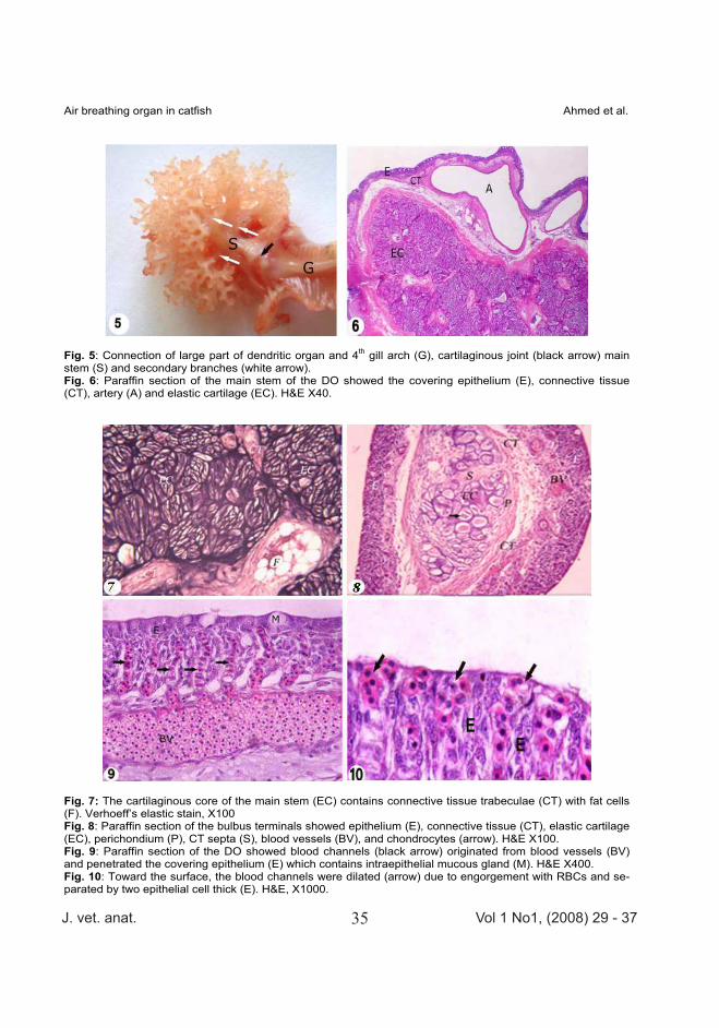
Fig. 5: Connection of large part of dendritic organ and 4th gill arch (G), cartilaginous joint (black arrow) main stem (S) and secondary branches (white arrow). Fig. 6: Paraffin section of the main stem of the DO showed the covering epithelium (E), connective tissue (CT), artery (A) and elastic cartilage (EC). H&E X40.
Fig. 7: The cartilaginous core of the main stem (EC) contains connective tissue trabeculae (CT) with fat cells (F). Verhoeff’s elastic stain, X100 Fig. 8: Paraffin section of the bulbus terminals showed epithelium (E), connective tissue (CT), elastic cartilage (EC), perichondium (P), CT septa (S), blood vessels (BV), and chondrocytes (arrow). H&E X100. Fig. 9: Paraffin section of the DO showed blood channels (black arrow) originated from blood vessels (BV) and penetrated the covering epithelium (E) which contains intraepithelial mucous gland (M). H&E X400. Fig. 10: Toward the surface, the blood channels were dilated (arrow) due to engorgement with RBCs and se-parated by two epithelial cell thick (E). H&E, X1000.

J. vet. anat. Vol 1 No1, (2008) 29 - 3736
Air breathing organ in catfish Ahmed et al.
Fig. 11: Epithelium of bulbus end (E) showed vascular papillae (arrow) and mucous gland (M). H&E X1000. Fig. 12: PAS positive intraepithelial mucous gland (arrow). PAS, X600
Fig. 13: Scanning electron micrograph of cauliflower-shaped DO showed stem (S) and bulbus end (B). Fig. 14: Higher magnification to the previous showed double parallel rows of respiratory lamellae (black and white arrow) on the stem (S) and bulbus end (B) respectively. Fig. 15: Higher magnification of double parallel rows of respiratory lamellae (arrow) on the stem (S) with epi-thelial surface (ES) in between contained mucus (white arrow head). Fig. 16: Higher magnification of double parallel rows of respiratory lamellae (RL) on the bulbus end and the intervening space in between (arrow).

J. vet. anat. Vol 1 No1, (2008) 29 - 3737
Air breathing organ in catfish Ahmed et al.
Fig. 17: Scanning electron micrograph of bulbus end showed the respiratory epithelium (RE) in between the respiratory lamellae (RL). Fig. 18: Higher magnification of the respiratory lamellae (RL) showed microvilli on its surface and opening of the mucous gland (arrow). Fig. 19: Scanning electron micrograph of cross section of the bulbus end showed connective tissue core (CC) and blood vessels (BV). Note the respiratory lamellae on the surface (arrow). Fig. 20: Higher magnification of figure 19 showed tongue-like protrusion (arrow) inside the blood vessels.
Address for correspondence Dr. Mohamed Kassab, Department of Cytology and Histology, Faculty of Veteri-nary Medicine, Kafr El Sheikh University, Kafr El Sheikh, Egypt