42 year old male presents after head trauma
TRANSCRIPT
42 year old male presents
after head trauma
Ryan Joyce, MD
Rafael Pacheco, MD


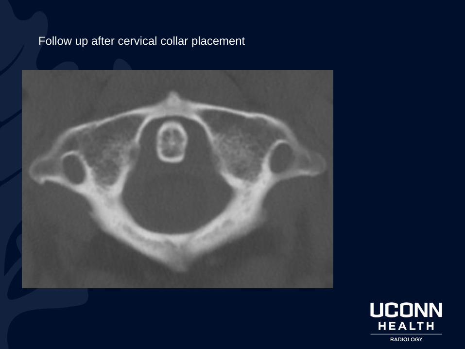
Follow up after cervical collar placement

Follow up after cervical collar placement

Follow up extension cervical spine radiograph

Follow up flexion
cervical spine
radiograph

Atlantoaxial instability
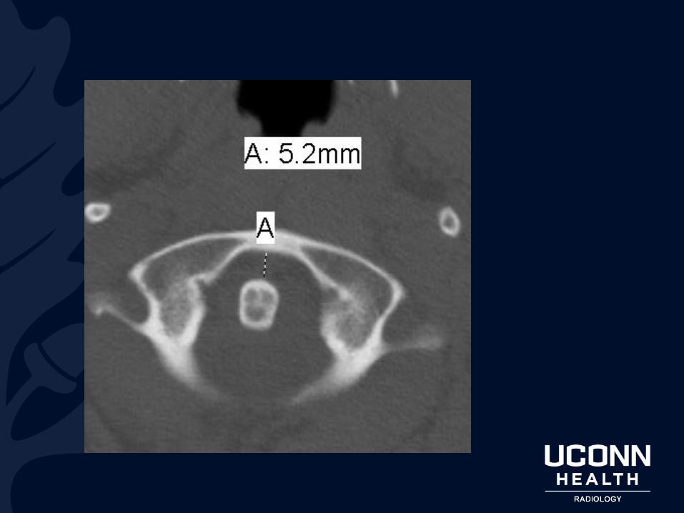
Imaging findings:• In adults, widening of the atlantoaxial interval >2 mm.
• In childen, >~5mm widening considered abnormal.
• Atlantoaxial interval defined as distance from posterior cortex of the anterior arch of the atlas to the anterior cortex of the dens.
• If suspected or borderline, perform neutral lateral, flexion lateral, and extension lateral radiographs of the cervical spine – if present, will reveal dynamic change in the atlantoaxial interval, indicating instability.
• Thin-section CT is most sensitive and specific for associated fractures.
• MR is most sensitive and specific for the characterization of associated ligamentous injuries.
• Can also have rotary subluxation/instability, though rare.

Atlantoaxial instability
Etiology:
• Related to ligamentous disruption or laxity.
• Primary ligamentous stability provided by the cruciate ligament and tectorial membrane.
• Related ligamentous stability of the atlanto-occipital joint provided by the alar ligaments. – Acquired:
• Trauma, Grisel syndrome, surgery, arthritides (rheumatoid, psoriatic, reactive, ankylosing spondylitis, SLE)
– Congenital:• Os odontoideum, Down syndrome, Morquio syndrome, Spongyloepithelial
dysplasia, Osteogenesis imperfecta, Marfan disease, NF1.

Atlantoaxial instability
Radiographics

Atlantoaxial instability
Radiographics

Atlantoaxial instability
Radiographics

Atlantoaxial instability
Significance:
• Instability and posterior subluxation of the dens relative to the atlas results in narrowing of the spinal canal with possible injury to the thecal sac and contents.
Treatment:
• Varies depending on severity and etiology. Importantly, in traumatic cases, mechanical stabilization with C1-C2 fusion typically performed to avoid cervical cord injury.

References
1. Imaging of Atlanto-Occipital and Atlantoaxial Traumatic Injuries: What the
Radiologist Needs to Know. Roy Riascos, Eliana Bonfante, Claudia Cotes,
Mary Guirguis, Reza Hakimelahi, Clark West. Radiographics. 2015 Nov-
Dec; 35(7): 2121–2134. doi: 10.1148/rg.2015150035
2. Statdx.com