3d slicer - johns hopkins bloomberg school of public health
TRANSCRIPT
3D Slicer
John Muschelli and Vadim Zipunnikov
Department of Biostatistics
November 18, 2011
John Muschelli and Vadim Zipunnikov (JHU) 3D-Slicer November 18, 2011 1 / 39
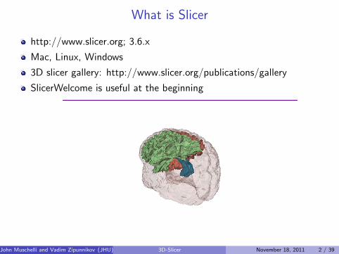
What is Slicer
http://www.slicer.org; 3.6.x
Mac, Linux, Windows
3D slicer gallery: http://www.slicer.org/publications/gallery
SlicerWelcome is useful at the beginning
John Muschelli and Vadim Zipunnikov (JHU) 3D-Slicer November 18, 2011 2 / 39

Step 1: Loading data
Brain atlas atlas.img/hdr (Analyze)
91 labeled regions1 medial front-orbital gyrus right
2 middle frontal gyrus right
3 lateral ventricle left
4 insula right
5 precentral gyrus right
6 lateral front-orbital gyrus right
7 cingulate region right
8 lateral ventricle right
9 medial frontal gyrus left
10 superior frontal gyrus right
11 globus palladus right
12 globus palladus left
14 putamen left
15 inferior frontal gyrus left
16 putamen right
17 frontal lobe WM right
19 angular gyrus right
23 subthalamic nucleus right
25 nucleus accumbens right
26 uncus right
27 cingulate region left
29 fornix left
30 frontal lobe WM left
32 precuneus right
33 subthalamic nucleus left
34 PLICICPL*
35 PLICICPR*
36 hippocampal formation right
37 inferior occipital gyrus left
38 superior occipital gyrus right
39 caudate nucleus left
41 supramarginal gyrus left
43 anterior limb of internal capsule left
45 occipital lobe WM right
50 middle frontal gyrus left
52 superior parietal lobule left
53 caudate nucleus right
54 cuneus left
56 precuneus left
57 parietal lobe WM left
59 temporal lobe WM right
60 supramarginal gyrus right
61 superior temporal gyrus left
62 uncus left
63 middle occipital gyrus right
64 middle temporal gyrus left
69 lingual gyrus left
70 superior frontal gyrus left
72 nucleus accumbens left
73 occipital lobe WM left
74 postcentral gyrus left
75 inferior frontal gyrus right
80 precentral gyrus left
83 temporal lobe WM left
85 medial front-orbital gyrus left
86 perirhinal cortex right
88 superior parietal lobule right
90 lateral front-orbital gyrus left
92 perirhinal cortex left
94 inferior temporal gyrus left
95 temporal pole left
96 entorhinal cortex left
97 inferior occipital gyrus right
98 superior occipital gyrus left
99 lateral occipitotemporal gyrus right
100 entorhinal cortex right
101 hippocampal formation left
102 thalamus left
105 parietal lobe WM right
108 insula left
110 postcentral gyrus right
112 lingual gyrus right
114 medial frontal gyrus right
118 amygdala left
119 medial occipitotemporal gyrus left
128 anterior limb of internal capsule right
130 middle temporal gyrus right
132 occipital pole right
133 corpus callosum
139 amygdala right
140 inferior temporal gyrus right
145 superior temporal gyrus right
154 middle occipital gyrus left
159 angular gyrus left
165 medial occipitotemporal gyrus right
175 cuneus right
196 lateral occipitotemporal gyrus left
203 thalamus right
243 background
251 occipital pole left
254 fornix right
255 subarachnoid cerebro-spinal fluid
John Muschelli and Vadim Zipunnikov (JHU) 3D-Slicer November 18, 2011 3 / 39

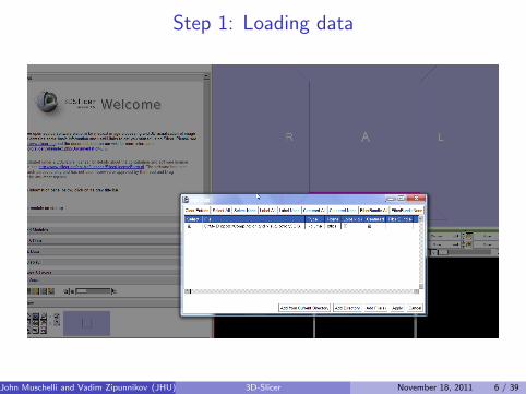
Step 1: Loading data
File ⇒ Add Data ⇒ choose atlas.hdr (check Centered if applicable)⇒ Click Apply.note: .img files always go with .hdr, at this step .hdr should bechosen.
Result: the data is loaded and you can see it in the three windows(directional)
John Muschelli and Vadim Zipunnikov (JHU) 3D-Slicer November 18, 2011 4 / 39

Step 1: Loading data
John Muschelli and Vadim Zipunnikov (JHU) 3D-Slicer November 18, 2011 5 / 39
Step 1: Loading data
John Muschelli and Vadim Zipunnikov (JHU) 3D-Slicer November 18, 2011 6 / 39

Step 1: Loading data
John Muschelli and Vadim Zipunnikov (JHU) 3D-Slicer November 18, 2011 7 / 39
Step 2: Creating Volume
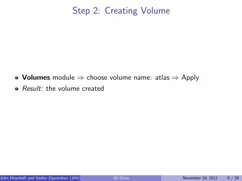
Volumes module ⇒ choose volume name: atlas ⇒ Apply
Result: the volume created
John Muschelli and Vadim Zipunnikov (JHU) 3D-Slicer November 18, 2011 8 / 39

Step 2: Creating Volume
John Muschelli and Vadim Zipunnikov (JHU) 3D-Slicer November 18, 2011 9 / 39
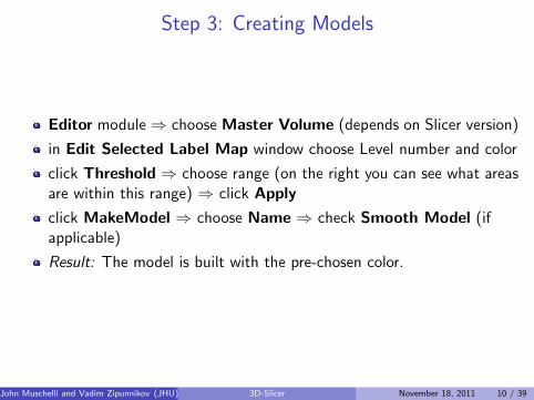
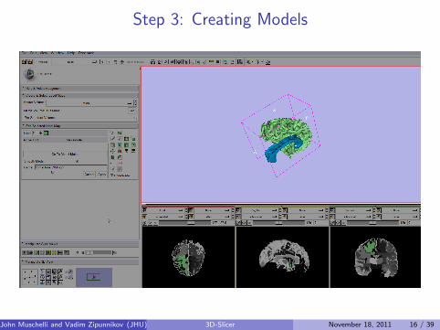
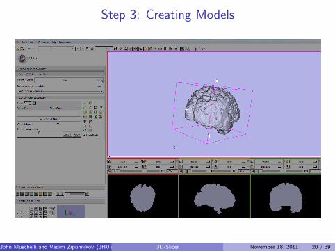
Step 3: Creating Models
Editor module ⇒ choose Master Volume (depends on Slicer version)
in Edit Selected Label Map window choose Level number and color
click Threshold ⇒ choose range (on the right you can see what areasare within this range) ⇒ click Apply
click MakeModel ⇒ choose Name ⇒ check Smooth Model (ifapplicable)
Result: The model is built with the pre-chosen color.
John Muschelli and Vadim Zipunnikov (JHU) 3D-Slicer November 18, 2011 10 / 39

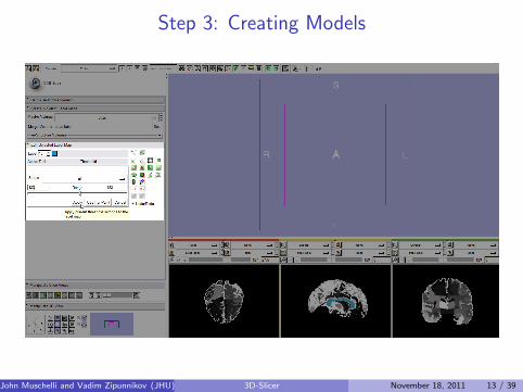
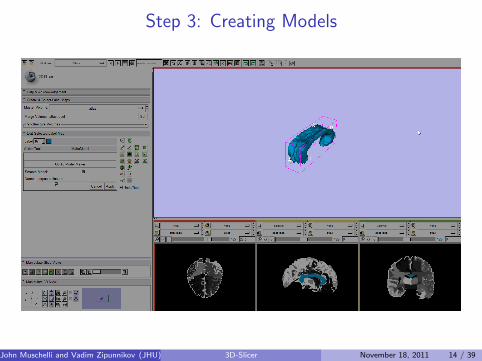
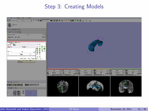
Step 3: Creating Models
The first model is corpus callosum labeled 133(Threshold: 133:133), color: blue
The second model is frontal lobe WM right labeled 17 (Threshold:17:17), color: green
The third model is frontal lobe WM left labeled 30 (Threshold:30:30), color: red
The last model is the whole brain (Threshold: 1:255), color: grey
John Muschelli and Vadim Zipunnikov (JHU) 3D-Slicer November 18, 2011 11 / 39

Step 3: Creating Models
John Muschelli and Vadim Zipunnikov (JHU) 3D-Slicer November 18, 2011 12 / 39
Step 3: Creating Models
John Muschelli and Vadim Zipunnikov (JHU) 3D-Slicer November 18, 2011 13 / 39
Step 3: Creating Models
John Muschelli and Vadim Zipunnikov (JHU) 3D-Slicer November 18, 2011 14 / 39
Step 3: Creating Models
John Muschelli and Vadim Zipunnikov (JHU) 3D-Slicer November 18, 2011 15 / 39
Step 3: Creating Models
John Muschelli and Vadim Zipunnikov (JHU) 3D-Slicer November 18, 2011 16 / 39

Step 3: Creating Models
John Muschelli and Vadim Zipunnikov (JHU) 3D-Slicer November 18, 2011 17 / 39

Step 3: Creating Models
John Muschelli and Vadim Zipunnikov (JHU) 3D-Slicer November 18, 2011 18 / 39

Step 3: Creating Models
John Muschelli and Vadim Zipunnikov (JHU) 3D-Slicer November 18, 2011 19 / 39
Step 3: Creating Models
John Muschelli and Vadim Zipunnikov (JHU) 3D-Slicer November 18, 2011 20 / 39

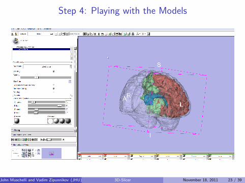
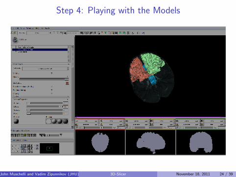
Step 4: Playing with the Models
Models module shows the created models
you can change visibility, opacity, set new color(in a much moreconvenient way), play with other things
The brain opacity can be set to see the other regions
John Muschelli and Vadim Zipunnikov (JHU) 3D-Slicer November 18, 2011 21 / 39

Step 4: Playing with the Models
John Muschelli and Vadim Zipunnikov (JHU) 3D-Slicer November 18, 2011 22 / 39
Step 4: Playing with the Models
John Muschelli and Vadim Zipunnikov (JHU) 3D-Slicer November 18, 2011 23 / 39
Step 4: Playing with the Models
John Muschelli and Vadim Zipunnikov (JHU) 3D-Slicer November 18, 2011 24 / 39
Step 5: Saving the Scene
Save ⇒ Select scene (.mrml) and the volumes(.vtk) included into thescene
Once the scene is saved you can load it later
John Muschelli and Vadim Zipunnikov (JHU) 3D-Slicer November 18, 2011 25 / 39

Step 5: Saving the Scene
John Muschelli and Vadim Zipunnikov (JHU) 3D-Slicer November 18, 2011 26 / 39

What is Slicer
Awesome, duh.
John Muschelli and Vadim Zipunnikov (JHU) 3D-Slicer November 18, 2011 27 / 39
Getting Data in
Bring in dataI DICOM/Analyze/NIFTI : File → Add VolumeI NIFTI : File → Add DataI You can bring in Analyze with Add Data if selecting .hdr file
Generally need a brain image (structural / functional / template) -needs to be in same space as labels
This makes up a scene (pretty much a project)
John Muschelli and Vadim Zipunnikov (JHU) 3D-Slicer November 18, 2011 28 / 39

Labels
Label map is surprisingly a map of labels.
We’ll be looking at categorical labels (thresholded or differentstructures)
We need to construct a “model”, which essentially is a 3Dconstruction of the data.
We go to Editor Module (upper left of panel, around 10 o’clock)
John Muschelli and Vadim Zipunnikov (JHU) 3D-Slicer November 18, 2011 29 / 39
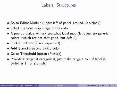
Labels: Structures
Go to Editor Module (upper left of panel, around 10 o’clock)
Select the label map image in the data
A pop-up dialog will ask you what label map (let’s just try genericcolors - which are not that good, but defaul)
Click structures (if not expanded)
Add Structures and pick a color
Go to Threshold button (Picture)
Provide a range: if categorical, just make range 1 to 1 if label iscoded as 1, for example
John Muschelli and Vadim Zipunnikov (JHU) 3D-Slicer November 18, 2011 30 / 39

Labels: Make some models
Once you’re done adding all your structures, then let’s build themodel!
Merge all
Merge and Build: there should be an image now in the 3D viewer.
John Muschelli and Vadim Zipunnikov (JHU) 3D-Slicer November 18, 2011 31 / 39
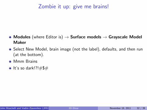
Zombie it up: give me brains!
Modules (where Editor is) → Surface models → Grayscale ModelMaker
Select New Model, brain image (not the label), defaults, and then run(at the bottom).
Mmm Brains
It’s so dark!?!#$#
John Muschelli and Vadim Zipunnikov (JHU) 3D-Slicer November 18, 2011 32 / 39

Tweak me
Modules (where Editor is) → Models
Grayscale Model (Scroll down)
Change opacity/diffusion
Try some presets, they are the shades spheres (come on, try it).
Bottom left corner - click the axes for different views
Click the eye to take off / put on axes and such
Click two check boxes to see things spin!
John Muschelli and Vadim Zipunnikov (JHU) 3D-Slicer November 18, 2011 33 / 39

Feeling Saucy? Record movie
gtk-recordMyDesktop for Linux
Jing for Mac
Windows? - google
John Muschelli and Vadim Zipunnikov (JHU) 3D-Slicer November 18, 2011 34 / 39

More applications: BOO
Slicer can read in 4D data, but I haven’t explored.
bioImageSuite - if trying to record a 4D movie, this does it
Originally for Cardiac 4D movies - so pretty good.
If you find something better, tell me!
John Muschelli and Vadim Zipunnikov (JHU) 3D-Slicer November 18, 2011 35 / 39
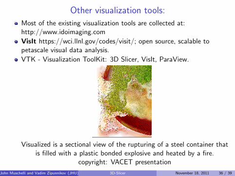
Other visualization tools:
Most of the existing visualization tools are collected at:http://www.idoimaging.com
VisIt https://wci.llnl.gov/codes/visit/; open source, scalable topetascale visual data analysis.
VTK - Visualization ToolKit: 3D Slicer, VisIt, ParaView.
Visualized is a sectional view of the rupturing of a steel container thatis filled with a plastic bonded explosive and heated by a fire.
copyright: VACET presentation
John Muschelli and Vadim Zipunnikov (JHU) 3D-Slicer November 18, 2011 36 / 39

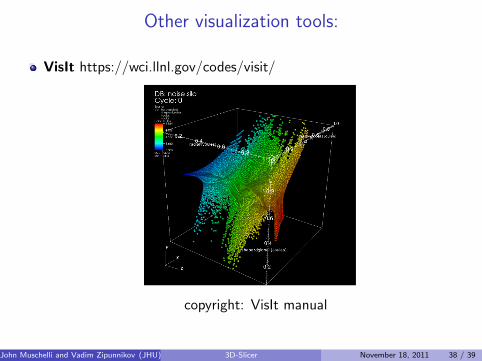
Other visualization tools:
VisIt https://wci.llnl.gov/codes/visit/
copyright: VACET presentation
John Muschelli and Vadim Zipunnikov (JHU) 3D-Slicer November 18, 2011 37 / 39
Other visualization tools:
VisIt https://wci.llnl.gov/codes/visit/
copyright: VisIt manual
John Muschelli and Vadim Zipunnikov (JHU) 3D-Slicer November 18, 2011 38 / 39

Other visualization tools:
ParaView www.paraview.org; open source
copyright: http://wiki.multiscaleflows.mecheng.strath.ac.uk
John Muschelli and Vadim Zipunnikov (JHU) 3D-Slicer November 18, 2011 39 / 39