3d in vitro model for drug efficiency testing
TRANSCRIPT
Potentials of Using 3D In Vitro Models for Drug
Efficiency Testing (in comparison to 2D)
By: Tiffany Ho, Shannen Ser, Arial Chan, Lim Ee Jing, Oo Ju Nn

Basic Principles of 2D & 3D Cell Cultures
Aspects 2D cultures 3D cultures
Growth condition Adhere and grow on a flat surface
Grow on a matrix or in suspension medium
Morphology Flat & stretched ; monolayer Form aggregates or spheroids
Cell status Mostly proliferating stage Mixture of cells at different stages
Limitation Does not adequately mimic the in vivo microenvironment
Core cells receive less oxygen, growth factors and nutrients from medium ; in quiescent or hypoxic state
Advantage Receive nutrients, growth factors and oxygen equally
Mimic in vivo microenvironment
Table 1 shows the comparison of principles of 2D & 3D cultures. (Edmondson 2014)

Potentials of 3D Cultures for Drug Testing
1. 2D cultured cells are stretched out in an unnatural state on flat substance, whereas cells in 3D cultures on
biological, synthetic or non-synthetic scaffold materials maintain a normal morphology.
- cells are relatively similar to the naturally-occurring cells in body.
2. Cellular response to drugs treatments in 3D cultures - more similar to what happen in vivo than 2D.
-studies have found that cells in 3D cultures are more resistant to anticancer drugs than 2D cultures.
3. 3D cultures have more stability and longer lifespan compared to cells cultured in 2D
-for long-term studies and long-term effects of the drugs.
4. Cells in 3D cultures are undisturbed whereas cells in 2D cultures have to be trypsinized when reach confluence.
(Antoni 2015)

Applications ➔ Study of tumor development
- Cancer cells response to host immune modulatory effect.
➔ Evaluation of anticancer drug sensitivity
➔ Drugs discovery
➔ High throughput screening
➔ 3D cell-based biosensors
- Investigate cell’s response to drugs
- Detect biological signals transmitted by the cell
➔ Microfluidic-based device: Organs-on-chips
- Serve as disease model; mimic human organ functions.
- Small device with hollow channels lined by living cells cultured with nutrient liquids flowing
through the channels.
Figure 1: How biosensors work (Vaghasiya n.d.)

Cancer cells response to host immune modulatory effect
1)Macrophages(recruited by solid tumors during their progression)
were integrated into a collagen-based 3D co-culture model system of
squamous cell carcinoma (SCCs) and showed tumor-promoting effects.
2)Melanoma cells cultured in spheroids showed immunomodulator
functions aside from the inhibition of mitogen-dependent T-
lymphocyte activation and proliferation. This method allows to study
the aggressiveness of certain malignancies and can be utilized to
investigate the poor responses of malignancies to various
immunotherapy drug treatments.
3) The expression of chemokine ligand 21 (CCL21) and interferon
gamma (IFNγ) that recruit and activate tumor specific T cells when in
3D scaffold model of breast cancer. In this method , IFNγ and CCL21
were delivered into tumor cells via plasmids, and transfected cells
were seeded to form spheroids on three-dimensional (3D) chitosan-
alginate (CA) scaffolds. This provides a useful breast cancer tumor
environment model to evaluate the T cell function.
Figure 2: Schematic illustration of a typical tumor microenvironment.
Adapted from (Joyce and Pollard, 2009; Koontongkaew, 2013).
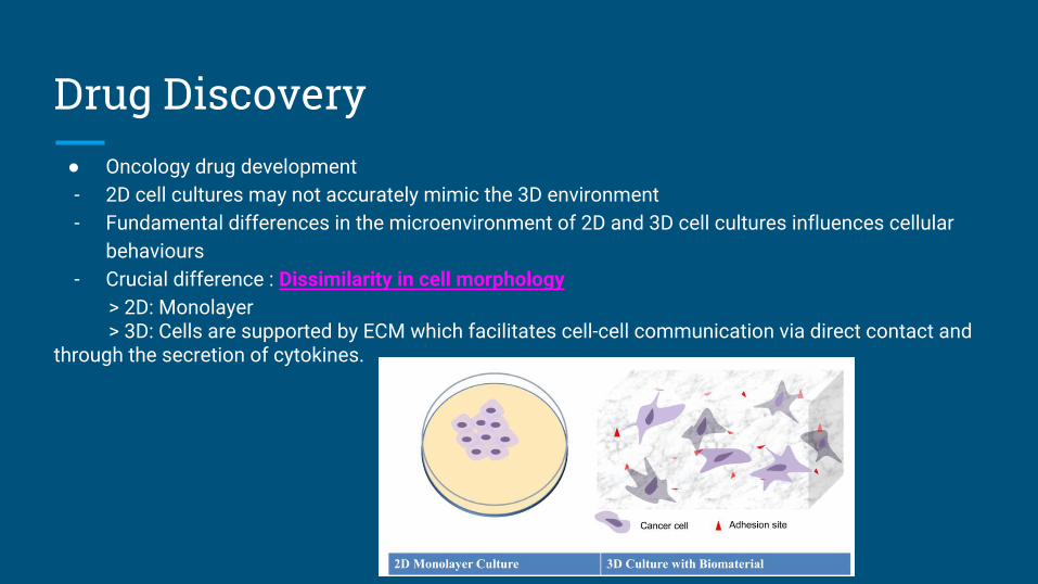
Drug Discovery● Oncology drug development- 2D cell cultures may not accurately mimic the 3D environment- Fundamental differences in the microenvironment of 2D and 3D cell cultures influences cellular
behaviours- Crucial difference : Dissimilarity in cell morphology
> 2D: Monolayer > 3D: Cells are supported by ECM which facilitates cell-cell communication via direct contact and through the secretion of cytokines.

High Throughput Screening (HTS)
● HTS assays using monolayer (2D) cultures still reflect a highly artificial cellular environment
- Limiting the predictive value for the clinical efficacy of a compound
● Optimize preclinical selection of the most active molecules from a large pool of potential effectors
● 3D cell culture systems:
- Spheroids are emphasized due to their advantages and potential for rapid development as HTS
systems
- 3D double network Hydrogels are similar to natural tissues and their chemical tunability which
impart abilities for response

❏ Mimics lungs physiology.
❏ Translucent design allows the viewing of inner workings of
human lungs.
❏ Contains tiny hollow channels lined with lung cells and
capillary cells separated by porous membrane.
❏ Vacuum pumps on either side of each channel expand and contract, thus imitating the action of a real alveolar sac. (Whitwam 2012)
Figure 3: Lung-on-a-chip (Anthony 2012)
Figure 4: Lung-on-a-chip as model for pulmonary edema (Whitewam 2012)
❏ Mimics inflammatory response triggered by microbial
pathogens.
e.g. WBCs migrate across capillary cells into the air space to
engulf bacteria.
❏ Used to model pulmonary edema by introducing IL-2 in
blood channel.

Biomaterials Technology ● Explores materials which are not passive and walled off by the body
● Actively participates in body’s effort to repair itself
● Biometric and bioactive materials are designed to mimic the body’s natural structures & functions from macro- to micro- to nano-levels.
● Give rise to tissue and organ development
● Replacement of animal testing using combined models(Ou & Hosseinkhani 2014)
Figure 5: Schematic illustration of tissue engineering based on 3D biomaterials technology.
- Regeneration of defective and injured tissue
Current Developments

3D Printing Technology
● Biological construct in small range (mm-cm), including several cell types and biomaterials at the same time
● Use 3D biomaterials printing and with cell patterning
● Constructing 3D scaffolds with living cells embedded in hydrogels
● Functional tissue is formed faster compared to classical tissue engineering methods
(Ou & Hosseinkhani 2014)
Figure 6: 3D printing technology for tissue engineering

Thank You!Questions?

References Antoni, D, Burckel, H, Josset, E & Noel, G, ‘Three-Dimensional Cell Culture : A breakthrough in Vivo’, International Journal of Molecular Science, vol. 7, no. 1, viewed 28 April 2016, <http://www.ncbi.nlm.nih.gov/pmc/articles/PMC4394490/>.
Dougherty, E 2010, Living, breathing human lung-on-a-chip: A potential drug-testing alternative, viewed 28 April 2016, <http://wyss.harvard.edu/viewpressrelease/36/living-breathing-human-lungonachip-a-potential-drugtesting-alternative>.
Edmondson, R, Broglie, J, Adcock, A & Yang, L, ‘Three Dimensional Cell Culture Systems and Their Applications in Drug Discovery and Cell-based Biosensors’, International Journal of Molecular Science, vol. 3, vol.1, viewed 28 April 2016, <http://www.ncbi.nlm.nih.gov/pmc/articles/PMC4026212/#B5>.
Tolikas, M 2014, Wyss Institute's technology translation engine launches 'Organs-on-Chips' company, viewed 28 April 2016, <http://wyss.harvard.edu/viewpressrelease/161>.
Ou, K & Hosseinkhani, H, 2014, ‘Development of 3D in Vitro Technology for Medical Applications’, IJMS, vol. 15, no. 10, pp.17938-17962.
Vaghasiya, K, Applications of Biosensors technology : Future trends development and new intervation in biotechnology, viewed 28 April 2016,<http://www.pharmatutor.org/articles/applications-of-biosensors-technology-future-trends-development-and-new-intervation-in-biotechnology>.
Whitwam, R 2012, Lung-on-a-chip could change the way disease is treated, viewed 28 April 2016, <http://www.geek.com/chips/lung-on-a-chip-could-change-the-way-disease-is-treated-1527521/>.