39th annual advanced us seminar disney yacht club & beach...
TRANSCRIPT
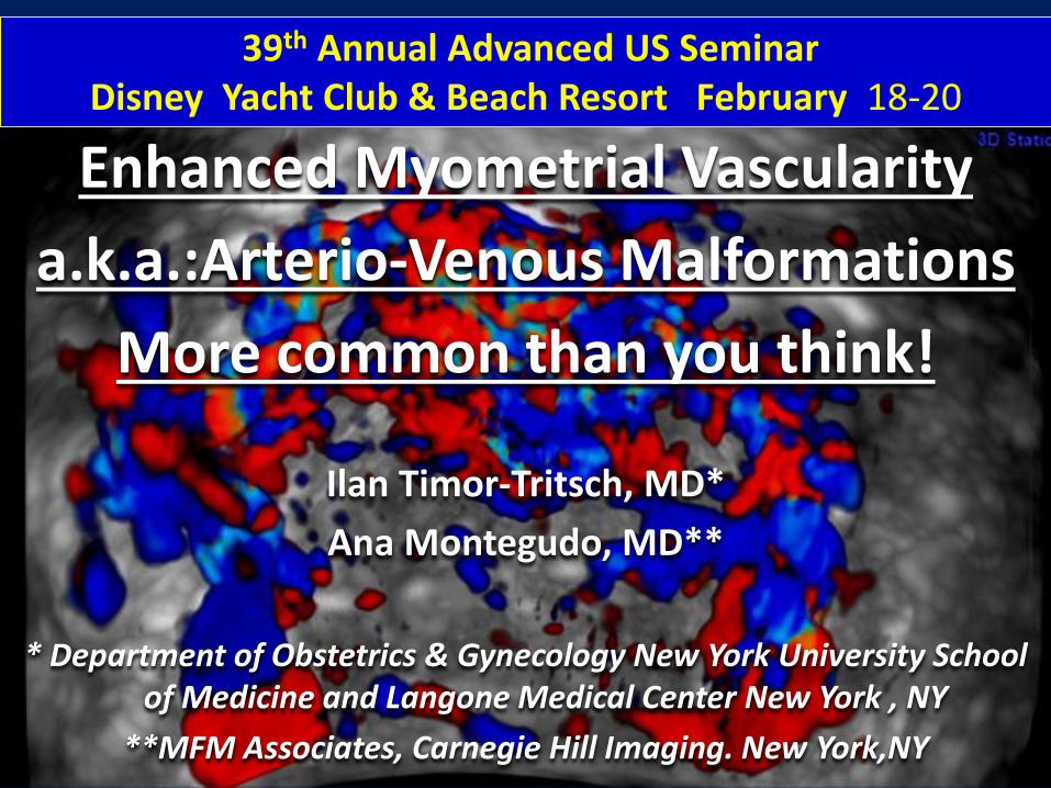
Enhanced Myometrial Vascularity
a.k.a.:Arterio-Venous Malformations
More common than you think!
Ilan Timor-Tritsch, MD*
Ana Montegudo, MD**
* Department of Obstetrics & Gynecology New York University School of Medicine and Langone Medical Center New York , NY
**MFM Associates, Carnegie Hill Imaging. New York,NY
•
39th Annual Advanced US SeminarDisney Yacht Club & Beach Resort February 18-20

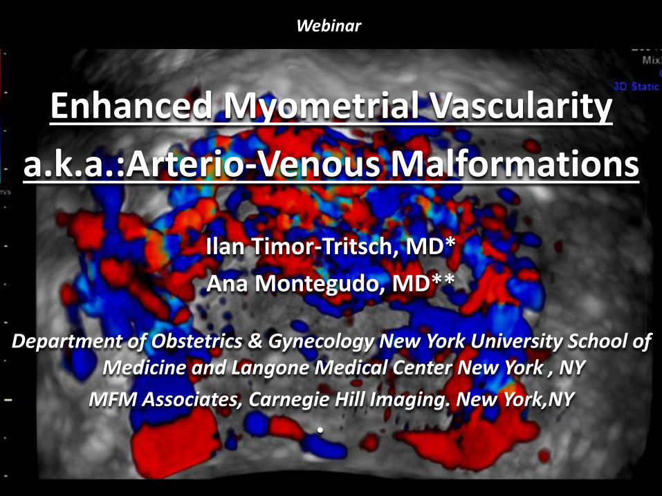
Enhanced Myometrial Vascularity
a.k.a.:Arterio-Venous Malformations
More common than you think!
Ilan Timor-Tritsch, MD*
Ana Montegudo, MD**
* Department of Obstetrics & Gynecology New York University School of Medicine and Langone Medical Center New York , NY
**MFM Associates, Carnegie Hill Imaging. New York,NY
•
12th ISUOG International Symposium Miami, February 17-20
Enhanced Myometrial Vascularity
a.k.a.:Arterio-Venous Malformations
Ilan Timor-Tritsch, MD*
Ana Montegudo, MD**
Department of Obstetrics & Gynecology New York University School of Medicine and Langone Medical Center New York , NY
MFM Associates, Carnegie Hill Imaging. New York,NY
•
Webinar

Disclosure
• Nothing to disclose

Objectives• The participants will be able to:• Realize the existence and clinical reality of
Enhanced Myometrial Vascularity aka AVM
• Learn the typical sonographic image of EMV
• Be able to make the diagnosis relying on using several Doppler techniques
• Use the US based information to triage patients according to a combination of clinical and sonographic presentation

Outline

EMV/AVM what is it?
• It is a clinical entity few are familiar with
• It is a pathology some deny its existence
• It is a disease that has different terms
What I want to achieve with this talk
• Familiarize Ob/Gyn’s with EMV/AVM
• Convince skepticals that it is real
• Provide an all inclusive term for it

• Failed 9 weeks IUP. Misopristol given for TOP
• Increasing amount of bleeding for last 1 week
• Referred for US. Here is the image:
Typical presentation
22mm
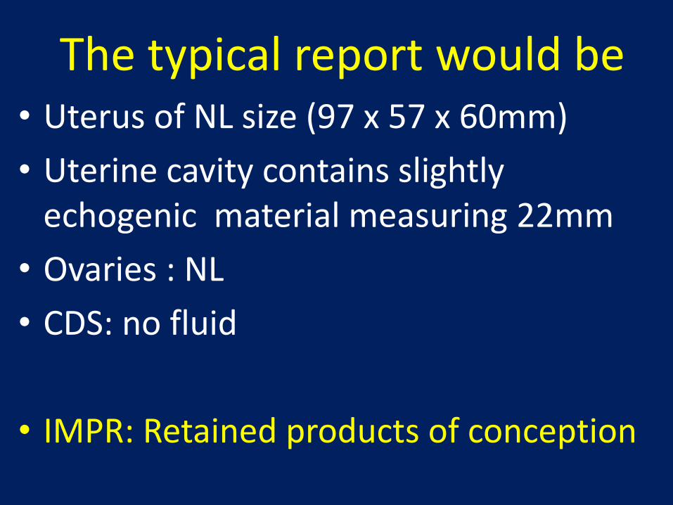
The typical report would be • Uterus of NL size (97 x 57 x 60mm)
• Uterine cavity contains slightly echogenic material measuring 22mm
• Ovaries : NL
• CDS: no fluid
• IMPR: Retained products of conception

Typical management of the “Impression” would be:
• D&C - Hysteroscopy under GA
• Removal of RPOC
• Unexpected “faucet-like” bleeding approximately 750ml
• Insertion of Foley balloon to tamponade

Fortunately there was a different scenario…

The astute sonographer took some more pictures using color Doppler
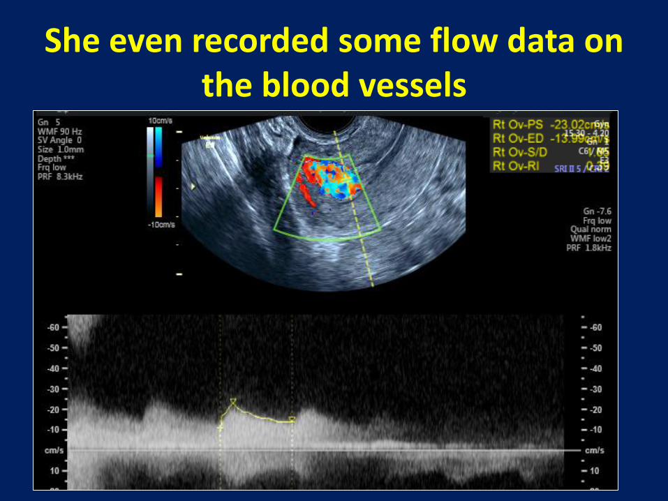
She even recorded some flow data on the blood vessels

……and searched until she found the highest blood flow velocity!!
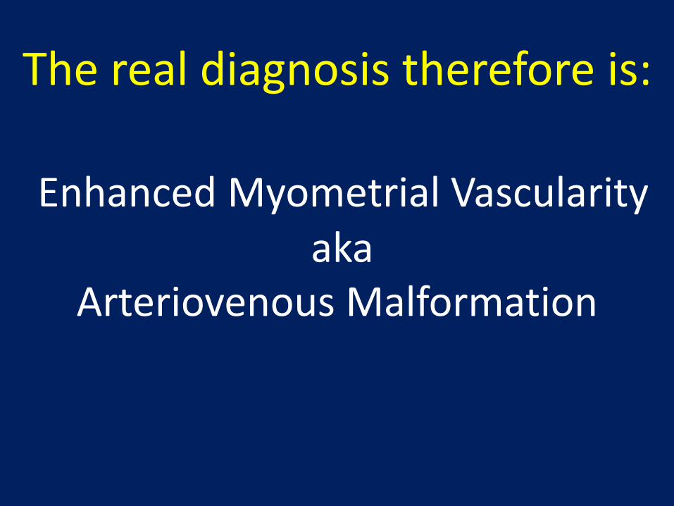
The real diagnosis therefore is:
Enhanced Myometrial Vascularityaka
Arteriovenous Malformation

New plan of action• Weekly hCG ordered until it drops to 0
• Weekly TV-US to evaluate trend of vascularity
• Intervene (UAE?) if significant vaginal bleeding, or if vascularity persists for weeks (give bleeding precautions!).
• If any RPOC’s seen after vascularity disappears: perform safe D&C.
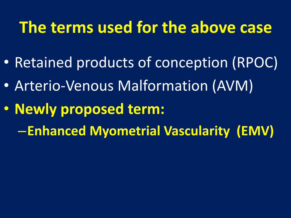
The terms used for the above case
• Retained products of conception (RPOC)
• Arterio-Venous Malformation (AVM)
• Newly proposed term:
–Enhanced Myometrial Vascularity (EMV)

The terms used in this presentation
• Referring to articles published BEFORE the proposal and introduction of the new term of EMV, I will use the original term of AVM.
• Anywhere else I will use EMV

What is a uterine AVM?• Clinically: uncommon vascular lesions;
may cause life-threatening hemorrhage
• Seen almost exclusively in reproductive years & rarely without Hx of IUP
• This may occur when the thin wall of the abnormal vessels are disrupted after menstruation, miscarriage or after uterine instrumentation.*-***
* Polat P, et al.. Radiographics 2002;/22:/4753.
** Vogelzang R et al. J Vasc Interv Radiol 1991;/2:/51722.
***Diwan R et al. J Clin Ultrasound 1983;/11:/2958.

Pathology of AVM• An arterio-venous malformation (or AVM) is a
pathological phenomenon described as a faulty “short circuit” of the blood stream between an organ’s arterial & venous supply.
• The blood stream assumes an unusually high velocity, rendering vessels into vascular fistulas
• They have been reported in patients between ages 18 and 72 years and are broadly classified as either congenital or acquired.
Vogelzang RL, Nemcek AA Jr, Skrtic Z, Gorrell J, Lurain JR. Uterine arteriovenousmalformations: primary treatment with therapeutic embolization. J Vasc Interv Radiol1991;2:517–522.

Schematic representation of congenital vascular malformation with dominant feeding arteryand multiple secondary feeders (top). After proximal ligation of major feeding artery, there is rapidhypertrophy of the multiple secondary feeders (bottom). (Reproduced with permission from RosenRJ, Riles T. Congenital vascular malformations and hemangiomas. Philadelphia: WB Saunders, 1989
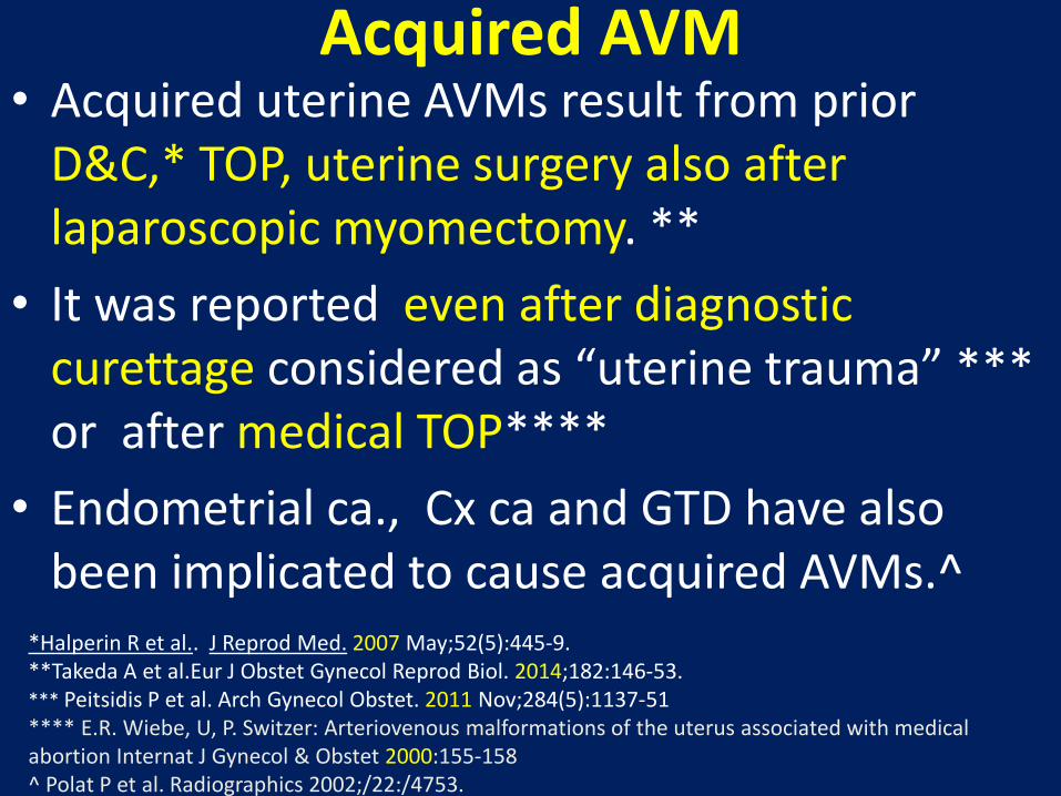
Acquired AVM• Acquired uterine AVMs result from prior
D&C,* TOP, uterine surgery also after laparoscopic myomectomy. **
• It was reported even after diagnostic curettage considered as “uterine trauma” *** or after medical TOP****
• Endometrial ca., Cx ca and GTD have also been implicated to cause acquired AVMs.^
*Halperin R et al.. J Reprod Med. 2007 May;52(5):445-9.**Takeda A et al.Eur J Obstet Gynecol Reprod Biol. 2014;182:146-53. *** Peitsidis P et al. Arch Gynecol Obstet. 2011 Nov;284(5):1137-51**** E.R. Wiebe, U, P. Switzer: Arteriovenous malformations of the uterus associated with medical abortion Internat J Gynecol & Obstet 2000:155-158^ Polat P et al. Radiographics 2002;/22:/4753.
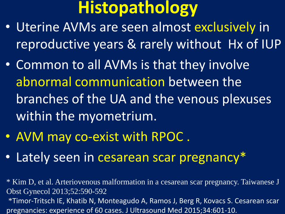
Histopathology• Uterine AVMs are seen almost exclusively in
reproductive years & rarely without Hx of IUP
• Common to all AVMs is that they involve abnormal communication between the branches of the UA and the venous plexuses within the myometrium.
• AVM may co-exist with RPOC .
• Lately seen in cesarean scar pregnancy*
* Kim D, et al. Arteriovenous malformation in a cesarean scar pregnancy. Taiwanese J
Obst Gynecol 2013;52:590-592
*Timor-Tritsch IE, Khatib N, Monteagudo A, Ramos J, Berg R, Kovacs S. Cesarean scarpregnancies: experience of 60 cases. J Ultrasound Med 2015;34:601-10.
Histopathology .
• It can be caused by the erosive property of the syncytiotrophoblastic tissue & chorionic villi during placentogenesis (NL or abnormal including CSP*)
• The faulty decidua induces the generation of abnormal connections among the above vascular structures
• AVM may co-exist with RPOC of trophoblasticproliferation.
* Kim D, et al. Arteriovenous malformation in a cesarean scar pregnancy.
Taiwanese J Obst Gynecol 2013;52:590-592

The histology resembles a hemangioma
Flemming et al Uterine vascular malformations. Obstet Gynecol 1989; 73:209

• AVMs demonstrate a muscular, thin walled capillary network of vessels of different proportions & sizes.
• Vessels have characteristics of both arteries and veins with prominent fibrous thickening with some elastin due to the high intraluminal blood pressure
Zorlu, Akar ME, Seker-Ari E, Yilmaz S, Sindel T. Uterine artery embolization to control bleeding after myomectomy. Acta Obstet Gynecol Scand. 2005 Jun;84(6):606-7.Grivell RM1,et al.Obstet Gynecol Survey 2005 Nov;60(11):761-7.
23. Mulliken JB, Glowacki J. Plast Reconstr Surg. 1982 Mar;69(3):412-22
(EVG stain x 25) Picture showing an arterialized vein. The elastica can be seen beneath the thickened intima

• It is also suggested that AVM arise when venous sinuses become incorporated into scar tissue after necrosis of chorionic villi.
• Mulliken and Glowacki view AVMs as errors in morphogenesis with stable cellularity not showing any spontaneous regression.
Zorlu, Akar ME, Seker-Ari E, Yilmaz S, Sindel T. Uterine artery embolization to control bleeding
after myomectomy. Acta Obstet Gynecol Scand. 2005 ;84(6):606-7.
Grivell RM1,et al.Obstet Gynecol Survey 2005 Nov;60(11):761-7.
Mulliken JB, Glowacki J. Plast Reconstr Surg. 1982 Mar;69(3):412-22

• These anomalies are composed of tortuous vascular channels of varying size and shape, lined by a continuous endothelium and surrounded by abnormal complement of mural cells.
• After TOP the remaining villi show variable vascularity and increasing fibrosis which may result for the diversity in the vascularity of RPOCs at Doppler testing.
• Timmerman suggest that the AVM represents a subinvolution of the placental bed with failed obliteration of its vessels in the absence of RPOCs after cessation of the pregnancy.
• This explains severe bleeding following a delayed postabortal hemorrhage, or (for
that matter) after D&C performed for a CSP.-Timmerman D, Van den Bosch T, Peeraer K, et al. Vascular malformations in the uterus: ultrasonographic diagnosis and conservative management. Eur J Obstet Gynecol Reprod Biol2000;92:171-8.*
- Timmerman D, Wauters J, Van Calenbergh S, et al. Color Doppler imaging is a valuable tool for the diagnosis and management of uterine vascular malformations. Ultrasound Obstet Gynecol2003;21:570-7.
Proposed change of term
• Following a scientific session at the ISUOG in Montreal (Oct 2015) it was proposed that the tem “AVM” should be changed to “Enhanced Myometrial Vascularity”*
* Van den Bosch T, Van Schoubroeck D, Timmerman D. Maximum Peak Systolic Velocity and Management of Highly Vascularized Retained Products of Conception. J Ultrasound Med 2015;34:1577-82.
Clinical aspects of EMV
Clinical aspects
• Clinical symptom is mostly heavy “faucet-like” or even prolonged vaginal bleeding after miscarriage, D&C or CSP
• May cause life-threatening gynecologic hemorrhage
• To repeat: Many claim that they never recognized it as a diagnostic entity

The diagnosis of EMV• Historically the Dx of EMV/AVM was made at
laparotomy.
• Subsequently angiography became the gold standard technique and is still used at the time of a Uterine Artery Embolization process.
Takeda A, Koike b M, Imoto S, Nakamura H. Conservative management of uterine artery pseudoaneurysm after laparoscopic-assisted myomectomy and subsequent pregnancy outcome: case series and review of the literature. European Journal of Obstetrics & Gynecology and Reproductive Biology 182 (2014) 146–153
The diagnosis of AVM
• Lately ultrasound has been used and proven to be effective in diagnosing as well determining clinical management of EMV
-Timmerman D, Van den Bosch T, Peeraer K, et al. Vascular malformations in the uterus: ultrasonographic diagnosis and conservative management. Eur J ObstetGynecol Reprod Biol 2000;92:171-8.*
Timmerman D, Wauters J, Van Calenbergh S, et al. Color Doppler imaging is a valuable tool for the diagnosis and management of uterine vascular malformations. Ultrasound Obstet Gynecol 2003;21:570-7.Van den Bosch T, Van Schoubroeck D, Timmerman D. Maximum Peak Systolic Velocity and
Management of Highly Vascularized Retained Products of Conception. J Ultrasound Med 2015;34:1577-82.

• In recent years TVS “gray scale” and color or power Doppler became the primary diagnostic tool, triaging as well as following patients with EMV leaving angiography as a therapeutic tool.
The diagnosis of EMV
Takeda A, Koike b M, Imoto S, Nakamura H. Conservative management of uterine artery pseudoaneurysm after laparoscopic-assisted myomectomy and subsequent pregnancy outcome: case series and review of the literature. European Journal of Obstetrics & Gynecology and Reproductive Biology 182 (2014) 146–153

The diagnosis of EMV• Gray scale (black and white) US characteristics are
nonspecific and include the presence of irregular hypoechogenic, tortuous, tubular structures within the myometrium.
• Without using Doppler interrogation they are easily missed.
-Maleux G, Timmerman D. et al
Eur Radiol 2006;16:299-306
-Kwon JH, Kim GS.
Radiographics 2002; 22: 35–46
-Hoffman MK, et al. Obstet
Gynecol Surv 1997; 52: 736–740.

The diagnosis of EMV• It is therefore that we consider a pelvic US as
incomplete without applying color or power Doppler interrogation.
• We do first a “panoramic” gray scale image, followed by Doppler focusing on suspicious areas for more information.
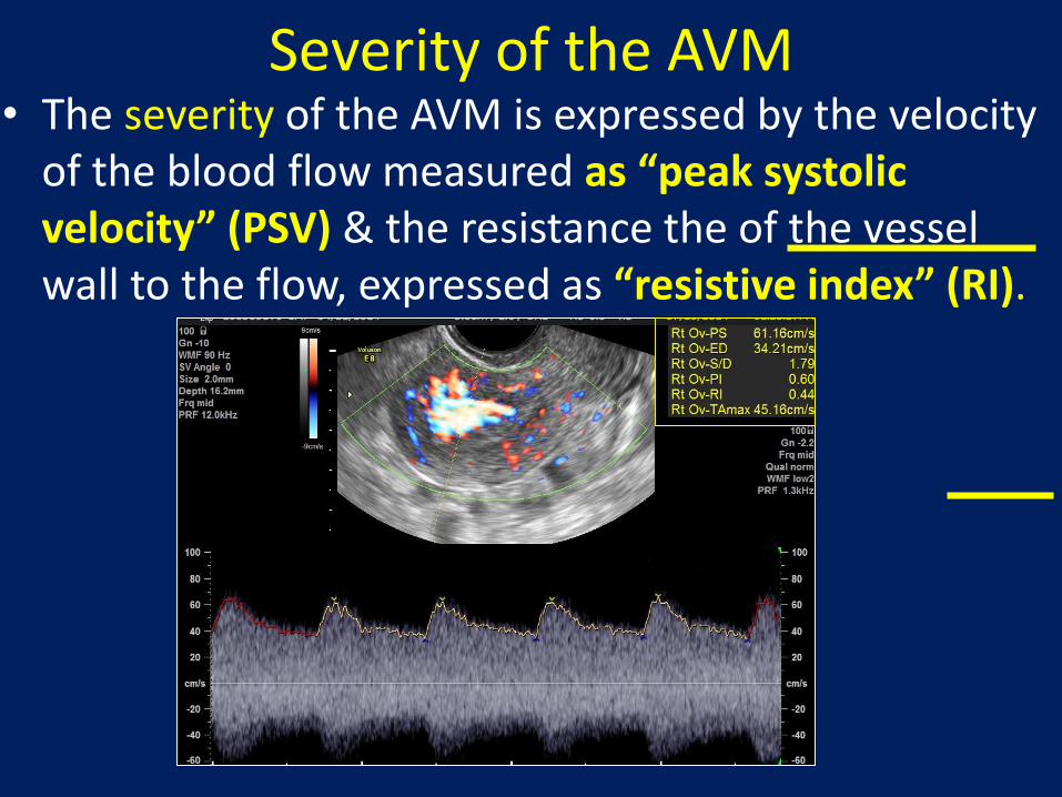
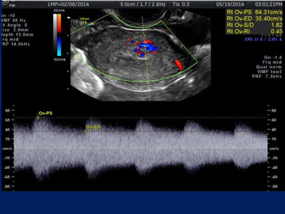
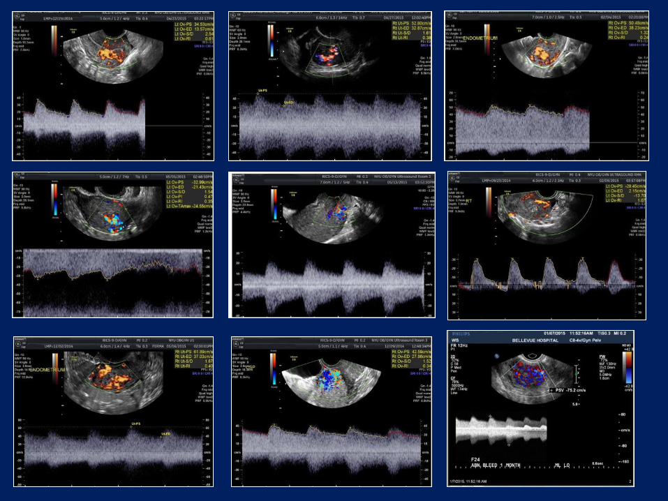
Severity of the AVM• The severity of the AVM is expressed by the velocity
of the blood flow measured as “peak systolic velocity” (PSV) & the resistance the of the vessel wall to the flow, expressed as “resistive index” (RI).

Where to measure to pick-up the highest
PSV?
O O
OO
This is the “trial-and-error” (‘fishing’) method. Time consuming. May be inaccurate missing
the highest PSV leading to inadequate management
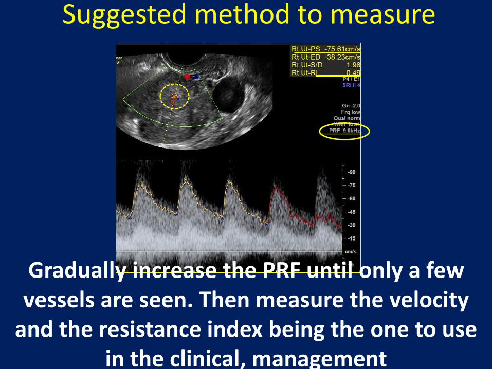
Gradually increase the PRF until only a few vessels are seen. Then measure the velocity
and the resistance index being the one to use in the clinical, management
Suggested method to measure
The effect of angle correction in measuring PSV

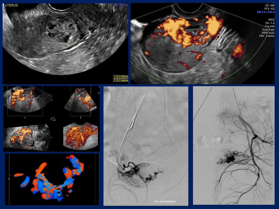
3D angio in the diagnosis of AVM
• The clinical necessity of 3D color or power Doppler angiographic rendering of the involved vessels may raise valid discussion.
• However in almost every case it provided us with a more specific image and presented a 3Dperspective of the “knotted” vessel complex.
• Angiographic rendering can direct the attention of the observer to the largest blood vessels and measure their flow characteristics.
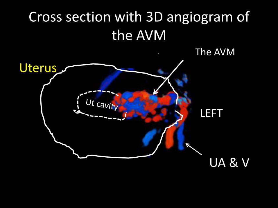
Cross section with 3D angiogram of the AVM
UterusThe AVM
LEFT
UA & V

Managing patients with EMV

Managing patients with EMV
• In order to render the least invasive and most successful treatment the correct diagnosis has to be established.
• This has to be performed swiftly using simple diagnostic means such as US.
• The patient’s hemodynamic status will dictate the urgency of the treatment.

Triage by severity of the EMVDepending on the PSV of the blood flow and the
vessel’s RI, as well as upon the severity of clinical symptoms, the care provider can triage patients at different levels of risk selecting the appropriate treatment. *
No bleeding Observation
Persistent high PSV; with or without bleeding ?UAE* Timmerman D, et al..
Ultrasound Obstet Gynecol
2003; 21: 570–577
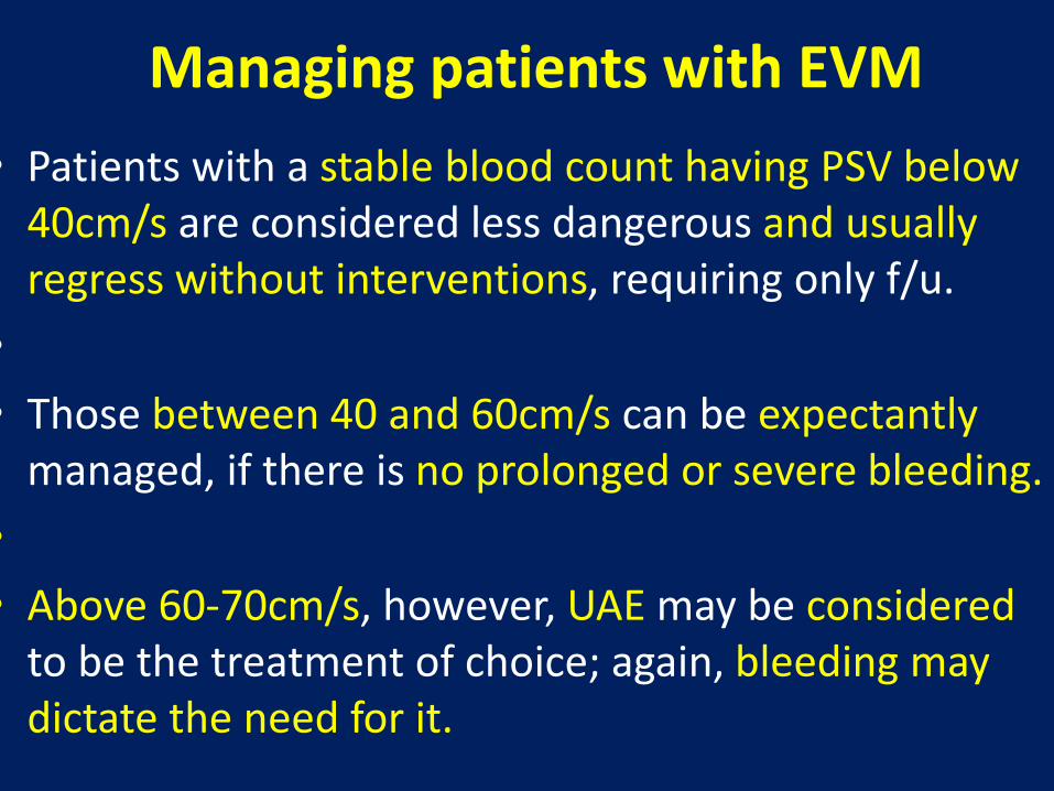
• Patients with a stable blood count having PSV below 40cm/s are considered less dangerous and usually regress without interventions, requiring only f/u.
•
• Those between 40 and 60cm/s can be expectantly managed, if there is no prolonged or severe bleeding.
•
• Above 60-70cm/s, however, UAE may be consideredto be the treatment of choice; again, bleeding may dictate the need for it.
Managing patients with EVM

• Since the severity of the pathology is shown to be linked to the quantitative measurement of blood flow velocities we suggest that such determinations are important for the clinical management of patient.
• Such approach will avoid overtreatment (i.e. UAE) of cases that can be managed by conservatively and conversely, to institute definitive treatment to those with significant pathology.
Managing patients with EMV

• REMEMBER: Patient with significant and life-threatening vaginal bleeding have to be treated regardless of their qualitative or quantitative US appearance and flow values.
• It also appears that an EMV on US with PSV <40cm/s (the “safe area” by Timmerman) with no or significant bleeding can be followed by serial US & clinical developments.
Managing patients with AVM

• EMV may be managed expectantly and will often regress, however patients with symptomatic bleeding often require treatment
• EMV with PSV over 60-70cm/s may need UAE
• Lately definitive treatment of EMV was replaced by UAE which is a highly successful and allows retaining fertility.
Managing patients with EMV
CSP and EMV
• The association between EMV and CSP deserves some more attention.
• This apparently strange phenomenon became evident only after the gradual global increase of CDs became obvious.
CSP and EMV• The first 3 cases were reported in 1999*
(Walter AJ et al. Obstet Gynecol 1999;93:846).
• The last 9 cases were reported in & after 2010 (Timor-Tritsch IE, Monteagudo A. AJOG 2012;207:14-29).
• Based upon the rate of CSPs that parallel the rate of CDs it is not unreasonable to expect more cases of EMVs in CSPs.
Kim D et Taiwanese J Obst Gynecol 2013;52:590-592
Kochhar PK et al J Reprod Med 2013;58:81-
Lui MW et al. Eur J Obstet Gynecol Reprod Biol. 2014 Apr;175:209-10.
Akbayir O et al. J Clin Ultrasound 2011;39:534-8
Tan WC et al. Singapore Med J. 2012 Oct;53(10):638-42.
*

CSP and EMV
• It is a fact that patients with RPOCs are at risk for EMV,** and since a treated CSP in which the gestation was left in place can be considered as a RPOC, it therefore can present a risk for developing an EMV.
** Goyal S et al. . J Obstet Gynaecol Res. 2014;40(1):271-4.
* Timor-Tritsch IE, Khatib N, Monteagudo A, Ramos J, Berg R, Kovacs S. Cesarean Scar Pregnancy (CSP): Experience of Sixty Cases. J Ultrasound Med 2015

Our experience at NYU
MATERIALS AND METHODS
• Retrospective study
• 1/1/2011 – 8/31/2014

MATERIALS AND METHODS• US Diagnostic criteria for the AVM were:
– 1. Subjectively an unusually rich vascular network with tortuous, irregular appearing blood vessels concentrated in a relatively small area of the myometrium and adjacent to the uterine cavity with or without clearly visible POCs on color /power Doppler

MATERIALS AND METHODS• US Diagnostic criteria for the AVM were:
– 2. Objectively: Demonstration of measurable high velocity blood flow within the vascular web with a peak systolic velocity of ≥20 cm/s
• Exclusion criteria included patients who were pregnant at the time of AVM diagnosis.
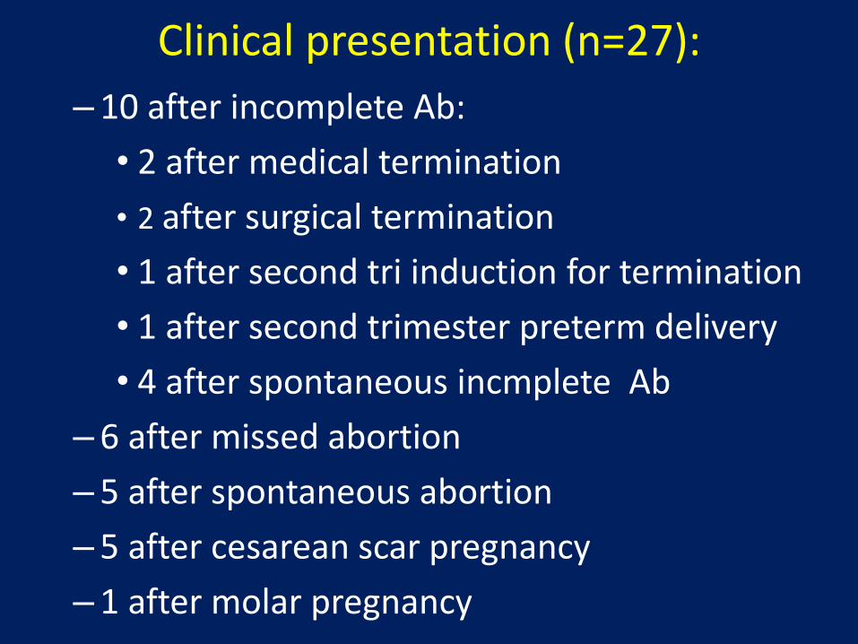
Clinical presentation (n=27):
–10 after incomplete Ab:
• 2 after medical termination
• 2 after surgical termination
• 1 after second tri induction for termination
• 1 after second trimester preterm delivery
• 4 after spontaneous incmplete Ab
–6 after missed abortion
–5 after spontaneous abortion
–5 after cesarean scar pregnancy
–1 after molar pregnancy
•
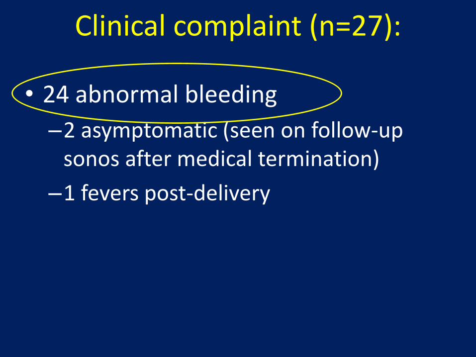
Clinical complaint (n=27):
• 24 abnormal bleeding
–2 asymptomatic (seen on follow-up sonos after medical termination)
–1 fevers post-delivery
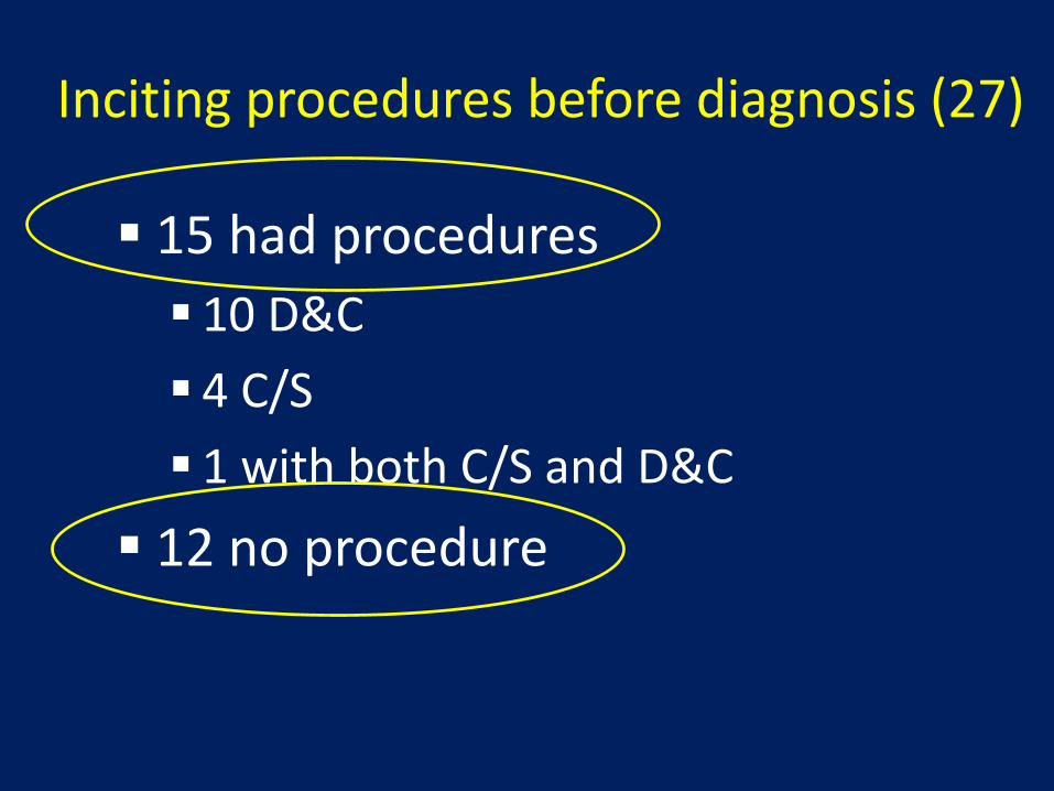
Inciting procedures before diagnosis (27)
15 had procedures
10 D&C
4 C/S
1 with both C/S and D&C
12 no procedure
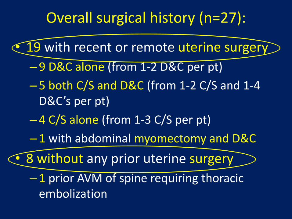
Overall surgical history (n=27):
• 19 with recent or remote uterine surgery
–9 D&C alone (from 1-2 D&C per pt)
–5 both C/S and D&C (from 1-2 C/S and 1-4 D&C’s per pt)
–4 C/S alone (from 1-3 C/S per pt)
–1 with abdominal myomectomy and D&C
• 8 without any prior uterine surgery
–1 prior AVM of spine requiring thoracic embolization

Peak Systolic Velocities (n=27)
• Overall range: 23-170cm/s
• Range of those who underwent UAE: 35-170cm/s
• Range of those who did not undergo UAE: 23-90cm/s
• P= 0.25
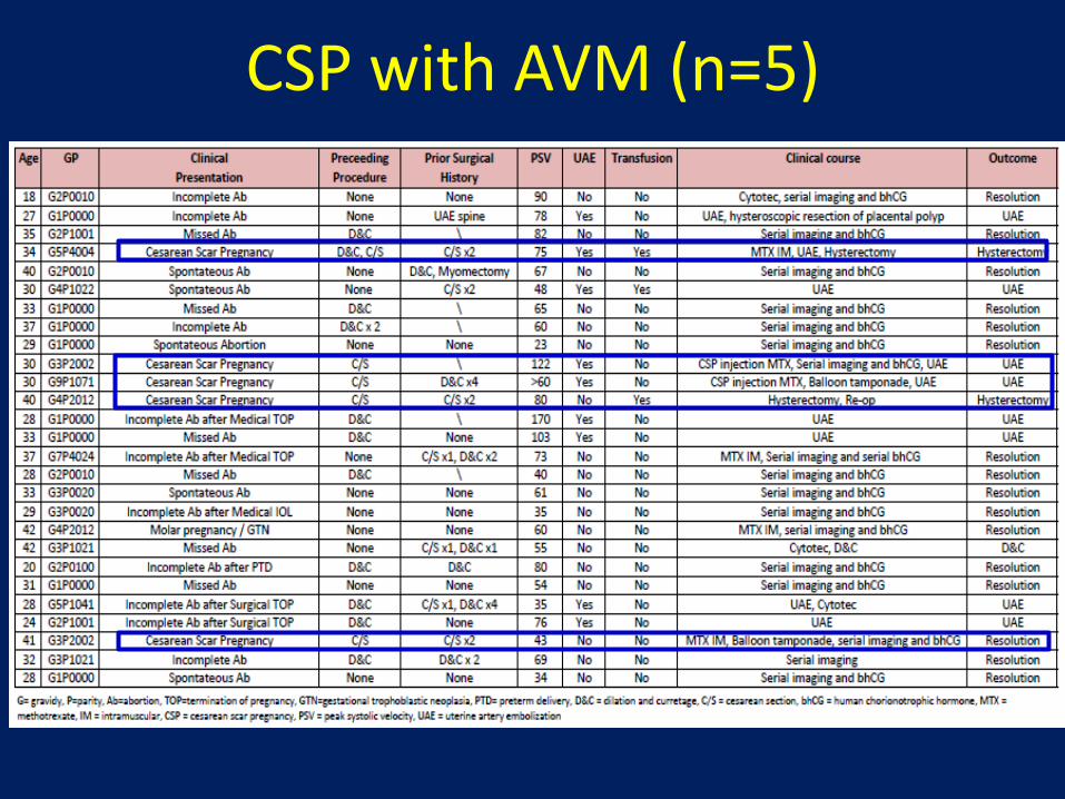
CSP with AVM (n=5)
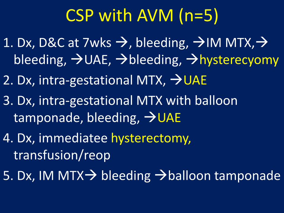
CSP with AVM (n=5)
1. Dx, D&C at 7wks , bleeding, IM MTX,bleeding, UAE, bleeding, hysterecyomy
2. Dx, intra-gestational MTX, UAE
3. Dx, intra-gestational MTX with balloon tamponade, bleeding, UAE
4. Dx, immediatee hysterectomy, transfusion/reop
5. Dx, IM MTX bleeding balloon tamponade

UAE (UAE (n=9)
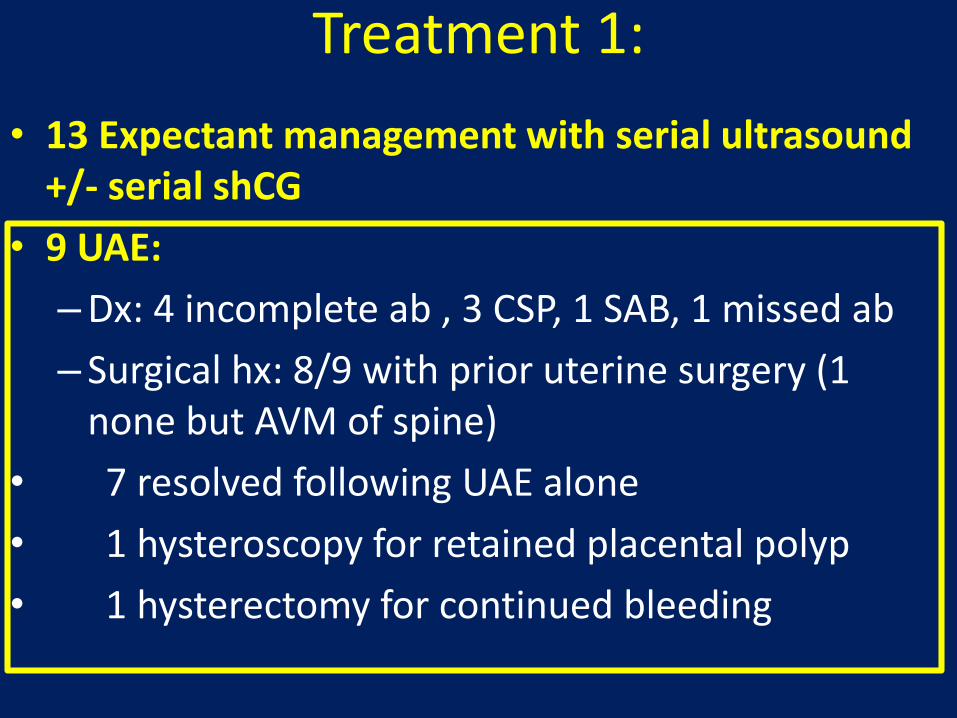
Treatment 1:
• 13 Expectant management with serial ultrasound +/- serial shCG
• 9 UAE:
–Dx: 4 incomplete ab , 3 CSP, 1 SAB, 1 missed ab
– Surgical hx: 8/9 with prior uterine surgery (1 none but AVM of spine)
• 7 resolved following UAE alone
• 1 hysteroscopy for retained placental polyp
• 1 hysterectomy for continued bleeding

Hysterectomy (n=2)

Treatment 2:• 6 MTX:
–4 with CSP (2 injected locally, 2 systemically):
–3 also needed UAE for continued bleeding, of whom 1 required hysterectomy
–2 required Foley balloon tamponade
• 1 for incomplete medical TOP
• 1 for GTD after molar pregnancy
• 2 Hysterectomy:
– 1 planned for CSP
– 1 after failed MTX/UAE as above
• 3 Cytotec (1 serial US, 1 D&C , and 1 UAE)
• 1 D&C

Time to resolution
• Range time from procedure to diagnosis of AVM: 2-10 weeks (avg5.5 weeks)
• Range time from diagnosis to resolution on ultrasound: 2-8 weeks (avg 4.5 weeks)
• Overall episode time: 2-15 weeks
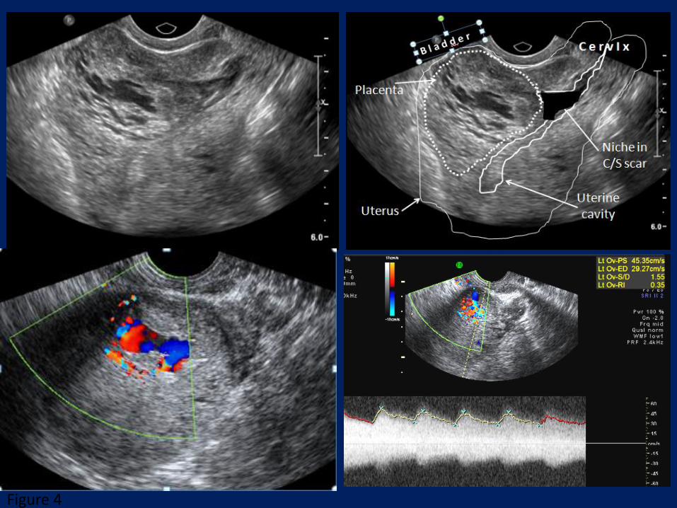
Figure 4

In conclusion• Acquired AVM, is relatively rare in early
pregnancy.
• It occurs following unsuccessful pregnancies and mostly triggered, or as a consequence of intrauterine treatment procedures.
• Spontaneous abortions, sharp uterine curettage and CSP seem to present the known risk for an acquired AVM.

In conclusion• TVS with gray scale and color Doppler US
evaluation is the simplest, best, as well as the most cost effective diagnostic imaging modality.
• Triage of patients for conservative follow-up or UAE based upon to their clinical picture and objective measurement of blood velocity measurement in the AVM appear to be the most successful clinical approach

In conclusion• Prevention of potentially life-threatening
hemorrhagic events and preservation of fertility are some of the main advantages that embolization for AVMs has over more definitive surgical options such as hysterectomy.
• Routine gray scale and color Doppler US evaluation of patients with early pregnancy failure or CSP is strongly indicated to detect a possible AVM as early as possible.

In conclusion
• Gynecologists providing surgical and medical abortions or encounter patients after miscarriage or CSP have to be aware of the existence of this entity to rule it out before a D&C or sucction aspiration is triggering severe uterine bleeding.
• Waiting for the hCG to receede and evaluating hemodynamic parameters is the way to go
• It is unclear if this pathology recurs in a subsequent pregnancy.

Since the acceptance of this article to the Am J Obstet Gynecol in December 2015, we had an additional 12 cases of AVM of which 2 were embolized due to
severe bleeding

Thank you for listening