3318 ieee transactions on nuclear science, vol. 60, no. 5
TRANSCRIPT

3318 IEEE TRANSACTIONS ON NUCLEAR SCIENCE, VOL. 60, NO. 5, OCTOBER 2013
3D Prior Image Constrained Projection Completionfor X-ray CT Metal Artifact Reduction
Abolfazl Mehranian, Mohammad Reza Ay, Member, IEEE, Arman Rahmim, Senior Member, IEEE, andHabib Zaidi, Senior Member, IEEE
Abstract—The presence of metallic implants in the body of pa-tients undergoing X-ray computed tomography (CT) examinationsoftenresults inseverestreakingartifacts thatdegrade imagequality.In this work, we propose a new metal artifact reduction (MAR) al-gorithm for 2D fan-beam and 3D cone-beamCT based on the max-imum a posteriori (MAP) completion of the projections corruptedby metallic implants. In this algorithm, the prior knowledge ob-tained from a tissue-classified prior image is exploited in the com-pletion of missing projections and incorporated into a new priorpotential function. The prior is especially designed to exploit andpromote the sparsity of a residual projection (sinogram) datasetobtained from the subtraction of the unknown target dataset fromthe projection dataset of the tissue-classified prior image. TheMAPcompletion is formulated as an equality-constrained convex opti-mization and solved using an accelerated projected gradient algo-rithm.Theperformanceof theproposedalgorithmiscomparedwithtwo state-of-the-art algorithms, namely 3D triangulated linear in-terpolation (LI) and normalized metal artifact reduction (NMAR)algorithmusing simulated and clinical studies. The simulations tar-geting artifact reduction in 2D fan-beam and 3D cone-beam CTdemonstrate that our algorithm can outperform its counterparts,particularly in cone-beam CT. In the clinical datasets, the perfor-mance of the proposed algorithm was subjectively and objectivelycomparedin termsofmetalartifactreductionofasequenceof2DCTslices. The clinical results show that the proposed algorithm effec-tively reducesmetal artifacts without introducing new artifacts duetoerroneous interpolationandnormalizationas in thecaseofLIandNMAR algorithms.
Index Terms—Metal artifact reduction, prior image, X-ray CT,3D projection completion.
I. INTRODUCTION
X -RAY computed tomography (CT) has experienced con-siderable technical advances over the past two decades
and has now emerged as a leading cross-sectional imaging
Manuscript received January 21, 2013; revised July 15, 2013; accepted July29, 2013. Date of publication September 04, 2013; date of current version Oc-tober 09, 2013. This work was supported in part by the Swiss National ScienceFoundation under grant SNSF 31003A-135576, in part by the Indo-Swiss JointResearch Programme ISJRP 138866, and in part by Geneva University Hospitalunder Grant PRD 11-II-1.A. Mehranian is with the Division of Nuclear Medicine and Molecular
Imaging, Geneva University Hospital, CH-1211 Geneva, Switzerland (e-mail:[email protected]).M. R. Ay is with the Department of Medical Physics and Biomedical
Engineering and the Research Center for Molecular and Cellular Imaging,Tehran University of Medical Sciences, Tehran, Iran (e-mail: [email protected]).A. Rahmim is with the Department of Radiology, School of Medicine, Johns
HopkinsUniversity,Baltimore,MD21287USA(e-mail: [email protected]).H. Zaidi is with the Division of Nuclear Medicine and Molecular Imaging,
Geneva University Hospital, CH-1211 Geneva, Switzerland, the NeuroscienceCenter, Geneva University, Geneva, Switzerland, and also with the Depart-ment of Nuclear Medicine and Molecular Imaging, University of Groningen,University Medical Center Groningen, Groningen, Netherlands (e-mail:[email protected]).Color versions of one or more of the figures in this paper are available online
at http://ieeexplore.ieee.org.Digital Object Identifier 10.1109/TNS.2013.2275919
technique for various diagnostic and therapeutic applications.However, the appearance of streaking metal artifacts in CT im-ages of patients bearing metallic implants can obscure crucialdiagnostic information and therefore reduce image quality andthe clinical relevance of this valuable imaging modality. Aspolychromatic X-ray beams used in CT pass through a patient,low energy (soft) X-ray photons, which are often of littleimportance to image formation, are preferentially absorbed toa greater extent than high energy photons. The outcome of thisselective absorption is that patient’s absorbed dose increasesand the X-ray beam gets richer in high energy photons andthus becomes harder [1]. Due to this so-called beam hardeningeffect, the log-processed transmission data will no longer be alinear function of tissue thickness. In the presence of stronglyattenuating objects, such as metallic implants, beam hardeningand Compton scattering become so severe that the detectorssensing the implants get starved of photons, and thus the rel-evant projection data become corrupted and inconsistent. Thefiltered backprojection (FBP) reconstruction algorithm, whichis widely used in CT image reconstruction, assumes a linearor monochromatic propagation model for the detected photonsand, as such fails to consider the non-linear beam hardeningand scattering effects [2]. Consequently, the reconstructedimages exhibit cupping artifacts, declined CT numbers behindbony structures [3] and contrast-enhanced regions [4], andstreaking artifacts around metallic objects [5]. Most currentgeneration commercial CT scanners, however, apply first-orderbeam hardening correction (water correction) algorithms tocompensate for beam hardening, but due to the incapabilityof these algorithms to calibrate the beam hardening of high-Zmaterials, streaking artifacts still appear in the reconstructedimages. The dark and bright streaking artifacts can obscurepathologic lesions and degrade the radiological manifestationof the surrounding tissues. Consequently, since the past threedecades, extensive efforts have been directed toward devel-oping efficient metal artifact reduction (MAR) algorithms inorder to compensate for the corrupted and missing projectiondata and hence to improve the diagnostic quality and confidenceof CT imaging.Typically, MAR algorithms comprise two steps: a) metal
trace identification, in which the projections corrupted bymetallic implants are identified and b) artifact reduction,through which the identified missing projections are compen-sated for or treated in such a way that the associated streakingartifacts are mitigated. Metal traces are conventionally identi-fied by segmentation of metallic implants in FBP reconstructedimages using thresholding [5]–[7] or clustering techniques [8],[9] followed by reprojection of the obtained metal-only imagesonto the projection or sinogram domain. Other approaches are
0018-9499 © 2013 IEEE

MEHRANIAN et al.: 3D PRIOR IMAGE CONSTRAINED PROJECTION COMPLETION FOR X-RAY CT METAL ARTIFACT REDUCTION 3319
based on segmentation of metal traces directly in raw sino-gram data using active contours [10], curve detection [11] andMarkov random field (MRF) [12] techniques. More recently,hybrids of these two approaches have also been proposed usingiterative metal-only image reconstruction and segmentation[13]–[16]. The second step of MAR methods has been mainlyexplored by two classes of algorithms: projection completionand iterative image reconstruction.Projection completion aims at interpolating the missing
projections from their neighbors through linear [5], cubic spline[6], [17], and wavelet [18] interpolations or iterative inpaintingtechniques using curvature-driven diffusion [19], [20], totalvariation (TV) [10], [21], [22] and wavelet regularization [23].Other approaches rely on replacing the missing projections withthe projections from nearby slices or opposite side angles [24],[25]. Bal and Spies proposed to replace the missing projectionsby the projections obtained from the forward projection of atissue-classified CT image, namely tissue-class model or priorimage [8]. The problem with this approach is that the priorsinogram projections over missing regions (metal traces) arenot well fitted with the projections of the original sinogramin immediate neighboring regions and hence, there is alwaysa risk for discontinuities and generation of new artifacts. Re-cently, Meyer et al. [26] proposed a promising method to solvethis fitness problem. In this method, referred to as normalizedMAR (NMAR), the original sinogram is normalized by thesinogram of prior image, thereby flattening neighboring pro-jections. Then, the missing data are linearly interpolated andthe resulting sinogram is de-normalized. Projection completionhas also been combined with algorithms that exploit the infor-mation hidden in low- and high-pass filtered sinograms [27]or low- and high-pass filtered reconstructed images [28]. Thisclass of algorithms is often fast and computationally appealing;however, if not efficiently implemented, these techniques mightproduce new artifacts. In fact, their efficiency depends on howrobustly they can exploit the still available projection data oreven a prior knowledge in the recovery of missing data.On the other hand, iterative reconstruction algorithms es-
tablish another class of algorithms that, unlike FBP, attemptto frame the reconstruction problem in a way that moreclosely resembles reality. In their evolution from algebraicto model-based statistical reconstruction techniques, thesealgorithms have allowed for a rich description of physical andstatistical mechanisms involved in the imaging process and alsofor incorporating a priori knowledge of the images to be recon-structed [29]. They can be adapted to missing data situationsby down-weighting [30], [31] or ignoring [13], [32], [33] thecontribution of the corrupted projections, or can be tailored topolychromatic propagation models in order to reduce both beamhardening and metallic artifacts [34], [35]. However, this classof algorithms cannot entirely eradicate severe metallic artifacts[36], hence their initiation [31], [37] and combination [38] withprojection completion techniques have also been investigated.Despite their advantages and the development of GPU-basedand parallelizable algorithms [2], iterative image reconstructiontechniques are still memory-demanding and computationallyintensive. To reduce the computational complexity of this classof MAR algorithms, Van Slambrouck et al. [40] proposed a
region-based iterative reconstruction method. In this method, afully polychromatic reconstruction model is used for metallicregions, while a simpler monochromatic model is used for otherregions. It is worth noting that model-based iterative algorithmshave also been successfully applied for sinogram restorationand beam hardening correction [41], [42]. Interested readersare referred to a recent review on MAR algorithms [39].In this study, we propose a three-dimensional (3D) projec-
tion completion MAR algorithm in a Bayesian framework forthe maximum a posteriori (MAP) completion of missing pro-jections. In this framework, we systematically exploit the sideinformation obtained from a tissue-classified prior image andalso prior knowledge about the unknown projections based onprevious works in the framework of compressed sensing andsparse signal recovery. In this context, the prior knowledge thata target signal or solution is sparse (i.e. having many zero com-ponents) or has a sparse and/or compressible representation in agiven transform domain is exploited to recover it from its sam-ples or incomplete measurements. Chen et al. proposed a priorimage constrained compressed sensing technique for reducingstreaking artifacts in CT image reconstruction from undersam-pled projection angles [43]. In this technique, the target imageis sparsified by subtraction from a prior image and then the sub-tracted image is further sparsified using a discrete gradient op-erator. Motivated by the concept of subtraction sparsification inthe context of compressed sensing [43], [44] and prior imageapplication in metal artifact reduction [8], [26], [45], we pro-pose a new prior function to exploit i) the sparsity of a residualsinogram obtained from the subtraction of a target sinogram anda prior sinogram and ii) a sparsity-promoting diffusivity func-tion defined on the prior sinogram for the recovery of missingprojections. Furthermore, we extend the proposed MAP projec-tion completion to three dimensions in order to interpolate themissing projections from all available projection data. The ideaof 3D interpolation has previously been studied for recovery ofmissing projections in flat-panel cone beam CT [45] and in a se-quence of 2D CT slices [46]. In the present work, we evaluatethe performance of the proposed MAR algorithm in compar-ison with NMAR and a 3D linear interpolation algorithm imple-mented on a triangulatedmesh grid using simulation and clinicalstudies and demonstrate that our MAR approach can potentiallyoutperform the above state-of-the-art algorithms.
II. MATERIALS AND METHODS
A. Problem Formulation
Let denote an observed CT projection (sinogram)dataset with projections corrupted by metallic implants over theset , namely missing or metal-trace set. In therecovery of the underlying uncorrupted projection dataset, , weformulate the following forward model:
(1)
where , , is a lossy operator that removesthe projections of over the set . is the observeddataset with removed or missing projections and representszero-mean Gaussian white noise with variance . The matrix
3320 IEEE TRANSACTIONS ON NUCLEAR SCIENCE, VOL. 60, NO. 5, OCTOBER 2013
is constructed in two steps: i) an diagonal matrix is de-fined with zero and one diagonal values. The rows and columnsalong which this matrix is zero are indexed by the set . ii) Thezero-rows of the matrix are then removed. In effect, the resulting
matrix removes the elements of over the set . In (1),the system of equations is underdetermined and therefore has aninfinite number of solutions. In order to regulate and confine thesolution space, we follow a Bayesian estimation approach. Inthis approach, one aims at finding a solution that maximizes thea posteriori probability density of given , which accordingto Bays’ rule is given by
(2)
where r.h.s densities are respectively the probability density ofgiven and the prior probability density of . Since the densityprobability for the observation of given is the density for
, we have
(3)
where . In this framework, the unknown istreated as a stochastic quantity with a prior probability density,
, where is Gibbs or prior energy. Thisdensity is in fact used to impose our prior knowledge on the es-timation. The MAP estimation is then obtained by maximizing
or equivalently minimizing the following a posteriorienergy:
(4)
In the above equation, the first termmeasures the proximity ofto if observed through , while the second term enforces thecompliance of to our prior knowledge. Generally, as the vari-ance of noise decreases, the proximity of to increases. Inthe limit where no noise is introduced as operates on (as isthe case in this work), the problem defined in (4) asymptoticallyreduces to the following constrained optimization problem:
(5)
where is a constraint set inside which the linear set of equa-tions defines the feasible set of solutions. Geometri-cally speaking, this set appears as a hyperplane whose intersec-tion with the ball of the prior defines the solution. Solving(5) is in fact achieved by decreasing the prior’s energy until itsball last touches the hyperplane.To impose our prior knowledge about the unknown , we
employ a prior function whose gradient at point is definedas follows:
(6)
where is a 3D derivative matrix (with symmetricboundary conditions) that approximates the gradient using first-order finite differences in horizontal, vertical and axial direc-tions, is the sinogram of a tissue-classified prior image (prior
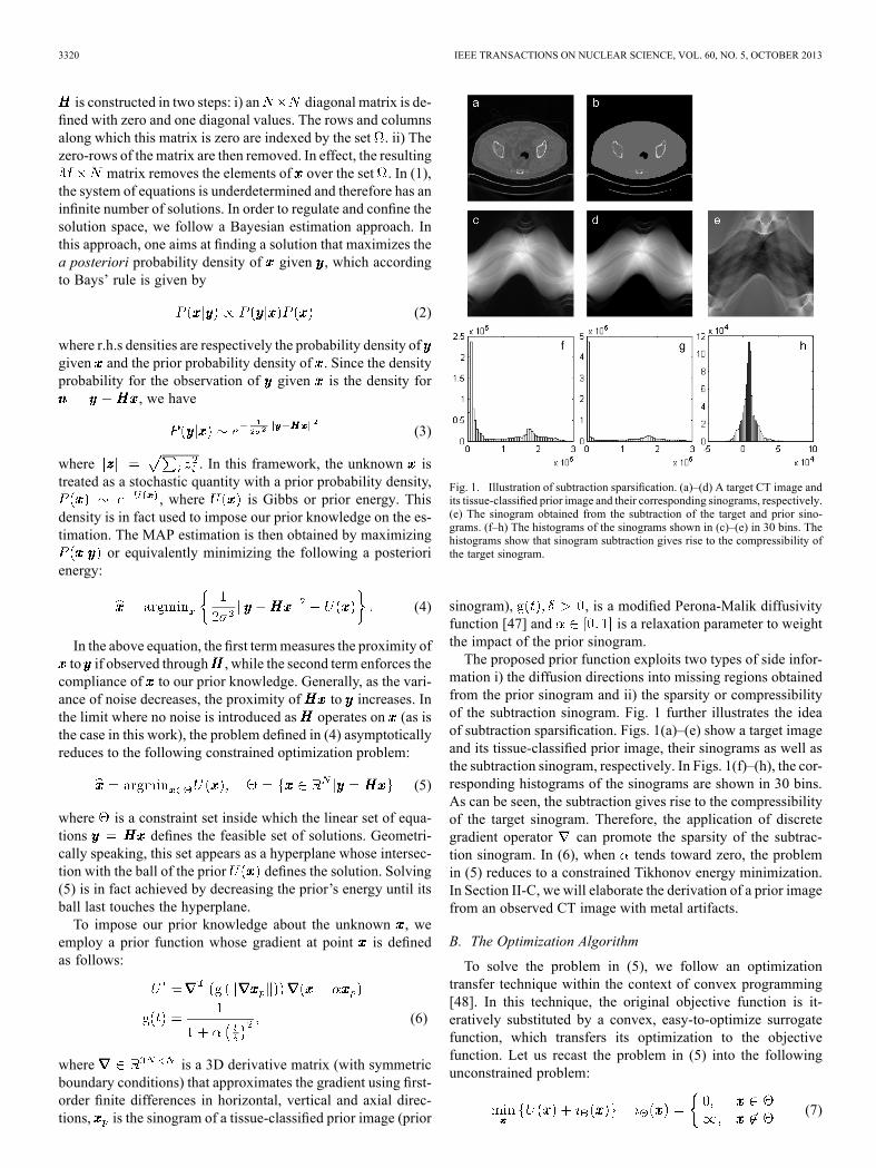
Fig. 1. Illustration of subtraction sparsification. (a)–(d) A target CT image andits tissue-classified prior image and their corresponding sinograms, respectively.(e) The sinogram obtained from the subtraction of the target and prior sino-grams. (f–h) The histograms of the sinograms shown in (c)–(e) in 30 bins. Thehistograms show that sinogram subtraction gives rise to the compressibility ofthe target sinogram.
sinogram), , is a modified Perona-Malik diffusivityfunction [47] and is a relaxation parameter to weightthe impact of the prior sinogram.The proposed prior function exploits two types of side infor-
mation i) the diffusion directions into missing regions obtainedfrom the prior sinogram and ii) the sparsity or compressibilityof the subtraction sinogram. Fig. 1 further illustrates the ideaof subtraction sparsification. Figs. 1(a)–(e) show a target imageand its tissue-classified prior image, their sinograms as well asthe subtraction sinogram, respectively. In Figs. 1(f)–(h), the cor-responding histograms of the sinograms are shown in 30 bins.As can be seen, the subtraction gives rise to the compressibilityof the target sinogram. Therefore, the application of discretegradient operator can promote the sparsity of the subtrac-tion sinogram. In (6), when tends toward zero, the problemin (5) reduces to a constrained Tikhonov energy minimization.In Section II-C, we will elaborate the derivation of a prior imagefrom an observed CT image with metal artifacts.
B. The Optimization Algorithm
To solve the problem in (5), we follow an optimizationtransfer technique within the context of convex programming[48]. In this technique, the original objective function is it-eratively substituted by a convex, easy-to-optimize surrogatefunction, which transfers its optimization to the objectivefunction. Let us recast the problem in (5) into the followingunconstrained problem:
(7)
MEHRANIAN et al.: 3D PRIOR IMAGE CONSTRAINED PROJECTION COMPLETION FOR X-RAY CT METAL ARTIFACT REDUCTION 3321
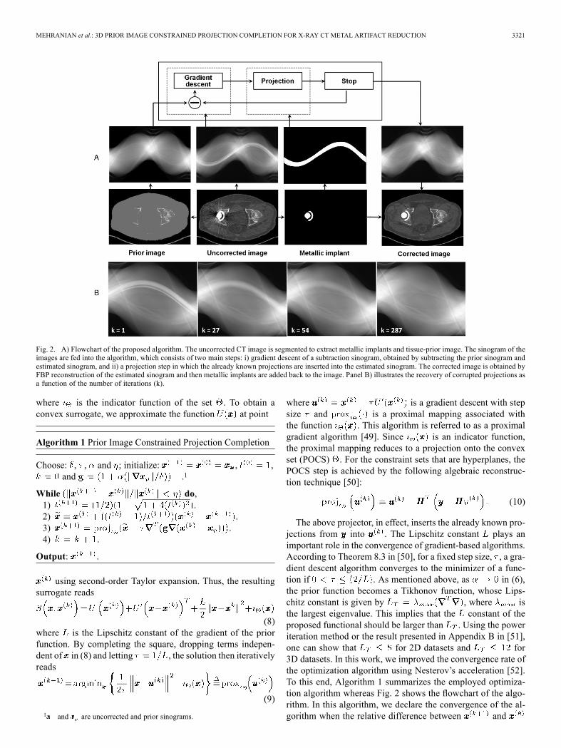
Fig. 2. A) Flowchart of the proposed algorithm. The uncorrected CT image is segmented to extract metallic implants and tissue-prior image. The sinogram of theimages are fed into the algorithm, which consists of two main steps: i) gradient descent of a subtraction sinogram, obtained by subtracting the prior sinogram andestimated sinogram, and ii) a projection step in which the already known projections are inserted into the estimated sinogram. The corrected image is obtained byFBP reconstruction of the estimated sinogram and then metallic implants are added back to the image. Panel B) illustrates the recovery of corrupted projections asa function of the number of iterations (k).
where is the indicator function of the set . To obtain aconvex surrogate, we approximate the function at point
Algorithm 1 Prior Image Constrained Projection Completion
Choose: , , and ; initialize: , ,and .1
While do,1) .2) .3) .4) .
Output: .
using second-order Taylor expansion. Thus, the resultingsurrogate reads
(8)where is the Lipschitz constant of the gradient of the priorfunction. By completing the square, dropping terms indepen-dent of in (8) and letting , the solution then iterativelyreads
(9)
1 and are uncorrected and prior sinograms.
where is a gradient descent with stepsize and is a proximal mapping associated withthe function . This algorithm is referred to as a proximalgradient algorithm [49]. Since is an indicator function,the proximal mapping reduces to a projection onto the convexset (POCS) . For the constraint sets that are hyperplanes, thePOCS step is achieved by the following algebraic reconstruc-tion technique [50]:
(10)
The above projector, in effect, inserts the already known pro-jections from into . The Lipschitz constant plays animportant role in the convergence of gradient-based algorithms.According to Theorem 8.3 in [50], for a fixed step size, , a gra-dient descent algorithm converges to the minimizer of a func-tion if . As mentioned above, as in (6),the prior function becomes a Tikhonov function, whose Lips-chitz constant is given by , where isthe largest eigenvalue. This implies that the constant of theproposed functional should be larger than . Using the poweriteration method or the result presented in Appendix B in [51],one can show that for 2D datasets and for3D datasets. In this work, we improved the convergence rate ofthe optimization algorithm using Nesterov’s acceleration [52].To this end, Algorithm 1 summarizes the employed optimiza-tion algorithm whereas Fig. 2 shows the flowchart of the algo-rithm. In this algorithm, we declare the convergence of the al-gorithm when the relative difference between and

3322 IEEE TRANSACTIONS ON NUCLEAR SCIENCE, VOL. 60, NO. 5, OCTOBER 2013
Fig. 3. Simulation of metal artifacts in a 2D bi-lateral hip prostheses phantom (top panel) and a 3D jaw phantom (bottom panel) based on the polychromaticpropagation of X-ray beams. In the hip phantom, an original CT image was segmented into bone, soft and normal tissues. A polychromatic data acquisitionwas performed on the segmented image to obtain an artifact-free reference image. To simulate metal artifacts, two metallic implants were superimposed on thesegmented CT image and the transmission data were acquired using a polychromatic X-ray CT transmission model considering the scattering due to metallicimplants. The jaw phantom was designed for 3D cone-beam CT and consists of several spheroids representing teeth and two simulated dental fillings within a largespheroidal soft tissue region. In this phantom, the metal artifacts were also simulated by considering a polychromatic CT model.
falls below a tolerance . In this work, we setand in (6) for all the datasets presented
in the Results section.
C. Prior and Metal-Only Images
To obtain a tissue-classified prior image from CT images withstreaking dark and bight artifacts, Bal and Spies [8] applied2D filtering to uncorrected CT images, tailored to reduce noiseand streaking artifacts, and classified them into air, soft tissue,normal tissue, bone, and metal regions using K-means clus-tering. An average CT number was then assigned to each region.In this work, we segmented the uncorrected CT images into air,bone, soft tissue and lung (if present in the field-of-view) using asimple thresholding technique [26], [45]. Following tissue clas-sification, CT numbers of air and soft tissue regions were set to1000 and 0 HU, respectively, and the numbers of bone as well
as lung regions were kept the same as the original image becauseof the inherent variability of bone and lung tissue densities andas such the corresponding CT numbers. In the segmentation ofuncorrected CT images into different tissues, severe dark andbright streaking artifacts can be falsely classified as air and bonein the segmented soft tissue and bone images, respectively. Fol-lowing the work of Karimi et al. [53] on the derivation of a priorimage, we applied a 3D close and open morphological filteringon the segmented classes to reduce these errors. In cases with se-vere artifacts, the residual misclassifications were interactivelyreduced using a graphical user interface. As suggested by Prellet al. [45], an alternative way would be to segment an imagepre-corrected using a linear interpolationMAR algorithm. How-ever, in some cases, we noticed that linear interpolation and
its improved 3D triangulated version fail to effectively reducestreaking artifacts. The segmentation of metallic implants andthus generation of a metal-only image was performed by simplethresholding at about 3000 HU for dental fillings and 2000 HUfor other implants. In the obtained prior image, we assigned theCT number of soft tissue to the segmented metal implants. Fol-lowing the generation of prior and metal-only images, a priorsinogram as well as metal traces (missing projections) were ob-tained by line-integral forward projections.
D. Simulation and Clinical Studies
The performance of the proposed MAR algorithm was com-pared with 3D linear interpolation (LI) implemented on a De-launay triangulated grid and the normalizedmetal artifact reduc-tion (NMAR) algorithms using simulated and clinical studies.To objectively evaluate the performance of algorithms with re-spect to a reference CT image (i.e. without metal artifacts), weretrospectively generated metal artifacts in artifact-free imagesof two simulated phantoms i) a patient with bilateral hip pros-theses and ii) a jaw phantom with dental fillings. These phan-toms were designed to evaluate the performance of the algo-rithm for both 2D fan-beam and 3D cone-beam geometries, re-spectively. As shown in Fig. 3, the hip phantomwas constructedby segmenting an original CT image into 3 classes, i.e. air, softtissue and bone plus iron prostheses. The jaw phantom was ana-lytically modeled from several spheroids, simulating teeth withradii ranging from 8 to 20 mm, and a large sphere simulatingthe head. For this phantom, we considered two dental fillings.To simulate beam hardening and the resulting streaking ar-
tifacts, we modeled the polychromatic propagation of X-ray
MEHRANIAN et al.: 3D PRIOR IMAGE CONSTRAINED PROJECTION COMPLETION FOR X-RAY CT METAL ARTIFACT REDUCTION 3323
TABLE ISUMMARY OF CT SCANNING PARAMETERS USED IN THE CLINICAL STUDIES
beams for the bilateral hip and jaw phantoms, according to thefollowing model [54]:
(11)where is the measured number of photons in projection bin, is the number of incident photons at th energy alongprojection line , is the energy-dependent attenuationmap for different tissue classes and accounts for the contri-bution of scatters. A polyenergetic X-ray spectrum was gener-ated using SpekCal software [55] for a tube voltage of 120 kVp,2.5 mm aluminum filtration, 10 degrees anode angle and a tubeoutput of 123.8 at 1 meter. The spectrum was uni-formly sampled for monoenergetic X-ray beams withan intensity and average energy calculated over each energy in-terval. For each tissue class, energy-dependent linear attenua-tion coefficients were derived and interpolated from the NISTXCOM photon cross section library [56]. The attenuation mapswere forward projected and then according to (11), the Poissonnoise realization of the transmission and scatter sinograms weresummed up to get a sinogram acquired under the conditionsof polychromatic propagation of X-ray beams. In this work,we considered a constant-level scatter for non-zero projectionbins [57]. The resulting sinogram was log-processed and re-constructed by FBP and FDK algorithms. As shown in Fig. 3,the reconstructed artificial CT images suffer from beam hard-ening and streaking artifacts in a similar way as in real CT ac-quisitions. In our simulations, non-linear partial volume effectwas not modeled. Following the generation of an artificially de-graded image, we obtained a prior image, metallic implants,missing projections in the sinograms resulting from the poly-chromatic propagation of X-ray beams. In addition, for eachdataset a reference image was obtained using the above-men-tioned procedure by considering the metallic implants as bonystructures and ignoring scatter. For metal artifact reduction inthe hip phantom and clinical datasets, we evaluated the perfor-mance of the proposed algorithm using artificial sinograms ob-tained from the fan-beam forward projection of uncorrected CTimages.To acquire artificial projection data under conditions closely
matching actual acquisition, we considered the fan-beam geom-etry of a simulated single-slice CT scanner with 888 detectorchannels, 984 angular samples over a 360 orbit, detector pitchof 1 mm, 949 mm source to detector distance, 541 mm sourceto iso-center distance, 408 mm iso-center to detector distance.The geometric systemmatrix describing this scanner was gener-ated by the Image Reconstruction Toolbox (IRT) [58], runningin MATLAB 2010a (The MathWorks, Inc., Natick, MA) on a
12-core workstation with 2.4 GHz Intel Xeon processors and32 GB memory.Line integrals were employed during forward projection to
obtain the Radon transform. For the evaluation of the algorithmfor the jaw phantom, we simulated a cone-beam flat-panel CTscanner with the following specifications: a flat panel detectorwith a matrix size of 384 320 and crystal size of
mm , 800 mm source to iso-center distance, 470 mm iso-center to detector distance and 360 projection angles. The
projection dataset of the jaw phantom was obtainedusing the IRT toolbox. Following the correction of the sino-grams of the hip phantom and clinical studies, the corrected im-ages were reconstructed using the FBP algorithmwith Ram-Lakfilter, for a resolution of 512 512 with pixel size of 0.97 mmand a 500-mm field-of-view. The Ram-Lak filter was chosento best preserve the sharpness of the reconstructed images. Thecorrected images of the jaw phantom were reconstructed usingthe FDK algorithm with a matrix size of andvoxel size of mm . For the clinical evaluation of theMAR algorithm, CT datasets of 8 patients were used. The datawere acquired in helical mode on the Biograph 64 True PointPET/CT and Sensation 16 CT scanners (Siemens Healthcare,Erlangen, Germany), equipped with 40- and 24-row detectors,respectively. The datasets include uni- and bi-lateral hip pros-theses, dental fillings, EEG electrodes, shoulder prosthesis andspine fixation with pedicle screws. Table I summarizes CT scan-ning parameters of the datasets.
E. Evaluation Metrics
The performance of the proposed algorithm was subjectivelyand objectively compared with 3D linear interpolation andNMAR algorithms. In simulation studies, the performanceof the algorithms in terms of reducing streaking artifacts wasobjectively evaluated with respect to a reference using region ofinterest (ROI) analysis. For this purpose, two ROIs were drawnon uncorrected images and the normalized root mean squaredifference (NRMSD) and mean absolute deviation (MAD)between corrected images and their reference image
were calculated for each ROI as follows:
(12)
HU (13)
For quantitative evaluation of the MAR algorithms for the clin-ical datasets, we calculated mean and standard deviation of CTnumbers in volumes of interests (VOI) defined on uncorrected

3324 IEEE TRANSACTIONS ON NUCLEAR SCIENCE, VOL. 60, NO. 5, OCTOBER 2013
Fig. 4. Comparison between the proposed and other MAR algorithms for the simulated bi-lateral hip prostheses dataset HU .
TABLE IIQUANTITATIVE EVALUATION OF THE MAR ALGORITHMS FOR THE SIMULATED HIP AND JAW PHANTOMS IN TERMS OF NORMALIZED ROOT MEAN SQUARE
DIFFERENCE (NRMSD) AND MEAN ABSOLUTE DEVIATION (MAD) METRICS
and corrected CT images over streaking artifacts. Owing to theabsence of a reference artifact-free image in the clinical datasets,we defined a reference VOI on uncorrected images far fromstreaking artifacts.
III. RESULTS
A. Simulation Studies
Fig. 4 compares the performance of MAR algorithms for thesimulated bilateral hip study. In this dataset, the algorithms havenoticeably reduced streaking artifacts. However, the visual com-parison reveals that the proposed algorithm results in the pro-duction of less new artifacts as pointed by the arrows. It shouldbe noted that both NMAR and the proposed algorithm depict asimilar dark streak over the bladder because both use the sameprior image. However, the proposed algorithm shows notice-able reduction of this artifact. For the objective comparison ofthe algorithms with respect to the reference image shown inFig. 3, two ROIs were defined on the uncorrected CT image(see Fig. 4): one large rectangular ROI, namely ROI 1, to cap-ture a large affected area, and one circular ROI, namely ROI 2,for local evaluation near the implants. Table II summarizes theNRMSD and MAD results.In the ROI-based evaluations, the regions ofmetallic implants
were excluded from the ROIs of (un-)corrected and reference
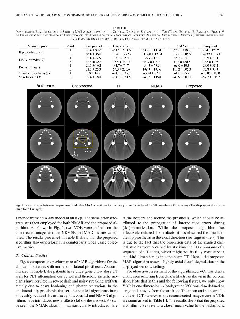
images, since the implants are finally added back to the cor-rected images. The results show that the proposed algorithmachieves a better local and global performance, which is con-sistent with the subjective evaluation.Fig. 5 compares the performance of MAR algorithms for the
cone-beam CT study. The figure also shows the simulated ref-erence and uncorrected images. As mentioned in Section II-D,in the reference image, the metallic implants were replaced bybones and the projection data were analytically acquired usingthe polychromatic X-ray propagation model defined in (11). Ascan be seen, the reference reconstructed images suffer fromstreaking artifacts between teeth and an overall cupping arti-fact due to beam hardening effect and the incapability of theFDK algorithm in considering the non-linear and selective ab-sorption of the X-ray photons. The subjective comparison of thecorrected images demonstrates that, contrary to 3D linear in-terpolation and NMAR, the proposed 3D MAR algorithm hasremarkably reduced metallic artifacts without introducing newartifacts. Note that the images were only corrected for metal ar-tifacts and as such, beam hardening artifacts between teeth arestill present. In this dataset, we performed the interpolation stepof the NMAR algorithm on sinogram views of the 3D projec-tions of the jaw phantom. A volumetric prior image was con-structed by replacing the metallic implants of the jaw phantomwith soft tissue and its corresponding projection obtained using
MEHRANIAN et al.: 3D PRIOR IMAGE CONSTRAINED PROJECTION COMPLETION FOR X-RAY CT METAL ARTIFACT REDUCTION 3325
TABLE IIIQUANTITATIVE EVALUATION OF THE STUDIED MAR ALGORITHMS FOR THE CLINICAL DATASETS, SHOWN ON THE TOP (T) AND BOTTOM (B) PANELS OF FIGS. 6–9,IN TERMS OF MEAN AND STANDARD DEVIATION OF CT NUMBERS WITHIN A VOLUME OF INTEREST DRAWN ON ARTIFACTUAL REGIONS (SEE THE FIGURES) AND
ON A BACKGROUND REFERENCE REGION FAR AWAY FROM THE ARTIFACTS
Fig. 5. Comparison between the proposed and other MAR algorithms for the jaw phantom simulated for 3D cone-beam CT imaging (The display window is thesame for all images).
a monochromatic X-ray model at 80 kVp. The same prior sino-gram was then employed for both NMAR and the proposed al-gorithm. As shown in Fig. 5, two VOIs were defined on theuncorrected images and the NRMSE and MAD metrics calcu-lated. The results presented in Table II show that the proposedalgorithm also outperforms its counterparts when using objec-tive metrics.
B. Clinical Studies
Fig. 6 compares the performance of MAR algorithms for theclinical hip studies with uni- and bi-lateral prostheses. As sum-marized in Table I, the patients have undergone a low-dose CTscan for PET attenuation correction and therefore metallic im-plants have resulted in severe dark and noisy streaking artifactsmainly due to beam hardening and photon starvation. In theuni-lateral hip prosthesis dataset, the studied algorithms havenoticeably reduced the artifacts; however, LI and NMAR algo-rithms have introduced new artifacts (follow the arrows). As canbe seen, the NMAR algorithm has particularly introduced flare
at the borders and around the prosthesis, which should be at-tributed to the propagation of interpolation errors during(de-)normalization. While the proposed algorithm haseffectively reduced the artifacts, it has obscured the details ofthe hip prosthesis in the axial direction (see sagittal view). Thisis due to the fact that the projection data of the studied clin-ical studies were obtained by stacking the 2D sinograms of asequence of CT slices, which might not be fully correlated inthe third dimension as in cone-beam CT. Hence, the proposedMAR algorithm shows slightly axial detail degradation in thedisplayed window setting.For objective assessment of the algorithms, a VOI was drawn
on the area suffering from dark artifacts, as shown in the coronalslice. Note that in this and the following figures, we only showVOIs in one dimension. A background VOI was also defined ona region far away from the artifacts. The mean and standard de-viation of CT numbers of the reconstructed image over the VOIsare summarized in Table III. The results show that the proposedalgorithm gives rise to a closer mean value to the background

3326 IEEE TRANSACTIONS ON NUCLEAR SCIENCE, VOL. 60, NO. 5, OCTOBER 2013
Fig. 6. Comparison between the proposed and other MAR algorithms for the clinical uni- and bi-lateral hip prostheses datasets HU .
mean value, while NMAR shows a noticeable difference withrespect to background. In the bi-lateral hip prostheses dataset(shown in Fig. 6 bottom panel), both NMAR and the proposedalgorithm have substantially reduced the artifacts, while linearinterpolation has introduced new artifacts. The subjective eval-uation of corrected images shows that the proposed algorithmhas reduced the artifacts without introducing bright streaksemanating from the prosthesis (see arrow). The VOI analysis(Table III), however shows that NMAR has more effectively re-duced the dark streaks between the hips (over the region shownin Fig. 6 on the coronal slice). The sizes of the projection data ofthese datasets were and 99. In the recoveryof missing projections, the proposed algorithm converged, onaverage, after 95 iterations with elapsed computation time of657 seconds. In order to reduce computation time, we trimmedthe radial bins of the projection data to those passing thoughthe patient body. This procedure reduces the matrix size of pro-jection datasets and therefore reduces the number of arithmeticoperations during the calculation of 3D finite differences.Fig. 7 compares the performance of MAR algorithms for two
patients with EEG electrodes. As can be seen in these datasets,
the proposed algorithm has more effectively reduced the arti-facts in comparison with LI and NMAR algorithms. In thesedatasets for which the metallic implants are outside the skull,we noticed that the NMAR algorithm introduces wide and se-vere bright and dark streaking artifacts at the borders near theelectrodes, which are due to the normalization of projection binsby small values at these regions. To practically reduce this ef-fect, we expanded the soft tissue region of prior images for thisalgorithm and also thresholded the very high-valued projectionsof de-normalized sinograms to a normal value. Furthermore, toavoid division by zero during normalization, we set zero binsin the sinogram of the prior image to 1. Small threshold valueshave been suggested byMeyer et al. [26]; however, these valuescan result in highly inaccurate values in the normalized sino-grams and hence can contribute to the appearance of severebright streaking artifacts in the EEG datasets.As indicated by the arrows in Fig. 7, the improved NMAR
still give rise to new artifacts, particularly in the bottom datasetwhich has more electrodes. The reduced streaks artifacts in theregions close to the electrodes is of importance in CT-basedattenuation correction of PET data, specifically in patients with
MEHRANIAN et al.: 3D PRIOR IMAGE CONSTRAINED PROJECTION COMPLETION FOR X-RAY CT METAL ARTIFACT REDUCTION 3327
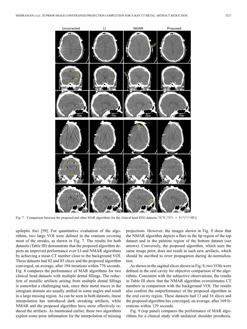
Fig. 7. Comparison between the proposed and other MAR algorithms for the clinical head EEG datasets HU .
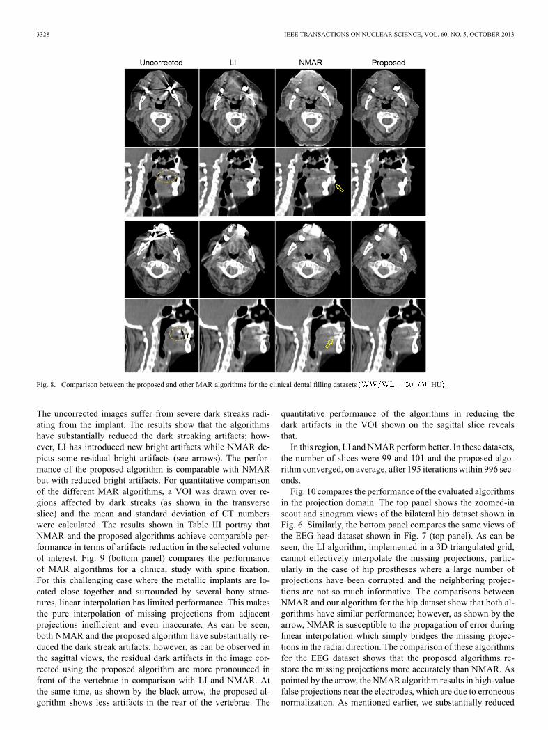
epileptic foci [59]. For quantitative evaluation of the algo-rithms, two large VOI were defined in the cranium coveringmost of the streaks, as shown in Fig. 7. The results for bothdatasets (Table III) demonstrate that the proposed algorithm de-picts an improved performance over LI and NMAR algorithmsby achieving a mean CT number close to the background VOI.These datasets had 82 and 83 slices and the proposed algorithmconverged, on average, after 194 iterations within 776 seconds.Fig. 8 compares the performance of MAR algorithms for twoclinical head datasets with multiple dental fillings. The reduc-tion of metallic artifacts arising from multiple dental fillingsis somewhat a challenging task, since their metal traces in thesinogram domain are usually unified in some angles and resultin a large missing region. As can be seen in both datasets, linearinterpolation has introduced dark streaking artifacts, whileNMAR and the proposed algorithm have more effectively re-duced the artifacts. As mentioned earlier, these two algorithmsexploit some prior information for the interpolation of missing
projections. However, the images shown in Fig. 8 show thatthe NMAR algorithm depicts a flare in the lip region of the topdataset and in the palatine region of the bottom dataset (seearrows). Conversely, the proposed algorithm, which uses thesame image prior, does not result in such new artifacts, whichshould be ascribed to error propagation during de-normaliza-tion.As shown in the sagittal slices shown in Fig. 8, twoVOIswere
defined in the oral cavity for objective comparison of the algo-rithms. Consistent with the subjective observations, the resultsin Table III show that the NMAR algorithm overestimates CTnumbers in comparison with the background VOI. The resultsalso confirm the outperformance of the proposed algorithm inthe oral cavity region. These datasets had 13 and 16 slices andthe proposed algorithm has converged, on average, after 169 It-erations within 129 seconds.Fig. 9 (top panel) compares the performance of MAR algo-
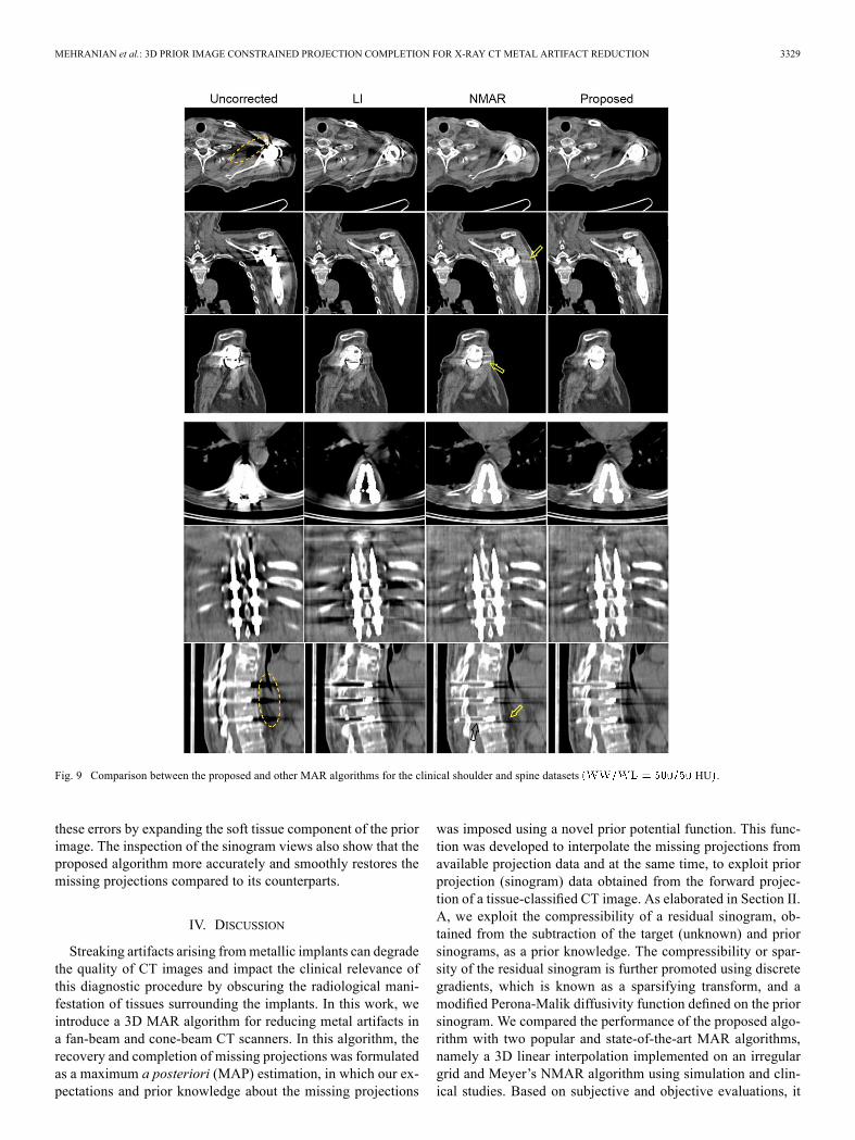
rithms for a clinical study with unilateral shoulder prosthesis.
3328 IEEE TRANSACTIONS ON NUCLEAR SCIENCE, VOL. 60, NO. 5, OCTOBER 2013
Fig. 8. Comparison between the proposed and other MAR algorithms for the clinical dental filling datasets HU .
The uncorrected images suffer from severe dark streaks radi-ating from the implant. The results show that the algorithmshave substantially reduced the dark streaking artifacts; how-ever, LI has introduced new bright artifacts while NMAR de-picts some residual bright artifacts (see arrows). The perfor-mance of the proposed algorithm is comparable with NMARbut with reduced bright artifacts. For quantitative comparisonof the different MAR algorithms, a VOI was drawn over re-gions affected by dark streaks (as shown in the transverseslice) and the mean and standard deviation of CT numberswere calculated. The results shown in Table III portray thatNMAR and the proposed algorithms achieve comparable per-formance in terms of artifacts reduction in the selected volumeof interest. Fig. 9 (bottom panel) compares the performanceof MAR algorithms for a clinical study with spine fixation.For this challenging case where the metallic implants are lo-cated close together and surrounded by several bony struc-tures, linear interpolation has limited performance. This makesthe pure interpolation of missing projections from adjacentprojections inefficient and even inaccurate. As can be seen,both NMAR and the proposed algorithm have substantially re-duced the dark streak artifacts; however, as can be observed inthe sagittal views, the residual dark artifacts in the image cor-rected using the proposed algorithm are more pronounced infront of the vertebrae in comparison with LI and NMAR. Atthe same time, as shown by the black arrow, the proposed al-gorithm shows less artifacts in the rear of the vertebrae. The
quantitative performance of the algorithms in reducing thedark artifacts in the VOI shown on the sagittal slice revealsthat.In this region, LI andNMARperform better. In these datasets,
the number of slices were 99 and 101 and the proposed algo-rithm converged, on average, after 195 iterations within 996 sec-onds.Fig. 10 compares the performance of the evaluated algorithms
in the projection domain. The top panel shows the zoomed-inscout and sinogram views of the bilateral hip dataset shown inFig. 6. Similarly, the bottom panel compares the same views ofthe EEG head dataset shown in Fig. 7 (top panel). As can beseen, the LI algorithm, implemented in a 3D triangulated grid,cannot effectively interpolate the missing projections, partic-ularly in the case of hip prostheses where a large number ofprojections have been corrupted and the neighboring projec-tions are not so much informative. The comparisons betweenNMAR and our algorithm for the hip dataset show that both al-gorithms have similar performance; however, as shown by thearrow, NMAR is susceptible to the propagation of error duringlinear interpolation which simply bridges the missing projec-tions in the radial direction. The comparison of these algorithmsfor the EEG dataset shows that the proposed algorithms re-store the missing projections more accurately than NMAR. Aspointed by the arrow, the NMAR algorithm results in high-valuefalse projections near the electrodes, which are due to erroneousnormalization. As mentioned earlier, we substantially reduced
MEHRANIAN et al.: 3D PRIOR IMAGE CONSTRAINED PROJECTION COMPLETION FOR X-RAY CT METAL ARTIFACT REDUCTION 3329
Fig. 9 Comparison between the proposed and other MAR algorithms for the clinical shoulder and spine datasets HU .
these errors by expanding the soft tissue component of the priorimage. The inspection of the sinogram views also show that theproposed algorithm more accurately and smoothly restores themissing projections compared to its counterparts.
IV. DISCUSSION
Streaking artifacts arising frommetallic implants can degradethe quality of CT images and impact the clinical relevance ofthis diagnostic procedure by obscuring the radiological mani-festation of tissues surrounding the implants. In this work, weintroduce a 3D MAR algorithm for reducing metal artifacts ina fan-beam and cone-beam CT scanners. In this algorithm, therecovery and completion of missing projections was formulatedas a maximum a posteriori (MAP) estimation, in which our ex-pectations and prior knowledge about the missing projections
was imposed using a novel prior potential function. This func-tion was developed to interpolate the missing projections fromavailable projection data and at the same time, to exploit priorprojection (sinogram) data obtained from the forward projec-tion of a tissue-classified CT image. As elaborated in Section II.A, we exploit the compressibility of a residual sinogram, ob-tained from the subtraction of the target (unknown) and priorsinograms, as a prior knowledge. The compressibility or spar-sity of the residual sinogram is further promoted using discretegradients, which is known as a sparsifying transform, and amodified Perona-Malik diffusivity function defined on the priorsinogram. We compared the performance of the proposed algo-rithm with two popular and state-of-the-art MAR algorithms,namely a 3D linear interpolation implemented on an irregulargrid and Meyer’s NMAR algorithm using simulation and clin-ical studies. Based on subjective and objective evaluations, it

3330 IEEE TRANSACTIONS ON NUCLEAR SCIENCE, VOL. 60, NO. 5, OCTOBER 2013
Fig. 10. Comparison of the scout scans and sinogram views of projection data completed by the studied MAR algorithms. Top to bottom panels: Projection dataof the bi-lateral hip prosthesis study (shown in Fig. 6, top panel) and the data of the EEG electrode study (shown in Fig. 7, top panel). As indicated by the arrows,the proposed algorithm is not susceptible to interpolation and normalization errors of LI and NMAR algorithms.
was found that the proposed algorithm can generally outper-form its counterparts for both 2D fan-beam and 3D cone-beamCT imaging. In the implementation of NMAR and the proposedalgorithm, we used the same prior image obtained by the proce-dure described in Section II-C. As demonstrated in our results,the proposed algorithm is not susceptible to interpolation andnormalization errors encountered in the NMAR algorithm, par-ticularly when metallic implants are at the surface of the bodyas in epileptic patients presenting with EEG electrodes. How-ever, both algorithms are susceptible to segmentation errors ofthe prior image. These errors are mostly due to the classificationof dark streak artifacts as air within the soft tissue componentof the prior image. As shown in [8], these errors can reappear inthe reconstructed images. Recently, several studies focused onaccurate segmentation of different tissues from metal artifactsin uncorrected CT images. Chen et al. [60] used non-local fil-tering and mutual information maximized segmentation to im-prove the performance of Bal and Spies’ method [8] for the clas-sification of biological tissues. Karimi et al. [53] proposed toapply close and open morphological operations on uncorrectedimages in order to reduce dark and bight artifacts. This proce-dure is then followed by a region growing segmentation guidedby a distance-dependent threshold that limits the grouping ofartifacts as anatomy. However, these approaches might fail incases with large or multiple closely-seated implants [53]. Tomore practically reduce segmentation errors, Prell et al. [45]suggested tissue classification on a CT image corrected by linearinterpolation. This idea has recently motivated some recent at-
tempts to iteratively improve the derivation of the prior image[61], [62]. There are also other approaches enabling to avoidsegmentation errors and possibly to improve the accuracy of re-covered projections by defining a prior image from statisticalanatomical atlases [63].In the proposed prior function defined in (6), we introduced
a relaxation parameter which can be used to control the im-pact of the prior image in the recovery of missing projections.In cases with severe segmentation errors in the prior image,this parameter can be set to a small value or zero, thereby theerrors are reduced or eliminated in the reconstructed image.However, as mentioned earlier, as decreases to zero, the pro-posed prior reduces to a Tikhonov quadratic prior. Therefore,the performance of the algorithm degrades to that of conven-tional MAR algorithms. The Perona-Malik diffusivity functiondefined in (6) includes the contrast parameter , which controlsthe amount of edge-enhancement. Since CT projection data areusually smooth, we set in this work for all useddatasets and found that this value is fairly small in order to guidethe completion ofmissing projections. Generally, smaller valuesof have a negative impact on the convergence of the employedprojected gradient algorithm. Nevertheless, we improved theconvergence rate and thus the computation time of the optimiza-tion algorithm using Nesterov’s acceleration, as formulated inAlgorithm 1. In terms of computation time, our results show thatthe 3D linear interpolation on a triangulated mesh and NMARare the most time-consuming and fastest MAR algorithms, re-spectively. In a dataset having size of , the

MEHRANIAN et al.: 3D PRIOR IMAGE CONSTRAINED PROJECTION COMPLETION FOR X-RAY CT METAL ARTIFACT REDUCTION 3331
elapsed computation times are about 1630 and 25 seconds, re-spectively, on our MATLAB-based implementations. Note thatsince the 3D LI algorithm is memory demanding for such adataset, it was implemented for every 10 slices. For the clin-ical studies used in this work, the performance of the variousMAR algorithms was evaluated using artificial projection dataobtained from the forward projection of uncorrected images,whereas for simulation studies, the algorithms were evaluatedon the original projection data. Joemai et al. reported that thecorrection of corrupted projections on original scanner-specificraw data is more effective than corrections performed on artifi-cial data [64]. However, we followed the latter generic methodfor the clinical studies while considering the geometry of a re-alistic fan-beam CT scanner and put more emphasis on the de-velopment of a newMAR algorithm that reduces metal artifactsas efficiently as or better than current state-of-the-art MAR al-gorithms.
V. CONCLUSION
In this study, a 3D MAR algorithm was proposed in themaximum a posteriori completion of missing projections in asequence of 2D CT slices and 3D cone-beam CT. In this algo-rithm, we exploited side information about missing projections,obtained from a tissue-classified prior CT image using a novelprior potential function. The prior was designed to exploit andpromote the sparsity of a residual projection dataset (sinogram)obtained from the subtraction of the unknown target datasetfrom the projection dataset of the tissue-classified prior image.The formulated MAP problem was casted as a constrained op-timization problem and solved using an accelerated projectedgradient algorithm. The proposed algorithm was compared withtwo state-of-the-art algorithms using simulation and clinicalstudies. In 2D fan-beam and 3D cone-beam simulations, itwas demonstrated that the proposed 3D algorithm outperformsits counterparts, particularly in cone-beam CT. In the clinicalstudies, the performance of the evaluated MAR algorithms wasevaluated using artificial sinograms of a sequence of 2D CTslices. It was found that the proposed algorithm effectivelyreduces metal artifacts without introducing new ones owing tomore accurate utilization of prior information in comparisonwith its state-of-the-art counterparts. Future work will focus onthe application of the proposed MAR algorithm in clinical 3Dcone beam CT imaging.
ACKNOWLEDGMENT
This work was supported by the Swiss National ScienceFoundation under grant SNSF 31003A-135576, the Indo-SwissJoint Research Programme ISJRP 138866, and Geneva Univer-sity Hospital under grant PRD 11-II-1.
REFERENCES[1] W. R. Hendee and E. R. Ritenour, Medical Imaging Physics, 4th ed.
Hoboken, NJ, USA: Wiley, 2002.[2] X. Pan, E. Y. Sidky, andM.Vannier, “Why do commercial CT scanners
still employ traditional, filtered back-projection for image reconstruc-tion?,” Inv. Probl., vol. 25, no. 12, p. 1230009, 2009.
[3] G. T. Herman, “Demonstration of beam hardening correction in com-puted tomography of the head,” J. Comput. Assist. Tomogr., vol. 3, no.3, pp. 373–378, 1979.
[4] K. Kitagawa, R. T. George, A. Arbab-Zadeh, J. A. Lima, and A. C.Lardo, “Characterization and correction of beam-hardening artifactsduring dynamic volume CT assessment of myocardial perfusion,” Ra-diology, vol. 256, no. 1, pp. 111–118, 2010.
[5] W. A. Kalender, R. Hebel, and J. Ebersberger, “Reduction of CT ar-tifacts caused by metallic implants,” Radiology, vol. 164, no. 2, pp.576–7, 1987.
[6] M. Bazalova, L. Beaulieu, S. Palefsky, and F. Verhaegen, “Correctionof CT artifacts and its influence on Monte Carlo dose calculations,”Med. Phys., vol. 34, pp. 2119–2132, 2007.
[7] M. Abdoli, J. R. de Jong, J. Pruim, R. A. J. O. Dierckx, and H. Zaidi,“Reduction of artefacts caused by hip implants in CT-based attenua-tion-corrected PET images using 2-D interpolation of a virtual sino-gram on an irregular grid,” Eur. J. Nucl. Med. Mol. Imag., vol. 38, pp.2257–2268, 2011.
[8] M. Bal and L. Spies, “Metal artifact reduction in CT using tissue-classmodeling and adaptive prefiltering,” Med. Phys., vol. 33, no. 8, pp.2852–2859, 2006.
[9] H. Yu, K. Zeng, D. K. Bharkhada, G. Wang, M. T. Madsen, O. Saba, B.Policeni, M. A. Howard, andW. R. K. Smoker, “A segmentation-basedmethod for metal artifact reduction,” Acad. Radiol., vol. 14, no. 4, pp.495–504, 2007.
[10] H. Xue, L. Zhang, Y. Xiao, Z. Chen, and Y. Xing, “Metal artifact reduc-tion in dual energy CT by sinogram segmentation based on active con-tour model and TV inpainting,” in Proc. IEEE Nuclear Science Symp.Conf. Rec., 2009, pp. 904–908.
[11] C. Xu, F. Verhaegen, D. Laurendeau, S. A. Enger, and L. Beaulieu,“An algorithm for efficient metal artifact reductions in permanent seedimplants,” Med. Phys., vol. 38, no. 1, pp. 47–56, 2011.
[12] W. J. H. Veldkamp, R. M. S. Joemai, A. J. van der Molen, and J.Geleijns, “Development and validation of segmentation and interpo-lation techniques in sinograms for metal artifact suppression in CT,”Med. Phys., vol. 37, no. 2, pp. 620–628, 2010.
[13] X. Zhang, J. Wang, and L. Xing, “Metal artifact reduction in X-raycomputed tomography (CT) by constrained optimization,”Med. Phys.,vol. 38, no. 2, pp. 701–711, 2011.
[14] B. Meng, J. Wang, and L. Xing, “Sinogram preprocessing and binaryreconstruction for determination of the shape and location of metal ob-jects in computed tomography (CT),” Med. Phys., vol. 37, no. 11, pp.5867–5875, 2010.
[15] J. Choi, K. S. Kim, M. W. Kim, W. Seong, and J. C. Ye, “Sparsitydriven metal part reconstruction for artifact removal in dental CT,” J.X-ray Sci. Tech., vol. 19, pp. 457–475, 2011.
[16] M. Meilinger, E. W. Lang, C. Schmidgunst, and O. Schutz, “Projec-tive segmentation of metal implants in Cone Beam computed tomo-graphic images,” in Proc. 7th Int. Symp. Image and Signal Processingand Analysis, Dubrovnik, 2011, pp. 507–512.
[17] M. Abdoli, M. R. Ay, A. Ahmadian, R. Dierckx, and H. Zaidi, “Re-duction of dental filling metallic artefacts in CT-based attenuation cor-rection of PET data using weighted virtual sinograms optimized by agenetic algorithm,” Med. Phys., vol. 37, pp. 6166–6177, 2010.
[18] S. Zhao, D. Robertson, G. Wang, and B. Whiting, “X-ray CT metalartifact reduction using wavelets: An application for imaging total hipprostheses,” IEEE Trans. Med. Imag., vol. 19, pt. 12, pp. 1238–1247,Dec. 2000.
[19] J. Gu, L. Zhang, G. Yu, Y. Xing, and Z. Chen, “X-ray CTmetal artifactsreduction through curvature based sinogram inpainting,” J. X-ray Sci.Tech., vol. 14, no. 2, pp. 73–82, 2006.
[20] Y. Zhang, Y. F. Pu, J. R. Hu, Y. Liu, Q. L. Chen, and J. L. Zhou, “Ef-ficient CT metal artifact reduction based on fractional-order curvaturediffusion,” Comput. Math. Meth. Med., p. 173748, 2011.
[21] X. Duan, L. Zhang, Y. Xiao, J. Cheng, Z. Chen, and Y. Xing, “Metalartifact reduction in CT images by sinogram TV inpainting,” in Proc.IEEE Nuclear Science Symp. Conf. Rec., 2008, pp. 4175–4177.
[22] Y. Zhang, Y. F. Pu, J. R. Hu, Y. Liu, and J. L. Zhou, “A newCT metal artifacts reduction algorithm based on fractional-ordersinogram inpainting,” J. X-ray Sci. Tech., vol. 19, no. 3, pp.373–384, 2011.
[23] A. Mehranian, M. R. Ay, A. Rahmim, and H. Zaidi, “X-ray CT metalartifact reduction using wavelet domain sparse regularization.,” IEEETrans Med Imag., to be published.
[24] M. Yazdi, M. A. Lari, G. Bernier, and L. Beaulieu, “An opposite viewdata replacement approach for reducing artifacts due to metallic dentalobjects,” Med. Phys., vol. 38, no. 4, pp. 2275–2281, 2011.
[25] S. Tohnak, M. A. J, M. Mahoney, and S. Crozier, “Dental CT metalartefact reduction based on sequential substitution,” Dentomaxillofac.Radiol., vol. 40, no. 3, pp. 184–190, 2011.

3332 IEEE TRANSACTIONS ON NUCLEAR SCIENCE, VOL. 60, NO. 5, OCTOBER 2013
[26] E. Meyer, R. Raupach, M. Lell, B. Schmidt, and M. Kachelrieß, “Nor-malized metal artifact reduction (NMAR) in Computed Tomography,”Med. Phys., vol. 37, pp. 5482–5493, 2010.
[27] K. Y. Jeong and J. B. Ra, “Metal artifact reduction based on sinogramcorrection in CT,” in Proc. IEEE Nuclear Science Symp. Conf. Rec.,2009, pp. 3480–3483.
[28] E. Meyer, R. Raupach, M. Lell, B. Schmidt, and M. Kachelriess, “Fre-quency split metal artifact reduction (FSMAR) in computed tomog-raphy,” Med. Phys., vol. 39, no. 4, pp. 1904–1916, 2012.
[29] J. Hsieh, Computed Tomography: Principles, Design, Artifacts, andRecent Advances, 2nd ed. Bellingham, WA: SPIE, 2009.
[30] J. Nuyts and S. Stroobants, “Reduction of attenuation correction arti-facts in PET-CT,” in Proc. IEEE Nuclear Science Symp. Conf. Rec.,2005, pp. 1895–1899.
[31] M. Oehler and T. M. Buzug, “Statistical image reconstruction for in-consistent CT projection data,” Meth. Inf. Med., vol. 46, pp. 261–269,2007.
[32] B. De Man, J. Nuyts, P. Dupont, G. Marchal, and P. Suetens, “Re-duction of metal streak artifacts in X-ray computed tomography usinga transmission maximum a posteriori algorithm,” IEEE Trans. Nucl.Sci., vol. 47, no. 3, pp. 977–981, Jun. 2000.
[33] L. Ritschl, F. Bergner, C. Fleischmann, and M. Kachelrieß, “Improvedtotal variation-based CT image reconstruction applied to clinical data,”Phys. Med. Biol., vol. 56, pp. 1545–1561, 2011.
[34] G. Van Gompel, K. Van Slambrouck, M. Defrise, K. J. Batenburg, J. deMey, J. Sijbers, and J. Nuyts, “Iterative correction of beam hardeningartifacts in CT,” Med. Phys., vol. 38, p. S36, 2011.
[35] B. De Man, J. Nuyts, P. Dupont, G. Marchal, and P. Suetens, “An it-erative maximum-likelihood polychromatic algorithm for CT,” IEEETrans. Med. Imag., vol. 20, no. 10, pp. 999–1008, Oct. 2001.
[36] J. F. Williamson, B. R. Whiting, J. Benac, R. J. Murphy, G. J. Blaine, J.A. O’Sullivan, D. G. Politte, and D. L. Snyder, “Prospects for quanti-tative computed tomography imaging in the presence of foreign metalbodies using statistical image reconstruction,”Med. Phys., vol. 29, no.10, pp. 2404–2418, 2002.
[37] H. Xue, L. Zhang, Y. Xing, and Z. Chen, “An iterative reconstructiontechnique for metal artifact reduction,” in Proc. 11th Int. Meet. FullyThree-Dimensional Image Reconstruction in Radiology and NuclearMedicine, 2011, pp. 199–202.
[38] C. Lemmens, D. Faul, and J. Nuyts, “Suppression of metal artifacts inCT using a reconstruction procedure that combines MAP and projec-tion completion,” IEEE Trans. Med. Imag., vol. 28, no. 2, pp. 250–260,Feb. 2009.
[39] M. Abdoli, R. A. Dierckx, and H. Zaidi, “Metal artifact reductionstrategies for improved attenuation correction in hybrid PET/CTimaging,” Med. Phys., vol. 39, no. 6, pp. 3343–3360, 2012.
[40] K. Van Slambrouck and J. Nuyts, “Metal artifact reduction in computedtomography using local models in an image block-iterative scheme,”Med. Phys., vol. 39, no. 11, pp. 7080–93, 2012.
[41] J. La Riviere, J. Bian, and P. A. Vargas, “Penalized-likelihood sinogramrestoration for computed tomography,” IEEE Trans. Med. Imag., vol.25, no. 8, pp. 1022–1036, Aug. 2006.
[42] G. V. Gompel, K. V. Slambrouck, M. Defrise, K. J. Batenburg, J. D.Mey, J. Sijbers, and J. Nuyts, “Iterative correction of beam hardeningartifacts in CT,” Med. Phys., vol. 38, no. S1, pp. S36–S49, 2011.
[43] G. H. Chen, J. Tang, and S. Leng, “Prior image constrained compressedsensing (PICCS): A method to accurately reconstruct dynamic CT im-ages from highly undersampled projection data sets,” Med. Phys., vol.35, no. 2, pp. 660–3, 2008.
[44] V. Cevher, A. Sankaranarayanan, M. F. Duarte, D. Reddy, R. G. Bara-niuk, and R. Chellappa, “Compressive sensing for background subtrac-tion,” in Proc. 10th Eur. Conf. Computer Vision: Part II, Marseille,France, 2008, pp. 155–168.
[45] D. Prell, Y. Kyriakou, M. Beister, and W. A. Kalender, “A novel for-ward projection-based metal artifact reduction method for flat-detectorcomputed tomography,” Phys. Med. Biol., vol. 54, no. 21, p. 6575,2009.
[46] B. Kratz, I. Weyers, and T. M. Buzug, “A fully 3D approach for metalartifact reduction in computed tomography,” Med. Phys., vol. 39, no.11, pp. 7042–7054, 2012.
[47] P. Perona and J. Malik, “Scale-space and edge detection usinganisotropic diffusion,” IEEE Trans. Pattern Anal. Mach. Intell., vol.12, no. 7, pp. 629–639, Jul. 1990.
[48] K. Lange, D. R. Hunter, and I. Yang, “Optimization transfer using sur-rogate objective functions,” J. Comput. Graph. Statist., vol. 9, pp. 1–20,2000.
[49] A. Beck and M. Teboulle, “Gradient-Based Algorithms with Applica-tions in Signal Recovery Problems,” in Convex Optimization in SignalProcessing and Communications, D. Palomar and Y. Eldar, Eds.Cambribge, U.K.: Cambribge Univ. Press, 2010, pp. 33–88.
[50] E. P. Chong and S. H. Zak, An Introduction to Optimization, 2nd ed.Hoboken, NJ, USA: Wiley, 2001.
[51] J. Dahl, P. C. Hansen, S. H. Jensen, and T. L. Jensen, “Algorithmsand software for total variation image reconstruction via first-ordermethods,” Numer. Alg., vol. 53, pp. 67–92, 2010.
[52] A. Beck and M. Teboulle, “A fast iterative shrinkage-thresholding al-gorithm for linear inverse problems,” SIAM J. Imag. Sci., vol. 2, no. 1,pp. 183–202, 2009.
[53] S. Karimi, P. Cosman, C. Wald, and H. Martz, “Segmentation of arti-facts and anatomy in CT metal artifact reduction,”Med. Phys., vol. 39,no. 10, pp. 5857–68, 2012.
[54] P. J. La Rivière, “Penalized-likelihood sinogram smoothing for low-dose CT,” Med. Phys., vol. 32, no. 6, pp. 1676–1683, 2005.
[55] G. Poludniowski, G. Landry, F. DeBlois, P. M. Evans, and F. Ver-haegen, “SpekCalc: A program to calculate photon spectra from tung-sten anode X-ray tubes,” Phys. Med. Biol., vol. 54, no. 19, p. N433,2009.
[56] M. J. Berger and J. H. Hubbell, “XCOM: Photon cross sections on apersonal computer,” Center for Radiation Research, National Bureauof Standards, 1987.
[57] B. De Man, J. Nuyts, P. Dupont, G. Marchal, and P. Suetens, “Metalstreak artifacts in X-ray computed tomography: A simulation study,” inProc. IEEE Nuclear Science Symp. Conf. Rec., 1998, pp. 1860–1865.
[58] J. Fessler, Image Reconstruction Toolbox, Univ. Michigan [Online].Available: http://www.eecs.umich.edu/~fessler/code/index.html, Ac-cessed 2012
[59] C. Lemmens, M.-L. Montandon, J. Nuyts, O. Ratib, P. Dupont, andH. Zaidi, “Impact of metal artefacts due to EEG electrodes in brainPET/CT imaging,” Phys. Med. Biol., vol. 53, no. 16, pp. 4417–4429,2008.
[60] Y. Chen, Y. Li, H. Guo, Y. Hu, L. Luo, X.Yin, J. Gu, and C. Toumoulin,“CT metal artifact reduction method based on image segmentation andsinogram in-painting,” Math. Prob. Eng., vol. 2012, 2012, Article ID786281, 18 pages.
[61] F. Morsbach, S. Bickelhaupt, G. A. Wanner, A. Krauss, B. Schmidt,and H. Alkadhi, “Reduction of metal artifacts from hip prostheses onCT images of the pelvis: Value of iterative reconstructions,”Radiology,2013, in press.
[62] Y. Zhang, H. Yan, X. Jia, J. Yang, S. B. Jiang, and X. Mou, “A hybridmetal artifact reduction algorithm for X-ray CT,” Med. Phys., vol. 40,no. 4, pp. 041910–17, 2013.
[63] O. Sadowsky, L. Junghoon, E. G. Sutter, S. J. Wall, J. L. Prince, andR. H. Taylor, “Hybrid cone-beam tomographic reconstruction: Incor-poration of prior anatomical models to compensate for missing data,”IEEE Trans. Med. Imag., vol. 30, no. 1, pp. 69–83, Jan. 2011.
[64] R. M. S. Joemai, P. W. D. Bruin, W. J. H. Veldkamp, and J. Geleijns,“Metal artifact reduction for CT: Development, implementation, andclinical comparison of a generic and a scanner-specific technique,”Med. Phys., vol. 39, no. 2, pp. 1125–1132, 2012.