31st annual combined orthopaedic spring symposium · aloha & welcome to the 31st annual...
TRANSCRIPT
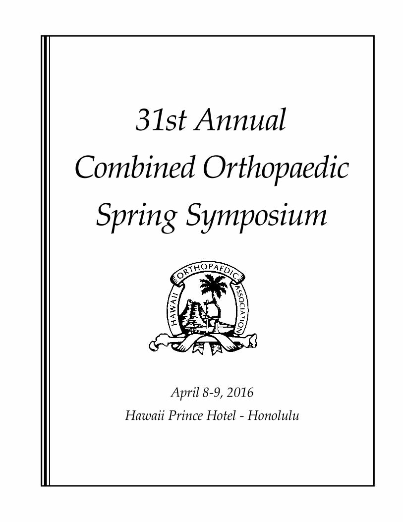
31st AnnualCombined Orthopaedic
Spring Symposium
April 8-9, 2016Hawaii Prince Hotel - Honolulu
Welcome from HOA President
2007 HOA Officers
Aloha & Welcome to the 31st Annual Combined OrthopaedicSpring Symposium! This annual event provides opportunities for
the orthopaedic community in Hawaii to learn about the latestadvances in orthopaedic surgery from nationally renownedexperts in their fields. The symposium features a constructive
forum for discussions among HOA members, residents, medicalstudents and allied health professionals from across our state. Italso provides a venue to feature the research being conducted
by University of Hawaii and Tripler Army Medical Centerresidents. And, don't forget the opportunities that thesymposium will provide to network with fellow specialists at the
awards banquet on Saturday, Apr. 9. This is truly a can't-missevent, providing opportunities to gain knowledge and earnCME credit, participate in discussions and catch-up with our
local orthopaedic ohana over the course of just a few days.Mahalo for joining us!
Darren Egami, MDHOA President & Symposium Chair
HOA Membership InformationContact HOA Executive Director Cathy Iwai at 808-630-1586 or [email protected] if
you are interested in becoming a member of the Hawaii Orthopaedic Association.
Hawaii Orthopaedic AssociationP.O. Box 61207Honolulu, HI 96839Fax: 808-536-4141.
Americans with Disability Act (ADA)Participants with special needs should contact Cathy Iwai at 808-630-1586 or [email protected] todiscuss desired accommodation(s).
PresidentDarren Egami, MD
Vice-PresidentWei Chin Chen, MD
Secretary/TreasurerJoseph Varcadipane, MD
Immediate Past PresidentJohn Jiuliano, MD
Board of CouncilorsJerry Van Meter, MD
Executive DirectorCathy Iwai
2016 HOAOfficers
1
CME CreditsThis activity has been planned and implemented in accordance with the Essential Areas and policies of the AccreditationCouncil for Continuing Medical Education through the joint sponsorship of the Hawai’i Consortium for Continuing MedicalEducation (HCCME) and the Hawaii Orthopaedic Association. The Hawai’i Consortium for Continuing Medical Education isaccredited by the Accreditation Council for Continuing Medical Education to provide continuing medical education for physi-cians.
The Hawaii Consortium for Continuing Medical Education designates this live activity for a maximum of 15.0 AMA PRACategory 1 credits™. Physicians should claim only the credit commensurate with the extent of their participation in the activity.
Disclosure DeclarationThe following have no relevant financial relationships with any commercial interest:
PlannersWei Chin Chen, MDDarren Egami, MDElizabeth Ignacio, MDCathy IwaiByron Izuka, MDJohn Jiuliano, MDCraig Ono, MDPaul M. Ryan, MDJerry Van Meter, MDJoseph Varcadipane, MD
Learning ObjectivesThe program content of the Hawaii Orthopaedic Association Spring Symposium is designed to identify and address theadvances and changes that occur throughout the many areas of orthopaedic surgery. The topics are intended to relate bothdirectly and indirectly to the practice of each practitioner. At the conclusion of this educational activity, participants will beable to:
1. Discuss and identify best practices in topics discussed.2. Learn how to improve patient outcomes through improving implementation of best practices.3. Continue to educate and supplement performance and competence in orthopedic surgery.
ResidentsChristopher Belyea, MDSean Brugman, MDEdward Chan, MDJames Deal, MDDaniel Derosa, MDJohn Dupaix, MDMichael Finnern, MDShawn Gee, MDMitch Harris, MDAdam Hines, MD
The following have relevant financial relationships to disclose:
Guest SpeakersCraig Bottoni, MDAndrew Kayes, MDDean Lorich, MDCharles Price, MD
2
John Daniel Johnson, MDChristopher Lau, MDNick Scarcella, MDJames Shaha, MDCole Turner, MDSteven Wilding, MDLiang Zhou, MD
Michael Ast, MD• Consultant, Speakers Bureau – Smith & Nephew• Consultant, Stock Equity – OrthAlign, Inc.• Committee Membership – Eastern Orthopaedic Association
Jack Farr, MD• Research or institutional support has been received from: Active Implant, Arthrocare a Smith & Nephew Company, Ceterix, Sanofi Company formally Genzyme
Biosurgery, Histogenics, Johnson and Johnson Companies, Depuy/Mitek, Knee Creations, LLC, Moximed, Inc., Nutech Medical, Inc., RTI Biologics, Inc,and Zimmer.
• Miscellaneous non-income support, commercially derived honoraria, or other nonresearch related funding has been received from: Advanced Biosurfaces,Arthrex, Sanofi Company formally Genzyme Biosurgery, Johnson and Johnson Companies, Knee Creations, LLC, MedShape, Inc., Regentis, RTI Biologics,Inc., and Zimmer.
• Royalties have been received from: Arthrex, Johnson and Johnson Companies, Depuy Orthopaedics, Nutech Medical,Inc., and SBM, Inc..• Stock or stock options held in: MedShape, Inc.• Consultant / Advisory Boards: Advanced Biosurfaces, Arthrex, Arthrocare, Ceterix, Orthopaedics, Exactech, Johnson and Johnson Companies, Knee Creations,
LLC, ISTO Technologies, Inc., MedShape, Inc., Moximed, Inc., NuOrtho Surgical, Inc., NuTech Medical, Ortho Regenerative Technologies, Inc., OsirisTherapeutics, Inc., RTI Biologics Cartilage Advisory Panel, SBM, Inc., Vericel formally Sanofi Company and Genzyme Biosurgery, and Zimmer.
• Board Member/Committee Appointments for a Society: Cartilage Research Foundation, Inc. Treasurer; ICRS Regulatory & Industry LiaisonCommittee 2010 – 2012; ICRS Chairman for Liaison Committee 2012 – Present; ICRS Education & Meeting Committee 2015 - Present; PatellofemoralFoundation Vice President; International Patellofemoral Study Group Treasurer; ISAKOS Patellofemoral Scoring Task Force 4/1/2013 to 3/31/2017; ISAKOSKnee: Arthroplasty & Alternatives Committee 20015-2019; Medical/Orthopaedic Publications Editorial/Governing Board; American Journal of Sports MedicinePrincipal Reviewer; Cartilage Associate Editor; Clinical Orthopaedics and Related Research Reviewer; Journal of Knee Surgery Reviewer, Guest AssociateEditor; Knee Surgery, Sports Traumatology, Arthroscopy Reviewer; and The Knee Reviewer.
• Books, Royalties Donated to Arthritis Foundation: "Cartilage Restoration: Practical Clinical Applications” Farr, J; Gomoll,A. Springer Science +BusinessMedia New York 2014; and “Articular Cartilage Injury of the Knee: Basic Science to Surgical Repair” Stannard,JP; Cook JL;Farr J. Thieme MedicalPublishers, Inc 2013
• Data Safety Monitor Board: Exactech CR09-006 2013 –July 2015; and Aesculap NovaCart 3D 2014 -present.
Jesse Jupiter, MD• Consultant - AO Foundation• Honoraria for Speaking - APTIS, DePuy Synthes, and Trimed
AcknowledgementsThank you for the Support of all of Our Exhibitors...
All Island Surgical
Advanced Imaging Institute
Arthrex
Automated Healthcare Solutions
Bioventus
Depuy Synthes (Johnson & Johnson)
Ferring Pharmaceuticals
Halyard Health
Invision Imaging
Special Thanks to...
Shriners Hospital for Children - Honolulu
Tripler Army Medical Center Orthopaedic Residency Program
University of Hawaii Orthopaedic Residency Program
...and a Big Mahalo to...
HOA Executive Director Cathy Iwai for all of your work
in overseeing another successful year!
Hawaii Diagnostic Radiology
ServicesMedartis, Inc.
NuVasive, Inc.
Smith & Nephew - Endo
Smith & Nephew - Navio & Wound
Smith & Nephew - Recon & Trauma
Wright Medical
Zimmer Biomet
Best Resident Paper AwardsRichardson Awards: The Richardson Fund was established in 1982 to honor the memory of B. AllenRichardson, MD. Dr. Richardson was one of the first Board-Certified Orthopaedic Surgeons in Honolulu,where he practiced for nearly 30 years. He was an active member of the teaching staff of the University ofHawaii Orthopaedic Residency Training Program from its inception in the mid-1960s, and was a staunchsupporter for the creation of the John A. Burns School of Medicine. The proceeds of the Richardson Fund areused to award first, second and third place prizes for the best resident papers presented at the Annual Com-bined Orthopaedic Spring Symposium.
Shriners Award: The Shriners Award is presented annually and was established to honor an orthopaedicresident who has completed a rotation at the Shriners Hospital for Children in Honolulu. Residents present theircompleted papers to medical staff and allied health professionals at the Shriners Hospital's patient care confer-ence. The paper must be written to meet standards for publishing in clinical publications.
3
31st Annual Combined Orthopaedic Spring SymposiumFriday, April 8, 2016
6:45 Registration / Continental Breakfast / Exhibits
7:00 Welcome and Opening Remarks - Darren Egami M.D.
7:10 Edward Chan MD - Bisphosphonate-Associated Atypical Femur Fractures Versus
Osteoporotic Femur Fractures: A Histologic Analysis
7:15 Shawn Gee MD - Characterization and Incidence of Missed Posterior Malleolus Fractures
on Plain Radiographs
7:20 Discussion
7:30 Dean Lorich MD - Ankle Fractures: How Little We Understand
8:15 Jesse Jupiter MD - Complex Fracture Dislocations About the Wrist
9:00 Discussion
9:10 Break and Exhibits / PLEASE VISIT EXHIBITS
9:40 Charles Price MD - Topics in Pediatric Orthopedics: Why Do We Operate on Pediatric Fractures?
10:25 Michael Ast MD - Same-Day Joint Replacements, the Outpatient Experience
11:10 Jack Farr MD - Current Status of Cartilage Restoration
11:55 Discussion
12:00 Lunch, Break and Exhibits, HMSA Presentation, PAC Discussion
1:00 Dean Lorich MD - Patella Fractures: An Unsolved Problem
1:45 Craig Bottoni MD - Glenohumeral Instability, Options for Treatment, Latarjet Results and Outcomes.
2:30 Jesse Jupiter MD - Tips on Management of Elbow Disasters
3:15 Discussion
3:30 Break and Exhibits / PLEASE VISIT EXHIBITS
3:45 Andrew Kayes MD - Complications of MRI Contrast
4:05 Charles Price MD - Topics in Pediatric Orthopedics: Nutrition and Vitamin D for Bone Healing
4:40 Michael Ast MD - Total Joint Revision
5:25 Discussion
4

31st Annual Combined Orthopaedic Spring SymposiumSaturday, April 9, 2016
7:00 Registration / Continental Breakfast / Exhibits7:10 John Dupaix MD - Patterns of Injuries Arising in Contralateral Limbs with Dismounted Complex
Blast Injuries Resulting in Lower Extremity Amputation7:15 Adam Hines MD - Risk Factors for Complications in Open Forearm Fractures in the Pediatric Patient7:20 Chris Belyea MD - Utility of Intraoperative Fluoroscopy for Increasing Accuracy of Acetabular
Component Placement in Total Hip Arthroplasty7:25 John Johnson MD - Pediatric Orthopedic Related Injuries Associated w/Recreational Trampoline Use7:30 Jesse Jupiter MD - The Qualifications Required to Be a Great Surgeon8:15 Charles Price MD - Hip Dysplasia: Thoughts for Prevention and Treatment9:00 Jack Farr MD: Treatment Options for Unicompartmental Arthritis9:45 Break and Exhibits / PLEASE VISIT EXHIBITS10:15 Dean Lorich MD - Femoral Neck Fractures: My Thoughts11:00 Michael Ast MD - Metal Wear Related Complications11:45 Jack Farr MD - Patellofemoral Pain and Instability Management12:30 Lunch / Break and Exhibits1:30 Nick Scarcella MD - Biomechanical Model of Differing Tension Band Techniques1:35 Cole Turner MD - Prospective comparison of Arthroscopic Versus Open Implantation of DeNovo1:40 Daniel Derosa MD - Incidence of Intra-Articular Loose Bodies in Patella Dislocation1:45 James Shaha MD - Preoperative Resilience is Predictive of Postoperative Return to Duty and
Functional Outcome Scores Following Arthroscopic Bankart Repair1:50 Discussion2:00 James Deal MD - Early versus Delayed Weight Bearing after Microfracture for Osteochondral
Defects of the Talus, a Prospective Randomized Controlled Trial2:05 Mitch Harris MD - The reliability of Kager’s Triangle in Detecting Acute Achilles Tendon Ruptures2:10 Christopher Lau MD - Collegiate Women’s Volleyball Injuries: Evaluating Injury Differences
Between Indoor and Beach Volleyball2:15 Liang Zhou MD - Long-term Clinical Outcomes Following Open and Arthroscopic Bankart Repair2:20 Discussion2:30 Michael Finnern MD - Occupational Outcomes of Flatfoot Reconstructive Surgery2:35 Steven Wilding MD - Redefining Optimal Medullary Canal Fill and Flexible Intramedullary
Nailing of Pediatric Femur Fractures2:40 Sean Brugman MD - Syrinx Associated Scoliosis vs.Comparison of Retrospective Matched Cohorts2:45 John Johnson MD - Is Ultrasound and Clinical Exam as Effective as MRI in Assessing Tendon
Approximation of Achilles Tendon Tears? A Clinical Study.2:50 Discussion3:00 Closing Remarks6:00 Banquet
5

Michael P. Ast, MD* Medical Director, Robotic Joint Replacement Program, Robert Wood Johnson University Hospital,
Hamilton, NJ* Director, Outpatient Joint Replacement, Mercer County Surgery Center, Lawrenceville, NJ
Craig R. Bottoni, MD* Chief of Sports Medicine, Tripler Army Medical Center, Honolulu, 2010 through Present* Graduate of West Point Sports Medicine Fellowship 2000* Chief of Surgery and Assistant Chief Medical Officer, Doha, Qatar 2007-2009* Recipient of the O’Donohue 2006 Excellence and Research for studies/sports medicine award, 2005* Recipient of the 2016 Hughston Award for the best study published in the American Journal of Sports
Medicine, 2015
Jack Farr II, MD* Sports Medicine Committee, OrthoIndy Orthopaedics, Indianapolis, IN* Board Member, International Cartilage Repair Society & Patellofemoral Foundation
Jesse B. Jupiter, MD* Hansjorg Wyss/AO Foundation Professor of Orthopaedic Surgery, Harvard Medical School,
Cambridge, MA* Orthopaedic Surgeon, Newton-Wellesley Hospital, Newton, MA
Andrew Kayes, MD* Private Practice Radiologist, Maui, Oahu, Honolulu* National Private Practice Representative for the American College of Radiology* Radiology Residency UCLA Medical Center, 2010-2005* Musculoskeletal Imaging Fellowship, Mayo Clinic, 2005 – 2006* Assistant Professor of Radiology at UCLA Medical Center
Dean G. Lorich, MD* Director, Department of Orthopaedic Surgery, Presbyterian Hospital, New York, NY* Associate Director, Orthopaedic Trauma Service, Hospital for Special Surgery, New York, NY
Charles T. Price, MD* Director, International Hip Dysplasia Institute, Orlando Health, Orlando, FL* Professor of Orthopedic Surgery, University of Central Florida College of Medicine, Orlando, FL
### 6
GUEST SPEAKERS

Sy
mp
os
ium
Ab
str
ac
ts -
Fri
da
y,
Apr
il 8
Bisphosphonate-Associated Atypical Femur FracturesVersus Osteoporotic Femur Fractures: A Histologic Analysis
Edward Chan MD, Jae You MD, Kevin Christensen MDUniversity of Hawaii Orthopaedic Residency Program
Honolulu, HI
BACKGROUND: Atypical fractures related to long-term bisphosphonate use have been reportedin both the subtrochanteric and diaphyseal regions of the femur. The pathophysiology behind theseatypical fractures is not well understood.
PURPOSE: The purpose of our study was to compare the histologic analyses of intramedullaryreamings between atypical, bisphosphonate-associated femur fractures and typical, osteoporoticfemur fractures.
METHODS: We prospectively enrolled patients over the age of 50 who sustained either a typical,osteoporotic femur fracture or atypical, bisphosphonate-associated femur fracture, and underwentintramedullary nail fixation. The intramedullary reamings were collected and sent to the pathologydepartment for histological analysis under hemaxoylin and eosin staining.
RESULTS: Twenty-one patients fitting the inclusion criteria have been enrolled so far. Seven had ahistory of bisphosphonate use, and four had atypical fractures, including one patient with bilateralfemur fractures. So far, no obvious consistent differences in the pathology reports have beenobserved between typical and atypical fractures. Final results are pending.
CONCLUSION: Pending
7###

Sy
mp
os
ium
Ab
str
ac
ts -
Fri
da
y,
Apr
il 8
8
Novel Radiographic View to Prevent Missed Posterior Malleolus Fractures
Shawn Gee, CPT, MC, USA, Tripler Army Medical CenterMitch Harris, CPT, MC, USA, Tripler Army Medical Center
Paul Ryan, LTC, MC, USA, Tripler Army Medical CenterClaude Anderson, CAPT, MC, USN, Tripler Army Medical Center
BACKGROUND: Posterior malleolus fractures are common and portend a poor prognosis inmany patients if left unrecognized and untreated. These fractures can be challenging to visualize onstandard radiographic views of the ankle (anteroposterior, lateral, and mortise views) because thefracture line travels obliquely, rather than perpendicular, to the standard lateral radiographic view.We propose a novel radiographic view to increase the sensitivity of radiographic detection ofposterior malleolar fractures.
RESULTS: Pending.
###
Sy
mp
os
ium
Ab
str
ac
ts -
Fri
da
y,
Apr
il 8
9
Ankle Fractures: How Little We Understand
Dean Lorich, MD
Abstract available upon request.
###

Sy
mp
os
ium
Ab
str
ac
ts -
Fri
da
y,
Apr
il 8
10
Complex Fracture Dislocations About the Wrist
Jesse Jupiter, MD
Abstract available upon request.
###

Sy
mp
os
ium
Ab
str
ac
ts -
Fri
da
y,
Apr
il 8
11
Topics in Pediatric Orthopedics:Why Do We Operate on Pediatric Fractures?
Charles Price, MD
Abstract available upon request.
###
Sy
mp
os
ium
Ab
str
ac
ts -
Fri
da
y,
Apr
il 8
###12
Same-Day Joint Replacements: The Outpatient Experience
Michael Ast, MD
Abstract available upon request.
Sy
mp
os
ium
Ab
str
ac
ts -
Fri
da
y,
Apr
il 8
###13
Current Status of Cartilage Restoration
Jack Farr, MD
Abstract available upon request.

Sy
mp
os
ium
Ab
str
ac
ts -
Fri
da
y,
Apr
il 8
14
Patella Fractures: An Unsolved Problem
Dean Lorich, MD
Abstract available upon request.
###

Sy
mp
os
ium
Ab
str
ac
ts -
Fri
da
y,
Apr
il 8
15
Glenohumeral Instability: Options for Treatment,Latarjet Results and Outcomes
Craig Bottoni, MD
Abstract available upon request.
###

Sy
mp
os
ium
Ab
str
ac
ts -
Fri
da
y,
Apr
il 8
16###
Tips on Management of Elbow Disasters
Jesse Jupiter, MD
Abstract available upon request.
Sy
mp
os
ium
Ab
str
ac
ts -
Fri
da
y,
Apr
il 8
###17
Complications of MRI Contrast
Andrew Kayes, MD
Abstract available upon request.

Sy
mp
os
ium
Ab
str
ac
ts -
Fri
da
y,
Apr
il 8
###18
Topics in Pediatric Orthopedics:Nutrition and Vitamin D for Bone Healing
Dean Lorich, MD
Abstract available upon request.

Sy
mp
os
ium
Ab
str
ac
ts -
Fri
da
y,
Apr
il 8
###19
Total Joint Revision
Michael Ast, MD
Abstract available upon request.
20###
Sy
mp
osi
um
Ab
stra
cts
- S
atur
da
y,
Apr
il 9
Patterns of Injuries Arising in Contralateral Limbs with Dismounted ComplexBlast Injuries Resulting in Lower Extremity Amputation
John Dupaix, MD, Department of Orthopaedic Surgery University of Hawaii,John A Burns School of Medicine, Honolulu, HI
Paul Ryan, MD, LTC, USArmy, Department of Orthopaedic Surgery,Tripler Army Medical Center, Honolulu, HI
OBJECTIVE: The purpose of this study is to evaluate the patterns of injury in the rear(contralateral) limb in dismounted troops sustaining single lower extremity amputation secondary toblast injury. It is expected that analysis may help predict pattern of injuries when such amputationsare observed and thus help guide acute trauma care.
METHODS: This study is a review of data collected prospectively from the US and UK JointTheater Trauma Registries (JTTR) of consecutive patients admitted to the UK Role 3 hospital atCamp Bastion, Afghanistan, from January 1, 2009, to February 29, 2012, with an injury caused byan IED blast while dismounted. Only military service members arriving alive were included in thisdatabase. Boards of the United Kingdom (UK) Joint Medical Command Academic Unit and theUnited States (US) Army’s Institute for Surgical Research approved this study. Database wassearched for those service members sustaining single leg amputations. Free text injuries were codedby injury type and location. Statistical analysis forthcoming. *Correlation to amputation level andrelative risk of types of injuries to be calculated. *Fisher’s exact test will be used to compareproportions of types of injuries in those sustaining dismounted blast injuries resulting in limbamputation versus no such injury.
RESULTS (preliminary and subject to change): There were 457 Service members in the data base.99.9% male 37.6% of whom sustained at least one extremity amputation. 116 had e” 2 limbsamputated, 56 had a single limb amputation, 55 were lower extremities (29 RLE, 26 LLE; 4 Symes,2 through ankle, 29 BKA, 13 through knee AND 7 AKA).
In terms of the contralateral lower limb there were 13 patients with foot fractures: 2 toe, 6 forefoot,8 midfoot, 5 hindfoot. For the lower leg 13 had isolated soft tissue injuries, 18 with tibial fractures(7 distal, 11 mid, 4 proximal), 16 with fibular fractures (8 distal, 8 mid, 3 proximal). For the upperleg there were 7 with femur fractures, 1 with a patellar fracture and 18 with significant isolated softtissue injuries. There were also 19 with groin wounds.
As to the contralateral upper limb there were 11 with skeletal injury: 8 with finger fractures, 4 withwrist/hand, 1 with radius and 1 with ulna fractures. There were 12 with significant soft tissueinjuries, 6 of which were isolated.
For the ipsilateral upper limb there were 11 with skeletal injury: 7 with finger fractures, 4 with wrist/hand, 1 with radius and 4 with ulnar fractures. There were 17 with significant soft tissue injury, 14 ofwhich were isolated.
CONCLUSION: To be determined following further analysis
21
Sy
mp
osi
um
Ab
stra
cts
- S
atur
da
y,
Apr
il 9
Risk Factors for Complications in Open Forearm Fracturesin the Pediatric Population
Hines AC, Elliot M, Smit K, Sucato D, Wimberly RL, Riccio ATripler AMC - Honolulu, HI
TSRH - Dallas, TX
INTRODUCTION: Complications such as infection and refracture are well reported following thetreatment of pediatric open both bone forearm fractures. The purpose of this paper is to determineif patient, injury and treatment characteristics can be used to predict the occurrence of infectiousand other complications following the surgical management of this common pediatric injury.
METHODS: This is an IRB-approved retrospective review at a single-institution Pediatric Level 1Trauma Center from 2007-2013. The trauma and billing databases were queried for all patientswith open forearm fractures. Medical records were reviewed to determine grade of the openfracture, time to administration of initial antibiotics, time from injury to surgery, type of fixation,length of immobilization, and complications. Radiographs were reviewed to document fracturecharacteristics. Statistical analysis was performed to identify any associations between injury ortreatment parameters and post-operative complications.
RESULTS: 262 patients met inclusion and exclusion criteria with an average age of 9.7 years.There were 219 Grade 1 open fractures, 39 Grade 2 fractures, and 4 Grade 3 fractures. Twenty-five (9.5%) patients experienced complications, including 9 Infections (3.4%) and 6 refractures(2.3%). Twenty-six (9.9%) patients required repeat operating room visits. Contaminated woundshad a greater chance of being infected (21% vs 2.2%, p=0.002). No difference in infection ratewas seen with regard to timing of antibiotics (p=0.87), timing to formal debridement (p=0.20),Grade 1 versus Grade 2/3 open fractures (3.4% vs 5.0%, p =0.64), burying intramedullary fixationor not (3.5% vs 4.2%, p>0.99), 24 hours vs 48 hours of post-operative IV antibiotics (5.2% vs3.5%, p =0.53), or when comparing diaphyseal, distal, and Monteggia fracture patterns (3.6 vs2.9% vs 5.9%, p=0.81). Rate of refracture was not increased based on Grade of open wound(p>0.99) or fracture type (0.4973), although 5 of the 6 refractures were in diaphyseal injuries.
DISCUSSION: In this large series of open pediatric both bone forearm fractures, the onlystatistically significant risk factor for infection was initial wound contamination. The rate of infectiondid not vary with timing of antibiotics or surgery, grade and type of open fracture, or length of post-operative antibiotics. Grade of the open wound or fracture pattern did not correlate to the risk ofrefracture. As wound contamination was the only predictor of infectious complications, surgeonsshould consider planned repeat irrigation and debridement for open forearm fractures withcontaminated wounds.
###

###22
Sy
mp
osi
um
Ab
stra
cts
- S
atur
da
y,
Apr
il 9
Utility of Intraoperative Fluoroscopy for Increasing Accuracy of AcetabularComponent Placement in Total Hip Arthroplasty
Christopher M Belyea, Jimmy Shaha, Duke YimTripler Army Medical Center
BACKGROUND: This study involves a retrospective review of over the course of 6 years (2009-2015) of consecutive patients who underwent total hip arthroplasty at a single military institution byone of two fellowship trained total joint arthroplasty (TJA) orthopaedic surgeons.
Data will be collected using intraoperative and postoperative radiographs to compare the accuracyof acetabular component placement as measured by acetabular inclination, leg length discrepancyand offset. The data will be subdivided into three groups 1) THAs performed by a newly fellowshiptrained TJA surgeon without the aid of intraoperative fluoroscopy 2) THAs performed by a newlyfellowship trained TJA surgeon with the aid of intraoperative fluoroscopy 3) THAs performed by aseasoned fellowship trained TJA surgeon without the aid of intraoperative fluoroscopy.
The study’s hypothesis is that the use of intraoperative fluoroscopy increases the accuracy ofacetabular component placement with results comparable to a seasoned fellowship trained TJAsurgeon.
23
Sy
mp
osi
um
Ab
stra
cts
- S
atur
da
y,
Apr
il 9
Pediatric Orthopaedic Related Injuries Associated withRecreational Trampoline Use
CPT John Johnson, DO, Tripler Army Medical Center, Honolulu, HIMAJ Jeffrey Knox, MD
INTRODUCTION: Since the development of the recreational trampoline in the 1950’s, its use bythe pediatric population was identified as a posing risk of injury. Due to its increased popularity inrecent years, orthopaedic surgeons nationwide are treating more injuries caused by this popularactivity among children. There has been increasing interest in and studies of injuries associated withtrampoline use but few large-scale studies are available in recent literature. In particular, little iswritten on the subset of patients requiring hospital admission due to such injuries. The purpose ofour study is to evaluate the characteristics and hospital charges associated with trampoline-relatedorthopaedic injuries.
METHODS: We examined the 2012 dataset of the HCUP-KID database , which compiles anationwide sample of pediatric admissions. The E-codes were evaluated to identify patients injuredfrom trampoline use and ICD-9 codes were used to identify those with a major orthopaedic injury.Injury details were determined from the ICD-9 code including injury type, location, and pattern.Demographic information was compiled as well as details of the hospital stay including length ofstay, occurrence of a major procedure, in-hospital complications and mortality, as well as hospitalcharges and primary payer. The included patients were divided into four groups based on age:Group 1: 1-4 years; Group 2: 5-9 years; Group 3: 10-14 years; and Group 4: 15-18 years. Datawas then compared between the different age groups using the Student’s t-test and ANOVAanalyses
RESULTS: There were a total of 341 patients that met our inclusion criteria with an average age of8.7 years. Overall, within the four age groups, 52.5 % of patients experienced an upper extremityinjury, followed by 40.7% lower extremity injury and 53% were male.Lower extremity injuries were significantly more prevalent among age group 1, representing78.69%. ( p < .0001) This group also had a significantly higher proportion of femur injuriesaccounting for 77% compared with other groups. ( p= 0.007) However, lower extremity injuriesshowed a decreasing incidence in older children. Upper extremity injuries were significantly morecommon in age group 2 (70.6%; p<0.005), which also had a decrease of incidence within olderchildren. Distal humerus injuries were also found to be significantly higher among group 2 (59%).Open fractures were the most common associated injury occurring in all age groups, but occur morefrequently within group 3 (18.5%). Spinal injuries appear more frequently among the older agegroups, specifically group 4, who had the highest proportion accounting for 31.7%
The average hospital stay for all patients was 1.7 days with the oldest age group (15-18 yrs)requiring twice the length of stay compared with the other groups. There were no deaths observedduring hospital stay for all groups. The overall average hospital charge for all patients was $21,609.The highest charges were observed in the oldest age group with an average of $44,289. An increaseof hospital expense was associated with the increase of patient age.
(continued on next page)

24
Sy
mp
osi
um
Ab
stra
cts
- S
atur
da
y,
Apr
il 9
###
DISCUSSION: The recreational use of trampolines in the U.S. poses a potential threat to thesafety of the pediatric population. We identified a significant burden of orthopaedic injuries causedby trampolines resulting in significant injury, long hospital stays, and significant cost to the healthcaresystem. The nature of injuries differed based on the age of the patient with more complex injuriesoccurring within older children (10-18 yrs). Promoting awareness of orthopaedic related injurieswill help prevent future orthopaedic related incidence.

25
Sy
mp
osi
um
Ab
stra
cts
- S
atur
da
y,
Apr
il 9
The Qualifications Required to be a Great Surgeon
Jesse Jupiter, MD
Abstract available upon request.
###

26
Sy
mp
osi
um
Ab
stra
cts
- S
atur
da
y,
Apr
il 9
Hip Dysplasia: Thoughts for Prevention and Treatment
Charles Price, MD
Abstract available upon request.
###

27
Sy
mp
osi
um
Ab
stra
cts
- S
atur
da
y,
Apr
il 9
Treatment Options for Unicompartmental Arthritis
Jack Farr, MD
Abstract available upon request.
###
###28
Sy
mp
osi
um
Ab
stra
cts
- S
atur
da
y,
Apr
il 9
Femoral Neck Fractures: My Thoughts
Dean Lorich, MD
Abstract available upon request.
Sy
mp
osi
um
Ab
stra
cts
- S
atur
da
y,
Apr
il 9
29
Metal Wear Related Complications
Michael Ast, MD
Abstract available upon request.
###
Sy
mp
osi
um
Ab
stra
cts
- S
atur
da
y,
Apr
il 9
###30
Patellofemoral Pain and Instability Management
Jack Farr, MD
Abstract available upon request.

Sy
mp
osi
um
Ab
stra
cts
- S
atur
da
y,
Apr
il 9
31###
Biomechanical Model of Differing Tension Band Techniques
Nick Scarcella MD, Byron Izuka MD, Scott Miller PhD,Bryce Adams, Brandon Chau, Sy Yoshida
University of HawaiiHonolulu, HI
OBJECTIVE: Tension band constructs are commonly used to repair transverse patellar fractures.A number of studies have tested the biomechanical properties of differing tension band constructs,including various configurations and fixating materials/materials.These studies have primarily utilize a3 point bending model in isolation. We plan to test 3 point bending with simultaneous tensionapplication for a more clinically relevant model.
METHODS: We designed a 3D model of a patella and matching trochlea to mimic a transversepatella fracture. Various tension band techniques with varying patterns and materials were tested tofailure with an instron machine with simultaneous 3 point bending and tension.
RESULTS: TBD
CONCLUSIONS: TBD
Sy
mp
osi
um
Ab
stra
cts
- S
atur
da
y,
Apr
il 9
###32
Comparative Outcomes for Treatment of Articular Cartilage Lesions in the Anklewith DeNovo® NT Natural Tissue Graft: Open vs. Arthroscopic Treatment
CPT Robert Turner, M.D., LTC Paul M. Ryan, M.D,LTC Adam T Groth, M.D., CAPT Claude D. Anderson, M.D.
BACKGROUND: Treatment of osteochondral defects of the talus with juvenile cartilage allograftis a relatively new procedure. Although other treatment options exist for large osteochondral defectsof the talus, the potential advantage of particulated juvenile allograft is the ability to perform theprocedure arthroscopically or through a minimal approach. While multiple case reports have beenpublished on techniques, there is only one outcome study published to date. No previous studieshave looked at the results of an arthroscopic approach and no previous studies have compared anarthroscopic technique to an open approach. The purpose of this study was to compare theoutcomes of an arthroscopic transfer technique to the previously published open technique.
METHODS: A total of 33 patients underwent treatment of talar cartilage lesions with DeNovo®NT Natural Tissue Graft. Nineteen of these were done arthroscopically and 14 were done with anopen arthrotomy. There was no statistically significant difference between the groups with respect toage, lesion width, lesion depth, lesion length, or operative time. Scores for 6 different validatedoutcome measures were recorded for patients in each group pre-operatively and subsequently at 6months, 1 year, 18 months, and 2 years.
RESULTS: Compared to pre-operative measures, there were statistically significant differencesfound at 2 years in Foot and Ankle Ability Measure - ADL Scale; and the SF12 Mental HealthScale. Both favored the arthroscopic approach over the open approach. There were no otherstatistically significant post-operative differences found between open and arthroscopic approacheswith regards to VAS Pain Scale, AOFAS Ankle-Hindfoot Scale, Foot and Ankle Ability Measure -Sport Scale, or SF12 Physical Health Scale.
CONCLUSIONS: Treatment of talar articular cartilage lesions with DeNovo® NT Natural TissueGraft is associated with improved outcomes at 2 years with regards to several validated outcomemeasures regardless of technique utilized. At 2 years follow up, there appears to be statisticallysignificant improved outcomes utilizing an arthroscopic technique versus open technique specificallyfor the ADL scale and SF12 Mental Health Scale. This data supports use of arthroscopicallydirected placement of DeNovo® NT Natural Tissue Graft over open placement.

Sy
mp
osi
um
Ab
stra
cts
- S
atur
da
y,
Apr
il 9
33###
Incidence of Intra-articular Loose Bodies in Acute Patellar Dislocations
CPT Daniel C. DeRosa, DOTripler Army Medical Center, Honolulu, HI
CPT Robert C. Turner, MD, CPT Jeremy R. McCallum, MD, Craig Bottoni, MD; CDR(ret)Douglas J. Rowles, MD, John M. Tokish, MD
INTRODUCTION: To retrospectively evaluate the incidence of intra-articular pathology bymagnetic resonance imaging (MRI) in patients following an acute patellar dislocation.
METHODS: We performed a retrospective review of all acute patellar dislocations at a singleinstitution over a ten-year period. Inclusion criteria consisted of isolated patellar dislocations withoutconcomitant ligamentous injuries. MRI findings, specifically osteochondral loose bodies, surgicalindications based upon MRI findings, and key demographics such as sex, age, and laterality weredocumented.
RESULTS: One hundred one patients sustained an isolated acute patellar dislocation, of which 62were male. Of the 101 patients, 17 (16.8%) were found to have a loose body on MRI. The averageage was 22.8 (r: 12 to 40), with 13 men and 4 women. Of the 17 patients with loose bodies onMRI, 14 (82.4%), underwent operative arthroscopy for loose body removal. An additional 16/101(15.8%) of patients underwent operative intervention at a later date related to patellar instability,including; nine tibial tubercle osteotomies, six medial patellofemoral ligament reconstructions, andone chondroplasty.
CONCLUSION: The management of an acute patellar dislocation remains controversial. We foundthat 16.8% of patients who sustained an isolated acute patella dislocation were diagnosed with anintra-articular loose body by MRI. Of these, 82.4% underwent arthroscopy to remove the loosebodies. An MRI after acute patellar dislocation can assist with an early diagnosis and intervention toprevent future cartilage damage. This is the first study to address the incidence of osteochondralloose bodies diagnosed by MRI that require surgery following an acute patellar dislocation.

Sy
mp
osi
um
Ab
stra
cts
- S
atur
da
y,
Apr
il 9
###34
Preoperative Resilience is Predictive of Postoperative Return to Duty andFunctional Outcome Scores Following Arthroscopic Bankart Repair
Shaha, JS, Shaha SH, Tokish JTTripler AMC, Honolulu, HI
INTRODUCTION: Resilience, which is a psychometric property related to “hardiness” or theability to respond to challenging situations, is a recognized predictor in many outcomes’ domains.This has been studied extensively in stressful situations such as military returning from deployment,serious disease, and traumatic injury. To date however, no study has assessed the role of patientresiliency with respect to surgical outcome. The purpose of this study was to assess the role ofpreoperative resiliency as calculated by the Brief Resiliency Score (BRS) on relevant surgicaloutcomes, including the time required to return to full unrestricted activity following an arthroscopicBankart repair. In addition, the correlation between pre-operative BRS with post-operative BRS,post-operative Western Ontario Instability Index (WOSI), American Shoulder and Elbow (ASES)and Single Assessment Numeric Evaluation (SANE) scores was also assessed.
METHODS: This is a retrospective review of prospectively gathered data on 25 consecutiveactive duty military patients undergoing an arthroscopic Bankart repair for instability. The meanfollow-up was 13.3 months (range, 11-15) as the primary outcome was return to unrestricted dutywhich occurs within the first year post-intervention. There were 24 males and 1 female. All patientswere on unrestricted active military duty prior to injuring the operative shoulder. All patientscompleted BRS, WOSI, ASES, and SANE questionnaires prior to operative intervention. Theythen completed the same questionnaires at the most recent follow-up as well as an additionalquestionnaire on military duty status (unrestricted duty, limited duty, medical separation from themilitary). Patients were divided into low resiliency and high resiliency groups based on a score ofXXX in the BRS, and their outcomes compared.
RESULTS: All patients had been cleared for return to full-duty or were undergoing a medicalseparation at final follow-up. Pre-operative BRS was significantly correlated with time to return tofull duty, need for medical separation from the military, post-operative WOSI, SANE and ASESscores and change between pre- and post-operative WOSI, ASES and SANE scores. Thosepatients with high resiliency returned to full duty significantly faster than the low resiliency group(4.4v 6.7 months, p<0.01), had better post-operative WOSI (285.5 v 1073.2, p<0.01), SANE (0.8 v2.8, p=0.03), ASES scores (91.5 v 67.6, p=0.03) and were 5 times less likely to be medicallyseparated from the military (7.7% v 38.5%, p<0.01). Also, patients with high resiliency hadsignificantly greater improvement comparing pre-operative to post-operative WOSI (942.0 v427.6, p=0.04), ASES (22.0 v 7.5, p=0.04) and SANE scores (2.5 v 1.3, p=0.01).
CONCLUSIONS: Preoperative resiliency was highly predictive of the time required to return tofull, unrestricted military duty. It was also predictive of post-operative subjective and objectiveoutcomes as well as overall improvement between pre- and post-operative outcomes scores. Highlyresilient patients were able to return to duty 2 months faster with significantly fewer requiring medicalseparation from the military than those lacking resiliency. Further study is needed to assess the roleof preoperative resilience optimization on those lacking resiliency.

Sy
mp
osi
um
Ab
stra
cts
- S
atur
da
y,
Apr
il 9
###
35
Early Versus Delayed Weightbearing after Microfracture forTalar Osteochondral Lesions: A Prospective Randomized Trial
Deal JB Jr, Patzkowski JC, Groth AT, Ryan PM
BACKGROUND: Bone marrow stimulation techniques, specifically arthroscopic microfracture,have become the therapy of choice for osteochondral lesions (OCLs) of the talar dome less than 15mm2 in size. Traditionally, these repairs have been protected with non-weight bearing period of 6-8weeks postoperatively. However, recent research has suggested postoperative early weight bearing(EWB) after microfracture may produce outcomes equivalent to delayed weight bearing (DWB).We performed a prospective, randomized trial to determine whether the results of EWB aftermicrofracture for talar OCLs are equivalent to DWB.
METHODS: We randomized 37 patients (37 ankles) undergoing arthroscopic microfracture forOCLs of the talar dome to either EWB or DWB protocols. All cases were performed by fellowshiptrained foot and ankle surgeons at Tripler Army Medical Center or San Antonio Military MedicalCenter. Demographic variables, Visual Analog Scale (VAS) scores, and American OrthopaedicFoot & Ankle Society (AOFAS) ankle-hindfoot scale scores were collected pre-operatively and at6 weeks, 3 months, 6 months, 1 year, and 2 years postoperatively.
PRELIMINARY RESULTS: The mean age at surgery was 34.1 years (range 21-50 years). Inthe EWB group, the mean VAS score was 3.76 ± 2.22 preoperatively, 2.00 ± 2.67 at 1 year, and3.67 ± 2.24 at 2 years. In the DWB group, the mean VAS score was 5.11 ± 2.98 preoperatively,2.40 ± 2.88 at 1 year, and 2.13 ± 2.23 at 2 years. In the EWB group, the mean AOFAS scorewas 69.65 ± 14.04 preoperatively, 87.50 ± 15.42 at 1 year, and 76.22 ± 15.91 at 2 years. In theDWB group, the mean AOFAS score was 67.32 ± 16.94 preoperatively, 74.20 ± 14.12 at 1 year,and 80.38 at 2 years. Results at 6 weeks, 3 months, and 6 months similarly showed no statisticallysignificant differences.
CONCLUSION: We demonstrate that in our patient population, EWB and DWB afterarthroscopic microfracture for talar OCLs < 1.5mm2 demonstrate no statistically significantdifference in VAS or AOFAS scores. This study suggests that delayed weightbearing may beunnecessary after microfracture for OCLs of the talar dome.
CLINICAL RELEVANCE: This study supports early weightbearing after arthroscopicmicrofracture for OCLs of the talar dome.

Sy
mp
osi
um
Ab
stra
cts
- S
atur
da
y,
Apr
il 9
###36
The Reliability of Kager’s Triangle in DetectingAcute Achilles Tendon Ruptures
CPT Mitchell C. Harris, M.D.MAJ Kevin P. Krul, M.D.
CAPT Claude D. Anderson M.D.LTC Paul M. Ryan M.D.
Tripler Army Medical CenterDepartment of Orthopaedics
INTRODUCTION: An acute Achilles tendon tear is common in the general military population.Unfortunately, misdiagnoses may delay treatment and complicate surgical outcome. As the historyand physical can be subtle, the use of readily available adjunctive studies is important to thepractitioner. Obscuration of Kager’s triangle has been described as a radiographic sign of Achillestendon rupture, however the sensitivity of this finding is poorly reported.
METHODS: A retrospective review of electronic patient medical records of 50 consecutivepatients who underwent acute Achilles tendon repair with lateral ankle radiographs taken within 3days of injury. A control group was created using 50 consecutive patients who underwent other footand ankle procedures by a single surgeon for chronic problems not involving the Achilles tendon.Radiographs and patient medical records were de-identified. Two fellowship trained foot and anklesurgeons were blinded to the diagnosis and separately reviewed and recorded their findings.
RESULTS: Radiographs for 100 ankles, 50 with Achilles tendon tears verified in surgery and 50with other chronic soft tissue tears were reviewed. In total the mean sensitivity of Kager’s trianglefor the detection of an Achilles tendon tear was 87%. The mean specificity was 80.5%. The tworeviewers agreed on the presence or absence of Kager’s triangle 80% of the time for an inter-observer reliability of 0.76.
CONCLUSION: Blunting or obscuration of Kager’s triangle provides a reliable and simplemethod of detecting an acute Achilles tendon rupture for a trained orthopaedic surgeon. To theauthor’s knowledge this is the first report of blinding of the surgeon when reading radiographs ofAchilles tendon tears. The ability to use this as a screening tool for difficult physical exams, fortelemedicine in remote areas, or as an adjunct to advanced imaging can avoid costly secondarystudies or evacuations and improve the detection of tears at the point of care.
Sy
mp
osi
um
Ab
stra
cts
- S
atur
da
y,
Apr
il 9
###37
Epidemiology of Collegiate Women’s Beach and Indoor VolleyballInjuries at a Single Center; with a Comparison of Injuries
Between Beach and Indoor Volleyball Injuries
Elizabeth Ignacio, MD, Alexander Garber, MD, Christopher Lau, MD, Ian Hasegawa
BACKGROUND: The NCAA first sanctioned women’s beach volleyball in 2012. Since then,according to the NCAA, women’s beach volleyball has been the fastest growing NCAA sport with50 colleges and universities sponsoring it as of January 2015. From 2012 to 2015, the University ofHawaii has accumulated game and practice injury data for women’s beach volleyball.
OBJECTIVE: To review (i.e. evaluate and compare) the University of Hawaii injury datacollection system from 2012 to 2015 (2016?) for the incidence of sports injuries in women’scollegiate volleyball athletes, comparing indoor and sand volleyball injuries, and to identify potentialareas for injury prevention initiatives. Upon recent literature review, there has been no prior studycomparing injuries sustained in women’s beach and indoor volleyball from a single institution.
METHODS: Injury collection data from a single institution (University of Hawaii at Manoa) wasretrospectively reviewed, reviewing for presence and type of injury, season of injury (pre-, in-, orpost-), treatment, and time of withdrawal. Data were analyzed with graphs and tables of injuryprevalence by sport (indoor vs sand), age, site of injury (body location). We also analyzed theaverage time of withdrawal of athletes.
MAIN RESULTS: Currently pending, but most injuries are related to repetitive jumping andoverhead hitting. The ankle is the most commonly injuries joint, but the knee, hip, lower back,shoulder, and fingers are also at risk during play.
CONCLUSIONS: Pending.
RECOMMENDATIONS: Pending. Currently level IV evidence, epidemiological study.
Sy
mp
osi
um
Ab
stra
cts
- S
atur
da
y,
Apr
il 9
###38
Long-Term Follow-up of Arthroscopic vs. Open AnteriorShoulder Stabilization Procedures
Johnson J, Shaha J, Raybin S, Zhou L, Bottoni CR
BACKGROUND: Recurrent shoulder instability following primary repair of Bankart lesions is well-documented in the literature. This aim of this prospective, randomized study was to evaluate clinicaloutcomes of arthroscopic and open Bankart repairs using 13-15 year follow-up data.
METHODS: We randomized 65 consecutive patients to either open or arthroscopic labral repairbetween 2001 and 2002. All cases were performed by a single sports medicine fellowship-trainedorthopaedic surgeon at our institution. Demographic variables, Western Ontario Shoulder Instability(WOSI) scores, Simple Shoulder Test (SST) scores, Single Assessment Numeric Evaluation(SANE) scores, and the rate of surgical failure (defined as > 1 re-dislocation event post-operatively)were collected both pre-operatively and post-operatively at the 13-15 year mark.
PRELIMINARY RESULTS: The mean age at surgery was 25.1 years (range 19-42 years), andthe mean follow-up was 14 years (range 13-15 years). The overall mean WOSI score was 672(560 for open group, 739 for arthroscopic group), the overall SANE score was 73.5 (75 for opengroup, 72.6 for arthroscopic group). There were 2 dislocations in both the open and arthroscopicgroups.
DISCUSSION & CONCLUSION: We demonstrate that in our active patient population,arthroscopic and open shoulder stabilization surgery yield comparable long-term outcomes, with nostatistically significant differences in WOSI, SAE or SST scores, or in surgical failure rates. Thisstudy reaffirms that arthroscopic Bankart repair is a suitable technique for operative management ofanterior shoulder instability.
Sy
mp
osi
um
Ab
stra
cts
- S
atur
da
y,
Apr
il 9
Occupational Outcomes of Reconstructive Surgery forAdult Acquired Flatfoot Deformity
Michael Finnern Jr, M.D., Paul Ryan, M.D., Claude Anderson, M.D.
BACKGROUND: Reconstruction of a symptomatic adult flatfoot is an involved operation with along recovery period. No previous studies have looked at the occupational or functional results ofathletically active patients who have undergone this surgery. In the United States Military, the ratesof return to un-restricted active duty are unknown.
METHODS: A retrospective review of all active duty military patients who underwent areconstructive surgery for adult acquired flatfoot surgery at a single institution from January 2001-2015 was performed. Surgical, inpatient, and outpatient databases were searched via CPT andICD9 codes. Approximately 1300 cases with potential flatfoot reconstructive surgery wereidentified. Only those patients with the diagnosis of flatfoot treated with both a boney procedureand a soft tissue procedure were included. Patients had to have at least one year follow-up orfollow-up to the point of maximum medical benefit as defined by the operative provider. Finaldisposition of the patients had to have been recorded in the medical record. Three possibleoutcomes were utilized in the review of this cohort: patient returned to duty without restrictions,patient returned to active duty with restrictions, or patient separated from active duty due all or inpart to this medical condition.
RESULTS: Twenty-three patients met inclusion criteria. One of 23 patients(4.3%) returned to fullduty without restrictions and 65.2% (12/23) were given permanent duty restrictions. 30.4% (10/23)underwent a Medical Evaluation Board (MEB) to separate from the military.
CONCLUSIONS: The sample size, while small, is the largest study to date of young activepatients who underwent surgical correction for symptomatic pes planus. The results demonstratethat a service member with symptomatic pes planus requiring surgery faces a 96 % chance of failureto return to his or her pre-injury level of function. Furthermore, while some patients (65%) wereable to remain on active duty with restrictions, there is a 30% chance that service members will faceseparation from the military due to their foot pain despite a year of surgery and recovery.
###
Sy
mp
osi
um
Ab
stra
cts
- S
atur
da
y,
Apr
il 9
Redefining Optimal Medullary Canal Fill in Flexible Intramedullary Nailing ofPediatric Femur Fractures
James S. Shaha, MD; J. Matthew Cage, DO; Sheena R. Black, MD; Robert L. Wimberly, MD;Steven H. Shaha, PhD; Anthony I. Riccio, MD
Childrens Medical Center / Texas Scottish Rite Hospital for Children, Dallas, TX
PURPOSE: To assess the relationship between percent canal fill and alignment at radiographicunion following flexible intrameduallary nailing (FIMN) of pediatric femoral shaft fractures.
METHODS: An IRB approved, retrospective review of a consecutive series of patients whosustained a femoral shaft fracture treated by retrograde, stainless steel FIMN was performed at asingle level 1 pediatric trauma center from 2005-2012. Medical and surgical records werereviewed and preoperative radiographs were analyzed to determine fracture pattern, fracturelocation, and isthmic canal diameter. Percent canal fill was calculated using the known nail diametersand the measured isthmic diameter. Radiographs at the time of bony union were reviewed tomeasure shortening, coronal angulation and sagittal angulation. Canal fill was analyzed to determinecorrelative factors and radiographic outcome with significance defined as p<0.05.
RESULTS: 274 children underwent retrograde FIMN at an average age of 8.3 years (range, 2.2-17.1 years). Canal fill of ?80% was seen in 108 (39.4%) patients. When compared to those with<80% canal fill, there were no significant differences in age (8.8 vs. 7.9 years), gender (76.8% vs.71.1% males), or BMI (18.5 vs. 17.2 kg/m2). There were significantly more length unstablefractures in the <80% canal fill group (49.3% vs. 29.6%, p<0.01). Radiographic outcome was nodifferent with respect to coronal angulation (2.6 vs. 3.0 degrees), sagittal angulation (3.0 vs. 3.1degrees), or shortening (2.4 vs. 4.0 millimeters). Canal fill of ?70% was seen in 185 (67.5%)patients and when compared to the <70% canal fill group, there were no differences in shortening(3.2 vs. 3.8 millimeters), coronal angulation (2.7 vs. 3.0 degrees) or sagittal angulation (2.9 vs. 3.3degrees). Finally, 7.5% of the population (20 patients) had less than 60% canal fill and did notdemonstrate a significant increase in shortening, coronal or sagittal angulation compared to groupswith higher percentages of canal fill. No group had an increased rate of infection, implant removal,nonunion or need for reoperation.
DISCUSSION: In a large series of consecutive patients treated with retrograde stainless steelFIMN there does not appear to be any correlation between the percent of canal fill andradiographic outcome. This calls into question the generally agreed upon principle that optimal canalfill of >80% is necessary for a successful result.
SIGNIFICANCE: Stainless steel flexible IM nails are able to maintain fracture alignment withoutan increase in complications at lower percentages of canal fill than previously reported as “optimal”.
###

Sy
mp
osi
um
Ab
stra
cts
- S
atur
da
y,
Apr
il 9
###
Radiographic Outcome of Operative Treatment For Spinal Deformity In PatientsWith Syringomyelia: A Comparison Study to Adolescent Idiopathic Patients
Sean Brugman, MD
BACKGROUND: Syringomyelia associated scoliosis (SAS) is relatively uncommon and there isonly sparse literature discussing fusion levels in severe cases and fusion levels have traditionally beenbased on adolescent idiopathic scoliosis (AIS) curve selection criteria. A few reports have observedcurve progression in the SAS group with the suggestion that fusion levels be extended in this group.Due to the rare occurrence of this pathology, these reports are small and retrospective. This study isthe first to compare this group of patients to adolescent idiopathic scoliosis and the largest series todate.
HYPOTHESIS: Using similar fusion level selection criteria curve progression will not significantlydiffer between operatively treated SAS and AIS patients
STUDY DESIGN: A retrospective cohort study , level 3 evidence.
METHODS: A retrospective review was conducted to identify all SAS patients with radiographsavailable for review at a single institution and matched by curve type and magnitude in a 1:2 ratiowith AIS patients. Demographic data, previous syrinx surgery and complications were alsoobtained. The choice of distal fusion level was established using AIS criteria in all cases. Pre-operative and post-operative radiographs were assessed for curve progression in the coronal andsagittal planes.
RESULTS: 61 SAS patients and 115 AIS patients were identified and matched. There were moremale patients in syringomyelia-associated scoliosis (34% vs 14.4%, p= 0.01). Syringomyelia-associated curves were stiffer on bending films, more often had a convex thoracic apex to the left(p<0.001), and had more thoracic kyphosis (p<0.001). Distal fusion levels were similar in bothgroups. Proximal fusion levels were more proximal in the SAS group (vertebrae level 2.9 vs 3.6,p=0.003). Percentage of curve correction was similar in both groups immediate post-operativelyand at two years. Complication rates including curve progression were similar between the twogroups.
CONCLUSION: Treatment of syringomyelia-associated scoliosis has a high likelihood ofsatisfactory outcome at 2 years when fusion levels are based on adolescent idiopathic principles.This was despite the fact that the syrinx patients had stiffer curvatures and more thoracic kyphosis.
Sy
mp
osi
um
Ab
stra
cts
- S
atur
da
y,
Apr
il 9
###
Are Ultrasounds and Clinical Exams as Effective as MRIs in Assessing TendonApproximation of Achilles Tendon Tears? A Clinical Study
John Johnson, MD
INTRODUCTION: Studies have attempted to clarify which acute Achilles tendon tears areamenable to nonoperative intervention by determining the amount of gapping at the rupture site onultrasound. To our knowledge, no studies have attempted to determine the reliability of ultrasoundand clinical exam in determining said gapping. The purpose of this study is to determine if ultrasoundand clinical exam are reliable, as compared to MRI, in determining tendon approximation of acuteAchilles ruptures.
METHODS: A retrospective review of consecutive patients presenting with a diagnosis of acute(<3 weeks) Achilles tendon rupture over a 1 year period were included. Patients were excluded ifthe tear was subacute (3-6 weeks) or chronic (>6 weeks), or if they had a known diagnosis oftendinosis/tear. Tears were diagnosed by physical examination. Each patient was then evaluated viathe orthopedist by ultrasound and palpation to determine the plantarflexion angle at which completeapproximation of the tendon ends occurred. A dorsally-based, short-legged splint was placed at thisdegree of equinus and an MRI was performed to assess for any residual gapping. Other variablesconsidered included mechanism of injury, age, sex, X-ray findings, amount of residual gapping (ifany), and time from presentation to exam/imaging.
RESULTS: Eighteen consecutive patients were included from 2014-2015. The average age was36 years and 16/18 were male while 13/18 were right-sided. All injuries occurred from an athleticevent with basketball being the most common. Diagnoses were made by clinical exam as palpabledefects, positive Thompson’s squeeze tests, positive Matle’s test, and/or inability to activelyplantarflex the ankle. Average equinus for splinting at which exam and ultrasound suggestedapproximation was 41 degrees (range 20-55). MRI’s were obtained at an average of 1.3 dayspost-injury in all patients. 10/18 (56%) patients demonstrated persistent gapping on MRI at anaverage of 2.0 cm (range 0.5-3.4 cm). The average time to MRI of gapped patients was 0.9 dayswhile the average time to MRI of those without gapping was 1.8 days.
CONCLUSIONS: In over half of our patients presenting with acute Achilles tendon rupture,clinical exam and ultrasound suggesting tendon approximation did not correlate with MRI findings ofapproximation. This could suggest that surgeons who utilize tendon approximation on ultrasound asa decisive factor for operative versus nonoperative intervention may not be adequately assessingtendon gapping.

In order to receive CME credit,
symposium participants must complete an online SurveyMonkey evaluation form.
Please see Cathy Iwai for details on how to access the form.
CME Requirement: Evaluation Form
Mahalo for attending the31st Annual Combined Orthopaedic Spring Symposium!