3-hydroxypropionyl–coenzyme a dehydratase and acryloyl ... · for the dehydratase purification,...
TRANSCRIPT

JOURNAL OF BACTERIOLOGY, July 2009, p. 4572–4581 Vol. 191, No. 140021-9193/09/$08.00�0 doi:10.1128/JB.00068-09Copyright © 2009, American Society for Microbiology. All Rights Reserved.
3-Hydroxypropionyl–Coenzyme A Dehydratase and Acryloyl-CoenzymeA Reductase, Enzymes of the Autotrophic 3-Hydroxypropionate/
4-Hydroxybutyrate Cycle in the Sulfolobales�
Robin Teufel, Johannes W. Kung, Daniel Kockelkorn, Birgit E. Alber,† and Georg Fuchs*Mikrobiologie, Fakultat Biologie, Universitat Freiburg, Freiburg, Germany
Received 19 January 2009/Accepted 5 May 2009
A 3-hydroxypropionate/4-hydroxybutyrate cycle operates in autotrophic CO2 fixation in various Cren-archaea, as studied in some detail in Metallosphaera sedula. This cycle and the autotrophic 3-hydroxypro-pionate cycle in Chloroflexus aurantiacus have in common the conversion of acetyl-coenzyme A (CoA) andtwo bicarbonates via 3-hydroxypropionate to succinyl-CoA. Both cycles require the reductive conversion of3-hydroxypropionate to propionyl-CoA. In M. sedula the reaction sequence is catalyzed by three enzymes.The first enzyme, 3-hydroxypropionyl–CoA synthetase, catalyzes the CoA- and MgATP-dependent forma-tion of 3-hydroxypropionyl–CoA. The next two enzymes were purified from M. sedula or Sulfolobus tokodaiiand studied. 3-Hydroxypropionyl–CoA dehydratase, a member of the enoyl-CoA hydratase family, elimi-nates water from 3-hydroxypropionyl–CoA to form acryloyl-CoA. Acryloyl-CoA reductase, a member of thezinc-containing alcohol dehydrogenase family, reduces acryloyl-CoA with NADPH to propionyl-CoA.Genes highly similar to the Metallosphaera CoA synthetase, dehydratase, and reductase genes were foundin autotrophic members of the Sulfolobales. The encoded enzymes are only distantly related to therespective three enzyme domains of propionyl-CoA synthase from C. aurantiacus, where this trifunctionalenzyme catalyzes all three reactions. This indicates that the autotrophic carbon fixation cycles in Chlo-roflexus and in the Sulfolobales evolved independently and that different genes/enzymes have been recruitedin the two lineages that catalyze the same kinds of reactions.
In the thermoacidophilic autotrophic crenarchaeum Metal-losphaera sedula, CO2 fixation proceeds via a 3-hydroxypropi-onate/4-hydroxybutyrate cycle (8, 23, 24, 28) (Fig. 1). A similarcycle may operate in other autotrophic members of the Sul-folobales and in mesophilic Crenarchaea (Cenarchaeum sp. andNitrosopumilus sp.) of marine group I. The cycle uses elementsof the 3-hydroxypropionate cycle that was originally discoveredin the phototrophic bacterium Chloroflexus aurantiacus (11, 16,17, 19, 20, 32, 33). It involves the carboxylation of acetyl-coenzyme A (CoA) to malonyl-CoA by the biotin-dependentacetyl-CoA carboxylase. Malonyl-CoA is reduced via malonatesemialdehyde to 3-hydroxypropionate (1), which is further re-ductively converted to propionyl-CoA (3). Propionyl-CoA iscarboxylated to (S)-methylmalonyl-CoA by a propionyl-CoAcarboxylase that is similar or identical to acetyl-CoA carboxy-lase. In fact, only one copy of the genes for the acetyl-CoA/propionyl-CoA carboxylase subunits is present in most Ar-chaea, suggesting that this is a promiscuous enzyme that actson both acetyl-CoA and propionyl-CoA (24). (S)-Methylmalo-nyl-CoA is epimerized to (R)-methylmalonyl-CoA, followed bycarbon rearrangement to succinyl-CoA by coenzyme B12-de-pendent methylmalonyl-CoA mutase.
In Chloroflexus succinyl-CoA is converted to (S)-malyl-CoA,
which is cleaved by (S)-malyl-CoA lyase to acetyl-CoA (thusregenerating the CO2 acceptor molecule) and glyoxylate (16).Glyoxylate is assimilated into cell material by a yet not com-pletely resolved pathway (37). In Metallosphaera succinyl-CoAis converted via 4-hydroxybutyrate to two molecules of acetyl-CoA (8), thus regenerating the starting CO2 acceptor moleculeand releasing another acetyl-CoA for biosynthesis. Hence, the3-hydroxypropionate/4-hydroxybutyrate cycle (Fig. 1) can bedivided into two parts. The first part transforms one acetyl-CoA and two bicarbonates into succinyl-CoA, and the secondpart converts succinyl-CoA to two acetyl-CoA molecules.
The reductive conversion of 3-hydroxypropionate to pro-pionyl-CoA requires three enzymatic steps: activation of3-hydroxypropionate to its CoA ester, dehydration of 3-hy-droxypropionyl–CoA to acryloyl-CoA, and reduction ofacryloyl-CoA to propionyl-CoA. In C. aurantiacus these threesteps are catalyzed by a single large trifunctional enzyme, pro-pionyl-CoA synthase (2). This 200-kDa fusion protein consistsof a CoA ligase, a dehydratase, and a reductase domain. At-tempts to isolate a similar enzyme from M. sedula failed.Rather, a 3-hydroxypropionyl–CoA synthetase was found (3),suggesting that the other two reactions may also be catalyzedby individual enzymes.
Here, we purified the missing enzymes 3-hydroxypropionyl–CoA dehydratase and acryloyl-CoA reductase from M. sedula,identified the coding genes in the genome of M. sedula andother members of the Sulfolobales, produced recombinant en-zymes as proof of function, and studied the enzymes in somedetail. A comparison with the respective domains of propionyl-CoA synthase from C. aurantiacus indicates that the conver-sion of 3-hydroxypropionate to propionyl-CoA via the 3-hy-
* Corresponding author. Mailing address: Mikrobiologie, Fakul-taet Biologie, Schaenzlestr. 1, D-79104 Freiburg, Germany. Phone:49 761 2032649. Fax: 49 761 2032626. E-mail: [email protected].
† Present address: Department of Microbiology, The Ohio StateUniversity, 484 West 12th Avenue, Columbus, OH 43210.
� Published ahead of print on 8 May 2009.
4572
on May 1, 2019 by guest
http://jb.asm.org/
Dow
nloaded from

droxypropionate route has evolved independently in these twophyla.
MATERIALS AND METHODS
Materials. Chemicals and biochemicals were obtained from Roche Diagnos-tics (Mannheim, Germany), Fluka (Neu-Ulm, Germany), Merck (Darmstadt,Germany), Roth (Karlsruhe, Germany), Sigma-Aldrich (Deisenhofen, Ger-many), Bio-Rad (Munich, Germany), and Genaxxon (Biberach, Germany).Gases were obtained from Sauerstoffwerke Friedrichshafen (Friedrichshafen,Germany), radioisotopes were from American Radiolabeled Chemicals Inc./Biotrend Chemikalien GmbH (Cologne, Germany). Enzymes and primers wereobtained from MBI Fermentas (St. Leon-Rot, Germany) and Genaxxon Bio-sciences GmbH (Biberach, Germany). Materials and equipment for proteinpurification were obtained from GE Healthcare (Munich, Germany), Millipore(Bedford, MA), Sigma-Aldrich (Deisenhofen, Germany), and Whatman Biosys-tems Ltd. (Madistone, Great Britain). Plasmids were obtained from Invitrogen(Karlsruhe, Germany), Novagene (Darmstadt, Germany), Fermentas (St. Leon-Rot, Germany), and Stratagene (La Jolla, CA).
Strains and culture conditions. M. sedula TH2 (DSM 5348) was grown au-totrophically at 75°C on a chemically defined medium (pH 2.0) under gassingwith a mixture of 19% CO2, 3% O2, and 78% H2 (generation time, 8 h) (21). Asa control, cells were grown aerobically and heterotrophically with 0.05% yeastextract (generation time, 16 h) (21). Sulfolobus tokodaii (DSMZ 16993) wasgrown aerobically and heterotrophically at 75°C on a chemically defined medium
(pH 3.0) with 1 g of glucose per liter (generation time, 6 h) (34). Cells werestored in liquid nitrogen until use. Escherichia coli strain DH5� and E. coli strainRosetta 2 (DE3) (Merck, Germany) were grown at 37°C in Luria-Bertani (LB)medium (30). Antibiotics were added to E. coli cultures to a final concentrationof 100 �g ampicillin ml�1 and 34 �g chloramphenicol ml�1.
Preparation of cell extracts. For the purification of the reductase, cells weresuspended in 1 volume of 100 mM Tris-HCl, pH 8.0, containing 5 mM MgCl2 and0.02 mg DNase I ml�1. For the dehydratase purification, cells were suspended in1 volume of 100 mM Tris-HCl, pH 7.2, containing 10 mM MgCl2, 5 mM 1,4-dithioerythritol (DTE), and 0.05 mg DNase I ml�1. The cell suspension waspassed through a French pressure cell at 137 MPa and ultracentrifuged (at100,000 � g) at 4°C for 1 h. The cell extract was used immediately or kept frozenat �70°C.
Enzyme assays. The enzyme activities were determined in coupled spectro-photometric assays, in which the oxidation of NAD(P)H was followed spectro-photometrically at 365 nm (ε365 for NAD(P)H of 3,400 M�1 cm�1) and 65°C.One unit corresponds to 1 �mol of substrate converted per min. The addition ofcell extract or (partially) purified enzyme started the reaction. The pH of thebuffers as indicated below was adjusted at room temperature, and the actualvalue at high temperature was calculated. Note that the pka values of the buffersmay deviate dramatically at high temperatures, and consequently their pH valuesand buffer capacities may also (10, 12, 15, 31). The variation of the pKa value ofthe buffer substance, d(pKa) per temperature unit dT, d(pKa)/dT, is �0.011 for2-(N-morpholino)ethanesulfonic acid (MES), �0.011 for 4-morpholinopropane-sulfonic acid (MOPS), and �0.028 for Tris.
FIG. 1. Proposed 3-hydroxypropionate/4-hydroxybutyrate cycle in M. sedula and other members of the Sulfolobales. Enzymes are the following:1, acetyl-CoA carboxylase; 2, malonyl-CoA reductase (NADPH); 3, malonate semialdehyde reductase (NADPH); 4, 3-hydroxypropionyl–CoAsynthetase (3-hydroxypropionate–CoA ligase, AMP forming); 5, 3-hydroxypropionyl–CoA dehydratase; 6, acryloyl-CoA reductase (NADPH); 7,propionyl-CoA carboxylase; 8, methylmalonyl-CoA epimerase; 9, methylmalonyl-CoA mutase; 10, succinyl-CoA reductase (NADPH); 11, succi-nate semialdehyde reductase (NADPH); 12, 4-hydroxybutyryl–CoA synthetase (4-hydroxybutyrate–CoA ligase, AMP-forming); 13, 4-hydroxybu-tyryl–CoA dehydratase; 14, crotonyl-CoA hydratase; 15, (S)-3-hydroxybutyryl–CoA dehydrogenase (NAD�); 16, acetoacetyl-CoA �-ketothiolase.The two steps of interest are highlighted.
VOL. 191, 2009 3-HYDROXYPROPIONATE CYCLE FOR AUTOTROPHIC CO2 FIXATION 4573
on May 1, 2019 by guest
http://jb.asm.org/
Dow
nloaded from

(i) Acryloyl-CoA reductase. Acryloyl-CoA was synthesized from acrylate,MgATP, and CoA by added 3-hydroxypropionyl–CoA synthetase (3-hydroxypro-pionate–CoA ligase; AMP forming) from M. sedula or S. tokodaii (3). Theacryloyl-CoA-dependent oxidation of NADPH was followed. The assay mixture(0.5 ml) contained 100 mM MES, pH 6, 10 mM MgCl2, 3 mM ATP, 0.1 mMCoA, 0.5 mM NADPH, 0.3 units of recombinant 3-hydroxypropionyl–CoA syn-thetase, and 10 mM acrylate. When the Km values for acryloyl-CoA and NADPHwere determined, the concentration of one substrate was varied (NADPH,0.0625 to 0.5 mM; acrylate, 0.001 to 10 mM), while the concentration of the othersubstrate was kept constant (acrylate, 0.1 mM; NADPH, 0.5 mM). The bufferused to determine the pH optimum was a mixture of sodium citrate, MES,4-(2-hydroxyethyl)piperazine-1-ethylsulfonic acid (HEPES), N-tris(hydroxy-methyl)methyl-4-aminobutanesulfonic acid, and glycine at a 100 mM concentra-tion each. The pH was adjusted to 4.5 to 9.5 at room temperature, and 20 mMMgCl2 was added. To test the effect of metal chelating agents, protein fractionswere incubated for 1 h at 30°C, 60°C, and 80°C with 0 and 100 mM EDTA. Theincubated fractions were measured with the coupled assay for reductase activity.
(ii) 3-Hydroxypropionyl–CoA dehydratase. 3-Hydroxypropionyl–CoA was syn-thesized from 3-hydroxypropionate, MgATP, and CoA by the addition of 3-hy-droxypropionyl–CoA synthetase from S. tokodaii (3). The formation of acryloyl-CoA by the dehydratase was followed by its NADPH-dependent reduction topropionyl-CoA by the addition of acryloyl-CoA reductase from S. tokodaii. Theassay mixture (0.5 ml) contained 100 mM Tris-HCl, pH 8.6 (alternatively, 100mM MES-NaOH, pH 6.0, during native purification of the enzyme), 20 mMMgCl2, 3 mM ATP, 0.1 mM CoA, 0.5 mM NADPH, 1 mM 3-hydroxypropionate,0.1 units of recombinant 3-hydroxypropionyl–CoA synthetase, and 0.2 units ofrecombinant acryloyl-CoA reductase. The Km value of 3-hydroxypropionyl–CoAwas determined by adding variable concentrations of 3-hydroxypropionyl–CoA(0.025 to 0.15 mM) to the coupled assay. The conversion of 3-hydroxybutyryl–CoA to crotonyl-CoA was measured at 40°C using recombinant crotonyl-CoAcarboxylase/reductase from Rhodobacter sphaeroides (13). The assay mixture (0.5ml) contained 100 mM MOPS-NaOH, pH 7.7, 20 mM MgCl2, 0.5 mM NADPH,0.2 mM (R)- or (S)-3-hydroxybutyryl–CoA, 30 mM NaHCO3, and 5 units ofrecombinant crotonyl-CoA carboxylase/reductase. The addition of purified re-combinant 3-hydroxypropionyl–CoA dehydratase started the reaction. Buffersused to determine the pH optimum were MES-NaOH (pH 5.9 to 7.0), MOPS-NaOH (pH 7.6 to 8.5), and Tris-Cl (pH 9.1 to 10.1).
(iii) Malate dehydrogenase. The oxaloacetate-dependent oxidation of NADHwas measured spectrophotometrically at 65°C. The reaction mixture (0.5 ml)contained 100 mM Tris-HCl, pH 8.0, 10 mM MgCl2, 0.5 mM NADH, and cellextract. In this case the reaction was started by the addition of 5 mM oxaloac-etate.
Purification of acryloyl-CoA reductase from M. sedula. All steps in the de-scribed purification protocols were performed at 4°C. The pH of buffers wasadjusted at room temperature.
(i) Preparation of cell extract. Autotrophically grown frozen cells (40 g, wetweight) were suspended in 40 ml of 100 mM Tris-HCl (pH 8.0)–5 mM MgCl2containing 0.8 mg DNase I and passed twice through a chilled French pressurecell at 137 MPa. The cell lysate was centrifuged at 20,000 � g for 15 min, and thesupernatant was centrifuged at 100,000 � g for 1 h.
(ii) Heat precipitation and dialysis. Since acryloyl-CoA reductase activitygradually became inactivated at 90°C and above, the supernatant from centrif-ugation at 100,000 � g (100,000 � g supernatant) was incubated for 15 min at85°C. The sample was then cooled on ice for 15 min to precipitate unwantedprotein, lipids, and pigments, followed by centrifugation (100,000 � g) at 4°C for60 min. The supernatant (42 ml) was dialyzed (exclusion size, 14 kDa) twiceovernight against 2 liters of 20 mM Tris-HCl (pH 8.5) buffer (buffer A).
(iii) Q-Sepharose chromatography. The extract from step ii (43 ml) was loadedat a flow rate of 1 ml min�1 onto a 40-ml Q-Sepharose column (GE Healthcare)equilibrated with buffer A. The active protein eluted with the flowthrough.
(iv) Carboxymethylcellulose chromatography. The flowthrough from step iii(87 ml) was applied onto a 40-ml carboxymethylcellulose (CM52) column (What-man) equilibrated with buffer A. The column was washed with buffer A anddeveloped with a 60-ml linear gradient of 0 to 100 mM KCl in buffer A at 1 mlmin�1. The active protein eluted at 30 mM KCl.
(v) Ammonium sulfate precipitation. To the active fraction of step iv (12.6 ml)saturated ammonium sulfate solution was added to a final saturation of 25%, andthe mixture was stirred for 10 min on ice and centrifuged at 20,000 � g for10 min.
(vi) Phenyl-Sepharose chromatography and dialysis. The supernatant fromstep v (16 ml) was loaded onto a 40-ml phenyl-Sepharose column (GE Health-care) equilibrated with 50 mM Tris-HCl, pH 7.9 (buffer B), containing 1 M(NH4)2SO4. After a wash with equilibration buffer, the column was developed
with a 200-ml linear gradient at 1 ml min�1 from 1 M to 0 M (NH4)2SO4 in bufferB. The peak of activity eluted at around 450 mM ammonium sulfate. The pooledactive fractions (20 ml) were dialyzed (exclusion size, 14 kDa) overnight twiceagainst 2 liters of 10 mM MOPS-NaOH, pH 7.0, buffer (buffer C).
(vii) Resource-S chromatography. The enzyme solution after dialyses (22 ml)was applied onto a 1-ml Resource-S column (GE Healthcare) equilibrated withbuffer C. The column was washed with buffer C and developed with a 20-mllinear gradient of 0 to 1 M KCl in buffer C at 1.0 ml min�1. Activity eluted ataround 30 mM KCl.
Heterologous expression of the acryloyl-CoA reductase gene from S. tokodaiiand production of the enzyme in E. coli. The gene encoding acryloyl-CoA re-ductase was amplified by PCR from S. tokodaii chromosomal DNA by using aforward primer (5�-TAAGTTTCATATGAAAGCAATTGTAGTTCC-3�) intro-ducing an NdeI site (underlined) at the initiation codon and a reverse primer(5�-CTTCACAATAGGATCCTATTCAGTTAA-3�) introducing a BamHI site(underlined) after the stop codon. PCR conditions were as follows: 25 cycles of1 min of denaturation at 94°C, 1 min of primer annealing at 47°C, and 2 min ofelongation at 72°C. Pfu polymerase (Gennaxon) was used. The PCR productswere isolated and cloned into pUC19 (Stratagene), resulting in plasmid pJK1.The sequence of the insert was determined for the control. Ligation of theNdeI/BamHI fragment of pJK1 into the similarly restricted pET16b (Novagen)resulted in plasmid pJK3. Introduction of the NdeI/BamHI fragment into theexpression vector pET16b results in the expression of an extended 5� codingregion, resulting in a N-terminal 10-His tag for the recombinant protein. Com-petent E. coli Rosetta 2 (DE3) cells were transformed with pJK3, grown at 37°Cin 2 liter of LB medium containing 100 �g ampicillin ml�1 and 34 �g chloram-phenicol ml�1, and induced at an optical density of 0.6 with 0.4 mM isopropylthiogalactopyranoside (IPTG). After additional growth for 3 h at 37°C, the cells(4.3 g, wet weight) were harvested and stored in liquid nitrogen until use.
Purification of recombinant acryloyl-CoA reductase from S. tokodaii. (i) Prep-aration of cell extract. Frozen E. coli cells (2 g, wet weight) were suspended in2 ml of 100 mM Tris-HCl (pH 8.0)–5 mM MgCl2 containing 0.04 mg of DNaseI and passed through a chilled French pressure cell at 138 MPa. The cell lysatewas centrifuged at 20,000 � g for 15 min.
(ii) Heat precipitation. Cell extract was incubated for 15 min at 85°C and thencooled on ice for 15 min to remove precipitated protein, lipids, and pigments,followed by centrifugation (100,000 � g) at 4°C for 60 min.
(iii) Affinity chromatography. For nickel affinity chromatography, 2 ml ofheat-precipitated cell extract was applied onto a 7-ml Ni2�-chelating Sepharoseaffinity column (GE Healthcare) equilibrated with 20 mM Tris-HCl (pH 7.9)–250mM KCl (buffer D). The column was washed with buffer D containing 100 mMimidazole and developed with a 30-ml step from 0.1 to 0.5 M imidazole in bufferD at 1.0 ml min�1. Active fractions (12 ml) were pooled, mixed with 50 ml ofbuffer A, and concentrated to 5.2 ml by ultrafiltration (Amicon YM 10 mem-brane; Millipore). The protein was stored at �20°C in 30% glycerol.
Purification of 3-hydroxypropionyl–CoA dehydratase from M. sedula. (i) Prep-aration of cell extract. Autotrophically grown frozen cells (8.2 g, wet weight)were suspended in 8 ml of 10 mM Tris-HCl (pH 7.2)–5 mM DTE containing 0.05mg of DNase I and passed twice through a chilled French pressure cell at 137MPa. The cell lysate was centrifuged at 100,000 � g for 1 h.
(ii) Heat precipitation and dialysis. The 100,000 � g supernatant (5 ml) wasincubated for 15 min at 65°C and then cooled on ice for 15 min, followed bycentrifugation (20,000 � g) at 4°C for 15 min. The supernatant (4.5 ml) wasdialyzed (exclusion size, 14 kDa) twice overnight against 2 liters of 20 mMTris-HCl (pH 8.5).
(iii) Q-Sepharose chromatography. The extract from step ii (4.7 ml) wasloaded at a flow rate 0.5 ml min�1 onto a 5-ml Q-Sepharose column (GEhealthcare) equilibrated with 20 mM Tris-HCl, pH 8.5. The column was washedwith 20 mM Tris-HCl, pH 8.5 (buffer E), and developed with a 90-ml lineargradient of 0 to 150 mM KCl in buffer E at 1.0 ml min�1. Activity eluted between30 and 70 mM KCl. Active fractions were pooled and concentrated to 5 ml byultrafiltration (Amicon YM 10 membrane; Millipore).
(iv) Dialysis. The protein fraction of step iii (5 ml) was dialyzed (exclusion size,14 kDa) twice overnight against 2 liters of 20 mM Tris-HCl, pH 6.5 (buffer F).
(v) Carboxymethylcellulose chromatography. The dialyzed extract from step iv(5 ml) was applied onto a 5-ml carboxymethylcellulose (CM52) column (What-man) equilibrated with buffer F at a flow rate of 0.5 ml min�1. The column waswashed with buffer F and developed with a 50-ml linear gradient of 0 to 150 mMKCl in buffer F at 1 ml min�1. The active protein eluted between 50 and 140 mMKCl. Active fractions were pooled and concentrated to 5 ml by ultrafiltration(Amicon YM 10 membrane; Millipore).
(vi) Affinity chromatography. Affinity chromatography was performed at roomtemperature. The concentrated protein solution obtained by step v (5 ml) was
4574 TEUFEL ET AL. J. BACTERIOL.
on May 1, 2019 by guest
http://jb.asm.org/
Dow
nloaded from
applied at a flow rate of 0.25 ml min�1 onto a 1-ml Cibacron blue 3GA agarose3000 CL column (Sigma-Aldrich) that had been equilibrated with 20 ml of 20mM MES-NaOH, pH 7.0 (buffer G). The column was washed with buffer G anddeveloped with 25 mM KCl steps in buffer G up to 250 mM KCl. Activity elutedbetween 175 mM KCl and 250 mM KCl. Active fractions were pooled (2 ml). Theprotein concentration of this pool was too low to be determined.
Heterologous expression of 3-hydroxypropionyl–CoA dehydratase gene fromM. sedula and production of the enzyme in E. coli. The gene encoding 3-hy-droxypropionyl–CoA dehydratase was amplified from M. sedula chromosomalDNA by PCR by using a forward primer (5�-CTCGTATCACATATGGAATTTGAAACAATAG-3�) introducing an NdeI site (underlined) at the initiationcodon and a reverse primer (5�-CGAAAGCTTTAATTCTAACCAGATTAATC-3�) introducing an HindIII site (underlined) after the stop codon. PCR con-ditions were as follows: 33 cycles of 45 s of denaturation at 94°C, 45 s of primerannealing at 52°C, and 2 min of elongation at 72°C. A mixture of Pfu and Taqpolymerase was used. The PCR products were isolated and cloned into pUC18,resulting in plasmid pUC18Msed_2001. Ligation of the NdeI/HindIII fragmentof pUC18Msed_2001 into the similarly restricted pET16b resulted in plasmidpET16bMsed_2001. The sequence of the insert was determined as a control.Introduction of the NdeI/HindIII fragment into the expression vector pET16bresults in the expression of an extended 5� coding region, resulting in a N-terminal 10-His tag for the recombinant protein. Competent E. coli Rosetta 2(DE3) cells were transformed with pET16Msed_2001, grown in a 12-liter fer-mentor at 37°C in LB medium containing 100 �g of ampicillin ml�1 and 34 �gof chloramphenicol ml�1, and induced at an optical density of 0.7 with 0.5 mMIPTG. After additional growth for 6 h at 30°C, the cells (28 g, wet weight) wereharvested and stored at �20°C until use.
Purification of heterologously expressed 3-hydroxypropionyl–CoA dehy-dratase from M. sedula. (i) Preparation of cell extract. Frozen E. coli cells (5.8 g,wet weight) were suspended in 6 ml of 10 mM Tris-HCl (pH 7.2)–5 mM DTEcontaining 0.05 mg of DNase I and passed twice through a chilled Frenchpressure cell at 137 MPa. The cell lysate was centrifuged at 100,000 � g for 1 h.
(ii) Heat precipitation. The supernatant (6 ml) was incubated for 15 min at85°C and then cooled on ice for 15 min, followed by centrifugation (20,000 � g)at 4°C for 15 min.
(iii) Affinity chromatography. For nickel affinity chromatography, the super-natant (4.8 ml) was applied onto a 1-ml His-Trap FF column (GE Healthcare)equilibrated with 20 mM Tris-HCl (pH 7.8)–100 mM KCl (buffer H). Thecolumn was washed with buffer H containing 100 mM imidazole and developedwith 0.5 M imidazole in buffer H at 1.0 ml min�1. Active fractions (3.5 ml) werepooled and stored at �20°C in 30% glycerol.
Protein analyzing methods. Protein was determined by the method of Brad-ford (9) using bovine serum albumin as the standard. Protein fractions wereanalyzed by sodium dodecyl sulfate (SDS)–12.5% polyacrylamide gel electro-phoresis (PAGE) (26). Proteins were visualized by Coomassie brilliant blueR-250 staining (38). In-gel protein digestion with trypsin and matrix-assistedlaser desorption ionization-mass spectrometry analysis of peptides was carriedout by TopLab (Martinsried, Germany). Proteins were identified using Pro-Found (Proteometrics) software and the NCBI database. The native molecularmass was determined on a 300-ml Superdex 200 (GE Healthcare) gel filtrationcolumn calibrated with ovalbumin (43 kDa), bovine serum albumin (67 and 134kDa), aldolase (158 kDa), and ferritin (440 kDa). Purified recombinant acryloyl-CoA reductase (1.0 mg) was analyzed for metals by inductively coupled plasma
optical emission spectroscopy in the Chemical Analysis Laboratory of R. Auxier(University of Georgia, Athens, GA).
Computational analysis. The BLAST searches were performed via the NCBIBLAST server (http://www.ncbi.nlm.nih.gov/BLAST/) (4, 7). The amino acidsequences were aligned using CLUSTAL W (35) implemented within BioEditsoftware (http://www.mbio.ncsu.edu/BioEdit/bioedit.html). The phylogenetictrees were reconstructed using neighbor-joining algorithms (29) in the TREE-CONW program package (36).
RESULTS
Reductive conversion of 3-hydroxypropionate by cell ex-tracts. A spectrophotometric enzyme assay was developed thatused excess of purified 3-hydroxypropionyl–CoA synthetase (3)for the synthesis of 3-hydroxypropionyl–CoA or acryloyl-CoA from 3-hydroxypropionate or acrylate in the presenceof MgATP and CoA. This synthetase acts nearly equally wellon either carboxylic acid. Cell extracts of M. sedula catalyzedthe additional NADPH-dependent transformation of 3-hy-droxypropionyl–CoA. The specific activity in autotrophicallygrown cells was 37 nmol min�1 mg�1 of protein (at 65°C, pH7.5 at room temperature), whereas in heterotrophically growncells this activity was 3 nmol min�1 mg�1 of protein. Whenacryloyl-CoA was tested as the substrate, the same results wereobtained. This indicates that extracts contained a 3-hy-droxypropionyl–CoA dehydratase, which formed acryloyl-CoA, and a NADPH-dependent acryloyl-CoA reductase form-ing propionyl-CoA. The reductase activity may limit theconversion rate and clearly is more than 10-fold downregulatedin heterotrophically grown cells. Whether the dehydratase isalso downregulated and whether a single enzyme or two en-zymes catalyze the two reactions cannot be decided based onthese experiments.
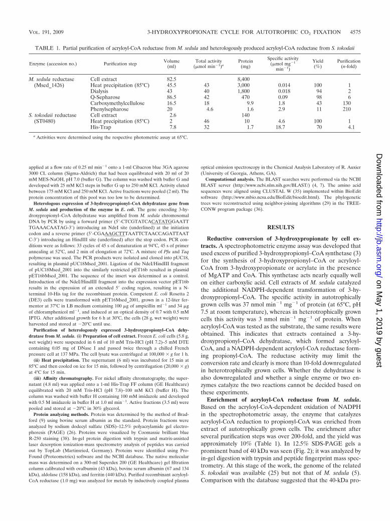
Enrichment of acryloyl-CoA reductase from M. sedula.Based on the acryloyl-CoA-dependent oxidation of NADPHin the spectrophotometric assay, the enzyme that catalyzesacryloyl-CoA reduction to propionyl-CoA was enriched fromextract of autotrophically grown cells. The enrichment afterseveral purification steps was over 200-fold, and the yield wasapproximately 10% (Table 1). In 12.5% SDS-PAGE gels aprominent band of 40 kDa was seen (Fig. 2); it was analyzed byin-gel digestion with trypsin and peptide fingerprint mass spec-trometry. At this stage of the work, the genome of the relatedS. tokodaii was available (25) but not that of M. sedula (5).Comparison with the database suggested that the 40-kDa pro-
TABLE 1. Partial purification of acryloyl-CoA reductase from M. sedula and heterologously produced acryloyl-CoA reductase from S. tokodaii
Enzyme (accession no.) Purification step Volume(ml)
Total activity(�mol min�1)a
Protein(mg)
Specific activity(�mol mg�1
min�1)
Yield(%)
Purification(n-fold)
M. sedula reductase Cell extract 82.5 8,400(Msed_1426) Heat precipitation (85°C) 45.5 43 3,000 0.014 100 1
Dialysis 43 40 1,800 0.018 94 2Q-Sepharose 86.5 42 470 0.09 98 6Carboxymethylcellulose 16.5 18 9.9 1.8 43 130Phenylsepharose 20 4.6 1.6 2.9 11 210
S. tokodaii reductase Cell extract 2.6 140(ST0480) Heat precipitation (85°C) 2 46 10 4.6 100 1
His-Trap 7.8 32 1.7 18.7 70 4.1
a Activities were determined using the respective photometric assay at 65°C.
VOL. 191, 2009 3-HYDROXYPROPIONATE CYCLE FOR AUTOTROPHIC CO2 FIXATION 4575
on May 1, 2019 by guest
http://jb.asm.org/
Dow
nloaded from

tein was the putative acryloyl-CoA reductase. The best hit wasfound in S. tokodaii (100% identity and 3% coverage; 11 iden-tical amino acids in a 334-amino-acid-containing protein; geneidentifiers gi 15920695, NP_376364, and ST0480).
Heterologous production of acryloyl-CoA reductase from S.tokodaii in E. coli. The putative reductase gene ST0480 from S.tokodaii coded for a hypothetical 36-kDa member of the zinc-containing alcohol dehydrogenase family (clusters of ortholo-gous groups of proteins family 1064). It was cloned into theexpression vector pET16b and expressed in E. coli Rosetta2(DE3). This strain carries a plasmid (pRARE2) with genesfor rare tRNA species. The His10 tag at the N terminus allowedan efficient purification of the enzyme after heat precipitationof most of the E. coli protein (Fig. 2 and Table 1). The over-produced enzyme was soluble, and the obtained preparationwas virtually pure and showed one single 39-kDa band (ex-pected value, 38.6 kDa) on SDS-PAGE gels (Fig. 2). Therecombinant His-tagged enzyme catalyzed the acryloyl-CoA-dependent NADPH oxidation and formation of propionyl-CoA, whereas it was inactive with 3-hydroxypropionyl–CoA,indicating that it was the wanted reductase. The enzyme couldbe stored for months in 30% glycerol at �20°C. In themeantime, the genome of M. sedula became available (5). Agene encoding a protein with 76% amino acid sequenceidentity to the identified acryloyl-CoA reductase of S. toko-daii is present in the genome of M. sedula (gi 146304192;Msed_1426). Reinterpretation of the fingerprint mass spec-trometry data identified the same gene (Msed_1426) in M.sedula as a best hit (100% identity and 73% coverage). It islocated upstream of a gene encoding succinic seminalde-hyde reductase (gi 146304190; Msed_1424), an enzyme alsoinvolved in autotrophic CO2 fixation (D. Kockelkorn and G.Fuchs, unpublished data).
Characterization of the recombinant acryloyl-CoA reduc-tase. The native molecular mass determined by gel filtrationwas 43 kDa, suggesting that the enzyme existed as a monomer.
The catalytic properties were analyzed at 65°C by the coupledspectrophotometric assay. Controls showed that the enzymeexhibited neither 3-hydroxypropionyl–CoA synthetase nor3-hydroxypropionyl–CoA dehydratase activity. Its optimumpH was 6.0 (65°C); half-maximal activity was obtained at pH7.5. The enzyme activity followed Michaelis-Menten kinetics,with apparent Km values for NADPH of 36 �M and for acryl-oyl-CoA of around 3 �M. The specific activity was 18.7 �molmin�1 mg�1, corresponding to a turnover number of 13 s�1.The enzyme did not act on either NADH or crotonyl-CoA.Incubation with 100 mM EDTA for 1 h at up to 80°C did notinactivate the enzyme. Addition of Zn2� or other divalentmetal ions to the enzyme assay did not stimulate or inactivateactivity. However, a metal analysis (27 elements) of acryloyl-CoA reductase by plasma emission spectroscopy revealed thepresence of 0.8 mol of Zn2� per mol of enzyme monomer,whereas other metals could not be detected. The UV-visiblespectrum showed only a protein absorption band near 280 nm.
Spectrophotometric assay for 3-hydroxypropionyl–CoA de-hydratase. The availability of recombinant 3-hydroxypropio-nyl–CoA synthetase and acryloyl-CoA reductase allowed thedesign of a spectrophotometric assay for 3-hydroxypropionyl–CoA dehydratase. Assays containing both auxiliary enzymes inexcess allowed detection and quantification of dehydratase ac-tivity in cell extract and protein fractions of M. sedula. Lowconcentrations of CoA (0.1 mM) were optimal and necessarybecause higher concentrations inhibited the assay. 3-Hy-droxypropionyl–CoA dehydratase activity in extracts of au-totrophically grown cells was 2.4 �mol min�1 mg�1 of cellprotein and was not downregulated in heterotrophically growncells (3.1 �mol min�1 mg�1 of cell protein). As a control,malate dehydrogenase was measured in both types of extracts.Its specific activity in autotrophically grown cells was 1.9 �molmin�1 mg�1 versus 3.5 �mol min�1 mg�1 in heterotrophicallygrown cells. Note that the heterotrophic growth rate was threetimes higher.
FIG. 2. SDS-PAGE (12.5%) of fractions obtained during purification of native and recombinant acryloyl-CoA reductase. Proteins were stainedwith Coomassie blue. (A) Enzyme fractions during purification of the native enzyme from M. sedula. Lane 1, cell extract of autotrophically growncells (20 �g); lane 2, after heat precipitation (20 �g); lane 3, after ultracentrifugation (20 �g); lane 4, after Q-Sepharose chromatography (20 �g);lane 5, after carboxymethylcellulose chromatography (20 �g); lane 6, after phenyl-Sepharose chromatography (10 �g; after an additionalpurification step using a Resource S column the marked band was cut out and sequenced); lane 7, molecular mass standard proteins. (B) Het-erologous expression of the acryloyl-CoA reductase gene from S. tokodaii in E. coli Rosetta 2 (DE3). Lane 1, whole cells before induction; lane2, whole cells after 3 h of induced growth; lane 3, cell extract after heat precipitation (20 �g); lane 4, purified recombinant acryloyl-CoA reductaseafter Ni2� affinity column (20 �g); lane 5, molecular mass standard proteins.
4576 TEUFEL ET AL. J. BACTERIOL.
on May 1, 2019 by guest
http://jb.asm.org/
Dow
nloaded from
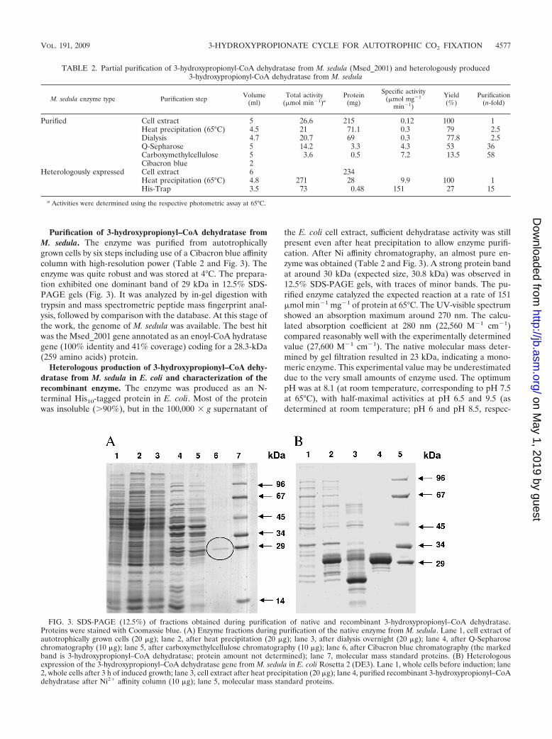
Purification of 3-hydroxypropionyl–CoA dehydratase fromM. sedula. The enzyme was purified from autotrophicallygrown cells by six steps including use of a Cibacron blue affinitycolumn with high-resolution power (Table 2 and Fig. 3). Theenzyme was quite robust and was stored at 4°C. The prepara-tion exhibited one dominant band of 29 kDa in 12.5% SDS-PAGE gels (Fig. 3). It was analyzed by in-gel digestion withtrypsin and mass spectrometric peptide mass fingerprint anal-ysis, followed by comparison with the database. At this stage ofthe work, the genome of M. sedula was available. The best hitwas the Msed_2001 gene annotated as an enoyl-CoA hydratasegene (100% identity and 41% coverage) coding for a 28.3-kDa(259 amino acids) protein.
Heterologous production of 3-hydroxypropionyl–CoA dehy-dratase from M. sedula in E. coli and characterization of therecombinant enzyme. The enzyme was produced as an N-terminal His10-tagged protein in E. coli. Most of the proteinwas insoluble (�90%), but in the 100,000 � g supernatant of
the E. coli cell extract, sufficient dehydratase activity was stillpresent even after heat precipitation to allow enzyme purifi-cation. After Ni affinity chromatography, an almost pure en-zyme was obtained (Table 2 and Fig. 3). A strong protein bandat around 30 kDa (expected size, 30.8 kDa) was observed in12.5% SDS-PAGE gels, with traces of minor bands. The pu-rified enzyme catalyzed the expected reaction at a rate of 151�mol min�1 mg�1 of protein at 65°C. The UV-visible spectrumshowed an absorption maximum around 270 nm. The calcu-lated absorption coefficient at 280 nm (22,560 M�1 cm�1)compared reasonably well with the experimentally determinedvalue (27,600 M�1 cm�1). The native molecular mass deter-mined by gel filtration resulted in 23 kDa, indicating a mono-meric enzyme. This experimental value may be underestimateddue to the very small amounts of enzyme used. The optimumpH was at 8.1 (at room temperature, corresponding to pH 7.5at 65°C), with half-maximal activities at pH 6.5 and 9.5 (asdetermined at room temperature; pH 6 and pH 8.5, respec-
TABLE 2. Partial purification of 3-hydroxypropionyl-CoA dehydratase from M. sedula (Msed_2001) and heterologously produced3-hydroxypropionyl-CoA dehydratase from M. sedula
M. sedula enzyme type Purification step Volume(ml)
Total activity(�mol min�1)a
Protein(mg)
Specific activity(�mol mg�1
min�1)
Yield(%)
Purification(n-fold)
Purified Cell extract 5 26.6 215 0.12 100 1Heat precipitation (65°C) 4.5 21 71.1 0.3 79 2.5Dialysis 4.7 20.7 69 0.3 77.8 2.5Q-Sepharose 5 14.2 3.3 4.3 53 36Carboxymethylcellulose 5 3.6 0.5 7.2 13.5 58Cibacron blue 2
Heterologously expressed Cell extract 6 234Heat precipitation (65°C) 4.8 271 28 9.9 100 1His-Trap 3.5 73 0.48 151 27 15
a Activities were determined using the respective photometric assay at 65°C.
FIG. 3. SDS-PAGE (12.5%) of fractions obtained during purification of native and recombinant 3-hydroxypropionyl–CoA dehydratase.Proteins were stained with Coomassie blue. (A) Enzyme fractions during purification of the native enzyme from M. sedula. Lane 1, cell extract ofautotrophically grown cells (20 �g); lane 2, after heat precipitation (20 �g); lane 3, after dialysis overnight (20 �g); lane 4, after Q-Sepharosechromatography (10 �g); lane 5, after carboxymethylcellulose chromatography (10 �g); lane 6, after Cibacron blue chromatography (the markedband is 3-hydroxypropionyl–CoA dehydratase; protein amount not determined); lane 7, molecular mass standard proteins. (B) Heterologousexpression of the 3-hydroxypropionyl–CoA dehydratase gene from M. sedula in E. coli Rosetta 2 (DE3). Lane 1, whole cells before induction; lane2, whole cells after 3 h of induced growth; lane 3, cell extract after heat precipitation (20 �g); lane 4, purified recombinant 3-hydroxypropionyl–CoAdehydratase after Ni2� affinity column (10 �g); lane 5, molecular mass standard proteins.
VOL. 191, 2009 3-HYDROXYPROPIONATE CYCLE FOR AUTOTROPHIC CO2 FIXATION 4577
on May 1, 2019 by guest
http://jb.asm.org/
Dow
nloaded from

tively, when extrapolated to 65°C). The extrapolated maximumrate (Vmax) at 65°C was 186 �mol min�1 mg�1, correspondingto a turnover number of 96 s�1; the apparent Km for 3-hy-droxypropionyl–CoA was 60 �M.
It was also tested whether the enzyme acted on 3-hydroxy-butyryl–CoA. In this assay crotonyl-CoA formation was fol-lowed spectrophotometrically at 40°C using crotonyl-CoAcarboxylase/reductase from R. sphaeroides (13). This auxil-iary enzyme catalyzes the following reaction: crotonyl-CoA �CO2 � NADPH � H�3 ethylmalonyl-CoA � NADP�. 3-Hy-droxypropionyl–CoA dehydratase acted nearly equally as wellon (S)-3-hydroxybutyryl–CoA as on 3-hydroxypropionyl–CoA.The enzyme did not convert the (R)-stereoisomer of 3-hy-droxybutyryl–CoA. The apparent Km value for (S)-3-hydroxy-butyryl–CoA was 75 �M; the extrapolated Vmax was 34 �molmin�1 mg�1. When extrapolated to 65°C assuming doubling ofthe rate per 10°C temperature increase, the Vmax value wasnearly same as that determined at 65°C for 3-hydroxypropio-nyl–CoA.
DISCUSSION
Role of the enzymes. We characterized the missing enzymes3-hydroxypropionyl–CoA dehydratase and acryloyl-CoA re-ductase that are required for the reductive conversion of 3-hy-droxypropionate to propionyl-CoA in the process of CO2
fixation in autotrophic members of the Sulfolobales. Threeseparate enzymes are required, 3-hydroxypropionyl–CoAsynthetase (3), 3-hydroxypropionyl–CoA dehydratase, andacryloyl-CoA reductase. In M. sedula cell extract the reductasewas the rate-limiting enzyme. The overall activity of 3-hy-droxypropionate reduction as well as the 3-hydroxypropionyl–CoA synthetase and the acryloyl-CoA reductase activities wereupregulated in autotrophically grown cells. These results areconsistent with the proposed function in autotrophic CO2 fix-ation. In Chloroflexus, all three steps are catalyzed by a largefusion protein with three enzyme domains (2). These enzymedomains are only distantly related to the three separate en-zyme entities in the Sulfolobales (see below).
The autotrophic 3-hydroxypropionate/4-hydroxybutyratecarbon fixation cycle leads from acetyl-CoA plus two bicarbon-ate molecules to succinyl-CoA and back to two molecules ofacetyl-CoA. The conversion of acetyl-CoA to succinyl-CoA via3-hydroxypropionate probably has evolved independently intwo genera in the order Sulfolobales (e.g., Sulfolobus and Me-tallosphaera sp.) and in the green non-sulfur bacteria (e.g.,Chloroflexus sp.). This is concluded from the finding that theMetallosphaera and Chloroflexus enzymes or enzyme domainshave been derived from genes that are only distantly related.The suggestion of a convergent evolution is further corrobo-rated by the completely different genes coding for malonyl-CoA reductase (1) and malonate semialdehyde reductase(Kockelkorn and Fuchs, unpublished). In support of this, twocompletely different solutions have been realized in these twolineages to regenerate the CO2 acceptor molecule acetyl-CoAfrom succinyl-CoA.
Enzyme properties and catalyzed reactions. The propertiesof the two enzymes are summarized in Table 3. Acryloyl-CoAreductase (E.C. 1.3.1.x) catalyzes the following irreversible re-action: acryloyl-CoA � NADPH � H� 3 propionyl-CoA �
TA
BL
E3.
Mol
ecul
aran
dca
taly
ticpr
oper
ties
ofre
com
bina
ntac
rylo
yl-C
oAre
duct
ase
from
S.to
koda
iian
d3-
hydr
oxyp
ropi
onyl
-CoA
dehy
drat
ase
from
M.s
edul
a
Org
anis
man
den
zym
eaSu
bstr
ates
Prod
ucts
Spec
ific
activ
ity(U
/mg)
bA
ppar
ent
Km
(�M
)O
ptim
umpH
Tur
nove
r(s
�1)
Nat
ive
mol
ecul
arm
ass
(kD
ac
alcu
late
d)c
Com
posi
tion
Spec
ifici
ty(%
)
S.to
koda
iiac
rylo
yl-
CoA
redu
ctas
eA
cryl
oyl-C
oA,
NA
DPH
,H�
Prop
iony
l-CoA
,N
AD
P�18
.7A
cryl
oyl-C
oA,�
10;
NA
DPH
,36
613
43(3
6)M
onom
erA
cryl
oyl-C
oA10
0;cr
oton
yl-C
oA,�
1M
.sed
ula
3-hy
drox
ypro
pion
yl-
CoA
dehy
drat
ase
3-H
ydro
xypr
opio
nyl-
CoA
,(S)
-3-
hydr
oxyb
utyr
yl-C
oA
Acr
yloy
l-CoA
,cro
tony
l-C
oA,H
2O15
13-
Hyd
roxy
prop
iony
l-C
oA,6
0;(S
)-3-
hydr
oxyb
utyr
yl-
CoA
,75
8.1
9623
(31)
Mon
omer
3-H
ydro
xypr
opio
nyl-
CoA
,100
;(S)
-3-
hydr
oxyb
utyr
yl-
CoA
,100
;(R
)-3-
hydr
oxyb
utyr
yl-
CoA
,�1
aA
cces
sion
num
bers
are
ST04
80fo
rth
eS.
toko
daii
redu
ctas
ean
dM
sed_
2001
for
the
M.s
edul
ade
hydr
atas
e.b
Act
iviti
esw
ere
dete
rmin
edus
ing
the
resp
ectiv
eph
otom
etri
cas
say
at65
°C.
cD
eter
min
edby
gelfi
ltrat
ion.
4578 TEUFEL ET AL. J. BACTERIOL.
on May 1, 2019 by guest
http://jb.asm.org/
Dow
nloaded from

NADP�. The enzyme does not require cofactors and does notuse NADH as an electron donor. Also it does not act oncrotonyl-CoA. This finding is important because crotonyl-CoAis an intermediate in the conversion of succinyl-CoA to twomolecules of acetyl-CoA (Fig. 1). Nonspecific reduction ofcrotonyl-CoA to butyryl-CoA would be disastrous, resulting ina dead-end product (butyryl-CoA) that traps CoA. An NADH-dependent, oxygen-sensitive acryloyl-CoA reductase has beenstudied in Clostridium propionicum that is involved in propi-onate fermentation (18). The complex enzyme contains anelectron-transferring flavoprotein and does not exhibit anysimilarity to the enzyme of M. sedula or to other trans-2-enoyl-CoA reductases.
3-Hydroxypropionyl–CoA dehydratase (E.C.4.2.1.17) cata-lyzes the following reversible reaction: 3-hydroxypropionyl–CoA 3 acryloyl-CoA � H2O. It also catalyzes the followingreversible reaction: crotonyl-CoA � H2O3 (S)-3-hydroxybu-tyryl–CoA. The (R)-stereoisomer is not accepted when testedin the dehydration reaction. Crotonyl-CoA hydration takespart in the conversion of succinyl-CoA to two molecules ofacetyl-CoA (Fig. 1), which also involves the (S)-stereoisomerrather than the (R)-stereoisomer of 3-hydroxybutyryl–CoA.Therefore, the dehydratase may fulfill a dual function.
Comparison with the corresponding domains of propionyl-CoA synthase from C. aurantiacus, distribution, and phyloge-netic trees. The genes coding for the two enzymes are notclustered on the Metallosphaera or Sulfolobus genome. Thegrouping of acryloyl-CoA reductase (Fig. 4) and of 3-hy-droxypropionyl–CoA dehydratase (Fig. 5) with related pro-teins in the database reveals interesting aspects. Similarreductases exist in other autotrophic members of the Sul-folobales (Metallosphaera and Sulfolobus spp.) and probably
fulfill the same function as in M. sedula. The function ofsimilar enzymes in other Crenarchaea (Caldivirga, Picrophi-lus, Thermoplasma, Pyrobaculum, and Thermoproteus spp.)and Eubacteria (Myxobacteria) cannot be predicted. The re-ductase domain of propionyl-CoA synthase of C. aurantia-cus shows only 28% amino acid sequence identity and 47%similarity over only one-fourth of the acryloyl-CoA reduc-tase from M. sedula. In fact, only the NADPH binding site ishighly conserved. Surprisingly, a similar gene product in theautotrophic marine group I Archaea (the Nitrosopumilus sp.Nmar_0523 and the Cenarchaeum sp. CENSYa_1407) showsonly moderate similarity with the Metallosphaera enzyme(27% amino acid sequence identity and 45% similarity);these archaea have been proposed to use a similar autotro-phic 3-hydroxypropionate/4-hydroxybutyrate cycle (8). Whetherthis discrepancy reflects large evolutionary distances betweenthe Sulfolobales and the marine group I Crenarchaea, conver-gent evolution of the pathway in these groups, or eventuallythe operation of two different autotrophic pathways cannot bedecided based on this study.
Also, similar dehydratases exist in other autotrophic mem-bers of the Sulfolobales and probably fulfill the same functionas in Metallosphaera. Many members of the Thermoproteales(Thermoproteus, Pyrobaculum, and Caldivirga) and Desulfuro-coccales (Aeropyrum sp.) contain similar genes whose functionis unknown. Furthermore, similar enzymes exist in Clostridialesand other Eubacteria. Again, the dehydratase domain of pro-pionyl-CoA synthase of C. aurantiacus shows only 40% aminoacid sequence identity and 57% similarity. A correspondinggene product in the autotrophic marine group I Archaea (Nitros-opumilus sp. Nmar_1308 and Cenarchaeum sp. CENSYa_0166)shows only moderate similarity with the Metallosphaera en-
FIG. 4. Phylogenetic tree of acryloyl-CoA reductase. The tree is based on amino acid sequence analysis and rooted with propionyl-CoAsynthase from C. aurantiacus. Tree topography and evolutionary distances are given by the neighbor-joining method with Poisson correction. Thescale bar represents a difference of 0.1 substitutions per site. Numbers at nodes indicate the percentage bootstrap values for the clade of this groupin 1,000 replications. The enzyme group from the order Sulfolobales is indicated.
VOL. 191, 2009 3-HYDROXYPROPIONATE CYCLE FOR AUTOTROPHIC CO2 FIXATION 4579
on May 1, 2019 by guest
http://jb.asm.org/
Dow
nloaded from

zyme (42% amino acid sequence identity and 65 to 69% sim-ilarity for Nmar_1308 and 65% CENSYa_0166). It should bestressed, however, that the enoyl-CoA hydratase family and theZn-containing alcohol dehydrogenase family comprise manyonly distantly related members and that the two reactionsunder study may well be catalyzed by members of other proteinfamilies.
Interestingly, Metallosphaera organisms as well as otherautotrophic members of the Sulfolobales and Proteales con-tain another enzyme that catalyzes the (S)-3-hydroxybu-tyryl–CoA dehydratase/crotonyl-CoA hydratase reaction(Kockelkorn and Fuchs, unpublished). Work is in progressto characterize this second enzyme, which consists of anenoyl-CoA hydratase and a 3-hydroxyacyl–CoA dehydroge-nase domain and which is twice as large as the enzymesunder study here. In Fig. 5 the (de)hydratase domain ofthese fusion proteins is shown in the phylogenetic tree.Because the members of the Thermoproteales use a similarconversion of succinyl-CoA to two acetyl-CoA in their au-totrophic dicarboxylate/4-hydroxybutyrate cycle (22), therole of this fusion protein is likely the same as in the Sul-folobales, i.e., the conversion of crotonyl-CoA via (S)-3-hydroxybutyryl–CoA to acetoacetyl-CoA. Cleavage of ace-toacetyl-CoA by �-ketothiolase forms two molecules ofacetyl-CoA, thus regenerating the CO2 acceptor and releas-ing another acetyl-CoA molecule for biosynthesis.
Related members of the Zn-containing alcohol dehydroge-nase and enoyl-CoA hydratase families and possible activesites. Acryloyl-CoA reductase belongs to the Zn-containingalcohol dehydrogenase enzyme family. It appears that the en-zyme contains Zn2� although it could not be inactivated byEDTA. A possible explanation for this apparent discrepancy
could be an insufficient accessibility of zinc by EDTA and/orhigh thermal stability of the reductase (27). An alignment ofthe acryloyl-CoA reductase with the well-studied horse liveralcohol dehydrogenase shows the presence of the strictly con-served sequence motif GHE of the catalytic zinc binding side(amino acid position 59 to 61) with two flanking cysteines(amino acids 38 and 146). In addition, a conserved sequencefor a structural zinc binding site DXCXXCXXXXXXXC(where X is any residue) can be found (amino acids 90, 93, 96,and 104). Here, amino acid 90 is changed from cysteine toaspartic acid, as already observed in Sulfolobus solfataricus andAeropyrum pernix (6, 14). Whether the native and active reduc-tases contain one or two zinc ions cannot be decided based onour data. 3-Hydroxypropionyl–CoA dehydratase belongs to theenoyl-CoA hydratase enzyme family. This class of enzymescatalyzes the syn-addition of a water molecule to a trans-�-�unsaturated CoA-activated acid. Since the OH� is added tothe �-position and the H� to the �-position from the 2-Re-sideof crotonyl-CoA, the S-stereoisomer results. In the case of thebest-studied crotonyl-CoA hydratase (crotonase) (S)-3-hydroxy-butyryl–CoA is formed. The amino acids in the active sites ofthese enzymes are well conserved in the Metallosphaera enzyme.
ACKNOWLEDGMENTS
This work was supported by the Deutsche Forschungsgemeinschaftand the Fonds der Chemischen Industrie.
We thank Gabor Igloi, Freiburg, for DNA sequencing and NasserGad’on, Freiburg, for cell culturing.
REFERENCES
1. Alber, B., M. Olinger, A. Rieder, D. Kockelkorn, B. Jobst, M. Hugler, and G.Fuchs. 2006. Malonyl-coenzyme A reductase in the modified 3-hydroxypro-pionate cycle for autotrophic carbon fixation in archaeal Metallosphaera andSulfolobus spp. J. Bacteriol. 188:8551–8559.
FIG. 5. Phylogenetic tree of 3-hydroxypropionyl–CoA dehydratase. The tree is based on amino acid sequence analysis and rooted withpropionyl-CoA synthase from C. aurantiacus. Tree topography and evolutionary distances are given by the neighbor-joining method withPoisson correction. The scale bar represents a difference of 0.1 substitutions per site. Numbers at nodes indicate the percentage bootstrapvalues for the clade of this group in 1,000 replications. The enzyme groups from the order Sulfolobales and from Clostridia are indicated.Fusion refers to the dehydratase domain of an archaeal fusion enzyme that contains an enoyl-CoA hydratase fused to a 3-hydroxyacyl–CoAdehydrogenase.
4580 TEUFEL ET AL. J. BACTERIOL.
on May 1, 2019 by guest
http://jb.asm.org/
Dow
nloaded from

2. Alber, B. E., and G. Fuchs. 2002. Propionyl-coenzyme A synthase fromChloroflexus aurantiacus, a key enzyme of the 3-hydroxypropionate cycle forautotrophic CO2 fixation. J. Biol. Chem. 277:12137–12143.
3. Alber, B. E., J. W. Kung, and G. Fuchs. 2008. 3-Hydroxypropionyl-coenzymeA synthetase from Metallosphaera sedula, an enzyme involved in autotrophicCO2 fixation. J. Bacteriol. 190:1383–1389.
4. Altschul, S. F., W. Gish, W. Miller, E. W. Myers, and D. J. Lipman. 1990.Basic local alignment search tool. J. Mol. Biol. 215:403–410.
5. Auernik, K. S., Y. Maezato, P. H. Blum, and R. M. Kelly. 2008. The genomesequence of the metal-mobilizing, extremely thermoacidophilic archaeonMetallosphaera sedula provides insights into bioleaching-associated metabo-lism. Appl. Environ. Microbiol. 74:682–692.
6. Auld, D. S., and T. Bergman. 2008. Medium- and short-chain dehydroge-nase/reductase gene and protein families: the role of zinc for alcohol dehy-drogenase structure and function. Cell. Mol. Life. Sci. 65:3961–3970.
7. Benson, D. A., I. Karsch-Mizrachi, D. J. Lipman, J. Ostell, and D. L.Wheeler. 2006. GenBank. Nucleic Acids Res. 34:D16–D20.
8. Berg, I. A., D. Kockelkorn, W. Buckel, and G. Fuchs. 2007. A 3-hydroxypro-pionate/4-hydroxybutyrate autotrophic carbon dioxide assimilation pathwayin Archaea. Science 318:1782–1786.
9. Bradford, M. M. 1976. A rapid and sensitive method for the quantitation ofmicrogram quantities of protein utilizing the principle of protein-dye bind-ing. Anal. Biochem. 72:248–254.
10. Dawson, R. M. C., D. C. Elliott, W. H. Elliott, and K. M. Jones. 1986. Datafor biochemical research, 3rd ed. Oxford University Press, New York, NY.
11. Eisenreich, W., G. Strauss, U. Werz, G. Fuchs, and A. Bacher. 1993. Retro-biosynthetic analysis of carbon fixation in the phototrophic eubacteriumChloroflexus aurantiacus. Eur. J. Biochem. 215:619–632.
12. Ellis, K. J., and J. F. Morrison. 1982. Buffers of constant ionic strength forstudying pH-dependent processes. Methods Enzymol. 87:405–426.
13. Erb, T. J., I. A. Berg, V. Brecht, M. Muller, G. Fuchs, and B. E. Alber. 2007.Synthesis of C5-dicarboxylic acids from C2-units involving crotonyl-CoAcarboxylase/reductase: the ethylmalonyl-CoA pathway. Proc. Natl. Acad. Sci.USA 104:10631–10636.
14. Esposito, L., F. Sica, C. A. Raia, A. Giordano, M. Rossi, L. Mazzarella, andA. Zagari. 2002. Crystal structure of the alcohol dehydrogenase from thehyperthermophilic archaeon Sulfolobus solfataricus at 1.85 Å resolution. J.Mol. Biol. 318:463–477.
15. Good, N. E., and S. Izawa. 1972. Hydrogen ion buffers. Methods Enzymol.24:53–68.
16. Herter, S., J. Farfsing, N. Gad’On, C. Rieder, W. Eisenreich, A. Bacher, andG. Fuchs. 2001. Autotrophic CO2 fixation by Chloroflexus aurantiacus: studyof glyoxylate formation and assimilation via the 3-hydroxypropionate cycle. J.Bacteriol. 183:4305–4316.
17. Herter, S., G. Fuchs, A. Bacher, and W. Eisenreich. 2002. A bicyclic autotro-phic CO2 fixation pathway in Chloroflexus aurantiacus. J. Biol. Chem. 277:20277–20283.
18. Hetzel, M., M. Brock, T. Selmer, A. J. Pierik, B. T. Golding, and W. Buckel.2003. Acryloyl-CoA reductase from Clostridium propionicum. An enzymecomplex of propionyl-CoA dehydrogenase and electron-transferring fla-voprotein. Eur. J. Biochem. 270:902–910.
19. Holo, H. 1989. Chloroflexus aurantiacus secretes 3-hydroxypropionate, a pos-sible intermediate in the assimilation of CO2 and acetate. Arch. Microbiol.151:252–256.
20. Holo, H., and R. Sirevåg. 1986. Autotrophic growth and CO2 fixation ofChloroflexus aurantiacus. Arch. Microbiol. 148:173–180.
21. Huber, G., C. Spinnler, A. Gambacorta, and K. O. Stetter. 1989. Metal-losphaera sedula gen. and sp. nov. represents a new genus of aerobic,metal mobilizing, thermoacidophilic Archaebacteria. Syst. Appl. Micro-biol. 12:38–57.
22. Huber, H., M. Gallenberger, U. Jahn, E. Eylert, I. A. Berg, D. Kockelkorn,W. Eisenreich, and G. Fuchs. 2008. A dicarboxylate/4-hydroxybutyrate au-totrophic carbon assimilation cycle in the hyperthermophilic archaeum Igni-coccus hospitalis. Proc. Natl. Acad. Sci. USA 105:7851–7856.
23. Hugler, M., H. Huber, K. O. Stetter, and G. Fuchs. 2003. Autotrophic CO2fixation pathways in archaea (Crenarchaeota). Arch. Microbiol. 179:160–173.
24. Hugler, M., R. S. Krieger, M. Jahn, and G. Fuchs. 2003. Characterization ofacetyl-CoA/propionyl-CoA carboxylase in Metallosphaera sedula. Carboxyl-ating enzyme in the 3-hydroxypropionate cycle for autotrophic carbon fixa-tion. Eur. J. Biochem. 270:736–744.
25. Kawarabayasi, Y., Y. Hino, H. Horikawa, K. Jin-no, M. Takahashi, M.Sekine, S. Baba, A. Ankai, H. Kosugi, A. Hosoyama, S. Fukui, Y. Nagai, K.Nishijima, R. Otsuka, H. Nakazawa, M. Takamiya, Y. Kato, T. Yoshizawa, T.Tanaka, Y. Kudoh, J. Yamazaki, N. Kushida, A. Oguchi, K. Aoki, S. Masuda,M. Yanagii, M. Nishimura, A. Yamagishi, T. Oshima, and H. Kikuchi. 2001.Complete genome sequence of an aerobic thermoacidophilic crenarchaeon,Sulfolobus tokodaii strain7. DNA Res. 8:123–140.
26. Laemmli, U. K. 1970. Cleavage of structural proteins during the assembly ofthe head of bacteriophage T4. Nature 227:680–685.
27. Magonet, E., P. Hayen, D. Delforge, E. Delaive, and J. Remacle. 1992.Importance of the structural zinc atom for the stability of yeast alcoholdehydrogenase. Biochem. J. 287:361–365.
28. Menendez, C., Z. Bauer, H. Huber, N. Gad’on, K. O. Stetter, and G. Fuchs.1999. Presence of acetyl coenzyme A (CoA) carboxylase and propionyl-CoAcarboxylase in autotrophic Crenarchaeota and indication for operation of a3-hydroxypropionate cycle in autotrophic carbon fixation. J. Bacteriol. 181:1088–1098.
29. Saitou, N., and M. Nei. 1987. The neighbor-joining method: a new methodfor reconstructing phylogenetic trees. Mol. Biol. Evol. 4:406–425.
30. Sambrook, J., E. F. Fritsch, and T. Maniatis. 1989. Molecular cloning: alaboratory manual, 2nd ed. Cold Spring Harbor Laboratory Press, ColdSpring Harbor, NY.
31. Stoll, V. S., and J. S. Blanchard. 1990. Buffers: principles and practice.Methods Enzymol. 182:24–38.
32. Strauss, G., W. Eisenreich, A. Bacher, and G. Fuchs. 1992. 13C-NMR studyof autotrophic CO2 fixation pathways in the sulfur-reducing Archaebacte-rium Thermoproteus neutrophilus and in the phototrophic Eubacterium Chlo-roflexus aurantiacus. Eur. J. Biochem. 205:853–866.
33. Strauss, G., and G. Fuchs. 1993. Enzymes of a novel autotrophic CO2fixation pathway in the phototrophic bacterium Chloroflexus aurantiacus, the3-hydroxypropionate cycle. Eur. J. Biochem. 215:633–643.
34. Suzuki, T., T. Iwasaki, T. Uzawa, K. Hara, N. Nemoto, T. Kon, T. Ueki, A.Yamagishi, and T. Oshima. 2002. Sulfolobus tokodaii sp. nov. (f. Sulfolobussp. strain 7), a new member of the genus Sulfolobus isolated from Beppu HotSprings, Japan. Extremophiles 6:39–44.
35. Thompson, J. D., D. G. Higgins, and T. J. Gibson. 1994. CLUSTAL W:improving the sensitivity of progressive multiple sequence alignment throughsequence weighting, position-specific gap penalties and weight matrix choice.Nucleic Acids Res. 22:4673–4680.
36. Van de Peer, Y., and R. De Wachter. 1994. TREECON for Windows: asoftware package for the construction and drawing of evolutionary trees forthe Microsoft Windows environment. Comput. Appl. Biosci. 10:569–570.
37. Zarzycki, J., A. Schlichting, N. Strychalski, M. Muller, B. E. Alber, and G.Fuchs. 2008. Mesaconyl-coenzyme A hydratase, a new enzyme of two centralcarbon metabolic pathways in bacteria. J. Bacteriol. 190:1366–1374.
38. Zehr, B. D., T. J. Savin, and R. E. Hall. 1989. A one-step, low backgroundCoomassie staining procedure for polyacrylamide gels. Anal. Biochem. 182:157–159.
VOL. 191, 2009 3-HYDROXYPROPIONATE CYCLE FOR AUTOTROPHIC CO2 FIXATION 4581
on May 1, 2019 by guest
http://jb.asm.org/
Dow
nloaded from