3-dimensional facial expression recognition in human using
TRANSCRIPT

RESEARCH ARTICLE Open Access
3-Dimensional facial expression recognitionin human using multi-points warpingOlalekan Agbolade1* , Azree Nazri1*, Razali Yaakob1, Abdul Azim Ghani2 and Yoke Kqueen Cheah3
Abstract
Background: Expression in H-sapiens plays a remarkable role when it comes to social communication. Theidentification of this expression by human beings is relatively easy and accurate. However, achieving the sameresult in 3D by machine remains a challenge in computer vision. This is due to the current challenges facing facialdata acquisition in 3D; such as lack of homology and complex mathematical analysis for facial point digitization.This study proposes facial expression recognition in human with the application of Multi-points Warping for 3Dfacial landmark by building a template mesh as a reference object. This template mesh is thereby applied to eachof the target mesh on Stirling/ESRC and Bosphorus datasets. The semi-landmarks are allowed to slide alongtangents to the curves and surfaces until the bending energy between a template and a target form is minimaland localization error is assessed using Procrustes ANOVA. By using Principal Component Analysis (PCA) for featureselection, classification is done using Linear Discriminant Analysis (LDA).
Result: The localization error is validated on the two datasets with superior performance over the state-of-the-artmethods and variation in the expression is visualized using Principal Components (PCs). The deformations showvarious expression regions in the faces. The results indicate that Sad expression has the lowest recognition accuracyon both datasets. The classifier achieved a recognition accuracy of 99.58 and 99.32% on Stirling/ESRC andBosphorus, respectively.
Conclusion: The results demonstrate that the method is robust and in agreement with the state-of-the-art results.
Keywords: Facial expression recognition, 3D faces, Multi-point warping, Automatic facial landmark, PCA, LDA
BackgroundEmotions in human face play a remarkable role when itcomes to social communication. The identification ofexpressions by human beings is relatively easy and ac-curate. However, achieving the same result by machineremains a challenge in computer vision. Human face isthe part that hosts the most crucial sensory organs. Italso acts as the central interface for appearance, communi-cation, expression and identification [1]. Therefore, acquir-ing its information digitally is important to researchers.This makes landmark-based geometric morphometricsmethods for facial expression a new insight into patterns ofbiological emotion variations [2]. Many advances havebeen proposed in the area of acquisition of facial landmarkbut with several challenges especially in three-dimensional
model. One of the challenges is the insufficient acqui-sition of 3D facial landmarks. Another challenge isthe lack of homology due to manual annotation. Whereascomplex mathematical analysis has made many works un-reproducible in 3D facial landmark acquisition.The use of three-dimensional face images in morpho-
metrics does not only give room to cover a wider area ofhuman facial region but also retains all the geometric in-formation of the object descriptors [3, 4]. In modalitycomparison, 3D face has higher detection rate than thatof 2D due to its higher intensity modality [5]. Further-more, during subjection to systematically increasingpitch and yaw rotation experiment performed in [6],there was a dropped in expression recognition perform-ance in 2D while that of 3D remained constant. This isas a result of occlusion effects substantial distortion inout-of-plane rotations. More so, in the area of featuretransformation and classification, 3D modality shows alittle improvement with higher confidence over 2D. But
© The Author(s). 2019 Open Access This article is distributed under the terms of the Creative Commons Attribution 4.0International License (http://creativecommons.org/licenses/by/4.0/), which permits unrestricted use, distribution, andreproduction in any medium, provided you give appropriate credit to the original author(s) and the source, provide a link tothe Creative Commons license, and indicate if changes were made. The Creative Commons Public Domain Dedication waiver(http://creativecommons.org/publicdomain/zero/1.0/) applies to the data made available in this article, unless otherwise stated.
* Correspondence: [email protected]; [email protected] of Computer Science, Faculty of Computer Science & IT,Universiti Putra Malaysia, Serdang, Selangor, MalaysiaFull list of author information is available at the end of the article
Agbolade et al. BMC Bioinformatics (2019) 20:619 https://doi.org/10.1186/s12859-019-3153-2

in terms of depth features, both show the same perform-ance; and the cost of 3D model in terms of processing ishigher than that of 2D [5].Below is the summary of the main contribution of this
work:
1) We developed an approach for 3D facial landmarkusing multi-points warping. This approach hasextended the computational deformation processingin [7] to improve the annotation performance usinga less complex pipeline. We used six iterations andhundred to 5 % exponential decay sliding step inour method to ensure convergence and optimumsmoothness.
2) Due to the easy detection, pose correction [8] andinvariant to facial expression of nose tip [9],Pronasale was selected as the most robust andprominent landmark point. Since the nose tip areacan be approximated as a semi-sphere of the humanface. This determines the location where the slidingpoints begin to spread across the facial surface.
3) We have tested the method on two public 3D facedatabases (Stirling/ESRC and Bosphorus) to validatethe precision of the annotation of the landmarkswith the state-of-the-art methods.
4) We have validated the usability of our approachthrough its application to soft-tissue facial expressionrecognition in 3D. By using PCA for feature selection,we classify six expressions on both datasets. So far, tothe best of our knowledge, sliding semi-landmarkapproach to facial landmarking has not been appliedto solve problem relating to soft-tissue facial expres-sion recognition in 3D.
Section one of this study focuses on the introduction,section two discusses the related studies. In section
three, the implementation of the methodology is pre-sented with supporting references where short explan-ation has been provided. Section four discusses theresults of the implementations. In section five, a moredetailed discussion is presented for the clarification ofthe result and comparison with state-of-the-art methods.The last section concludes the study and presents thelimitations and future direction. Figure 1 shows thearchitectural diagram of the application of multi-pointswarping to the analysis of facial expression recognitionin 3D.
Literature reviewThe term “Morphometrics” was coined by Robert E.Blackith more than 50 years ago, who applied multivari-ate statistical methods to the basic carapace morphologyof grasshoppers [10]. Morphometrics is the study ofshape variation and its covariation with other variables[7, 11]. According to DC Adams, et al. [12], morphomet-rics was traditionally the application of multivariate stat-istical analyses to a sets of quantitative variables such aslength, width, height and angle. But advances in mor-phometrics have shifted focus to the Cartesian coordi-nates of anatomical points that might be used to definemore traditional measurements. Morphometrics exam-ines shape variation, group differences in shape, the cen-tral tendency of shape, and associations of shape withextrinsic factors [13]. This is directly based on the digi-tized x,y, (z)-coordinate positions of landmarks, pointsrepresenting the spatial positions of putatively homolo-gous structures in two or three dimensions; whereasconventional morphometric studies utilize distances asvariables [7, 11, 14]. The landmark was described in LFMarcus, et al. [15] as a point in a bi- or three-dimensional space that corresponds to the position of aparticular trait in an object. This set of points, one on
Fig. 1 Architecture of the proposed method
Agbolade et al. BMC Bioinformatics (2019) 20:619 Page 2 of 15

each form, are operationally defined on an individual bylocal anatomical features and must be consistent withsome hypothesis of biological homology. But the formallandmark definitions were provided by anthropometricstudies in [16]. This work by LG Farkas [16] has beenprovided as the standard for head and face landmarkdefinitions through the study of thousands of subjectsfrom different races. These have produced a large num-ber of anthropometric studies in the head and faceregions.A flexible and mathematically rigorous interpolation
technique of D’Arcy Thompson’s transformation grids[17], called Thin Plate-Spline (TPS), was brought intomorphometrics. This ensures that the correspondingpoints of the starting and target form appear precisely incorresponding positions in relation to the transformedand untransformed grids [18]. With the application of It-erative Closest Point (ICP), landmark correspondencecan iteratively be registered in the vicinity of a landmarkwith a re-weighted error function. Morphometrically, somestudies have been proposed which computed localizationerrors of facial landmarks on Bosphorus dataset. A novel3D constrained Local Models (CLM) approach facial land-mark detection in 3D images is proposed in [19], whichcapitalizes on the Independent Component Analysis (ICA)properties in order to define appropriate face Point Distri-bution Model (PDM) tailored to the mesh manifold modal-ity. Each sample contains 24 manually annotated faciallandmarks. While the PDM includes 33 landmarks and 14of them are part of the ground truth set tested on Bos-phorus database. An automatic method for facial landmarklocalization relying on geometrical properties of 3D facialsurface was proposed in [20], working on complete facesdisplaying different emotions and in presence of occlu-sions. The method extracts the landmark one-by-one.While the geometrical condition remains unchanged, themethod double-checks to ascertain whether pronasale,nasion and alare are correctly localized, otherwise theprocess starts afresh. The method is deterministic and isbackboned by a thresholding technique designed by study-ing the behavior of each geometrical descriptor in corres-pondence to the locus of each landmark, experimented onBosphorus database.Though facial landmarks are known to be specific
points with an anatomical meaning which has beendescribed in Table 1; since a considerable amount ofbiological variability cannot be assessed using only ana-tomical landmarks [21], in order to quantify complexshapes, sliding semi-landmarks have been developed whichcan be placed on surfaces [22] or curves [7, 22]. This ap-proach generates landmarks that are spatially homologousafter sliding [23] which may be optimized by minimizingbending energy [24, 25] or Procrustes distance [26, 27].Since sliding semi-landmarks have not been implemented
in analysing facial expression for soft-tissue in 3D, we havedecided to investigate the expression recognition using theapplication of multi-points warping approach.Emotion or expression recognition using facial analysis
has been the current trend in computer vision but thediversity of human facial expression has made the emotionrecognition somehow difficult [28]. Moreover, asides un-identifiable lighting challenges, the fairly significant differ-ences in age, skin colour and appearance of individualplaced additional burden on machine learning. When facesubjects are transformed into feature vectors, any classifiercan be used for expression recognition such as neuralnetwork, support vector machines, random forest, lineardiscriminant analysis, etc. But the uniqueness is the appli-cation of facial image information [29]. Due to the sensi-tivity of the change in head posture and illumination, theuse of static 2D image is unstable for expression recogni-tion. The use of 3D does not only play safe in the area ofillumination and pose change but also enables the use ofmore image information. This is because facial expres-sions are generated by facial muscle contractions. It resultsin temporary facial deformations in both texture and facialgeometry which is detectable in 3D and 4D [30]. The samesuccesses achieved in 3D face recognition could still benaturally adopted for expression recognition [31]. Ac-cording to M Pantic and LJ Rothkrantz [32] on facialexpression analyser, facial expression follows the generalproperties for solving computer vision problems: face de-tection, landmark localisation, recognition or classifica-tion. As 3D databases are becoming more and moreavailable in the computer vision community, differentmethods are being proposed to tackle the challenges fa-cing facial expression recognition. Most of these studiesare based on six fundamental expression classes or less:anger, fear, disgust, sadness, happiness, and surprise [33].Many also focus on the use of local features whichretrieves the topological and geometrical properties of theface expression [29, 34].Linear discriminant analysis and many other classifiers
have been used for classification in many face expression
Table 1 Procrustes ANOVAs for facial shape on Stirling andBosphorus datasets
Effect SS MS DF F P
Stirling
Expression 0.178563 2.39E-05 7465 8.36 <.0001
Individual 1.02071 2.86E-06 356,827 1.86 <.0001
Error 0.041283 1.54E-06 26,874
Bosphorus
Expression 0.308295 4.13E-05 7465 16.4 <.0001
Individual 0.65404 2.52E-06 259,782 0.72 1
Error 0.09455 3.52E-06 26,874
Agbolade et al. BMC Bioinformatics (2019) 20:619 Page 3 of 15

recognitions. A learn sparse features from spatio-temporallocal cuboids extracted from human face was proposed in[35]. This has application of conditional random field clas-sifiers for training and testing the model. In H Tang andTS Huang [36], similar distance feature was exploredusing automatic feature selection technique. This wasdone by maximizing the average relative entropy of mar-ginalized class-conditional feature distributions. Using 83landmarks, less than 30 features were selected. The fea-tures distance are subtracted from the features of theexpressive scan on the neutral scan which they classifiedby Naive Bayes, Neural network and Linear DiscriminantAnalysis on BU-3DFE dataset. To approximate the con-tinuous surface at each vertex of an input mesh, YL WangJun, Wei Xiaozhou, Sun Yi [6] proposed a cubic-orderpolynomial functions. It estimated coefficient at a particu-lar vertex, formed the weingarten matrix for the local sur-face path. The eigenvectors and eigenvalues of the matrixcould be derived by normal direction along the gradientmagnitude. The facial region was described using 64 land-marks to overcome the lack of correspondence betweenthe meshes. Their best performance was obtained usingLDA; no rigid transformation is required due to the geo-metrical invariance of curvature-based features. To dealwith issue of deformation of facial geometry which resultsfrom expression changes, C Li and A Barreto [37] pro-posed a framework that is composed of three subsystems:expressional face recognition system, neutral face recogni-tion system and expression recognition system. This wastested on 30 subjects and was classified using LDA, butused only two expression groups.H Li, et al. [38] proposed a novel method using fine-
grained matching of 3D key-point descriptors by extend-ing the SIFT-like matching framework to mesh data. Toaccount average for reconstruction error of probe facedescriptors, multi-task sparse representation algorithmwas used. The approach was evaluated on Bosphorusdatabase for expression recognition, pose invariant andocclusion. A comprehensive comparative evaluation wasperformed on Gavab, UND/FRGC, and Bosphorus in[39] by using local shape descriptor. The method cap-tured distinguishing traits on the face by extracting 3Dkey-points. Similarity expression on faces was evaluatedby comparing local shape descriptors across inlier pairsof matching key-points between gallery scans and probe.Using a Key-point-based Multiple Triangle Statistics(KMTS) with a Two-Phase Weighted Collaborative Rep-resentation Classification (TPWCRC), a robust to partialdata, large facial expression and pose variations was pro-posed in [40]. The method was experimented on six da-tabases including Bosphorus which achieved a promisingresult on occlusions, pose variation and expressions. A 3Dface augmentation technique was proposed in [41], whichsynthesizes a number of different facial expressions from a
single 3D face scan. The method showed excellent per-formance on BU-3DFE, 3D-TEC, and Bosphorus datasets,without application of hand-crafted features. A novel geo-metric framework for analysing 3D faces was proposed in[42] with the goals of averaging face shapes and compar-ing matching. The method presented facial surfaces byradial curves emanating from the nose tips, which wasexperimented on FRGCv2, GavabDB, and Bosphorus.Furthermore, in order to address the issue of 2D coun-
terpart and the handling of large intra-class and inter-class variability for human facial expression, W Hariri,et al. [43] proposed the use of covariance matrices ofdescriptors rather than using the descriptors themselves.Their work focused on application of manifold-basedclassification which was tested on BU-3DFE and Bos-phorus databases. While extended local binary patternswas proposed in [44] for facial expression recognitionfrom 3D depth map images where the results on Bos-phorus showed better performance by the combinationof 3D and 3D curvature.
Experiment resultsAfter the step-by-step methods in facial surface deform-ation of semi-landmark, the error assessment, the ana-lysis, visualisation and classification of the experimentwere performed using MorphoJ 1.06d [45], PAST 3.0[46] and R 5.1 [47].
Landmarks significanceThe use of landmarks evolves when locating biologicalor anatomical features on human faces. Its validity isdrawn from the morphometric analysis which dependson the biological justification for designation of the land-marks as stated in [3]. But not all the facial anatomicallandmarks always indicate a meaningful significantmeasure. On Stirling dataset, the overall landmarks aretested using one way ANOVA to see the significant ofthe variation on each expression group, each group hav-ing the same degree of freedom (df = 1499). Angry: F =133.9, p < 0.00001; Disgust: F = 120.9, p < 0.00001; Fear:F = 132.9, p < 0.00001; Sad: F = 130.2, p < 0.00001; Happy:F = 184.3, p < 0.00001; and Surprise: F = 117, p < 0.00001.Subsequently, same test was computed for Bosphorus oneach expression group, each group having the same de-gree of freedom (df = 1499). Angry: F = 2507, p <0.00001; Disgust: F = 1552, p < 0.00001; Fear: F = 3899,p < 0.00001; Sad: F = 2543, p < 0.00001; Happy: F = 2435,p < 0.00001; and Surprise: F = 1582, p < 0.00001. Further-more, we conducted PERMANOVA (Non-ParametricMANOVA) which is a non-parametric test of the signifi-cant difference between the expression groups, based onthe distance measured [48] with F = 7.76 and P = 0.0001for Stirling dataset and F = 115.5 and P = 0.0001 for Bos-phorus dataset. The large positive of F value indicates
Agbolade et al. BMC Bioinformatics (2019) 20:619 Page 4 of 15
that there is a significant difference between the expres-sion groups.
Procrustes ANOVAFor the assessment of localization errors of the land-marks; the deviations of each landmark is obtained bysimply calculating the amount of displacement from theaverage position calculated from all digitization and thevariation accounts for the smallest portion of the totalvariation using Procrustes ANOVA. The localization er-rors accounts for only 0.041 and 0.095 for Stirling andBosphorus, respectively, from the total variation (Table 1).
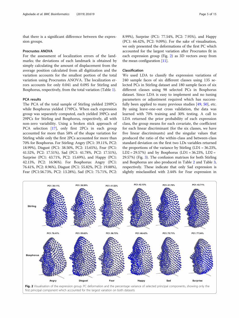
PCA resultsThe PCA of the total sample of Stirling yielded 239PCswhile Bosphorus yielded 179PCs. When each expressiongroup was separately computed, each yielded 39PCs and29PCs for Stirling and Bosphorus, respectively, all withnon-zero variability. Using a broken stick approach ofPCA selection [17], only first 2PCs in each groupaccounted for more than 58% of the shape variation forStirling while only the first 2PCs accounted for more than70% for Bosphorus. For Stirling: Angry (PC1: 39.11%, PC2:18.99%), Disgust (PC1: 38.50%, PC2: 15.65%), Fear (PC1:41.52%, PC2: 17.51%), Sad (PC1: 41.78%, PC2: 17.51%),Surprise (PC1: 43.71%, PC2: 15.69%), and Happy (PC1:42.13%, PC2: 16.96%). For Bosphorus: Angry (PC1:76.41%, PC2: 8.96%), Disgust (PC1: 55.82%, PC2: 19.88%),Fear (PC1:56.73%, PC2: 13.28%), Sad (PC1: 75.71%, PC2:
8.99%), Surprise (PC1: 77.54%, PC2: 7.95%), and Happy(PC1: 66.42%, PC2: 9.09%). For the sake of visualisation,we only presented the deformations of the first PC whichaccounted for the largest variation after Procrustes fit ineach expression group (Fig. 2) as 3D vectors away fromthe mean configuration [11].
ClassificationWe used LDA to classify the expression variations of240 sample faces of six different classes using 135 se-lected PCs in Stirling dataset and 180 sample faces of sixdifferent classes using 98 selected PCs in Bosphorusdataset. Since LDA is easy to implement and no tuningparameters or adjustment required which has success-fully been applied to many previous studies [49, 50], etc.By using leave-one-out cross validation, the data waslearned with 70% training and 30% testing. A call toLDA returned the prior probability of each expressionclass, the group means for each covariate, the coefficientfor each linear discriminant (for the six classes, we havefive linear discriminants) and the singular values thatproduced the ratio of the within-class and between-classstandard deviation on the first two LDs variables returnedthe proportions of the variance by Stirling (LD1 = 36.23%,LD2 = 29.57%) and by Bosphorus (LD1 = 36.23%, LD2 =29.57%) (Fig. 3). The confusion matrixes for both Stirlingand Bosphorus are also produced in Table 2 and Table 3,respectively. These indicate that only Sad expression isslightly misclassified with 2.44% for Fear expression in
Fig. 2 Visualisation of the expression group. PC deformation and the percentage variance of selected principal components, showing only thefirst principal component which accounted for the largest variation on both datasets
Agbolade et al. BMC Bioinformatics (2019) 20:619 Page 5 of 15

Stirling dataset while only the same Sad expression isslightly misclassified with 4.55% for Surprise in Bosphorusdataset.
LDA model performanceIn this scheme, the dataset was divided into 70% trainingand 30% testing for both Stirling and Bosphorus. Thescheme performance was measured using precision,recall and specificity.
Sensitivity=Recall ¼ TP= TP þ FNð Þ � 100 ð1ÞSpecificity ¼ TN= FP þ TNð Þ � 100 ð2Þ
Accuracy ¼ TP þ TN= TP þ FP þ TN þ FNð Þ� 100 ð3Þ
Where TP is the true positive, TN is true negative, FPis false positive, FN is false negative. The accuracy showsoverall prediction performance; sensitivity is the capacityof features to accurately recognize an expression whilespecificity is the feature capacity to recognise a true
expression. The classifier produced the percentage preci-sion, sensitivity, specificity and accuracy of 99.70, 99.60,99.90 and 99.58%, respectively for Stirling dataset and99.20, 99.30, 99.90 and 99.32%, respectively for Bos-phorus dataset. The performance metrics are displayedin Table 4, showing precision, recall and specificity.
DiscussionsThe Procrustes ANOVA suggests a modest but appre-ciable variation in facial shape. Shape differences arestatistically significant even after averaging faces withinexpression. Small localization errors for both datasetsshow that the landmarks can be annotated with precisionusing the proposed method. Table 5 demonstrated super-iority of our method on localization error when comparedwith state-of-the-art methods. Though, many approachesare available in addressing measurement error. Discussingsuch at length is beyond the scope of this study, more andextended details can be found in [51]. The expression rec-ognition accuracy demonstrated superiority when com-pared with state-of-the-art methods (Table 6 and Table 7).
Fig. 3 Scatterplot of Expression group. Separability and distribution of expression group using scatter plot. a Stirling dataset. b Bosphorus dataset
Table 2 Confusion matrix for six group facial expressionrecognition on Stirling dataset
% Ang Dis Fea Sad Hap Sur
Ang 100 0 0 0 0 0
Dis 0 100 0 0 0 0
Fea 0 0 100 0 0 0
Sad 0 0 2.44 97.56 0 0
Hap 0 0 0 0 100 0
Sur 0 0 0 0 0 100
Table 3 Confusion matrix for six group facial expressionrecognition in Bosphorus dataset
% Ang Dis Fea Hap Sad Sur
Ang 100 0 0 0 0 0
Dis 0 100 0 0 0 0
Fea 0 0 100 0 0 0
Hap 0 0 0 100 0 0
Sad 0 0 0 0 95.45 4.55
Sur 0 0 0 0 0 100
Agbolade et al. BMC Bioinformatics (2019) 20:619 Page 6 of 15

There is agreement and consistency in our work withmost of the state-of-the-art studies, which carried outsimilar work on Bosphorus dataset using different methods.In [52], a differential evolution based optimization was pre-sented by first transforming 3D faces in to 2D plane usingconformal mapping and selecting optimal features usingSpeed Up Robust Features (SURF). The method was testedon Bosphorus dataset and classified by SVM containing sixbasic expressions. The results indicated that Sad expressionhas the lowest recognition accuracy of 67.50%. The use ofcovariance matrices of descriptors proposed in [43] testedon Bosphorus dataset indicted that Sad expression has thelowest recognition rate of 79.75%. Though both results arein agreement with our study, yet our method performedbetter in the Sad expression with recognition rate of 95.45%on Bosphorus dataset.The scatter plot of the expressions along the first two
linear discriminants produced maximal separation be-tween all groups; these linear discriminants are linearcombinations of the original variables as in principalcomponent analysis, which indicates amount of variationexplained by these linear discriminants. The classifierclassified the expression groups with accuracy of 99.58and 99.32% for both Stirling and Bosphorus, respectively.Though some Sad faces were misclassified as Fear facesin Stirling dataset. This indicates that it is possible tomisrepresent a Sad expression with Fear expression.While Sad faces were misclassified as Surprise in Bos-phorus dataset. This also indicates that it is possible tomisrepresent a Sad expression with Surprise expression.In the visualization of the expression using PCs, the de-formations show various expression regions in the faces.
In Stirling dataset, Surprise shows more expression inmouth region, Happy shows more expression in thecheek region, Angry and Disgust show more expressionboth in mouth and eyes regions. Only Sad seems to bevery close to the neutral expression but slightly showexpression in the whole facial regions. Whereas in Bos-phorus dataset, Surprise shows more expression in cheekregion, Sad and Fear show more expression both inmouth and eyes regions, Angry show more expression inthe cheek region. While Happy and Disgust show moreexpression in the whole facial region.To the best of our knowledge, there is currently no facial
landmark annotation analysis and expression recognitionperformed using Stirling/ESRC dataset. Therefore, this isthe first facial expression study using Stirling/ESRC dataset.According to T Fang, et al. [29, 53] who reported that add-itional 3D datasets in expression recognition with differentmodalities, plus some examples of spontaneous and naturalbehaviour captured in 3D are needed for researchers toevaluate their methods. We believe that, in the future thisdataset will be used for many research benchmarks espe-cially in the field of facial expression in 3D.We strongly advise not to rely on broken stick of scree
plot decision on PCA when it comes to classification ormachine learning, further data wrangling must be per-formed. Note also that the features were never standar-dised during learning as the data has already beenProcrustes-fitted in PAST software, as covariance matrixis always affected when such happens. Whereas there isno effect on covariance matrix for mean centering andvariables scaling.
ConclusionsThis method combines pragmatic solutions to configurean optimized pipeline for high-throughput multi-pointsfacial signature in three-dimensional. Only the referencesurfaces and curves were warped to each sample facesusing automatic warping approach and the errors wereassessed using Procrustes ANOVA. The result acquiredwas further used in the selection of features for classifica-tion using PCA; and LDA was used to classified
Table 4 Performance metrics reports for facial expression on Stirling and Bosphorus dataset
Stirling Dataset Bosphorus Dataset
Exp Precision Sensitivity Specificity Precision Sensitivity Specificity
Ang 1 1 1 1 1 1
Dis 1 1 1 1 1 1
Fea 1 0.975 1 1 1 1
Sad 0.976 1 0.995 0.954 1 0.992
Hap 1 1 1 1 1 1
Sur 1 1 1 1 0.96 1
Avg/Total 0.997 0.996 0.999 0.992 0.993 0.999
Table 5 Comparison of mean localization error with state-of-the-art method on Bosphorus datasets
Author Method Landmark Mean error (mm)
[19] CLM-ICA-GGD 33 2.71
[20] Geometric Descriptor 13 4.75
This work Multi-points warping 500 0.094
Agbolade et al. BMC Bioinformatics (2019) 20:619 Page 7 of 15

expressions. Such a high-throughput and accurate pheno-typic facial data like this is not only valuable for facial ex-pression recognition but also in forensic studies of humanfacial morphology, sexual dimorphism, anthropology, dis-ease diagnosis and prediction, statistical shape or imageanalysis, face recognition and age estimation. In the feature,the method can be further improved by automaticallyapplying the reference model to all the targets at oncewithout applying to each target one after the other. Fur-thermore, ViewBox 4.0 does not work well in the annota-tion of eyeball when the eyes are opened. Though it doesnot affect the annotation and measurement of endo-canthion and exocanthion as they lie at the tissue edges ofthe eyeballs; this will be addressed in the future studies.
MethodsDataset & DescriptionThe first dataset is acquired from Stirling/ESRC 3D facedatabase captured by a Di3D camera system [54]. Theimage format used for this study is in wavefront obj filecontaining 240 faces which were randomly selected fromdifferent expression positions: Angry (40), Disgust (40),Fear (40), Happy (40), Sad (40), and Surprise (40). This isintended to facilitate research in sexual dimorphism, facerecognition, facial expression recognition and perception.The dataset is being used as a test set for a competitionon 3D face reconstruction from 2D images, with the 3Dscans acting as ‘ground truth’ in IEEE conference. Thesecond dataset is the Bosphorus database, which was
intended for research on 3D and 2D human face process-ing tasks. A total of 180 subjects are rondomly selected forthis study: Angry (30), Disgust (30), Fear (30), Happy (30),Sad (30), and Surprise (30). The dataset was acquiredusing structured-light based 3D system. The subjects wereinstructed to sit at a 1.5 m distance with sensor resolutionin x, y and z depth of 0.3 mm, 0.3mm, and 0.4mm, re-spectively, with a high-resolution color texture [5, 55].
Creating template meshThe template mesh was created by manually locating six-teen anatomical points on a 3D face (Fig. 4) with neutralexpression called fixed points according to facial landmarkstandard [56] (details in Table 8). The anchor landmarkswere not subjected to sliding but were used for establishingthe warping fields that would be used for minimizing thebending energy. Due to the easy detection, pose correction[8] and invariant to facial expression of nose tip [9], Prona-sale has been selected as the most robust and prominentlandmark point. Since the nose tip area can be approxi-mated as a semi-sphere of the human face. This is wherethe sliding points begin to spread across the facial surface.Using this anchor point (Pronasale), 484 semi-landmarkswere automatically generated overlapping on Pronasaleshowing in blue color. These were first randomly placed onthe facial mesh before they were uniformly distributed onthe selected facial surface using the locational position ofthe anchor anatomical points with 1.5mm radius to accom-modate all the 500 points (see Additional file 1: Table S1
Table 6 Comparison of classification rates with state-of-the-art method on Stirling and Bosphorus datasets
Author Method Dataset Classifier Accuracy (%)
[39] Local shape descriptor Bosphorus RANSAC 93.40
[40] KMTS Bosphorus TPWCRC 98.90
[41] Face augmentation technique Bosphorus CNN 99.20
[42] Geometric framework Bosphorus – 87.06
[38] Extended SIFT-like matching Bosphorus – 98.80
[6] 3D-PSFD – LDA 83.60
[52] Differential Evolution based optimization Bosphorus SVM 84.00
[44] Extended LBP Bosphorus SVM 76.98
[43] Covariance matrices of descriptors Bosphorus SVM 86.17
This work Multi-points warping Stirling/ESRC LDA 99.58
Bosphorus LDA 99.32
Table 7 Comparison of classification rates by each expression with state-of-the-art method on Bosphorus datasets
Author Hap (%) Fea (%) Dis (%) Ang (%) Sad (%) Sur (%) Overall (%)
[52] 97.50 86.25 90.00 82.50 67.50 83.75 84.10
[43] 93.00 81.00 85.25 86.25 79.75 90.50 86.17
This work 100 100 100 100 95.45 100 99.32
Agbolade et al. BMC Bioinformatics (2019) 20:619 Page 8 of 15

Fig. 4 A three-dimensional mesh template with the location of the prominent point at the center of the face for pose-invariant correction. The16 fixed anatomical landmarks are shown in red color. The blue color on the Pronasale indicates the point where the semi-landmarks begin thesliding process
Table 8 Anchor anatomical points and descriptions
No Anchor Landmarks 3D Notation Description
1 Endocanthion left enl Left most medial point of the palpebral fissure, at the inner commissure of the eye
2 Exocanthion left exl Left most lateral point of the palpebral fissure, at the outer commissure of the eye
3 Exocanthion right exr Right most lateral point of the palpebral fissure, at the outer commissure of the eye
4 Endocanthion right enr Right most medial point of the palpebral fissure, at the inner commissure of the eye
5 Sellion se Deepest midline point of the nasofronal angle
6 Pronasale pr The most anteriorly protruded point of the apex nasi
7 subnasale su Median point at the junction between the lower border of the nasal septum and the philtrum area
8 Alare left all Left most lateral point on the nasal ala
9 Alare right alr Right most lateral point on the nasal ala
10 Cheilion left chl Left outer corners of the mouth where the outer edges of the upper and lower vermilions meet
11 Cheilion right chr Right outer corners of the mouth where the outer edges of the upper and lower vermilions meet
12 Labiale superius ls Midpoint of the vermilion border of the upper lip
13 Labiale inferius li Midpoint of the vermilion border of the lower lip
14 Gnathion gn Median point halfway between pogonion and menton
15 Obelion left obl Left median point where the sagittal suture intersects with a transverse line connecting parietal foramina
16 Obelion right obr Right median point where the sagittal suture intersects with a transverse line connecting parietal foramina
Agbolade et al. BMC Bioinformatics (2019) 20:619 Page 9 of 15
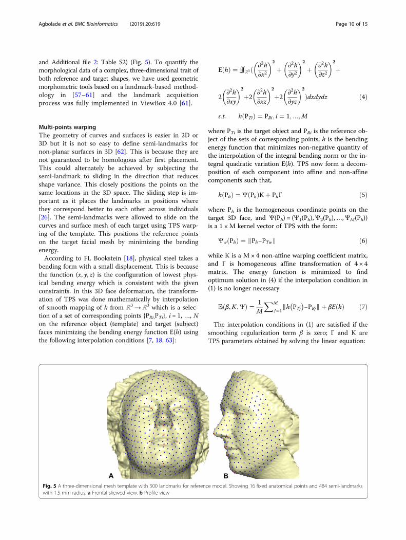
and Additional file 2: Table S2) (Fig. 5). To quantify themorphological data of a complex, three-dimensional trait ofboth reference and target shapes, we have used geometricmorphometric tools based on a landmark-based method-ology in [57–61] and the landmark acquisitionprocess was fully implemented in ViewBox 4.0 [61].
Multi-points warpingThe geometry of curves and surfaces is easier in 2D or3D but it is not so easy to define semi-landmarks fornon-planar surfaces in 3D [62]. This is because they arenot guaranteed to be homologous after first placement.This could alternately be achieved by subjecting thesemi-landmark to sliding in the direction that reducesshape variance. This closely positions the points on thesame locations in the 3D space. The sliding step is im-portant as it places the landmarks in positions wherethey correspond better to each other across individuals[26]. The semi-landmarks were allowed to slide on thecurves and surface mesh of each target using TPS warp-ing of the template. This positions the reference pointson the target facial mesh by minimizing the bendingenergy.According to FL Bookstein [18], physical steel takes a
bending form with a small displacement. This is becausethe function (x, y, z) is the configuration of lowest phys-ical bending energy which is consistent with the givenconstraints. In this 3D face deformation, the transform-ation of TPS was done mathematically by interpolationof smooth mapping of h from ℝ3→ℝ3 which is a selec-tion of a set of corresponding points {ΡRi,ΡTi}, i = 1, …, Non the reference object (template) and target (subject)faces minimizing the bending energy function Ε(h) usingthe following interpolation conditions [7, 18, 63]:
Ε hð Þ ¼ ∭ℝ3ð ∂2h∂x2
� �2
þ ∂2h∂y2
� �2
þ ∂2h∂z2
� �2
þ
2∂2h∂xy
� �2
þ2∂2h∂xz
� �2
þ2∂2h∂yz
� �2
Þdxdydz ð4Þ
s:t: h ΡTið Þ ¼ ΡRi; i ¼ 1;…;M
where ΡTi is the target object and ΡRi is the reference ob-ject of the sets of corresponding points, h is the bendingenergy function that minimizes non-negative quantity ofthe interpolation of the integral bending norm or the in-tegral quadratic variation Ε(h). TPS now form a decom-position of each component into affine and non-affinecomponents such that,
h Ρhð Þ ¼ Ψ Ρhð ÞΚþ ΡhΓ ð5Þ
where Ρh is the homogeneous coordinate points on thetarget 3D face, and Ψ(Ρh) = (Ψ1(Ρh),Ψ2(Ρh),…,ΨM(Ρh))is a 1 ×M kernel vector of TPS with the form:
Ψw Ρhð Þ ¼ ∥Ρh−ΡTw∥ ð6Þ
while Κ is a M × 4 non-affine warping coefficient matrix,and Γ is homogeneous affine transformation of 4 × 4matrix. The energy function is minimized to findoptimum solution in (4) if the interpolation condition in(1) is no longer necessary.
E β;K ;Ψð Þ ¼ 1M
XM
J¼1∥h ΡTj
� �−ΡRj∥þ βE hð Þ ð7Þ
The interpolation conditions in (1) are satisfied if thesmoothing regularization term β is zero; Γ and Κ areTPS parameters obtained by solving the linear equation:
Fig. 5 A three-dimensional mesh template with 500 landmarks for reference model. Showing 16 fixed anatomical points and 484 semi-landmarkswith 1.5 mm radius. a Frontal skewed view. b Profile view
Agbolade et al. BMC Bioinformatics (2019) 20:619 Page 10 of 15

Ψ ΡR
ΡTR 0
� �KΓ
� �¼ ΡT
0
� �ð8Þ
Ψ is a M ×M matrix with the component Ψwl = ∥ ΡTw− ΡTl ∥ and ΡR is a M × 4 matrix with each row being thehomogeneous coordinate of the point ΡRi, i = 1, …, M.Using (2), the target facial mesh ΡTi is deformed to thereference mesh ΡRi. Applying the bending energy, theprocess was iterated specified number of cycles (6) tohave optimum sliding of the points on the facial surfacewhich gives points relaxed. This changed the bendingenergy from initial value Ei to final value Ef after acomplete iteration. This makes the semi-landmarks tobe treated the same as homologous landmarks with re-spect to downstream analyses. Because the warping mayresult in points that do not lie directly on the facial sur-face on the target mesh, the transferred points were pro-jected on the closest point on the mesh surface. Thiswas done using Iterative Closest Point (ICP) method [8],which aims to iteratively minimize the mean squareerror between two point sets. If the distance between thetwo points is within the acceptable threshold, then theclosest point is determined as the corresponding point[64]. The homologous landmark warping HKΓ after a sixcomplete iterations is, therefore:
HKΓ ¼ E f −iKΓ
� �ð9Þ
Where
KΓ
� �¼ Ψ ΡR
ΡTR 0
� �−1ΡT0
� �; ð10Þ
is the linear TPS equation obtained during deformationsurface of the target mesh to the reference mesh beforeconvergence was finally reached and Ef − i = Ef − Ei of sixcomplete iterations. The first iteration showed a partialdistribution of sliding points on the target surface mesh(Fig. 6). This was automatically repeated until optimumhomologous result was achieved using exponential decaysliding step of hundred to 5 %. During the relaxation ofthe spline, the semi-landmarks slid along the surface andthe curve tangent structures, and not on the surfaces orthe curves which reduced the computational effort. Thismakes the minimization problem become linear, as slid-ing along the tangents lets the semi-landmarks slip offthe data [22]. The target surface mesh is now treated ashomologous points (Fig. 7). Note that we did not build anew deformable mathematical equation from scratch butextended the standard deformable method that has beenestablished in [7].In assessing error, 18 subjects (three from each expres-
sion) from each dataset were randomly selected; eachone belonging to a different individual, distinct from thetemplate subject. Each was digitized twice following thesame method to account for digitization error. The re-sults were analyzed using Procrustes ANOVA [65, 66]which has been implemented in morphometrics toanalyze measurement error in MorphoJ [67–69]. This isdone by the minimization of the squared sum of the dis-tance of all objects and the consensus configuration [51].
Fig. 6 Partially warped 500 sliding point on target facial surface. a Angry. b Disgust. c Fear. d Sad. e Surprise. f Happy
Agbolade et al. BMC Bioinformatics (2019) 20:619 Page 11 of 15

Feature selection with PCAThe features were selected by dimensionality reductionusing Principal Components Analysis (PCA). Here, thedata is represented as matrix M = [m1,m2,…mn], wheremi is the ith column vector representing the ith trainingdata. The covariance matrix K = cov (M) =MMT, we thencarried out eigenvalue decomposition on the matrix M toproduce highest ranking eigenvectors known as PrincipalComponents (PCs) with the help of their correspondingeigenvalues. We chose x eigenvectors (p1, p2,…, pn) thatbest described the data with projection onto the space
spanned by these vectors such that X ¼ ½p1; p2;…; pn�Tm ;where X is the n dimensional vector used as features dur-ing the training and classification process. The total PCscomputed during reduction process is 239PCs and179PCs for Stirling and Bosphorus, respectively (see Add-itional file 3: Table S3 and Additional file 4: Table S4).Among these, only 135PCs from Stirling and 98PCs fromBosphorus which have been observed to have the highestranking eigenvectors were selected for classification usingBartlett’s test for the first principal component method[70, 71]. In other to establish total PCs that expressedmeaningful variation in each expression group, a brokenstick was used [70, 72]. This is based on the eigenvaluesfrom random data of the principal components.
Linear discriminant analysis (LDA)The method used multi-class LDA to classify the fea-tures. This is one of the supervised learning methods for
classification. It operates by maximizing the ratio ofbetween-class variance to that of within-class variance ina dataset, thereby guaranteeing maximum separability. Ithas been widely applied to many applications such asmicroarray data classification [73], face recognition [74],and image retrieval [75]. LDA comes with singularityproblem [76] which has given room to many extensionsto LDA such as regularized LDA [77], pseudo-inverseLDA [78], and subspace LDA [79]. In order to overcomethe singularity issue of classical LDA, PCA was appliedas an intermediate dimensionality reduction.Computing LDA for multi-class is slightly different
from two-class. The multi-class requires the applicationof multiple discriminant analysis [80]. The maximizationof ratio of within-class scatter to between-class scatter isdone among the competing classes [81]. The multi-classcan also be called Canonical Variates Analysis (CVA)but the major assumption for LDA is that the variance–covariance matrices are all equal [82]. To simplify thecomputational process, we first computed the within-class matrix for n classes (n = 6 for this study) suchthat:
Σ̂w ¼ S1 þ…þ Sn
¼Xn
i¼1
XΧ∈ci
X−Xi� �
X−Xi� �0 ð11Þ
followed by between-class matrix, given by:
Σ̂b ¼Xn
i¼1mi Xi−X
� �Xi−X� �0 ð12Þ
Fig. 7 Complete and homologous 500 warped points on target mesh. a Angry. b Disgust. c Fear. d Sad. e Surprise. f Happy
Agbolade et al. BMC Bioinformatics (2019) 20:619 Page 12 of 15

where mi is the number of samples for each class, Xi isthe mean vector for each class and X is the summedmean vector computed as X ¼ 1
m
Pni¼1miXi:
By obtaining the within-class and between-class matri-
ces (Σ̂w and Σ̂bÞ, we now obtained the transformation Φby solving generalized eigenvalue problem:
Σ̂bΦ ¼ λ Σ̂wΦ ð13ÞOnce the transformation Φ is solved, the classification
is then performed based on distance metrics in trans-formed space. Here, Euclidean distance is applied suchthat:
d x; yð Þ ¼ffiffiffiffiffiffiffiffiffiffiffiffiffiffiffiffiffiffiffiffiffiffiffiffiX
ixi−yið Þ2
qð14Þ
and cosine measure
d x; yð Þ ¼ 1−
PixiyiffiffiffiffiffiffiffiffiffiffiffiffiffiffiffiffiPix
2i y
2i
p ; ð15Þ
we arrive at a new instance z, which classified into argmindðzΦ;Xk ΦÞ; where Xk is the centroid of k-th class.The advantage of multiple discriminant analysis over sin-gle discriminant analysis is that it produces an elegantclassification with the use of discriminant features [81].
Supplementary informationSupplementary information accompanies this paper at https://doi.org/10.1186/s12859-019-3153-2.
Additional file 1: Table S1. Raw three-dimensional digitized data forStirling dataset expression group.
Additional file 2: Table S2. Raw three-dimensional digitized data forBosphorus dataset expression group.
Additional file 3: Table S3. PCs scores for all subjects used in theStirling dataset.
Additional file 4:. Table S4. PCs scores for all subjects used in theBosphorus dataset
AcknowledgmentsWe acknowledge Stirling/ESRC (University of Stirling) and Bosphorus(Bogazici University) for prompt agreement to use their dataset. Furthermore,credit goes to the Computer Laboratory of the Faculty of Computer Science& Information Technology, Universiti Putra Malaysia.
Authors’ contributionsOA and AN conceived the idea and data acquisition; OA developed themethodology; AAG prepared original draft; YKC reviewed and edit themanuscript; OA and AN prepared the visualization and performed analysis;RY supervised the entire work and approved the manuscript. We alsodeclared that all authors have read and approved the final manuscript.
FundingThis work was supported by Putra Grant Scheme, Malaysia – Code:9538100 and Fundamental Research Grant Scheme, Malaysia - Code:5524959. The funders had no role in study design, data collection andanalysis, decision to publish, or preparation of the manuscript.
Availability of data and materialsRaw three-dimensional digitized data for each expression group and princi-pal component analysis scores of the entire subjects. Table S1 and S2: Three-
dimensional raw data for each expression group in different sheet for Stirlingand Bosphorus, respectively. Table S3 and S4: PCs scores for all subjects usedin the experiment for Stirling and Bosphorus, respectively. Note that the 3Dface dataset is not permitted to be shared by third party according to thelicense agreement.
Ethics approval and consent to participateThe use of human subject was approved by the committee in charge ofStirling database headed by Peter Hancock ([email protected]) andBosporus database headed by Prof. Bulent Sankur ([email protected]). The informed consent to participate was obtained by written and signed.
Consent for publicationFor the purpose of method demonstration, only subject with ID F1002 wasvisualized in the study who has indicated her consent by written and signedfor publication on Stirling dataset; while no image was visualized onBosphorus dataset.
Competing interestsThe authors declare that they have no competing interests.
Author details1Department of Computer Science, Faculty of Computer Science & IT,Universiti Putra Malaysia, Serdang, Selangor, Malaysia. 2Department ofSoftware Engineering, Faculty of Computer Science & IT, Universiti PutraMalaysia, Serdang, Selangor, Malaysia. 3Department of Biomedical Science,Faculty of Medicine and Health Sciences, Universiti Putra Malaysia, Serdang,Selangor, Malaysia.
Received: 2 May 2019 Accepted: 11 October 2019
References1. Peng S, Tan J, Hu S, Zhou H, Guo J, Jin L, Tang K. Detecting genetic
association of common human facial morphological variation using highdensity 3D image registration. PLoS Comput Biol. 2013;9(12):e1003375.
2. Anies OS, Torres MAJ, Manting MM, Demayo CG. Landmark-basedgeometric Morphometrics in describing facial shape of the Sama-Banguingui tribe from the Philippines. J Med Bioengineering. 2013;2(2):131–6.
3. Bookstein FL. Morphometric tools for landmark data: geometry and biology:Cambridge University Press; 1997. https://onlinelibrary.wiley.com/doi/abs/10.1002/bimj.4710350416.
4. Dean D. Three-dimensional data capture and visualization. In: Advances inmorphometrics: Springer; 1996. p. 53–69. https://link.springer.com/chapter/10.1007/978-1-4899-0092-0_12.
5. Savran A, Sankur B, Bilge MT. Comparative evaluation of 3D vs. 2D modalityfor automatic detection of facial action units. Pattern Recogn. 2012;45(2):767–82.
6. Wang Jun YL, Xiaozhou W, Yi S. 3D facial expression recognition based onprimitive surface feature distribution. In: 2006 IEEE computer societyconference on computer vision and pattern recognition (CVPR'06): IEEE;2006. p. 1399–406. http://www.cs.binghamton.edu/~lijun/Research/3DFE/Yin_cvpr06.pdf.
7. Bookstein FL. Landmark methods for forms without landmarks-morphometrics of group differences in outline shape. Med Image Anal.1997;1(3):225–43.
8. Creusot C, Pears N, Austin J. 3D face landmark labelling. In: Proceedings ofthe ACM workshop on 3D object retrieval: ACM; 2010. p. 27–32. https://dl.acm.org/citation.cfm?id=1877815.
9. Colombo A, Cusano C, Schettini R. 3D face detection using curvatureanalysis. Pattern Recogn. 2006;39(3):444–55.
10. Elewa AM, Elewa AM. Morphometrics for nonmorphometricians, vol. 124:Springer; 2010. https://www.springer.com/gp/book/9783540958529.
11. Dryden IL, Mardia KV. Statistical shape analysis, vol. 4. Chichester: Wiley;1998.
12. Adams DC, Rohlf FJ, Slice DE. Geometric morphometrics: ten years ofprogress following the ‘revolution’. Italian J Zoology. 2004;71(1):5–16.
13. Slice DE. Geometric Morphometrics. Annu Rev Anthropol. 2007;36(1):261–81.14. Rohlf FJ. Relative warp analysis and an example of its application to
mosquito. In: Contributions to morphometrics, vol. 8; 1993. p. 131.
Agbolade et al. BMC Bioinformatics (2019) 20:619 Page 13 of 15

15. Marcus LF, Bello E, García-Valdecasas A, Museo Nacional de Ciencias N.Contributions to morphometrics: Consejo Superior de InvestigacionesCientíficas; 1993.
16. Farkas LG. Anthropometry of the head and face: Raven Pr; 1994. https://www.sciencedirect.com/science/article/pii/0278239195902082?via%3Dihub.
17. Klingenberg CP. Visualizations in geometric morphometrics: how to readand how to make graphs showing shape changes. Hystrix Italian JMammalogy. 2013;24(1):15–24.
18. Bookstein FL. Principal warps: thin-plate splines and the decomposition ofdeformations. IEEE Trans Pattern Anal Mach Intell. 1989;11(6):567–85.
19. El Rai MC, Tortorici C, Al-Muhairi H, Werghi N, Linguraru M. Facial landmarksdetection using 3D constrained local model on mesh manifold. In: Circuitsand Systems (MWSCAS), 2016 IEEE 59th International Midwest Symposiumon: IEEE; 2016. p. 1–4. https://ieeexplore.ieee.org/document/7869954.
20. Vezzetti E, Marcolin F, Tornincasa S, Ulrich L, Dagnes N. 3D geometry-basedautomatic landmark localization in presence of facial occlusions. MultimedTools Appl. 2017:1–29.
21. Botton-Divet L, Houssaye A, Herrel A, Fabre A-C, Cornette R. Tools forquantitative form description; an evaluation of different software packagesfor semi-landmark analysis. PeerJ. 2015;3:e1417.
22. Gunz P, Mitteroecker P, Bookstein FL. Semilandmarks in threedimensions. In: Modern morphometrics in physical anthropology:Springer; 2005. p. 73–98. https://link.springer.com/chapter/10.1007/0-387-27614-9_3.
23. Parr W, Wroe S, Chamoli U, Richards H, McCurry M, Clausen P, McHenry C.Toward integration of geometric morphometrics and computationalbiomechanics: new methods for 3D virtual reconstruction and quantitativeanalysis of finite element models. J Theor Biol. 2012;301:1–14.
24. Cornette R, Baylac M, Souter T, Herrel A. Does shape co-variation betweenthe skull and the mandible have functional consequences? A 3D approachfor a 3D problem. J Anat. 2013;223(4):329–36.
25. Fabre AC, Goswami A, Peigné S, Cornette R. Morphological integration inthe forelimb of musteloid carnivorans. J Anat. 2014;225(1):19–30.
26. Philipp Mitteroecker PG, Sonja Windhagerc, Katrin Schaefer: A brief reviewof shape, form, and allometry in geometric morphometrics, withapplications to human facial morphology. Hystrix Italian J Mammalogy2013;24(1):59-66.
27. Perez SI, Bernal V, Gonzalez PN. Differences between sliding semi-landmarkmethods in geometric morphometrics, with an application to humancraniofacial and dental variation. J Anat. 2006;208(6):769–84.
28. Li H, Wen G. Sample awareness-based personalized facial expressionrecognition. Appl Intell. 2019:1–14.
29. Fang T, Zhao X, Ocegueda O, Shah SK, Kakadiaris IA. 3D facial expressionrecognition: A perspective on promises and challenges. In: Face andGesture 2011: IEEE; 2011. p. 603–10. https://ieeexplore.ieee.org/abstract/document/5771466.
30. Zhen Q, Huang D, Wang Y, Chen L. Muscular movement model-basedautomatic 3D/4D facial expression recognition. IEEE Trans Multimedia. 2016;18(7):1438–50.
31. Kakadiaris IA, Passalis G, Toderici G, Murtuza MN, Lu Y, Karampatziakis N,Theoharis T. Three-dimensional face recognition in the presence of facialexpressions: an annotated deformable model approach. IEEE Trans PatternAnal Mach Intell. 2007;29(4):640–9.
32. Pantic M, Rothkrantz LJ. Automatic analysis of facial expressions: the state ofthe art. IEEE Trans Pattern Analysis Machine Int. 2000;12:1424–45.
33. Ekman P, Friesen WV. Constants across cultures in the face and emotion. JPers Soc Psychol. 1971;17(2):124.
34. Tabia H, Daoudi M, Vandeborre J-P, Colot O. A new 3D-matching methodof nonrigid and partially similar models using curve analysis. IEEE TransPattern Anal Mach Intell. 2011;33(4):852–8.
35. Shao J, Gori I, Wan S, Aggarwal J. 3D dynamic facial expressionrecognition using low-resolution videos. Pattern Recogn Lett. 2015;65:157–62.
36. Tang H, Huang TS. 3D facial expression recognition based on automaticallyselected features. In: 2008 IEEE computer society conference on computervision and pattern recognition workshops: IEEE; 2008. p. 1–8. https://ieeexplore.ieee.org/document/4563052.
37. Li C, Barreto A. An integrated 3D face-expression recognition approach. In:2006 IEEE international conference on acoustics speech and signalprocessing proceedings: 2006: IEEE. p. III. https://ieeexplore.ieee.org/document/1660858.
38. Li H, Huang D, Morvan J-M, Wang Y, Chen L. Towards 3D face recognitionin the real: a registration-free approach using fine-grained matching of 3Dkeypoint descriptors. Int J Comput Vis. 2015;113(2):128–42.
39. Berretti S, Werghi N, Del Bimbo A, Pala P. Matching 3D face scans usinginterest points and local histogram descriptors. Comput Graph. 2013;37(5):509–25.
40. Lei Y, Guo Y, Hayat M, Bennamoun M, Zhou X. A two-phase weightedcollaborative representation for 3D partial face recognition with singlesample. Pattern Recogn. 2016;52:218–37.
41. Kim D, Hernandez M, Choi J, Medioni G. Deep 3D face identification. In:2017 IEEE International Joint Conference on Biometrics (IJCB): IEEE; 2017. p.133–42. https://ieeexplore.ieee.org/document/8272691.
42. Drira H, Amor BB, Srivastava A, Daoudi M, Slama R. 3D face recognitionunder expressions, occlusions, and pose variations. IEEE Trans Pattern AnalMach Intell. 2013;35(9):2270–83.
43. Hariri W, Tabia H, Farah N, Benouareth A, Declercq D. 3D facial expressionrecognition using kernel methods on Riemannian manifold. Eng Appl ArtifIntell. 2017;64:25–32.
44. Chun S-Y, Lee C-S, Lee S-H. Facial expression recognition using extendedlocal binary patterns of 3D curvature. In: Multimedia and UbiquitousEngineering: Springer; 2013. p. 1005–12. https://link.springer.com/chapter/10.1007/978-94-007-6738-6_124.
45. Klingenberg CP. MorphoJ: an integrated software package for geometricmorphometrics. Mol Ecol Resour. 2011;11:353–7.
46. Hammer Ø, Harper D, Ryan P. Paleontological statistics software: packagefor education and data analysis. Palaeontol Electron. 2001;4.
47. Team RC: R: a language and environment for statistical computing. 2013.48. Anderson MJ. A new method for non-parametric multivariate analysis of
variance. Austral Ecol. 2001;26(1):32–46.49. Gilani SZ, Rooney K, Shafait F, Walters M, Mian A. Geometric facial gender
scoring: objectivity of perception. PLoS One. 2014;9(6):e99483.50. Bekios-Calfa J, Buenaposada JM, Baumela L. Revisiting linear discriminant
techniques in gender recognition. IEEE Trans Pattern Anal Mach Intell. 2011;33(4):858–64.
51. Fruciano C. Measurement error in geometric morphometrics. Dev GenesEvol. 2016;226(3):139–58.
52. Azazi A, Lutfi SL, Venkat I, Fernández-Martínez F. Towards a robust affectrecognition: automatic facial expression recognition in 3D faces. Expert SystAppl. 2015;42(6):3056–66.
53. Georgia Sandbach SZ, Pantic M, Yin L. Static and dynamic 3D facialexpression recognition: a comprehensive survey. Image Vis Comput. 2012;30(2012):683–97.
54. Stirling-ESRC 3D Face Database [http://pics.stir.ac.uk/ESRC/3d_images.htm].55. Savran A, Alyüz N, Dibeklioğlu H, Çeliktutan O, Gökberk B, Sankur B, Akarun
L. Bosphorus database for 3D face analysis. In: European Workshop onBiometrics and Identity Management: Springer; 2008. p. 47–56.
56. Caple J, Stephan CN. A standardized nomenclature for craniofacial andfacial anthropometry. Int J Legal Med. 2016;130(3):863–79. https://link.springer.com/article/10.1007%2Fs00414-015-1292-1.
57. Zelditch ML, Swiderski DL, Sheets HD. Geometric morphometrics forbiologists: a primer: academic press; 2012.
58. Klingenberg CP, Zaklan SD. Morphological integration betweendevelopmental compartments in the Drosophila wing. Evolution. 2000;54(4):1273–85.
59. Kouli A, Papagiannis A, Konstantoni N, Halazonetis DJ, Konstantonis D. Ageometric morphometric evaluation of hard and soft tissue profile changesin borderline extraction versus non-extraction patients. Eur J Orthod. 2018;41(3):264–72.
60. Yong R, Ranjitkar S, Lekkas D, Halazonetis D, Evans A, Brook A, Townsend G.Three-dimensional (3D) geometric morphometric analysis of humanpremolars to assess sexual dimorphism and biological ancestry in Australianpopulations. Am J Phys Anthropol. 2018;166(2):373–85.
61. Viewbox 4 - Cephalometric Software [http://dhal.com/viewboxindex.htm].62. Huanca Ghislanzoni L, Lione R, Cozza P, Franchi L. Measuring 3D shape
in orthodontics through geometric morphometrics. Prog Orthod. 2017;18(1):38.
63. Corner BD, Lele S, Richtsmeier JT. Measuring precision of three-dimensionallandmark data. J Quant Anthropol. 1992;3(4):347–59.
64. Mian AS, Bennamoun M, Owens R. Keypoint detection and local featurematching for textured 3D face recognition. Int J Comput Vis. 2008;79(1):1–12.
Agbolade et al. BMC Bioinformatics (2019) 20:619 Page 14 of 15

65. Klingenberg CP, McIntyre GS. Geometric morphometrics of developmentalinstability: analyzing patterns of fluctuating asymmetry with Procrustesmethods. Evolution. 1998;52(5):1363–75.
66. Klingenberg CP, Barluenga M, Meyer A. Shape analysis of symmetricstructures: quantifying variation among individuals and asymmetry.Evolution. 2002;56(10):1909–20.
67. Leamy LJK, Peter C, Sherratt E, Wolf JB, Cheverud JM. The geneticarchitecture of fluctuating asymmetry of mandible size and shape in apopulation of mice: another look. Symmetry. 2015;7(1):146–63.
68. Singh N, Harvati K, Hublin J-J, Klingenberg CP. Morphological evolutionthrough integration: a quantitative study of cranial integration in Homo,Pan, Gorilla and Pongo. J Hum Evol. 2012;62(1):155–64.
69. Klingenberg C, Wetherill L, Rogers J, Moore E, Ward R, Autti-Rämö I,Fagerlund Å, Jacobson S, Robinson L, Hoyme H. Prenatal alcohol exposurealters the patterns of facial asymmetry. Alcohol. 2010;44(7–8):649–57.
70. Peres-Neto PR, Jackson DA, Somers KM. How many principal components?Stopping rules for determining the number of non-trivial axes revisited.Comp Stat Data Analysis. 2005;49(4):974–97.
71. Bartlett M. A further note on the multiplying factors for various X2approximations in factor analysis. J R Stat Soc. 1954;16:296–8.
72. Jackson DA. Stopping rules in principal components analysis: A comparisonof heuristical and statistical approaches, vol. 74. Brooklyn: Ecology; 1993. p. 8.
73. Dudoit S, Fridlyand J, Speed TP. Comparison of discrimination methods forthe classification of tumors using gene expression data. J Am Stat Assoc.2002;97(457):77–87.
74. Belhumeur PN, Hespanha JP, Kriegman DJ. Eigenfaces vs. fisherfaces:recognition using class specific linear projection. IEEE Trans Pattern AnalMach Intell. 1997;19(7):711–20.
75. Swets DL, Weng JJ. Using discriminant eigenfeatures for image retrieval.IEEE Trans Pattern Anal Mach Intell. 1996;18(8):831–6.
76. Ye J, Janardan R, Li Q. Two-dimensional linear discriminant analysis. In:Advances in neural information processing systems; 2005. p. 1569–76.
77. Guo Y, Hastie T, Tibshirani R. Regularized linear discriminant analysis and itsapplication in microarrays. Biostatistics. 2006;8(1):86–100.
78. Krzanowski W, Jonathan P, McCarthy W, Thomas M. Discriminant analysiswith singular covariance matrices: methods and applications tospectroscopic data. J R Stat Soc: Ser C: Appl Stat. 1995;44(1):101–15.
79. Zhao W, Chellappa R, Phillips PJ. Subspace linear discriminant analysis forface recognition: Citeseer; 1999.
80. Johnson RA, Wichern DW. Applied multivariate statistical analysis, vol. 5. NJ:Prentice hall Upper Saddle River; 2002. https://searchworks.stanford.edu/view/6804286.
81. Li T, Zhu S, Ogihara M. Using discriminant analysis for multi-class classification:an experimental investigation. Knowl Inf Syst. 2006;10(4):453–72.
82. Peterson LE, Coleman MA. Machine learning-based receiver operatingcharacteristic (ROC) curves for crisp and fuzzy classification of DNAmicroarrays in cancer research. Int J Approx Reason. 2008;47(1):17–36.
Publisher’s NoteSpringer Nature remains neutral with regard to jurisdictional claims inpublished maps and institutional affiliations.
Agbolade et al. BMC Bioinformatics (2019) 20:619 Page 15 of 15