β3-adrenoreceptors control mitochondrial dormancy in
TRANSCRIPT

Research Articleβ3-Adrenoreceptors Control Mitochondrial Dormancy inMelanoma and Embryonic Stem Cells
Maura Calvani ,1 Lorenzo Cavallini,1,2 Annalisa Tondo ,1 Valentina Spinelli ,3
Luisa Ricci ,2 Amada Pasha ,1,4 Gennaro Bruno ,1,4 Daniela Buonvicino ,4
Elisabetta Bigagli ,3 Marina Vignoli ,1,4 Francesca Bianchini ,5 Laura Sartiani ,3
Maura Lodovici ,3 Roberto Semeraro,2 Filippo Fontani ,1,4 Francesco De Logu ,4
Massimo Dal Monte ,6 Paola Chiarugi,5 Claudio Favre ,1 and Luca Filippi7
1Oncohematology Unit, Department of Pediatric Oncology, A. Meyer Children’s University Hospital, Florence, Italy2Department of Experimental and Clinical Medicine, University of Florence, Florence, Italy3Department of NEUROFARBA, Section of Pharmacology and Toxicology, University of Florence, Florence, Italy4Department of Health Sciences, University of Florence, Florence, Italy5Department of Experimental and Clinical Biomedical Sciences, University of Florence, Florence, Italy6Department of Biology, University of Pisa, Italy7Neonatal Intensive Care Unit, Medical Surgical Fetal-Neonatal Department, A. Meyer Children’s University Hospital, Florence, Italy
Correspondence should be addressed to Maura Calvani; [email protected]
Received 1 June 2018; Accepted 20 September 2018; Published 13 November 2018
Academic Editor: Ilaria Peluso
Copyright © 2018 Maura Calvani et al. This is an open access article distributed under the Creative Commons Attribution License,which permits unrestricted use, distribution, and reproduction in any medium, provided the original work is properly cited.
The early phases of embryonic development and cancer share similar strategies to improve their survival in an inhospitableenvironment: both proliferate in a hypoxic and catecholamine-rich context, increasing aerobic glycolysis. Recent studiesshow that β3-adrenergic receptor (β3-AR) is involved in tumor progression, playing an important role in metastasis.Among β-adrenergic receptors, β3-AR is the last identified member of this family, and it is involved in cancer cell survival andinduction of stromal reactivity in the tumor microenvironment. β3-AR is well known as a strong activator of uncouplingprotein 1 (UCP1) in brown fat tissue. Interestingly, β3-AR is strongly expressed in early embryo development and in manycancer tissues. Induction of uncoupling protein 2 (UCP2) has been related to cancer metabolic switch, leading to acceleratedglycolysis and reduced mitochondrial activity. In this study, for the first time, we demonstrate that β3-AR is able to promotethis metabolic shift in both cancer and embryonic stem cells, inducing specific glycolytic cytoplasmic enzymes and a sort ofmitochondrial dormancy through the induction of UCP2. The β3-AR/UCP2 axis induces a strong reduction of mitochondrialactivity by reducing ATP synthesis and mitochondrial reactive oxygen species (mtROS) content. These effects are reverted bySR59230A, the specific β3-AR antagonist, causing an increase in mtROS. The increased level of mtROS is neutralized by astrong antioxidant activity in embryonic stem cells, but not in cancer stem cells, where it causes a dramatic reduction in tumorcell viability. These results lead to the possibility of a selective antitumor therapeutic use of SR59230A. Notably, we demonstratethe presence of β3-AR within the mitochondrial membrane in both cell lines, leading to the control of mitochondrial dormancy.
1. Introduction
Both fetal development and tumor growth, despite, respec-tively, physiological and pathological events with feedbackfar away from each other, are triggered by heterogeneousinterrelated pathways. Embryo and cancer, among hypoxic
active stromal tissues and high stem cell potential [1], sharea high aerobic glycolytic rate [2, 3] and a catecholamine-rich environment [4, 5]. Aerobic glycolysis occurs in the earlypreimplantation of mammalian embryo [6, 7], during earlypregnancy [8], and in tumors [4], by increasing the con-sumption of glucose and production of lactate. Aerobic
HindawiOxidative Medicine and Cellular LongevityVolume 2018, Article ID 6816508, 10 pageshttps://doi.org/10.1155/2018/6816508

glycolysis, which converts glucose to lactate, is the majorsource of energy of these cells, instead of mitochondrial oxi-dative phosphorylation [4, 9, 10]. Both cancer cells andembryos need to increase the uptake of glucose, the expres-sion of glycolytic enzymes, and the export of lactate to obtainenergy for growth [6–11]. Glycolytic metabolism confers aproliferative advantage to both tumor cells and embryos,since this metabolism produces a large number of usefulintermediates to secondary biosynthetic pathways [5, 12].
Several studies suggest that stress-related catecholaminerelease accelerates cancer progression, so the targeting ofβ-adrenergic receptors (β-ARs) has been proposed as apotential therapeutic approach to cancer and, in particular,melanoma [13, 14]. At the beginning, attention was mainlyfocused on β2-AR [15], but, more recently, β3-AR wasfound overexpressed in tumors, particularly in melanomacell lines and tumor microenvironments [16–18]. Currently,it is known that β3-AR expression correlates with cancerprogression, angiogenesis, and tumor stromal cell reactivity[16–18]. Interestingly, high expression ofβ3-ARhas also beenrevealed in the pregnant myometrium [19] and during earlyembryo development [20, 21], confirming the similaritiesbetween the environments surrounding tumor and embryo.
β3-AR is well known to control thermogenesis by activat-ing uncoupling protein 1 (UCP1). At low temperature, asignal is transmitted to brown adipocytes by catecholaminerelease and β3-AR activation, resulting in increased UCP1expression and activity. The UCPs are members of the mito-chondrial anion carrier family. While UCP1 is predomi-nantly expressed in brown adipocyte tissue [22], UCP2 isubiquitously expressed in cancer derived fromdifferent tissues[23, 24]. Interestingly, UCP2 appears to be highly expressed incancer cells (such as leukemia and pancreatic cancer) and innondifferentiated cells with low amounts of mitochondrialtissues, which rely on glycolysis rather than oxidative phos-phorylation for their energy production [25–28].
In this study, we investigated the role of the β3-AR/UCP2axis on the regulation of the Warburg metabolism in cancerstem cells (CSC) and in mouse embryonic stem cells (ESC).
2. Materials and Methods
2.1. Cell Culture and ES Cell Differentiation. Melanoma cells(A375) and mouse ESC (CGR8) were purchased from theEuropean Collection of Cell Cultures (ECACC,Wiltshire, UK).
A375 cells were cultured in high-glucose DMEM con-taining 10% FBS, while CGR8 were cultured in a propaga-tion medium containing a BHK21 medium (Gibco), 1%nonessential amino acids (Gibco), 10−3M sodium pyruvate(Gibco), 10−7M beta-mercaptoethanol (Sigma), 2× 10⁻³Mglutamine (Sigma) 1% penicillin-streptomycin (Gibco), 10%fetal bovine serum (Gibco), and 1000U/ml recombinantmouse leukemia inhibitory factor (Chemicon International)in a humidified 5% CO2 atmosphere at 37°C [29]. Differenti-ation of ES cells was performed by the hanging drop method[30]. Briefly, embryoid bodies (EBs) were formed for 2days in hanging drops containing 450–600 cells/20μl ofa differentiation medium, whose composition was similarto that used for cell propagation except for fetal bovine
serum (20%, HyClone) and without recombinant mouse leu-kemia inhibitory factor. After 4 days in suspension, EBs wereplated on gelatin-coated plates. Starting from day 8 of differ-entiation, the number of beating EBs was counted by phase-contrast microscopy.
2.2. Cell Treatments. A375 cells were grown to 70% conflu-ence in complete medium DMEM high-glucose for 48 h.Then the medium was removed, and after washing in PBSsolution, cells were serum-starved overnight with a starvationmedium, without FCS, in order to promote the cells’ entryinto a quiescent G0 phase, thereby better evaluating cells’responsiveness to exogenous treatments. After 24 h, cellswere treated with a single dose of BRL, a β3-adrenergic ago-nist, and after 30 minutes with the β3-antagonist SR59230Aand with genipin, UCP2’s antagonist.
2.3. Melanosphere Formation Assay. A375 cells were incu-bated for 72 h with a conditioned medium (CM) and thendetached using Accutase (Sigma). Single A375 cells wereplated at 150 cells/cm2 on a low-attachment 100mm platein a Dulbecco’s modified Eagle’s medium/F12 supplementedwith B27 and N2 media, 5 g/ml insulin, 20 ng/ml fibroblastGF-2, and 20ng/ml epidermal GF. A375 cells were grownunder these conditions for 15–20days and formed nonadher-ent P0 spheres termed prostaspheres. For the evaluation ofself-renewal, a single prostasphere was dissociated in singlecells with Accutase, and a dilution of one cell per well into96-well low-attachment plates was performed in order toisolate individual P1 prostaspheres. Single-cell cloningwas confirmed by microscopic analysis, and single cloneswere counted.
2.4. Western Blot Analysis. The buffer used was RIPA lysisbuffer (50mM Tris-HCl, pH7.5, 150mM NaCl, 1% TritonX-100, 2mM EGTA, 1mM sodium orthovanadate, 1mMphenylmethanesulphonyl fluoride, 10mg/ml aprotinin, and10mg/ml leupeptin). Twenty micrograms of total proteinswas loaded on SDS-PAGE, separated, and transferred ontonitrocellulose. The immunoblots were incubated in 3%bovine serum albumin, 10mmol/l Tris-HCl (pH7.5),1mmol/l EDTA, and 0.1% Tween 20 for 1 h at room temper-ature, probed first with specific antibodies and then withappropriate secondary antibodies. Bound antibodies weredetected using the Novex ECL, HRP ChemiluminescentSubstrate Reagent Kit (Life Technologies). Filters wereautoradiographed, and images were acquired through theBioSpectrum Imaging System (Ultra-Violet Products Ltd.,Cambridge, UK). The following antibodies were used:UCP2 (sc-6525), β-actin (sc-1615), HKII (ab-76959), MCT-4 (376140), GLUT1 (ab-209449), SOX2 (sc-16320), totalOXPHOS (ab-110143), and β3-AR (h-160).
2.5. Metabolic Assays. The lactate extrusion assay was per-formed using the Lactate Colorimetric Assay Kit II, followingthe manufacturer’s instructions (BioVision®).
The glucose uptake assay was performed using the Glu-cose Uptake Cell-Based Assay Kit (item n. 600470), followingthe manufacturer’s instructions (Cayman Chemicals®).
2 Oxidative Medicine and Cellular Longevity

ATP production assay was performed using the ATPliteLuminescence Assay System, following the manufacturer’sinstructions (PerkinElmer®).
All experiments were collected after 48 h, and the lumi-nescence and fluorescence were measured with a Flexspectrophotometer.
2.6. ROSCytofluorimetric Analysis.The cells were stainedwith1μl of MitoSOX and were incubated for 15 minutes, thenwashed with PBS one time; the supernatant was aspirated;and the cells were incubated with Accutase. Finally, thedetached cells were suspended in a final volume of 300μl andwere analyzed using MACSQuant FACS (Miltenyi Biotec).
2.7. DilC(5) Membrane Potential. Membrane potential wasperformed using the MitoProbe™ DiIC1(5) Assay Kit forFlow Cytometry M34151, following the manufacturer’sinstructions (Thermo Fisher®).
2.8. Frap Assay. The ferric reducing antioxidant power(FRAP) assay, which is a measure of the antioxidant effectsof nonenzymatic defences in plasma, was performed accord-ing to the method by Benzie and Strain [31].
2.9. Confocal Microscopy. After washing with PBS, the cellswere fixed with a 3.7% formaldehyde solution in PBS for20min at 4°C. Then, after extensive washes in PBS, thecells were permeabilized with 0.1% Triton X-100 in PBSand then stained with a 50 μg/ml fluorescent phalloidinconjugate solution in PBS, phalloidin-TRITC, for 1 h atroom temperature with anti-β3-AR antibody, MitoProbe,and DAPI, then analyzed with the related laser. Afterseveral washes with PBS, the coverslips were mountedwith glycerol plastine and then observed under a confocalfluorescence microscope (Leica).
2.10. Mitochondrial Isolation. To isolate the mitochondriafrom A375 stem cells and ESC, the Mitochondria IsolationKit (Miltenyi Biotec®) was used. At the end of the kit proce-dure, after the centrifugation at 13,000xg for 2 minutes at4°C, we aspirated the supernatant and resuspended the mito-chondria pellet in lysis buffer forWestern blot analysis and inPBS for FACS analysis.
2.11. MTT Assay. Viability of tumor cells, in all conditions,was evaluated using an MTT (3-[4,5-dimethylthiazol-2-yl]-2,5-diphenyltetrazolium bromide; thiazol blue) assay. It is atest of cell viability based on the reduction of the MTT dyeby mitochondrial succinate dehydrogenase enzyme, whichis active only in living cells. The enzyme cuts the tetrazoliumring of MTT (yellow substance) with the formation of a bluesalt that is exocitated and precipitates.
The cells were maintained in MTT for 1 h, then theformation of needle-like crystals near the membrane wasvisible (if the cell is not vital, the process is reduced).Then, the cell membrane was lysed and the dye solubilizeddeveloping a color whose intensity was proportional to cellviability. The absorbance was evaluated at 570nm using aspectrophotometer.
2.12. Bioinformatic Analysis. To assess the bioinformaticanalysis, we chose four widely used methods: Hum-mPLoc3.0, FUEL-mLoc, WegoLoc, and COMPARTMENTS. Allthe selected predictors are available as Web servers that areintended for eukaryotic proteins or are specific for humanproteins. These predictors take advantage from the usageof the Gene Ontology (GO) terms, representing geneproduct properties. GO-based methods use the correlationbetween the annotations (usually functional annotations)for a protein and its subcellular location. Methods suchas FUEL-mLoc, WegoLoc, and Hum-mPLoc 3.0 adopt dif-ferent databases. WegoLoc and COMPARTMENTS extractthe GO terms from the UniProtKB database. Hum-mPLoc3.0 uses the SWISS-PROT portal, and FUEL-mLoc adoptstwo newly created compact databases, namely, ProSeq andProSeq-GO, that allow consuming much less memory andmake predictions faster.
By unifying these information with evidence on pro-tein localization from curated knowledge, high-throughputexperiments, text mining, and sequence-based predictionmethods, these tools infer on the subcellular localization ofour β3-AR protein.
2.13. Statistical Analysis. In vitro data are presented asmeans ± standarddeviation (SD) from at least three experi-ments. Results were normalized versus control expression levels.
Statistical analysis was performed using GraphPad Prismsoftware (GraphPad, San Diego CA, USA) by one-wayanalysis variance (ANOVA), followed by a Bonferroni posthoc analysis.
3. Results and Discussion
Since embryos and cancer are enriched in nondifferentiatedcells (embryonic and cancer stem cells), we investigatedwhether β3-AR affected CSC and ESC metabolism. Weenriched the melanoma stem cell compartment by growingmelanospheres in vitro. Interestingly, melanospheres revealedhigher levels of β3-AR compared with A375 parental popula-tions (Figure 1(a)). Usually, cells with an accelerated glycolysisrate display elevated glucose uptake, lactate overproduction,and overexpression of specific markers such as hexokinase2 (HKII), monocarboxylate transporter-4 (MCT-4), andglucose transporter-1 (Glut-1) [32]. Here, we report thatmelanospheres expressed higher levels of HKII, MCT-4,and Glut-1 compared with the A375 parental population,thus demonstrating that A375 melanospheres rely on anaccelerated glycolytic metabolism (Figure 1(a)). Elevated gly-colytic metabolism in melanospheres was confirmed by ahigher glucose uptake, lactate export, and decreased ATPsynthesis, when compared with the A375 parental popula-tion (Figures 1(b) and 1(c)).
Selective β3-AR antagonism with SR59230A decreasedthe ability of A375 cells to form melanospheres underBRL37344 (selective β3-AR agonist) stimulations, as demon-strated by the reduction of P1 populations and reduction ofstem cell markers CD133 and SOX2 (Figures 1(d) and1(e)). To verify the metabolic effect of β3-AR stimulation,CSC and ESC were treated with BRL37344, inducing an
3Oxidative Medicine and Cellular Longevity

Ctrl BRL
SR59230A
Melanospheres 21% O2
N° o
f mel
anos
pher
es P
1 80
60
40
20
0
Ctrl
BRL
A37
5
A37
5
A37
5
�훽-Actin
GLUT-1
MCT-4
HKII
�훽3-AR
A375 melanospheres
(nm
ol) l
acta
te
Lactate export Glucose uptake
14C
gluc
ose C
PM/p
rote
in
ATP
ATP
prod
uctio
n
Ctrl
SR59
230A
+ BR
L
BRL
Glucose uptake
Ctrl
SR59
230A
+ BR
L
BRL
Lactate export
Lact
ate (
nmol
)
BRL
SR59
230A
+ B
RLCtrl
SOX-2
�훽-Actin
5.0 × 10⁶
1.0 × 10⁷
1.5 × 10⁷
0.0
2
1
0
3
4
210
345
2
1
0
3
SR59
230A
+ BR
L
Ctrl
BRL
SR59
230A
+ BR
L
Embryonic stem cells
�훽-Actin
HKII
MCT-4
Ctrl
BRL
SR59
230A
+ BR
L
2
1
0
3
Lact
ate (
nmol
)
Ctrl
BRL
SR59
230A
+ BR
L
0
5000
10000
15000
Fluo
resc
ence
at 5
35 n
m
Glucose uptakeLactate export
A375 melanospheres
HKII
MCT-4
�훽-Actin
BRL
SR59
230A
+ BR
L
Ctrl
% o
f CD
133+
cells 80
604020
0
100
Fluo
resc
ence
at 5
35 n
m
0
5000
10000
15000
⁎⁎
⁎⁎⁎⁎⁎
⁎⁎
⁎⁎⁎⁎
⁎⁎
⁎⁎⁎⁎⁎⁎
BRL
SR59
230A
+ BR
L
Ctrl
(a)
(d) (e)
(f) (g)
(h) (i)
(b) (c)
Mel
anos
pher
es
Mel
anos
pher
es
Mel
anos
pher
es
A37
5
Mel
anos
pher
es
Figure 1: β3-AR affects the glycolytic metabolism of both A375 melanospheres and embryonic stem cell. WB analysis of β3-AR, hexokinaseII (HKII), monocarboxylate transporter-4 (MCT-4), and GLUT-1 markers (a). Analysis of the lactate export and glucose uptake (b). ATPproduction assay (c). A375 melanospheres and mouse ES cells treated with BRL37344 (10 μM) and BRL37344 (10 μM)+ SR59230A(10 μM). The following experiments were performed: representative random figure of melanospheres in each condition and clonogenesisassay quantification (d); analysis of CD133+ cells by FACS and WB of stem cell marker SOX-2 (e); glucose uptake and lactate export inA375 melanospheres and ES cells (f, g); WB analysis of HKII and MCT-4 in A375 melanospheres (h) and ES cells (i). Data arerepresentative of at least three experiments (mean and SD). ∗∗P < 0 01 and ∗∗∗P < 0 001.
4 Oxidative Medicine and Cellular Longevity

accelerated aerobic glycolysis (Warburg effect), as con-firmed by the increased expression of HKII and MCT-4,lactate export, and glucose uptake. These effects weredramatically reduced by SR59230A in both CSC and ESC(Figures 1(f)–1(i)). Supplementary Figure 1 shows that pro-pranolol (unselective β1-/β2-AR antagonist) had no effecton the reduction of the Warburg metabolism in CSC. Thus,these results indicate that CSC and ESC share similarmetabolic pathways (Warburg effect) and that this shiftis mediated by β3-AR. The extrusion of lactate, promotedby β3-AR, enhanced the similarities between cancer andembryonic environment. During embryo development, thedramatic production and extracellular transport of lactatereduce the pH in the microenvironment, promoting the dis-aggregation of uterine tissues and facilitating the trophoblastinvasion [11]. The lactate extrusion, together with the pro-motion of angiogenesis, cell migration, and metastatization,is probably also useful for the growth and infiltration of thetumor. Interestingly, lactate contributes significantly to theimmune escape and further analogy between the tumor andthe embryonic environment [33].
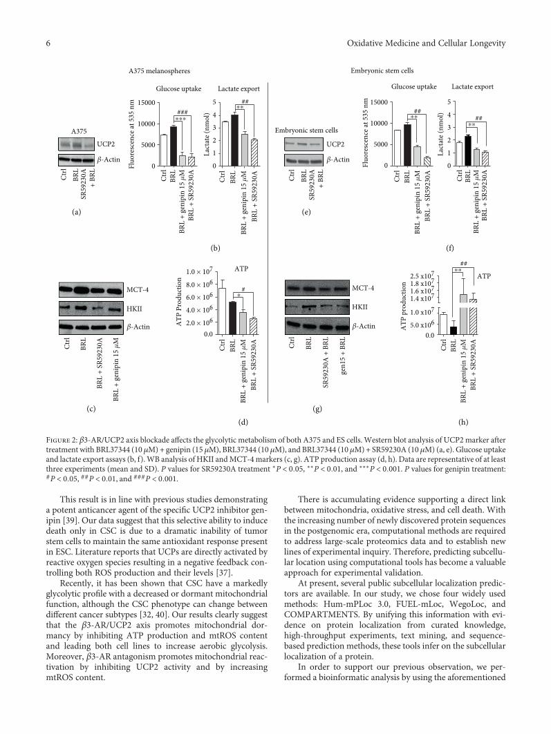
β3-AR's role has been well clarified in white and brownadipocytes. Selective, pharmacological activation of β3-ARhas been shown to have strong effects on adipose tissue mor-phology and metabolism. The sympathetic nervous system,through β3-AR stimulations, is the main trigger of UCP1induction and activation in brown fat mitochondria, leadingto uncoupling of respiration and adaptive thermogenesis.UCP1, localized on the inner mitochondrial membrane,uncouples the activity of the respiratory chain from ATP syn-thesis, thereby releasing energy as heat [34]. Administrationof CL-316,243, a potent and highly selective β3-AR agonist,leads to a marked increase in thermogenesis in brown adi-pose tissue (BAT) and an acute decrease in food consump-tion [35]. At the same time, the role of UCPs in cancer hasbeen extensively studied, even though the effect of UCP2 oncellular energy balance in cancer cells remains unclear [23].Current evidence demonstrates a link between UCP2 andthe Warburg effect. Colon cancer cells stably overexpressingUCP2 produce progressively more lactate than do control-transfected cells, indicating higher rates of glycolysis [36].Leukemia cells overexpressing UCP2 increase lactate produc-tion, decreasing the entry of pyruvate into the Krebs cycle,thereby inducing the Warburg effect [26, 27]. To assess apossible involvement of UCP2 in the β3-AR-induced War-burg effect, we used SR 59230A (specific β3-AR antagonist)and genipin (specific UCP2 inhibitor) under BRL37344 stim-ulations, in both CSC and ESC. UCP2 Western blottingexpression analysis and lactate export assay at different con-centrations of genipin were performed to evaluate which ofthem could have a similar effect to SR59230A treatment(Supplementary Figure 2).
Intriguingly, we demonstrated that SR59230A inhibitedUCP2 expression in both CSC and ESC (Figures 2(a) and2(e)). Performing functional metabolic assays, we observedthat genipin, as well as SR59230A, decreased the lactateexport and glucose consumption induced by BRL37344, bothin CSC (Figure 2(b)) and in ESC (Figure 2(f)). Moreover,HKII and MCT-4 were impaired by genipin treatment
(Figures 2(c) and 2(g)). The results obtained with genipinwere comparable with those obtained with SR59230A,suggesting that CSC and mouse ESC share an acceleratedβ3-AR/UCP2-mediated glycolytic pathway.
Interestingly, the results regarding ATP productionwere not similar in ESC and CSC: the treatment withSR59230A and genipin induced a reduction in ATP synthe-sis only in A375 CSC but not in ESC, where ATP synthesisdramatically increased, indicating that only embryonic cellsare able to shift to an aerobic metabolism (Figures 2(d) and2(h)). These data confirm the hypothesis that β3-AR playsa crucial role in mitochondria UCP2 function and alsohighlight a different regulation of the β3-AR/UCP2 axisin CSC and ESC.
The similarity in the impairment of glycolytic metabo-lism between the two cell lines and the difference in ATPproduction suggest a diverse regulation of cell survival path-ways mediated by β3-AR. It has already been shown thatSR59230A impairs cancer cell viability by inducing cyto-chrome C release [16], and this study shows that SR59230Aand genipin dramatically reduced cell viability only in CSC(Figures 3(a) and3(f)). SinceUCP2 is implicated inmitochon-drial ROS (mtROS) content modulation [37], we revealedthat SR59230A and genipin increased mtROS content inboth CSC (Figure 3(b)) and ESC (Figure 3(g)) treated withBRL37344, and the increase was markedly relevant in CSC(Figure 3(b)). Notably, the basal mtROS levels were definitelylower in ESC (Figure 3(g)) than in CSC (Figure 3(b)). Thiseffect suggests a possible different antioxidant ability betweenthe two cell lines. Results of this study indicate that the anti-oxidant ability induced by BRL37344 is definitely higher inESC than in CSC, as confirmed by an increased FRAP func-tional assay in both cell lines (Figures 3(c) and 3(h)) [31]. InESC, neither treatment with SR59230A nor treatment withgenipin reduced SOD2 expression, if compared with CSC(Figures 3(d) and 3(i)). Therefore, after SR59230A and geni-pin treatment, ESC maintained a higher antioxidant abilitycompared to CSC, and this difference might explain the dif-ferent effect on cell viability.
The addition of the ectopic antioxidant β-mercaptoetha-nol (β-ME) in the culture media recovered cancer cell deathmediated by SR59230A and genipin in CSC (Figures 3(e)and 3(j)). These data demonstrate that the reduced viabilityof CSC after these treatments was due to their lower abilityto counteract mtROS. In fact, the inhibition of the UCP2-mediated mtROS modulation ability in the CSC was criticalfor the survival of the A375 cell line. The antagonism of β3-AR dramatically increased A375 cell death compared toembryonic stem cells, demonstrating a strong selectivity forcancer cells. SR59230A, by inhibiting the β3-AR/UCP2 axis,strongly increases mtROS levels mainly in CSC, due to theirinability to maintain antioxidant response compared to ESC.These results confirm data already present in literature,where β3-ARs and UCP2 are indicated to have a strong anti-oxidant role [37, 38]. The expression of β3-ARs and UCP2in both cancer and embryonic stem cells is not surprising.Both types of cells need a strong ability to proliferate andstrong protection from reactive oxygen species and presenta highly β3-AR expression.
5Oxidative Medicine and Cellular Longevity
This result is in line with previous studies demonstratinga potent anticancer agent of the specific UCP2 inhibitor gen-ipin [39]. Our data suggest that this selective ability to inducedeath only in CSC is due to a dramatic inability of tumorstem cells to maintain the same antioxidant response presentin ESC. Literature reports that UCPs are directly activated byreactive oxygen species resulting in a negative feedback con-trolling both ROS production and their levels [37].
Recently, it has been shown that CSC have a markedlyglycolytic profile with a decreased or dormant mitochondrialfunction, although the CSC phenotype can change betweendifferent cancer subtypes [32, 40]. Our results clearly suggestthat the β3-AR/UCP2 axis promotes mitochondrial dor-mancy by inhibiting ATP production and mtROS contentand leading both cell lines to increase aerobic glycolysis.Moreover, β3-AR antagonism promotes mitochondrial reac-tivation by inhibiting UCP2 activity and by increasingmtROS content.
There is accumulating evidence supporting a direct linkbetween mitochondria, oxidative stress, and cell death. Withthe increasing number of newly discovered protein sequencesin the postgenomic era, computational methods are requiredto address large-scale proteomics data and to establish newlines of experimental inquiry. Therefore, predicting subcellu-lar location using computational tools has become a valuableapproach for experimental validation.
At present, several public subcellular localization predic-tors are available. In our study, we chose four widely usedmethods: Hum-mPLoc 3.0, FUEL-mLoc, WegoLoc, andCOMPARTMENTS. By unifying this information with evi-dence on protein localization from curated knowledge,high-throughput experiments, text mining, and sequence-based prediction methods, these tools infer on the subcellularlocalization of a protein.
In order to support our previous observation, we per-formed a bioinformatic analysis by using the aforementioned
Glucose uptake
Ctrl
BRL
+ SR
5923
0ABRL
BRL
+ ge
nipi
n 15
�휇M
Fluo
resc
ence
at 5
35 n
m
Lactate export Glucose uptake
Fluo
resc
ence
at 5
35 n
m
Lact
ate (
nmol
)
Ctrl
BRL
+ SR
5923
0ABRL
BRL
+ ge
nipi
n 15
�휇M
Embryonic stem cells
�훽-Actin
5.0 x1061.0 x107
1.6 x10⁷
0.0
1.8 x10⁷ ATPATP
6.0 × 1068.0 × 1061.0 × 107
0.0
ATP
Pro
duct
ion
Ctrl
BRL
BRL
+ ge
nipi
n 15
�휇M
BRL
+ SR
5923
0A
4.0 × 10⁶
2.0 × 10⁶HKII
MCT-4
�훽-Actin
HKII
MCT-4
�훽-Actin
BRL
Ctrl
BRL
SR59
230A
+ B
RLCtrl
gen1
5 +
BRL
UCP2
Ctrl
BRL
SR59
230A
+ BR
L
�훽-Actin
UCP2
Ctrl
BRL
SR59
230A
+ BR
L
A375
0
1
2
3
4
5
0
5000
10000
15000BR
L +
geni
pin
15 �휇
M
BRL
+ SR
5923
0A
Ctrl
BRL
+ SR
5923
0ABRL
BRL
+ ge
nipi
n 15
�휇M
0
5000
10000
15000
Embryonic stem cells
#####
#
####
A375 melanospheres
Ctrl
BRL
+ SR
5923
0ABRL
BRL
+ ge
nipi
n 15
�휇M
Lact
ate (
nmol
)
0
1
2
3
4
5
Lactate export
Ctrl
BRL
BRL
+ ge
nipi
n 15
�휇M
BRL
+ SR
5923
0A
ATP
pro
duct
ion
2.5 x10⁷
1.4 x10⁷
##
⁎⁎⁎
⁎⁎
⁎⁎⁎⁎
⁎
⁎⁎
(a)
(b) (f)
(e)
(g)(h)
(c)(d)
Figure 2: β3-AR/UCP2 axis blockade affects the glycolytic metabolism of both A375 and ES cells. Western blot analysis of UCP2marker aftertreatment with BRL37344 (10 μM)+ genipin (15 μM), BRL37344 (10 μM), and BRL37344 (10 μM)+ SR59230A (10 μM) (a, e). Glucose uptakeand lactate export assays (b, f). WB analysis of HKII andMCT-4markers (c, g). ATP production assay (d, h). Data are representative of at leastthree experiments (mean and SD). P values for SR59230A treatment ∗P < 0 05, ∗∗P < 0 01, and ∗∗∗P < 0 001. P values for genipin treatment:#P < 0 05, ##P < 0 01, and ###P < 0 001.
6 Oxidative Medicine and Cellular Longevity

Web tools. The results shown in Supplementary Figure 3(a)represent the sum of ranked probability, estimated by eachmethod. Surprisingly, all predictors we used designate themitochondria as a potential candidate. As expected, theorganelle does not appear as the best hit. This result is partlyattributable to the different total number of proteins locatedin the different compartments; e.g., the plasma membranecontains more proteins than the mitochondria. A closer lookat these results also shows that COMPARTMENTS assignsthe best score to the mitochondria. This result is supportedby the text mining Z-score computed by COMPART-MENTS, which designates the mitochondrion as the secondbest hit (Supplementary Figure 3(b)).
Surprisingly, we found that β3-AR is expressed on mito-chondria of both ESC (Supplementary Figure 3(c)) and CSC,as shown in the WB analysis and in the confocal images(Figures 4(a) and 4(b)). Moreover, through functional
analysis, we demonstrated that SR59230A treatment is ableto modify ATP production and mtROS levels in theisolated mitochondria (Figures 4(c) and 4(d)).
In summary, this study highlighted the parallelismbetween embryonic and cancer stem cells in the regulationof the β3-AR/UCP2-mediated Warburg metabolism andenhanced the different ability of the two cell lines to respondto SR59230A that induced cell death only in CSC by inhibit-ing antioxidant response (Figure 4(e)). An important resultof this study is the presence of β3-AR in the mitochondria,suggesting a possible cooperation with β3-AR located in theplasmatic membrane. We suggest that β3-AR could work assensor of redox state of cells directly influencing mitochon-dria bioenergetic functions. The presence of β3-AR in iso-lated mitochondria could be explained by its protective roleagainst the induction of apoptosis, in both cancer and embry-onic stem cells. Here, we clearly demonstrated a functional
FRA
P �휇
m/ p
rote
in co
nc.
FRAP assayFRAP assay
BRL
Ctrl
BRL
+ ge
nipi
n 15
�휇M
BRL
+ SR
5923
0A
0.0
0.5
1.0
1.5
2.0
BRL
Ctrl
BRL
+ ge
nipi
n 15
�휇M
BRL
+ SR
5923
0A
BRL
Ctrl
BRL
+ ge
nipi
n 15
�휇M
BRL
+ SR
5923
0A
0
20
40
60
80
100
BRL
Ctrl
BRL
+ ge
nipi
n 15
�휇M
BRL
+ SR
5923
0A
Mito
chon
dria
l RO
S le
vels
BRL
Ctrl
BRL
+ ge
nipi
n 15
�휇M
BRL
+ SR
5923
0A
SOD2
�훽-Actin
SOD2
�훽-Actin
⁎⁎##
##
A375 melanospheres
0
20
40
60
80
100
Ctrl
BRL
+ SR
5923
0ABRL
BRL
+ ge
nipi
n 15
�휇M
Abs
orba
nce a
t 570
nm
0.0
0.5
1.0
1.5
2.0MTT
0.0
0.5
1.0
1.5
2.0
Abs
orba
nce a
t 570
nm
Ctrl
BRL
+ SR
5923
0ABRL
BRL
+ ge
nipi
n 15
�휇M
MTT
Embryonic stem cells
MTT+ �훽-ME
0.0
0.5
1.0
1.5
2.0
Ctrl
BRL
+ SR
5923
0ABRL
BRL
+ ge
nipi
n 15
�휇M
ns
0.0
0.5
1.0
1.5
2.0
Abs
orba
nce a
t 570
nm
Ctrl
BRL
+ SR
5923
0ABRL
BRL
+ ge
nipi
n 15
�휇M
+ �훽-MEns ns
MTT
⁎⁎
nsns
⁎⁎
⁎
⁎⁎10
25
#
BRL
Ctrl
BRL
+ ge
nipi
n 15
�휇M
BRL
+ SR
5923
0A
Mito
chon
dria
l RO
S le
vels
0
1
⁎⁎⁎
⁎
FRA
P �휇
m/ p
rote
in co
nc.
(a)
(d) (e)
(f)
(i) (j)
(g) (h)(b) (c)
Figure 3: The antioxidant ability promotes survival in embryo with respect to tumor. The A375 melanospheres and ES cells were treated as inFigure 1. MTT survival experiment on each condition (a, f), mitochondrial mtROS measurement (b, g), FRAP assay (c, h), WB analysis ofSOD-2 antioxidant marker (d, i), and MTT survival experiment on each condition in the presence of 100 μM β-ME (e, j). Data arerepresentative of at least three experiments (mean and SD). NS: not significant. P values for SR59230A treatment: ∗P < 0 05, ∗∗P < 0 01,and ∗∗∗P < 0 001; P values for genipin treatment: #P < 0 05 and ##P < 0 01.
7Oxidative Medicine and Cellular Longevity

role of β3-AR blockade in isolated mitochondria by its abilityto decrease ATP production and to increase mtROS levels.
Further experiments need to be performed to better clar-ify and elucidate the role of β3-AR in mitochondria, but theresults of this study have shown a new and different possiblefunction of this receptor in a new compartment.
Finally, we report that SR59230A is extremely selectivein reducing the viability of CSC by blocking the Warburgmetabolism and by inducing high mtROS levels. Theseresults suggest a potential role of SR59230A as a strongand selective agent that could be used in cancer therapy.
Data Availability
All data generated or analyzed during this study are includedin this article (and its supplementary files). Requests formaterial should be made to the corresponding author.
Conflicts of Interest
The authors declare that there is no conflict of interestregarding the publication of this paper.
Authors’ Contributions
Maura Calvani and Lorenzo Cavallini are fisrt name equalcontributors to the work, Claudio Favre and Luca Filippiare last name equal contributors to the work.
Acknowledgments
This work was supported by the Meyer Foundation Onlus toC. Favre and M. Calvani. We would like to thank Prof. E.Cerbai and Dr. Luigi Ippolito for their collaboration.
Supplementary Materials
Supplementary Figures: (1) metabolic assay of propranololtreatment; (2) metabolic assay with dose/response of genipin;(3) bioinformatic analysis of β3-AR presence prediction indifferent cell compartments. (Supplementary Materials)
References
[1] B. H. Fryer andM. C. Simon, “Hypoxia, HIF and the placenta,”Cell Cycle, vol. 5, no. 5, pp. 495–498, 2006.
[2] O. Warburg, “On the origin of cancer cells,” Science, vol. 123,no. 3191, pp. 309–314, 1956.
[3] D. G. Smith and R. G. Sturmey, “Parallels between embryo andcancer cell metabolism,” Biochemical Society Transactions,vol. 41, no. 2, pp. 664–669, 2013.
[4] A. Greenough, K. H. Nicolaides, and H. Lagercrantz, “Humanfetal sympathoadrenal responsiveness,” Early Human Devel-opment, vol. 23, no. 1, pp. 9–13, 1990.
[5] S. A. Thomas, A. M. Matsumoto, and R. D. Palmiter, “Nor-adrenaline is essential for mouse fetal development,” Nature,vol. 374, no. 6523, pp. 643–646, 1995.
Merge �훽3-AR & MitoTRACKERMito TRACKER�훽3-AR
�훽3-AR
Tota
l OXP
HO
S
Mito
chon
dria
Cyto
sol
A375
CTRL BR
L
BRL
+ ge
nipi
n 15
�휇M
AD
P
0
25
50
75
100
ADP 50 �휇M
BRL
BRL
+ ge
nipi
n 15
�휇M
BRL
+ SR
5923
0A
CTRL
BRL
+ SR
5923
0A
ATP
prod
uctio
n
Mito
chon
dria
l RO
S le
vels⁎#
⁎ #
A375 melanospheres
02468
10
↓ glucose
↓ pyruvate
↑ mtROSnot neutralized↓ lactate
↓ glut-1
↑ M
CT4
↓ la
ctat
e
Respiratory chain ↓ UCP2
↓ HK II
↓ glucose 6 P
�훽3
�훽3
�훽3-
AR
anta
goni
smin
canc
er st
em ce
ll Apoptosis↓ glucose
↓ pyruvate
↓ lactate
↓ glut-1
No apoptosis
Respiratory chain ↓ UCP2
↓ HK II
↓ glucose 6 P
�훽3�훽3-
ARs
anta
goni
smin
embr
yoni
c ste
m ce
ll
↑ ATPmtROS neutralized
↑ M
CT4
↓ la
ctat
e
�훽3�훽3
�훽3
Figure 4: Functional β3-AR in mitochondria: a new receptor for an old compartment. WB analysis of β3-AR on mitochondria proteins (a).Confocal representative images of β3-ARs, MitoTracker, and merge of both markers (b). ATP production measured on isolated mitochondriaas in Figure 1(d). Ectopic ADP was added to the reaction mix (c). mtROS measured as in Figure 3(a) (d). Summary scheme of the study (e).Data are representative of at least three experiments (mean and SD). P values for SR59230A treatment: ∗P < 0 05; P values for genipintreatment: #P < 0 05.
8 Oxidative Medicine and Cellular Longevity

[6] L. Fridhandler, “Pathways of glucose metabolism in fertilizedrabbit ova at various pre-implantation stages,” ExperimentalCell Research, vol. 22, pp. 303–316, 1961.
[7] B. K. Redel, A. N. Brown, L. D. Spate, K. M. Whitworth, J. A.Green, and R. S. Prather, “Glycolysis in preimplantation devel-opment is partially controlled by the Warburg effect,”Molecu-lar Reproduction and Development, vol. 79, no. 4, pp. 262–271,2012.
[8] R. J. Zuo, X. W. Gu, Q. R. Qi et al., “Warburg-like glycolysisand lactate shuttle in mouse decidua during early pregnancy,”Journal of Biological Chemistry, vol. 290, no. 35, pp. 21280–21291, 2015.
[9] M. G. Vander Heiden, L. C. Cantley, and C. B. Thompson,“Understanding the Warburg effect: the metabolic require-ments of cell proliferation,” Science, vol. 324, no. 5930,pp. 1029–1033, 2009.
[10] C. Yang, L. Jiang, H. Zhang, L. A. Shimoda, R. J. DeBerardinis,and G. L. Semenza, “Analysis of hypoxia-induced metabolicreprogramming,” Methods in Enzymology, vol. 542, pp. 425–455, 2014.
[11] D. K. Gardner, “Lactate production by the mammalian blasto-cyst: manipulating the microenvironment for uterine implan-tation and invasion?,” BioEssays, vol. 37, no. 4, pp. 364–371,2015.
[12] R. L. Krisher and R. S. Prather, “A role for the Warburgeffect in preimplantation embryo development: metabolicmodification to support rapid cell proliferation,” MolecularReproduction and Development, vol. 79, no. 5, pp. 311–320,2012.
[13] S. W. Cole and A. K. Sood, “Molecular pathways: beta-adrenergic signaling in cancer,” Clinical Cancer Research,vol. 18, no. 5, pp. 1201–1206, 2012.
[14] V. De Giorgi, M. Grazzini, S. Benemei et al., “Propranolol foroff-label treatment of patients with melanoma: results from acohort study,” JAMA Oncology, vol. 4, no. 2, article e172908,2018.
[15] S. Moretti, D. Massi, V. Farini et al., “β-Adrenoceptors areupregulated in human melanoma and their activation releasespro-tumorigenic cytokines and metalloproteases in melanomacell lines,” Laboratory Investigation, vol. 93, no. 3, pp. 279–290,2013.
[16] M. Dal Monte, G. Casini, L. Filippi, G. P. Nicchia, M. Svelto,and P. Bagnoli, “Functional involvement of β3-adrenergicreceptors in melanoma growth and vascularization,” Journalof Molecular Medicine, vol. 91, no. 12, pp. 1407–1419, 2013.
[17] F. Sereni, M. Dal Monte, L. Filippi, and P. Bagnoli, “Role ofhost β1- and β2-adrenergic receptors in a murine model ofB16 melanoma: functional involvement of β3-adrenergicreceptors,” Naunyn-Schmiedeberg's Archives of Pharmacology,vol. 388, no. 12, pp. 1317–1331, 2015.
[18] M. Calvani, F. Pelon, G. Comito et al., “Norepinephrinepromotes tumor microenvironment reactivity through β3-adrenoreceptors during melanoma progression,” Oncotarget,vol. 6, no. 7, pp. 4615–4632, 2015.
[19] C. Rouget, M. Bardou, M. Breuiller-Fouché et al., “β3-Adreno-ceptor is the predominant β-adrenoceptor subtype in humanmyometrium and its expression is up-regulated in pregnancy,”The Journal of Clinical Endocrinology & Metabolism, vol. 90,no. 3, pp. 1644–1650, 2005.
[20] M. Fujinaga and J. C. Scott, “Gene expression of catecholaminesynthesizing enzymes and beta adrenoceptor subtypes during
rat embryogenesis,” Neuroscience Letters, vol. 231, no. 2,pp. 108–112, 1997.
[21] T. A. Slotkin, J. T. Auman, and F. J. Seidler, “Ontogenesis of β-adrenoceptor signaling: implications for perinatal physiologyand for fetal effects of tocolytic drugs,” Journal of Pharmacol-ogy and Experimental Therapeutics, vol. 306, no. 1, pp. 1–7,2003.
[22] P. Puigserver, C. Picó, M. J. Stock, and A. Palou, “Effect ofselective β-adrenoceptor stimulation on UCP synthesis in pri-mary cultures of brown adipocytes,” Molecular and CellularEndocrinology, vol. 117, no. 1, pp. 7–16, 1996.
[23] G. Baffy, “Uncoupling protein-2 and cancer,” Mitochondrion,vol. 10, no. 3, pp. 243–252, 2010.
[24] V. Ayyasamy, K. M. Owens, M. M. Desouki et al., “Cellularmodel of Warburg effect identifies tumor promoting functionof UCP2 in breast cancer and its suppression by genipin,” PLoSOne, vol. 6, no. 9, article e24792, 2011.
[25] A. Rupprecht, D. Sittner, A. Smorodchenko et al., “Uncouplingprotein 2 and 4 expression pattern during stem cell differenti-ation provides new insight into their putative function,” PLoSOne, vol. 9, no. 2, article e88474, 2014.
[26] I. Samudio, M. Fiegl, T. McQueen, K. Clise-Dwyer, andM. Andreeff, “The Warburg effect in leukemia-stroma cocul-tures is mediated by mitochondrial uncoupling associated withuncoupling protein 2 activation,” Cancer Research, vol. 68,no. 13, pp. 5198–5205, 2008.
[27] I. Samudio, M. Fiegl, and M. Andreeff, “Mitochondrial uncou-pling and the Warburg effect: molecular basis for the repro-gramming of cancer cell metabolism,” Cancer Research,vol. 69, no. 6, pp. 2163–2166, 2009.
[28] A. Vozza, G. Parisi, F. de Leonardis et al., “UCP2 transports C4metabolites out of mitochondria, regulating glucose and glu-tamine oxidation,” Proceedings of the National Academy ofSciences of the United States of America, vol. 111, no. 3,pp. 960–965, 2014.
[29] J. Li, M. Pucéat, C. Perez-Terzic et al., “Calreticulin reveals acritical Ca2+ checkpoint in cardiac myofibrillogenesis,” TheJournal of Cell Biology, vol. 158, no. 1, pp. 103–113, 2002.
[30] V. A. Maltsev, A. M. Wobus, J. Rohwedel, M. Bader, andJ. Hescheler, “Cardiomyocytes differentiated in vitro fromembryonic stem cells developmentally express cardiac-specific genes and ionic currents,” Circulation Research,vol. 75, no. 2, pp. 233–244, 1994.
[31] I. F. F. Benzie and J. J. Strain, “The ferric reducing ability ofplasma (FRAP) as a measure of “antioxidant power”: the FRAPassay,”Analytical Biochemistry, vol. 239, no. 1, pp. 70–76, 1996.
[32] M. Peiris-Pagès, U. E. Martinez-Outschoorn, R. G. Pestell,F. Sotgia, and M. P. Lisanti, “Cancer stem cell metabolism,”Breast Cancer Research, vol. 18, no. 1, p. 55, 2016.
[33] I. San-Millán and G. A. Brooks, “Reexamining cancer metabo-lism: lactate production for carcinogenesis could be the pur-pose and explanation of the Warburg effect,” Carcinogenesis,vol. 38, no. 2, pp. 119–133, 2017.
[34] A. Fedorenko, P. V. Lishko, and Y. Kirichok, “Mechanism offatty-acid-dependent UCP1 uncoupling in brown fat mito-chondria,” Cell, vol. 151, no. 2, pp. 400–413, 2012.
[35] J. Himms-Hagen, J. Cui, E. Danforth Jr et al., “Effect of CL-316,243, a thermogenic beta 3-agonist, on energy balanceand brown and white adipose tissues in rats,” American Jour-nal of Physiology-Regulatory, Integrative and ComparativePhysiology, vol. 266, no. 4, pp. R1371–R1382, 1994.
9Oxidative Medicine and Cellular Longevity

[36] Z. Derdak, N. M. Mark, G. Beldi, S. C. Robson, J. R. Wands,and G. Baffy, “The mitochondrial uncoupling protein-2 pro-motes chemoresistance in cancer cells,” Cancer Research,vol. 68, no. 8, pp. 2813–2819, 2008.
[37] R. J. Mailloux and M. E. Harper, “Uncoupling proteins and thecontrol of mitochondrial reactive oxygen species production,”Free Radical Biology &Medicine, vol. 51, no. 6, pp. 1106–1115,2011.
[38] T. Hadi, R. Douhard, A. M. M. Dias et al., “Beta3 adrenergicreceptor stimulation in human macrophages inhibits NAD-PHoxidase activity and induces catalase expression via PPARγactivation,” Biochimica et Biophysica Acta (BBA) - MolecularCell Research, vol. 1864, no. 10, pp. 1769–1784, 2017.
[39] I. Dando, R. Pacchiana, E. D. Pozza et al., “UCP2 inhibitioninduces ROS/Akt/mTOR axis: role of GAPDH nuclear trans-location in genipin/everolimus anticancer synergism,” FreeRadical Biology & Medicine, vol. 113, pp. 176–189, 2017.
[40] B. L. Emmink, A. Verheem, W. J. van Houdt et al., “The secre-tome of colon cancer stem cells contains drug-metabolizingenzymes,” Journal of Proteomics, vol. 91, pp. 84–96, 2013.
10 Oxidative Medicine and Cellular Longevity

Stem Cells International
Hindawiwww.hindawi.com Volume 2018
Hindawiwww.hindawi.com Volume 2018
MEDIATORSINFLAMMATION
of
EndocrinologyInternational Journal of
Hindawiwww.hindawi.com Volume 2018
Hindawiwww.hindawi.com Volume 2018
Disease Markers
Hindawiwww.hindawi.com Volume 2018
BioMed Research International
OncologyJournal of
Hindawiwww.hindawi.com Volume 2013
Hindawiwww.hindawi.com Volume 2018
Oxidative Medicine and Cellular Longevity
Hindawiwww.hindawi.com Volume 2018
PPAR Research
Hindawi Publishing Corporation http://www.hindawi.com Volume 2013Hindawiwww.hindawi.com
The Scientific World Journal
Volume 2018
Immunology ResearchHindawiwww.hindawi.com Volume 2018
Journal of
ObesityJournal of
Hindawiwww.hindawi.com Volume 2018
Hindawiwww.hindawi.com Volume 2018
Computational and Mathematical Methods in Medicine
Hindawiwww.hindawi.com Volume 2018
Behavioural Neurology
OphthalmologyJournal of
Hindawiwww.hindawi.com Volume 2018
Diabetes ResearchJournal of
Hindawiwww.hindawi.com Volume 2018
Hindawiwww.hindawi.com Volume 2018
Research and TreatmentAIDS
Hindawiwww.hindawi.com Volume 2018
Gastroenterology Research and Practice
Hindawiwww.hindawi.com Volume 2018
Parkinson’s Disease
Evidence-Based Complementary andAlternative Medicine
Volume 2018Hindawiwww.hindawi.com
Submit your manuscripts atwww.hindawi.com