28 29 30 31 32 33 stramenopiles. 1 3 4 5 stramenopiles: autotrophs (“ochrophytes”) brown algae...
TRANSCRIPT

2829
30 31
32
33
Stramenopiles

13
45
STRAMENOPILES: Autotrophs(“Ochrophytes”)
Brown Algae
Yellow-Green Algae
Synurans
Golden-Brown Algae
2
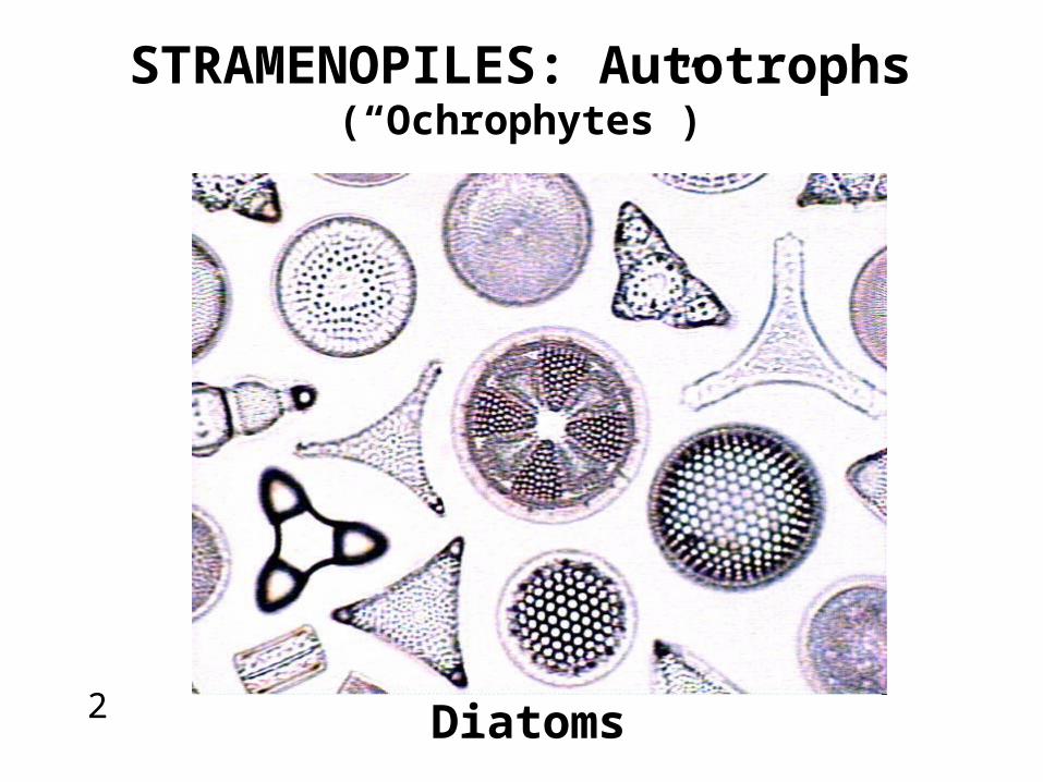
STRAMENOPILES: Autotrophs(“Ochrophytes”)
Diatoms

67
Water Molds
STRAMENOPILES: Heterotrophs
Saprolegnia

STRAMENOPILES: Heterotrophs
Late Potato Blight Grape Downy MildewDamping Off Disease,Root Rot
PlasmoporaPythiumPhytopthora
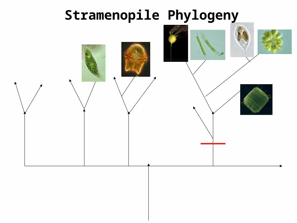
Stramenopile Phylogeny
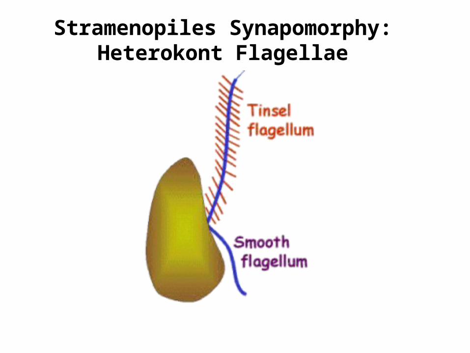
Stramenopiles Synapomorphy:Heterokont Flagellae

13
45
Brown Algae
Yellow-Green Algae
Synurans
Golden-Brown Algae
Diatoms
Stramenopiles: Ochrophytes
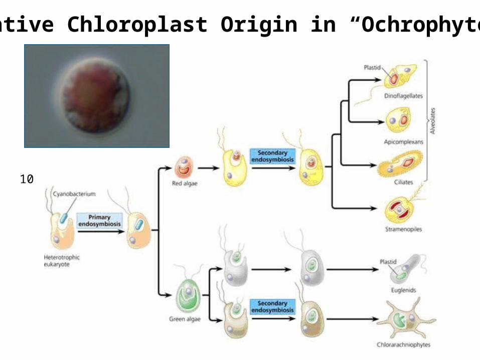
Putative Chloroplast Origin in “Ochrophytes”
10
11

2
3
1
Diatoms

(Marine, Freshwater)Diatom Habitat
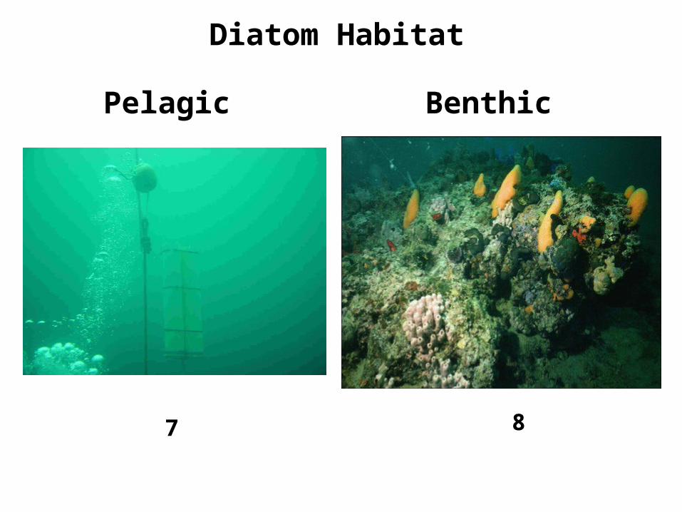
7 8
Pelagic Benthic
Diatom Habitat

24
Plankton Periphyton
Diatom Substrates

9Epizoic
Endozoic
11(Foraminfera)
Epiphytic
Endophytic/Endosymbiotic
Epilithic
(Cnidaria)
Diatom Substrates

Ocean Sediments
14

13
Primary Productivity: 20% of Global Carbon Fixation

26
Diatomaceous Earth
27

Pseudo-nitzschiaAmnesiac Shellfish Poisoning
15
Neurotoxin: Domoic Acid(Binds to Kainate Receptors Membranes of Nervous System)

Frustule

18Silica (SiO2, Silicon Dioxide)
19
• Abundant in Water
• Inert to Enzymatic Attack
• pH buffer; maintains Carbonic Anhydrase Activity

Frustule Components

Fig. 12-16 in Graham and Wilcox 2007
Epivalve, Epicingulum, Hypovalve, Hypocingulum

Girdle = Cingulum

Girdle View
2220
Valve View

Areolae
Fig. 12-19 in Graham and Wilcox 2007

Fig. 12-18 in Graham and Wilcox 2007
Loculate and Poroid Areolae (and Velum)

STRIAE
Fig. 12-20 in Graham and Wilcox 2000
Striae

Ocellus: Mucilage Secretion
Figs. 12-21, 12-22 in Graham and Wilcox 2007

Fig. 12-14 in Graham and Wilcox 2000
Tubes: Labiate Processes

Tubes: Strutted Processes
Figs. 12-14, 12-23 in Graham and Wilcox 2007

Forms of Extracellular Mucilage
Fig. 13.10 in Lee 1999
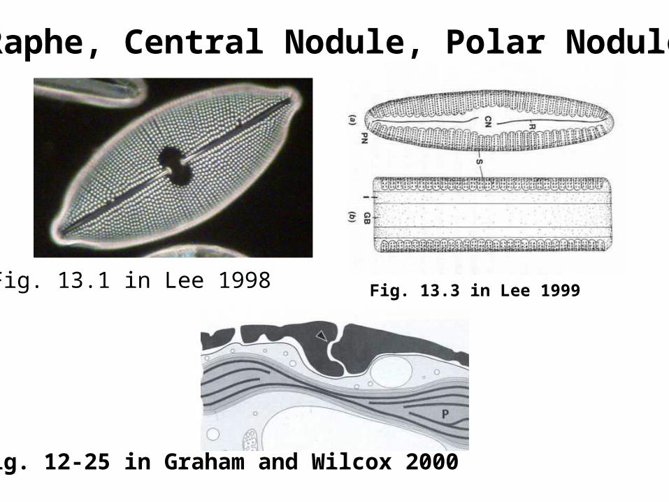
Raphe, Central Nodule, Polar Nodules
Fig. 12-25 in Graham and Wilcox 2000
Fig. 13.1 in Lee 1998 Fig. 13.3 in Lee 1999
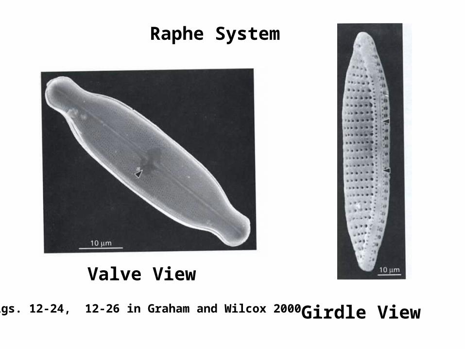
Raphe System
Figs. 12-24, 12-26 in Graham and Wilcox 2000
Valve View
Girdle View

Diatom Structural Diversity:Centric, Araphic Pennate, Raphic Pennate
CENTRIC
ARAPHIC PENNATE
RAPHIC PENNATE
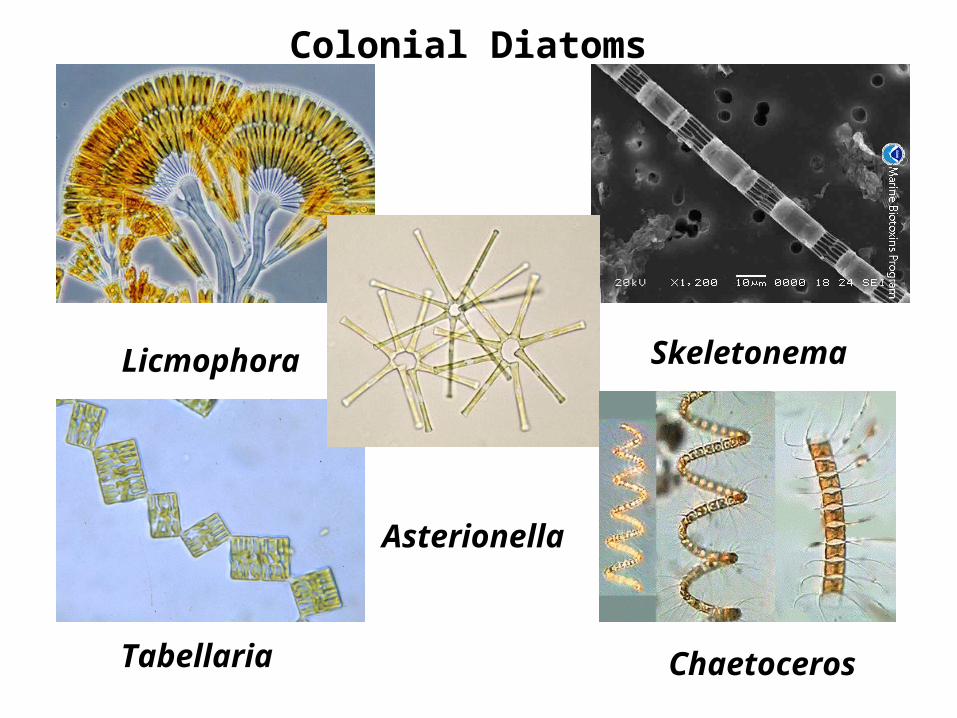
Licmophora Skeletonema
Tabellaria
Asterionella
Chaetoceros
Colonial Diatoms

Periphytic Diatoms
Gomphonema
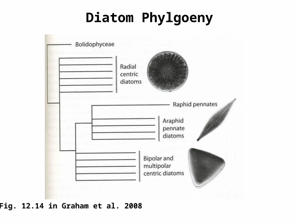
Diatom Phylgoeny
Fig. 12.14 in Graham et al. 2008

Chloroplast Origin
34
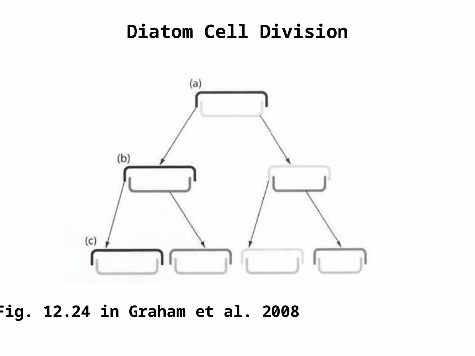
Fig. 12.24 in Graham et al. 2008
Diatom Cell Division

Centric Sex
Fig. 12.25 in Graham et al. 2008

Pennate Sex
Fig. 12.26 in Graham et al. 2008
1 http://www.daviddarling.info/images/diatoms.jpg
2 http://www.bhikku.net/archives/03/img/diatoms.JPG
3 http://www.zarzora.com/albums/Great-Sand-Sea/silica.jpg
4 http://mynasadata.larc.nasa.gov/images/upwelling.gif
5 http://library.thinkquest.org/5194/picts/ocean%20map.JPEG
6 http://www.islandnet.com/~see/weather/graphics/photos0506/ laketurnover.gif
7 http://www.mholmer.biology.sdu.dk/grafik/snapshot/Pelagic% 20trap.JPG
8 http://dept.bio.unipd.it/~casellato/research/foto/benthic1.jpg
9 http://home.hiroshima-u.ac.jp/fishlab/Dhaku/epizoic%20diatom/Falcula %20hyalina/1st%20ante%20em.jpg
11 http://fce.lternet.edu/publications/presentations/FCEslides/images/Slide17.jpg
12 http://www.epa.vic.gov.au/water/coasts/contentpix/shore.jpg
13 http://www.fbs.leeds.ac.uk/gradschool/BCMSc/graphics/wifs.gif
14 http://hjs.geol.uib.no/marinemicro/maps/0-0-2-map-sediments-facies.jpg
15 http://www.arpa.emr.it/daphne/progetto_mare/images/im_ Pseudo_nitzschia_seriata.jpg
16 http://www.astrographics.com/GalleryPrints/Display/GP2131.jpg
17 http://www.ualg.pt/adiac/intro/frustule.GIF
18 http://www.microscope-microscope.org/applications/sand/LakePowell3.jpg

19 http://wwwdelivery.superstock.com/WI/223/1597/PreviewComp/SuperStock_1597-27246.jpg
20 http://www.astrographics.com/GalleryPrints/Display/GP2131.jpg
21 http://www.priweb.org/ed/pgws/systems/images/diatom.jpg
22 http://www.priweb.org/ed/pgws/systems/images/diatom.jpg
23 http://www.microscopy-uk.org.uk/mag/imgapr02/db2sa.jpg
24 http://www-biol.paisley.ac.uk/bioref/Chromista/Vaucheria_epiphytes.jpg
25 http://www.scienceprogress.org/wp-content/uploads/2008/10/plankton_591.jpg
26 http://courses.bio.psu.edu/fall2005/biol110/tutorials/tutorial30_files/ diatomaceous_earth.jpg
27 http://www.holocaust-history.org/auschwitz/zyklonb/Sample3-diatomaceousearth.jpg
28 http://www.carolina.com/images/en_US//local/products/altview_large/152940_lp.jpg
29 http://www.glerl.noaa.gov/seagrant/GLWL/Protozoa/Ochromonas2.jpg
31 http://www.marlin.ac.uk/imgs/Species/Chromophycota/ o_lamhyp.jpg
32 http://herbarium.biosci.ohio-state.edu/Diatom.jpg
33 http://botit.botany.wisc.edu/toms_fungi/images/saprolegnia.jpg
34 http://myweb.dal.ca/jmarchib/pic.secondary-endo.jpg