2135 normal and abnormal us findings womens … and abnormal... · diagnosis of early pregnancy and...
TRANSCRIPT
WO
MEN
’S IMA
GIN
G2135
Normal and Abnormal US Findings in Early First-Trimester Pregnancy: Review of the Society of Radiolo-gists in Ultrasound 2012 Consen-sus Panel Recommendations1
Since being introduced more than 30 years ago, endovaginal ul-trasonography (US) and quantitative testing of serum levels of the beta subunit of human chorionic gonadotropin have become the standard means of establishing the presence of normal intrauterine pregnancy (IUP), failed IUP, and ectopic pregnancy. Appropriate use of these powerful tools requires clear, standardized interpreta-tions based on conservative criteria to protect both the pregnancy and the mother. Since diagnoses are assigned earlier and avail-able medical treatments for ectopic pregnancy and failed IUP are expanding, emphasis must carefully shift toward watchful waiting when the mother is clinically stable and a definitive location for the pregnancy cannot be established with US. To this end and to prevent inadvertent harm to early normal pregnancies, the Society of Radiologists in Ultrasound convened a consensus panel of radi-ologists, obstetricians, and emergency medicine physicians in 2012 with the goal of reviewing current literature and clinical practices and formulating modern criteria and terminology for the various first-trimester outcomes.
©RSNA, 2015 • radiographics.rsna.org
Shuchi K. Rodgers, MD Crystal Chang, MD John T. DeBardeleben, MD Mindy M. Horrow, MD
Abbreviations: b-hCG = beta subunit of hu-man chorionic gonadotropin, CRL = crown-rump length, IUP = intrauterine pregnancy, MSD = mean sac diameter
RadioGraphics 2015; 35:2135–2148
Published online 10.1148/rg.2015150092
Content Codes: 1From the Department of Radiology, Einstein Medical Center, 5501 Old York Rd, Philadel-phia, PA 19141. Presented as an education ex-hibit at the 2014 RSNA Annual Meeting. Re-ceived April 5, 2015; revision requested May 11; final revision received June 23; accepted July 17. For this journal-based SA-CME activity, the au-thors, editor, and reviewers have disclosed no rel-evant relationships. Address correspondence to S.K.R. (e-mail: [email protected]).
©RSNA, 2015
After completing this journal-based SA-CME activity, participants will be able to:
■ Describe issues related to safe interpre-tation of US findings in first-trimester pregnancy, including definitely normal findings, definitely abnormal findings, and indeterminate findings that require follow-up.
■ List criteria that are diagnostic for pregnancy failure and suspicious for preg-nancy failure.
■ Identify the correct management strat-egy for a pregnancy of unknown location with normal or near-normal adnexa.
See www.rsna.org/education/search/RG.
SA-CME LEARNING OBJECTIVES
IntroductionPelvic ultrasonography (US) and testing of the beta subunit of human chorionic gonadotropin (b-hCG) serum levels are key to diagnosis of early pregnancy and guide management of its associated complications. US imaging in early pregnancy should be primarily endovaginal, with transabdominal imaging used for adnexal masses high in the pelvis and documentation of the amount of free fluid. These tests allow distinction among the diagnostic possibilities of early pregnancy—intrauterine pregnancy (IUP) versus ectopic pregnancy, viable versus nonviable IUP, IUP of uncertain viability, and pregnancy of unknown location—and have contributed to the marked decline in mortality from ectopic pregnancy since the 1980s (1). However, misuse of these tests and misinterpretation of the find-ings can lead to unintentional harm to potentially viable pregnan-cies, such as administration of methotrexate for suspected ectopic pregnancy when, in fact, an early IUP is present but not recognized, resulting in embryonic demise or clinically significant birth defects (2). In several case reports, the teratogenic effects of methotrex-ate in fetuses have been documented (3–5), but inappropriate use of methotrexate due to misdiagnosis is likely underreported in the
This copy is for personal use only. To order printed copies, contact [email protected]
2136 November-December 2015 radiographics.rsna.org
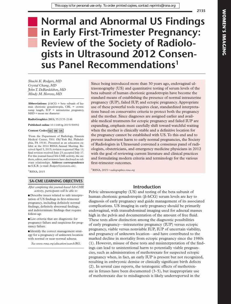
Figure 1. Endovaginal US image demonstrates the intra-decidual sign in a pregnant woman with pelvic pain. A 2-mm round gestational sac (arrow) is embedded within the decidua, adjacent to the collapsed endometrial cavity (arrowhead). The MSD is 2 mm, projecting to a gestational age of 4 weeks 4 days.
The role of follow-up pelvic US and monitoring of b-hCG levels is reviewed.
Normal Development of Early IUP between 4
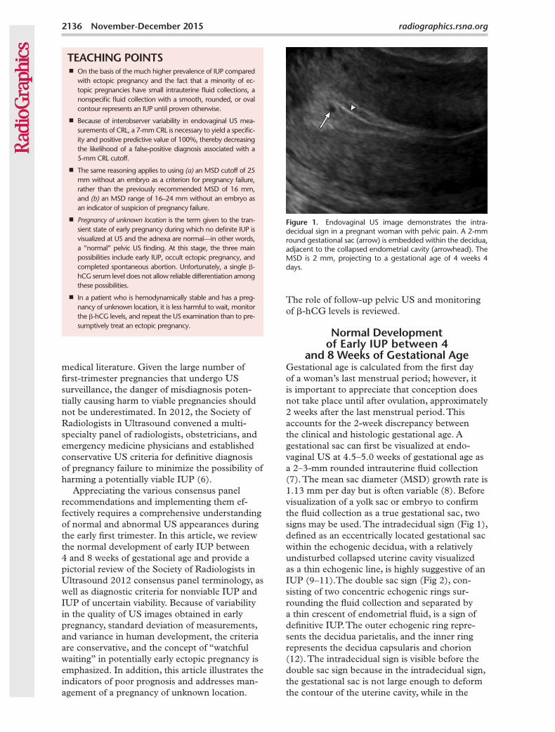
and 8 Weeks of Gestational AgeGestational age is calculated from the first day of a woman’s last menstrual period; however, it is important to appreciate that conception does not take place until after ovulation, approximately 2 weeks after the last menstrual period. This accounts for the 2-week discrepancy between the clinical and histologic gestational age. A gestational sac can first be visualized at endo-vaginal US at 4.5–5.0 weeks of gestational age as a 2–3-mm rounded intrauterine fluid collection (7). The mean sac diameter (MSD) growth rate is 1.13 mm per day but is often variable (8). Before visualization of a yolk sac or embryo to confirm the fluid collection as a true gestational sac, two signs may be used. The intradecidual sign (Fig 1), defined as an eccentrically located gestational sac within the echogenic decidua, with a relatively undisturbed collapsed uterine cavity visualized as a thin echogenic line, is highly suggestive of an IUP (9–11).The double sac sign (Fig 2), con-sisting of two concentric echogenic rings sur-rounding the fluid collection and separated by a thin crescent of endometrial fluid, is a sign of definitive IUP. The outer echogenic ring repre-sents the decidua parietalis, and the inner ring represents the decidua capsularis and chorion (12). The intradecidual sign is visible before the double sac sign because in the intradecidual sign, the gestational sac is not large enough to deform the contour of the uterine cavity, while in the
medical literature. Given the large number of first-trimester pregnancies that undergo US surveillance, the danger of misdiagnosis poten-tially causing harm to viable pregnancies should not be underestimated. In 2012, the Society of Radiologists in Ultrasound convened a multi-specialty panel of radiologists, obstetricians, and emergency medicine physicians and established conservative US criteria for definitive diagnosis of pregnancy failure to minimize the possibility of harming a potentially viable IUP (6).
Appreciating the various consensus panel recommendations and implementing them ef-fectively requires a comprehensive understanding of normal and abnormal US appearances during the early first trimester. In this article, we review the normal development of early IUP between 4 and 8 weeks of gestational age and provide a pictorial review of the Society of Radiologists in Ultrasound 2012 consensus panel terminology, as well as diagnostic criteria for nonviable IUP and IUP of uncertain viability. Because of variability in the quality of US images obtained in early pregnancy, standard deviation of measurements, and variance in human development, the criteria are conservative, and the concept of “watchful waiting” in potentially early ectopic pregnancy is emphasized. In addition, this article illustrates the indicators of poor prognosis and addresses man-agement of a pregnancy of unknown location.
TEACHING POINTS ■ On the basis of the much higher prevalence of IUP compared
with ectopic pregnancy and the fact that a minority of ec-topic pregnancies have small intrauterine fluid collections, a nonspecific fluid collection with a smooth, rounded, or oval contour represents an IUP until proven otherwise.
■ Because of interobserver variability in endovaginal US mea-surements of CRL, a 7-mm CRL is necessary to yield a specific-ity and positive predictive value of 100%, thereby decreasing the likelihood of a false-positive diagnosis associated with a 5-mm CRL cutoff.
■ The same reasoning applies to using (a) an MSD cutoff of 25 mm without an embryo as a criterion for pregnancy failure, rather than the previously recommended MSD of 16 mm, and (b) an MSD range of 16–24 mm without an embryo as an indicator of suspicion of pregnancy failure.
■ Pregnancy of unknown location is the term given to the tran-sient state of early pregnancy during which no definite IUP is visualized at US and the adnexa are normal—in other words, a “normal” pelvic US finding. At this stage, the three main possibilities include early IUP, occult ectopic pregnancy, and completed spontaneous abortion. Unfortunately, a single b-hCG serum level does not allow reliable differentiation among these possibilities.
■ In a patient who is hemodynamically stable and has a preg-nancy of unknown location, it is less harmful to wait, monitor the b-hCG levels, and repeat the US examination than to pre-sumptively treat an ectopic pregnancy.
RG • Volume 35 Number 7 Rodgers et al 2137
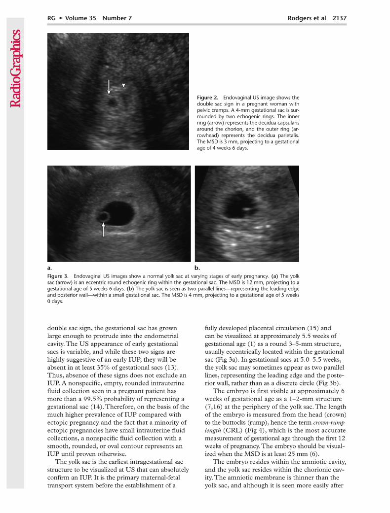
Figure 3. Endovaginal US images show a normal yolk sac at varying stages of early pregnancy. (a) The yolk sac (arrow) is an eccentric round echogenic ring within the gestational sac. The MSD is 12 mm, projecting to a gestational age of 5 weeks 6 days. (b) The yolk sac is seen as two parallel lines—representing the leading edge and posterior wall—within a small gestational sac. The MSD is 4 mm, projecting to a gestational age of 5 weeks 0 days.
fully developed placental circulation (15) and can be visualized at approximately 5.5 weeks of gestational age (1) as a round 3–5-mm structure, usually eccentrically located within the gestational sac (Fig 3a). In gestational sacs at 5.0–5.5 weeks, the yolk sac may sometimes appear as two parallel lines, representing the leading edge and the poste-rior wall, rather than as a discrete circle (Fig 3b).
The embryo is first visible at approximately 6 weeks of gestational age as a 1–2-mm structure (7,16) at the periphery of the yolk sac. The length of the embryo is measured from the head (crown) to the buttocks (rump), hence the term crown-rump length (CRL) (Fig 4), which is the most accurate measurement of gestational age through the first 12 weeks of pregnancy. The embryo should be visual-ized when the MSD is at least 25 mm (6).
The embryo resides within the amniotic cavity, and the yolk sac resides within the chorionic cav-ity. The amniotic membrane is thinner than the yolk sac, and although it is seen more easily after
double sac sign, the gestational sac has grown large enough to protrude into the endometrial cavity. The US appearance of early gestational sacs is variable, and while these two signs are highly suggestive of an early IUP, they will be absent in at least 35% of gestational sacs (13). Thus, absence of these signs does not exclude an IUP. A nonspecific, empty, rounded intrauterine fluid collection seen in a pregnant patient has more than a 99.5% probability of representing a gestational sac (14). Therefore, on the basis of the much higher prevalence of IUP compared with ectopic pregnancy and the fact that a minority of ectopic pregnancies have small intrauterine fluid collections, a nonspecific fluid collection with a smooth, rounded, or oval contour represents an IUP until proven otherwise.
The yolk sac is the earliest intragestational sac structure to be visualized at US that can absolutely confirm an IUP. It is the primary maternal-fetal transport system before the establishment of a
Figure 2. Endovaginal US image shows the double sac sign in a pregnant woman with pelvic cramps. A 4-mm gestational sac is sur-rounded by two echogenic rings. The inner ring (arrow) represents the decidua capsularis around the chorion, and the outer ring (ar-rowhead) represents the decidua parietalis. The MSD is 3 mm, projecting to a gestational age of 4 weeks 6 days.
2138 November-December 2015 radiographics.rsna.org
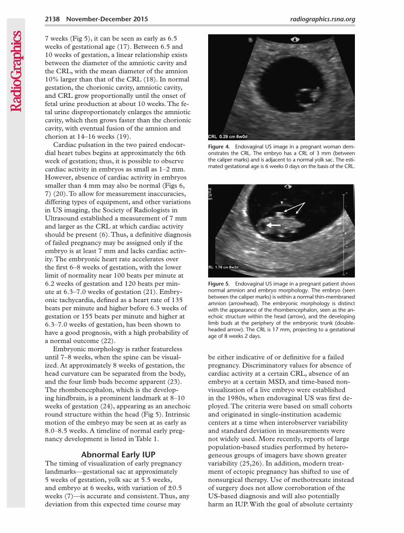
Figure 5. Endovaginal US image in a pregnant patient shows normal amnion and embryo morphology. The embryo (seen between the caliper marks) is within a normal thin-membraned amnion (arrowhead). The embryonic morphology is distinct with the appearance of the rhombencephalon, seen as the an-echoic structure within the head (arrow), and the developing limb buds at the periphery of the embryonic trunk (double-headed arrow). The CRL is 17 mm, projecting to a gestational age of 8 weeks 2 days.
Figure 4. Endovaginal US image in a pregnant woman dem-onstrates the CRL. The embryo has a CRL of 3 mm (between the caliper marks) and is adjacent to a normal yolk sac. The esti-mated gestational age is 6 weeks 0 days on the basis of the CRL.
7 weeks (Fig 5), it can be seen as early as 6.5 weeks of gestational age (17). Between 6.5 and 10 weeks of gestation, a linear relationship exists between the diameter of the amniotic cavity and the CRL, with the mean diameter of the amnion 10% larger than that of the CRL (18). In normal gestation, the chorionic cavity, amniotic cavity, and CRL grow proportionally until the onset of fetal urine production at about 10 weeks. The fe-tal urine disproportionately enlarges the amniotic cavity, which then grows faster than the chorionic cavity, with eventual fusion of the amnion and chorion at 14–16 weeks (19).
Cardiac pulsation in the two paired endocar-dial heart tubes begins at approximately the 6th week of gestation; thus, it is possible to observe cardiac activity in embryos as small as 1–2 mm. However, absence of cardiac activity in embryos smaller than 4 mm may also be normal (Figs 6, 7) (20). To allow for measurement inaccuracies, differing types of equipment, and other variations in US imaging, the Society of Radiologists in Ultrasound established a measurement of 7 mm and larger as the CRL at which cardiac activity should be present (6). Thus, a definitive diagnosis of failed pregnancy may be assigned only if the embryo is at least 7 mm and lacks cardiac activ-ity. The embryonic heart rate accelerates over the first 6–8 weeks of gestation, with the lower limit of normality near 100 beats per minute at 6.2 weeks of gestation and 120 beats per min-ute at 6.3–7.0 weeks of gestation (21). Embry-onic tachycardia, defined as a heart rate of 135 beats per minute and higher before 6.3 weeks of gestation or 155 beats per minute and higher at 6.3–7.0 weeks of gestation, has been shown to have a good prognosis, with a high probability of a normal outcome (22).
Embryonic morphology is rather featureless until 7–8 weeks, when the spine can be visual-ized. At approximately 8 weeks of gestation, the head curvature can be separated from the body, and the four limb buds become apparent (23). The rhombencephalon, which is the develop-ing hindbrain, is a prominent landmark at 8–10 weeks of gestation (24), appearing as an anechoic round structure within the head (Fig 5). Intrinsic motion of the embryo may be seen at as early as 8.0–8.5 weeks. A timeline of normal early preg-nancy development is listed in Table 1.
Abnormal Early IUPThe timing of visualization of early pregnancy landmarks—gestational sac at approximately 5 weeks of gestation, yolk sac at 5.5 weeks, and embryo at 6 weeks, with variation of ±0.5 weeks (7)—is accurate and consistent. Thus, any deviation from this expected time course may
be either indicative of or definitive for a failed pregnancy. Discriminatory values for absence of cardiac activity at a certain CRL, absence of an embryo at a certain MSD, and time-based non-visualization of a live embryo were established in the 1980s, when endovaginal US was first de-ployed. The criteria were based on small cohorts and originated in single-institution academic centers at a time when interobserver variability and standard deviation in measurements were not widely used. More recently, reports of large population-based studies performed by hetero-geneous groups of imagers have shown greater variability (25,26). In addition, modern treat-ment of ectopic pregnancy has shifted to use of nonsurgical therapy. Use of methotrexate instead of surgery does not allow corroboration of the US-based diagnosis and will also potentially harm an IUP. With the goal of absolute certainty
RG • Volume 35 Number 7 Rodgers et al 2139
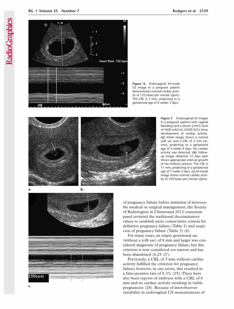
Figure 6. Endovaginal M-mode US image in a pregnant patient demonstrates normal cardiac activ-ity of 122 beats per minute (bpm). The CRL is 3 mm, projecting to a gestational age of 6 weeks 3 days.
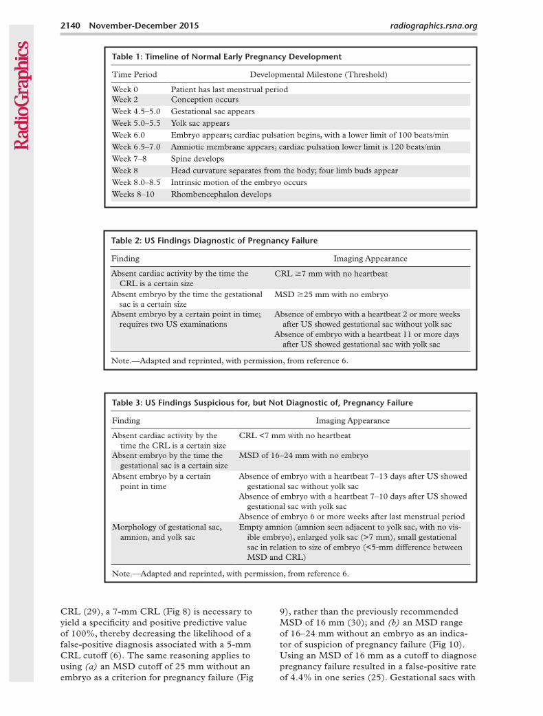
Figure 7. Endovaginal US images in a pregnant patient with vaginal bleeding and a serum b-hCG level of 5420 mIU/mL (5420 IU/L) show development of cardiac activity. (a) Initial image shows a normal yolk sac and a CRL of 2 mm (ar-row), projecting to a gestational age of 5 weeks 6 days. No cardiac activity was detected. (b) Follow-up image obtained 12 days later shows appropriate interval growth of the embryo (arrow). The CRL is 11 mm, projecting to a gestational age of 7 weeks 3 days. (c) M-mode image shows normal cardiac activ-ity of 150 beats per minute (bpm).
of pregnancy failure before initiation of irrevoca-ble medical or surgical management, the Society of Radiologists in Ultrasound 2012 consensus panel revisited the traditional discriminatory values to establish more conservative criteria for definitive pregnancy failure (Table 2) and suspi-cion of pregnancy failure (Table 3) (6).
For many years, an empty gestational sac (without a yolk sac) of 8 mm and larger was con-sidered diagnostic of pregnancy failure, but this criterion is now considered too narrow and has been abandoned (6,25–27).
Previously, a CRL of 5 mm without cardiac activity fulfilled the criterion for pregnancy failure; however, in one series, this resulted in a false-positive rate of 8.3% (25). There have also been reports of embryos with a CRL of 6 mm and no cardiac activity resulting in viable pregnancies (28). Because of interobserver variability in endovaginal US measurements of
2140 November-December 2015 radiographics.rsna.org
CRL (29), a 7-mm CRL (Fig 8) is necessary to yield a specificity and positive predictive value of 100%, thereby decreasing the likelihood of a false-positive diagnosis associated with a 5-mm CRL cutoff (6). The same reasoning applies to using (a) an MSD cutoff of 25 mm without an embryo as a criterion for pregnancy failure (Fig
9), rather than the previously recommended MSD of 16 mm (30); and (b) an MSD range of 16–24 mm without an embryo as an indica-tor of suspicion of pregnancy failure (Fig 10). Using an MSD of 16 mm as a cutoff to diagnose pregnancy failure resulted in a false-positive rate of 4.4% in one series (25). Gestational sacs with
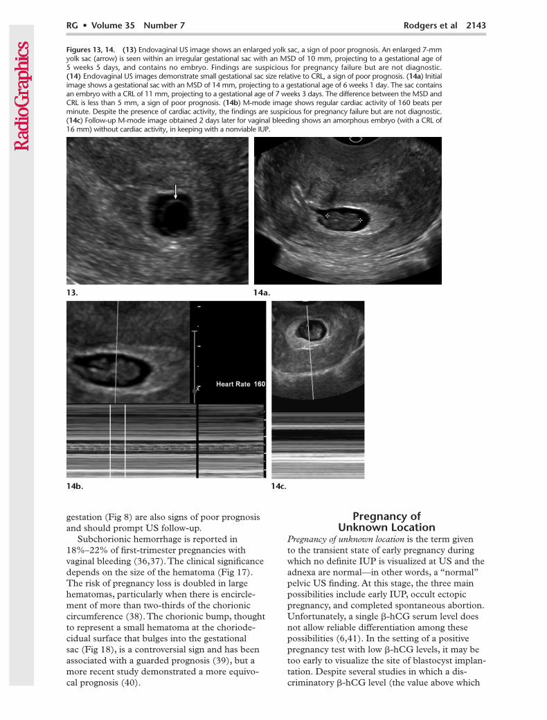
Table 1: Timeline of Normal Early Pregnancy Development
Time Period Developmental Milestone (Threshold)
Week 0 Patient has last menstrual periodWeek 2 Conception occursWeek 4.5–5.0 Gestational sac appearsWeek 5.0–5.5 Yolk sac appearsWeek 6.0 Embryo appears; cardiac pulsation begins, with a lower limit of 100 beats/minWeek 6.5–7.0 Amniotic membrane appears; cardiac pulsation lower limit is 120 beats/minWeek 7–8 Spine developsWeek 8 Head curvature separates from the body; four limb buds appearWeek 8.0–8.5 Intrinsic motion of the embryo occursWeeks 8–10 Rhombencephalon develops
Table 2: US Findings Diagnostic of Pregnancy Failure
Finding Imaging Appearance
Absent cardiac activity by the time the CRL is a certain size
CRL 7 mm with no heartbeat
Absent embryo by the time the gestational sac is a certain size
MSD 25 mm with no embryo
Absent embryo by a certain point in time; requires two US examinations
Absence of embryo with a heartbeat 2 or more weeks after US showed gestational sac without yolk sac
Absence of embryo with a heartbeat 11 or more days after US showed gestational sac with yolk sac
Note.—Adapted and reprinted, with permission, from reference 6.
Table 3: US Findings Suspicious for, but Not Diagnostic of, Pregnancy Failure
Finding Imaging Appearance
Absent cardiac activity by the time the CRL is a certain size
CRL <7 mm with no heartbeat
Absent embryo by the time the gestational sac is a certain size
MSD of 16–24 mm with no embryo
Absent embryo by a certain point in time
Absence of embryo with a heartbeat 7–13 days after US showed gestational sac without yolk sac
Absence of embryo with a heartbeat 7–10 days after US showed gestational sac with yolk sac
Absence of embryo 6 or more weeks after last menstrual periodMorphology of gestational sac,
amnion, and yolk sacEmpty amnion (amnion seen adjacent to yolk sac, with no vis-
ible embryo), enlarged yolk sac (>7 mm), small gestational sac in relation to size of embryo (<5-mm difference between MSD and CRL)
Note.—Adapted and reprinted, with permission, from reference 6.

RG • Volume 35 Number 7 Rodgers et al 2141
Figure 10. Endovaginal US images show findings suspicious for but not diagnostic of pregnancy failure at ini-tial US and findings of nonviable IUP at follow-up US. (a) Initial findings are suspicious for pregnancy failure but not diagnostic. There is an irregular gestational sac (arrowheads) with an MSD of 17 mm, an enlarged empty amnion (arrows), and no embryo or yolk sac. (b) Follow-up image obtained 10 days later shows a nonviable IUP. There is a lack of appropriate interval growth of the gestational sac and no embryo. Note the hydropic changes in the chorionic villi (arrow). The MSD is 19 mm, projecting to a gestational age of 6 weeks 6 days.
Figure 8. Endovaginal US image shows a nonviable IUP. An amorphous embryo (arrowhead) is seen with a CRL of 20 mm, projecting to a gestational age of 8 weeks 4 days, but there was no cardiac activity. These findings are consistent with a nonvi-able IUP because the CRL measures at least 7 mm. Note the ir-regular gestational sac contour (arrow), a sign of poor prognosis.
Figure 9. Endovaginal US image demonstrates a nonviable IUP. There is an empty gestational sac with an MSD of 29 mm. Fine linear echogenic debris is noted in the sac, but there is no yolk sac or embryo. The estimated gestational age is 8 weeks 1 day. The findings are in keeping with a nonviable IUP because the MSD measures at least 25 mm.
mean diameters between 17 and 21 mm and no visible embryo have resulted in viable pregnan-cies (25,26). Because of interobserver variability in endovaginal US measurements, an MSD cut-off of 25 mm increases the specificity to 100% (29). Not all failed or potentially nonviable intrauterine pregnancies demonstrate a 7-mm CRL without cardiac activity or a 25-mm MSD with no embryo, necessitating additional criteria based on nonvisualization of a live embryo by a certain time interval (Fig 11).
Morphologic assessment of the individual components of a pregnancy—including the gesta-tional sac, the yolk sac, the amnion, the embryo, cardiac activity, and the decidua—is helpful in
evaluating the prognosis of the pregnancy (Table 4). Additional findings that are suspicious for pregnancy failure in the consensus panel crite-ria include an empty amniotic sac, an enlarged yolk sac, and small gestational sac size relative to embryo size. Given the similar length of the amniotic cavity to the CRL during 6.5–10 weeks of gestation in a normal pregnancy, the pres-ence of an “empty amnion” with no identifiable embryo adjacent to a yolk sac is an indication of poor prognosis (Figs 10a, 12) (31) and should prompt US follow-up. An enlarged yolk sac larger than 7 mm (Fig 13) (15) and small gestational sac size relative to embryo size (defined as less than a 5-mm difference between the MSD and
2142 November-December 2015 radiographics.rsna.org
Figure 12. Endovaginal US im-age demonstrates an “empty amnion,” a sign of poor progno-sis. An empty amnion (arrow) is seen adjacent to a normal yolk sac (arrowhead). The MSD is 2.2, projecting to an estimated gesta-tional age of 7 weeks 2 days. An embryo should be present within the amnion in a normal IUP.
Figure 11. Endovaginal US images show findings of uncertain pregnancy viability at initial US and a nonviable IUP at follow-up US. (a) Initial image shows a round gestational sac that contains a yolk sac (arrow) and a possible adjacent embryo. The MSD is 14 mm, projecting to a gestational age of 6 weeks 1 day. (b) Follow-up image obtained 13 days later shows lack of ap-propriate growth of the gestational sac, with an MSD of 16 mm, projecting to a gestational age of 6 weeks 3 days. There is a 4-mm embryo (single arrowhead) within an expanded amnion (arrow). No cardiac activity was detected. A yolk sac is present (double arrowhead). Findings are diagnostic of pregnancy failure.
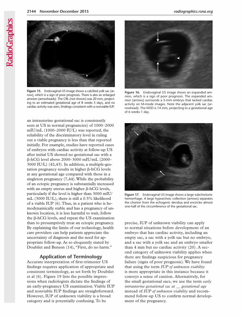
the CRL) (Fig 14) have also been associated with poor pregnancy outcome (32). An irregular gestational sac (lack of a smooth contour and/or presence of a distorted sac shape) is highly suggestive of an abnormal IUP. In one series, this finding had a 100% specificity and a 100% posi-tive predictive value for an abnormal IUP, but it had a low sensitivity of 10% (Fig 10a) (33). The presence of a calcified yolk sac (Fig 15) suggests that the embryonic demise is likely of a relatively
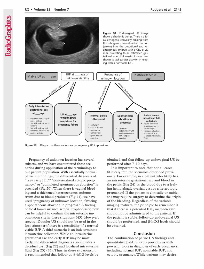
long-standing duration of 2 weeks or longer (34). An enlarged or expanded amnion (amnion too large for the size of the embryo) (17,18) (Figs 15, 16), embryonic bradycardia of 85 beats per minute or less (35), degenerative hydropic changes (Fig 10b) within the chorionic villi, and amorphous shape of the embryo at 7–8 weeks of
Table 4: US Indicators of Poor Prognosis in Early Pregnancy
Feature Imaging Appearance
Gestational sac Irregular contour, low-lying positionYolk sac Calcified, larger than 7 mmAmnion Empty, enlarged, or expandedEmbryo Amorphous shapeCardiac activity Bradycardia of 85 beats/min or lessChorionic villi Hydropic changeSubchorionic hemorrhage Large, particularly if it encircles at least two-
thirds of the gestational sac circumference
RG • Volume 35 Number 7 Rodgers et al 2143
Figures 13, 14. (13) Endovaginal US image shows an enlarged yolk sac, a sign of poor prognosis. An enlarged 7-mm yolk sac (arrow) is seen within an irregular gestational sac with an MSD of 10 mm, projecting to a gestational age of 5 weeks 5 days, and contains no embryo. Findings are suspicious for pregnancy failure but are not diagnostic. (14) Endovaginal US images demonstrate small gestational sac size relative to CRL, a sign of poor prognosis. (14a) Initial image shows a gestational sac with an MSD of 14 mm, projecting to a gestational age of 6 weeks 1 day. The sac contains an embryo with a CRL of 11 mm, projecting to a gestational age of 7 weeks 3 days. The difference between the MSD and CRL is less than 5 mm, a sign of poor prognosis. (14b) M-mode image shows regular cardiac activity of 160 beats per minute. Despite the presence of cardiac activity, the findings are suspicious for pregnancy failure but are not diagnostic. (14c) Follow-up M-mode image obtained 2 days later for vaginal bleeding shows an amorphous embryo (with a CRL of 16 mm) without cardiac activity, in keeping with a nonviable IUP.
gestation (Fig 8) are also signs of poor prognosis and should prompt US follow-up.
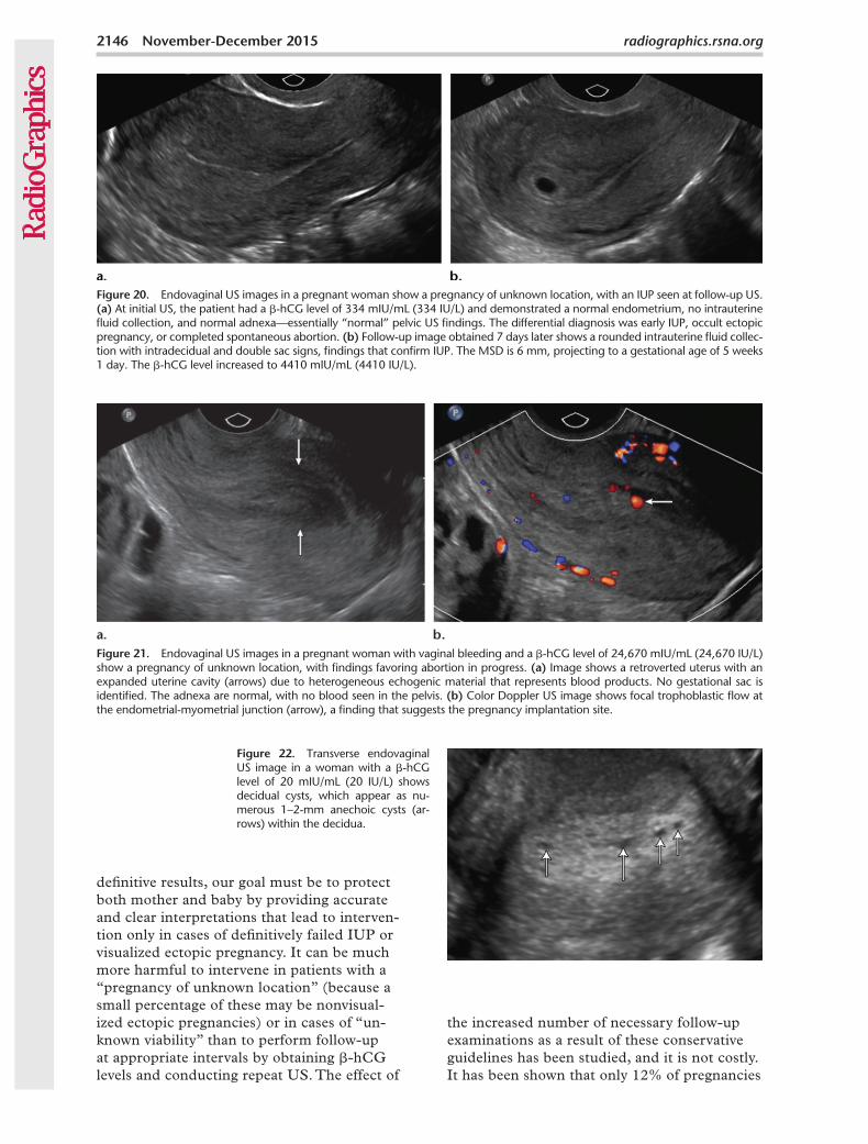
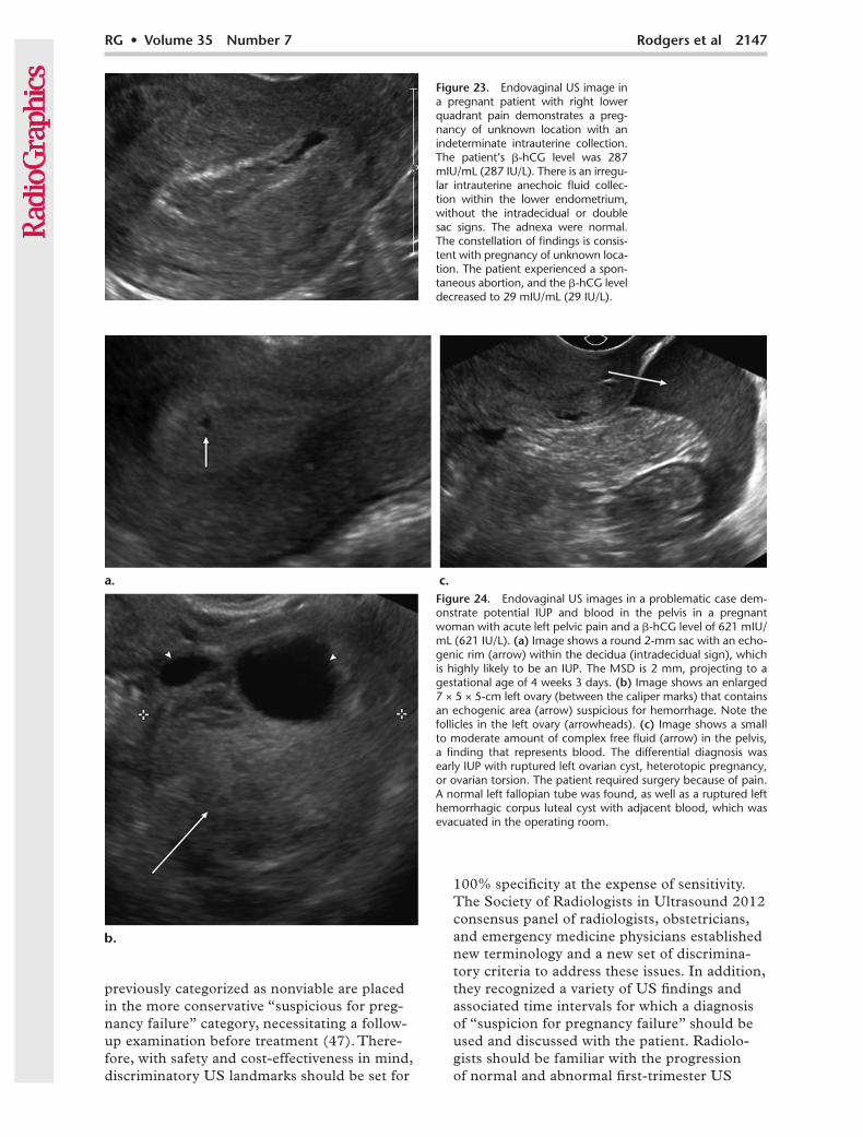
Subchorionic hemorrhage is reported in 18%–22% of first-trimester pregnancies with vaginal bleeding (36,37). The clinical significance depends on the size of the hematoma (Fig 17). The risk of pregnancy loss is doubled in large hematomas, particularly when there is encircle-ment of more than two-thirds of the chorionic circumference (38). The chorionic bump, thought to represent a small hematoma at the choriode-cidual surface that bulges into the gestational sac (Fig 18), is a controversial sign and has been associated with a guarded prognosis (39), but a more recent study demonstrated a more equivo-cal prognosis (40).
Pregnancy of Unknown Location
Pregnancy of unknown location is the term given to the transient state of early pregnancy during which no definite IUP is visualized at US and the adnexa are normal—in other words, a “normal” pelvic US finding. At this stage, the three main possibilities include early IUP, occult ectopic pregnancy, and completed spontaneous abortion. Unfortunately, a single b-hCG serum level does not allow reliable differentiation among these possibilities (6,41). In the setting of a positive pregnancy test with low b-hCG levels, it may be too early to visualize the site of blastocyst implan-tation. Despite several studies in which a dis-criminatory b-hCG level (the value above which
2144 November-December 2015 radiographics.rsna.org
Figure 17. Endovaginal US image shows a large subchorionic hemorrhage. A large hypoechoic collection (arrows) separates the chorion from the echogenic decidua and encircles almost one-half of the circumference of the gestational sac.
Figure 16. Endovaginal US image shows an expanded am-nion, which is a sign of poor prognosis. The expanded am-nion (arrows) surrounds a 5-mm embryo that lacked cardiac activity on M-mode images. Note the adjacent yolk sac (ar-rowhead). The MSD is 14 mm, projecting to a gestational age of 6 weeks 1 day.
an intrauterine gestational sac is consistently seen at US in normal pregnancies) of 1000–2000 mIU/mL (1000–2000 IU/L) was reported, the reliability of the discriminatory level in ruling out a viable pregnancy is less than that reported initially. For example, studies have reported cases of embryos with cardiac activity at follow-up US after initial US showed no gestational sac with a b-hCG level above 2000–3000 mIU/mL (2000–3000 IU/L) (42,43). In addition, a multiple-ges-tation pregnancy results in higher b-hCG levels at any gestational age compared with those in a singleton pregnancy (7,44). While the probability of an ectopic pregnancy is substantially increased with an empty uterus and higher b-hCG levels, particularly if the level is higher than 3000 mIU/mL (3000 IU/L), there is still a 0.5% likelihood of a viable IUP (6). Thus, in a patient who is he-modynamically stable and has a pregnancy of un-known location, it is less harmful to wait, follow the b-hCG levels, and repeat the US examination than to presumptively treat an ectopic pregnancy. By explaining the limits of our technology, health care providers can help patients appreciate the uncertainty of diagnosis and the need for ap-propriate follow-up. As so eloquently stated by Doubilet and Benson (14), “First, do no harm.”
Application of TerminologyAccurate interpretation of first-trimester US findings requires application of appropriate and consistent terminology, as set forth by Doubilet et al (6). Figure 19 lists the possible impres-sions when radiologists dictate the findings of an early-pregnancy US examination. Viable IUP and nonviable IUP findings are straightforward. However, IUP of unknown viability is a broad category and is potentially confusing. To be
precise, IUP of unknown viability can apply to normal situations before development of an embryo that has cardiac activity, including an empty sac, a sac with a yolk sac but no embryo, and a sac with a yolk sac and an embryo smaller than 4 mm but no cardiac activity (20). A sec-ond category of unknown viability applies when there are findings suspicious for pregnancy failure (signs of poor prognosis). We have found that using the term IUP of unknown viability is more appropriate in this instance because it conveys a sense of caution. Alternatively, for the small gestational sacs, we use the term early intrauterine gestational sac at __ gestational age instead of IUP of unknown viability and recom-mend follow-up US to confirm normal develop-ment of the pregnancy.
Figure 15. Endovaginal US image shows a calcified yolk sac (ar-row), which is a sign of poor prognosis. There is also an enlarged amnion (arrowheads). The CRL (not shown) was 20 mm, project-ing to an estimated gestational age of 8 weeks 5 days, and no cardiac activity was seen, findings consistent with a nonviable IUP.
RG • Volume 35 Number 7 Rodgers et al 2145
Figure 19. Diagram outlines various early-pregnancy US impressions.
Pregnancy of unknown location has several subsets, and we have encountered these sce-narios during application of the terminology to our patient population. With essentially normal pelvic US findings, the differential diagnosis of “very early IUP,” “nonvisualized ectopic preg-nancy,” or “completed spontaneous abortion” is provided (Fig 20). When there is vaginal bleed-ing and a thickened heterogeneous endome-trium due to blood products (Fig 21), we have used “pregnancy of unknown location, favoring a spontaneous abortion in progress.” A finding of focal low-resistance arterial trophoblastic flow can be helpful to confirm the intrauterine im-plantation site in these situations (45). However, spectral Doppler US should not be used in the first trimester if there is a possibility of a normal viable IUP. A third scenario is an indeterminate intrauterine collection. While an intrauterine gestational sac and early IUP may be most likely, the differential diagnosis also includes a decidual cyst (Fig 22) and localized intrauterine fluid (Fig 23) (46). Thus, in these situations, it is recommended that follow-up b-hCG levels be
obtained and that follow-up endovaginal US be performed after 7–10 days.
It is important to note that not all cases fit nicely into the scenarios described previ-ously. For example, in a patient who likely has an intrauterine gestational sac and blood in the pelvis (Fig 24), is the blood due to a leak-ing hemorrhagic ovarian cyst or a heterotopic pregnancy? If the patient is clinically unstable, she may require surgery to determine the origin of the bleeding. Regardless of the variable imaging features, the principle to remember is that if there is a potential IUP, methotrexate should not be administered to the patient. If the patient is stable, follow-up endovaginal US should be performed, and b-hCG levels should be obtained.
ConclusionThe combination of pelvic US findings and quantitative b-hCG levels provides us with powerful tools in diagnosis of early pregnancy, including normal IUP, nonviable IUP, and ectopic pregnancy. While patients may desire
Figure 18. Endovaginal US image shows a chorionic bump. There is a fo-cal echogenic convexity bulging from the echogenic choriodecidual reaction (arrow) into the gestational sac. An amorphous embryo with a CRL of 20 mm, projecting to an estimated ges-tational age of 8 weeks 4 days, was shown to lack cardiac activity, in keep-ing with a nonviable IUP.
2146 November-December 2015 radiographics.rsna.org
Figure 21. Endovaginal US images in a pregnant woman with vaginal bleeding and a b-hCG level of 24,670 mIU/mL (24,670 IU/L) show a pregnancy of unknown location, with findings favoring abortion in progress. (a) Image shows a retroverted uterus with an expanded uterine cavity (arrows) due to heterogeneous echogenic material that represents blood products. No gestational sac is identified. The adnexa are normal, with no blood seen in the pelvis. (b) Color Doppler US image shows focal trophoblastic flow at the endometrial-myometrial junction (arrow), a finding that suggests the pregnancy implantation site.
definitive results, our goal must be to protect both mother and baby by providing accurate and clear interpretations that lead to interven-tion only in cases of definitively failed IUP or visualized ectopic pregnancy. It can be much more harmful to intervene in patients with a “pregnancy of unknown location” (because a small percentage of these may be nonvisual-ized ectopic pregnancies) or in cases of “un-known viability” than to perform follow-up at appropriate intervals by obtaining b-hCG levels and conducting repeat US. The effect of
the increased number of necessary follow-up examinations as a result of these conservative guidelines has been studied, and it is not costly. It has been shown that only 12% of pregnancies
Figure 20. Endovaginal US images in a pregnant woman show a pregnancy of unknown location, with an IUP seen at follow-up US. (a) At initial US, the patient had a b-hCG level of 334 mIU/mL (334 IU/L) and demonstrated a normal endometrium, no intrauterine fluid collection, and normal adnexa—essentially “normal” pelvic US findings. The differential diagnosis was early IUP, occult ectopic pregnancy, or completed spontaneous abortion. (b) Follow-up image obtained 7 days later shows a rounded intrauterine fluid collec-tion with intradecidual and double sac signs, findings that confirm IUP. The MSD is 6 mm, projecting to a gestational age of 5 weeks 1 day. The b-hCG level increased to 4410 mIU/mL (4410 IU/L).
Figure 22. Transverse endovaginal US image in a woman with a b-hCG level of 20 mIU/mL (20 IU/L) shows decidual cysts, which appear as nu-merous 1–2-mm anechoic cysts (ar-rows) within the decidua.
RG • Volume 35 Number 7 Rodgers et al 2147
previously categorized as nonviable are placed in the more conservative “suspicious for preg-nancy failure” category, necessitating a follow-up examination before treatment (47). There-fore, with safety and cost-effectiveness in mind, discriminatory US landmarks should be set for
Figure 23. Endovaginal US image in a pregnant patient with right lower quadrant pain demonstrates a preg-nancy of unknown location with an indeterminate intrauterine collection. The patient’s b-hCG level was 287 mIU/mL (287 IU/L). There is an irregu-lar intrauterine anechoic fluid collec-tion within the lower endometrium, without the intradecidual or double sac signs. The adnexa were normal. The constellation of findings is consis-tent with pregnancy of unknown loca-tion. The patient experienced a spon-taneous abortion, and the b-hCG level decreased to 29 mIU/mL (29 IU/L).
Figure 24. Endovaginal US images in a problematic case dem-onstrate potential IUP and blood in the pelvis in a pregnant woman with acute left pelvic pain and a b-hCG level of 621 mIU/mL (621 IU/L). (a) Image shows a round 2-mm sac with an echo-genic rim (arrow) within the decidua (intradecidual sign), which is highly likely to be an IUP. The MSD is 2 mm, projecting to a gestational age of 4 weeks 3 days. (b) Image shows an enlarged 7 × 5 × 5-cm left ovary (between the caliper marks) that contains an echogenic area (arrow) suspicious for hemorrhage. Note the follicles in the left ovary (arrowheads). (c) Image shows a small to moderate amount of complex free fluid (arrow) in the pelvis, a finding that represents blood. The differential diagnosis was early IUP with ruptured left ovarian cyst, heterotopic pregnancy, or ovarian torsion. The patient required surgery because of pain. A normal left fallopian tube was found, as well as a ruptured left hemorrhagic corpus luteal cyst with adjacent blood, which was evacuated in the operating room.
100% specificity at the expense of sensitivity. The Society of Radiologists in Ultrasound 2012 consensus panel of radiologists, obstetricians, and emergency medicine physicians established new terminology and a new set of discrimina-tory criteria to address these issues. In addition, they recognized a variety of US findings and associated time intervals for which a diagnosis of “suspicion for pregnancy failure” should be used and discussed with the patient. Radiolo-gists should be familiar with the progression of normal and abnormal first-trimester US
2148 November-December 2015 radiographics.rsna.org
findings and develop an understanding of the accepted terminology to use in their interpreta-tions, so that referring physicians will clearly un-derstand our intent and treat their patients safely.
References 1. Creanga AA, Shapiro-Mendoza CK, Bish CL, Zane S, Berg CJ,
Callaghan WM. Trends in ectopic pregnancy mortality in the United States: 1980–2007. Obstet Gynecol 2011;117(4):837–843.
2. Nurmohamed L, Moretti ME, Schechter T, et al. Outcome following high-dose methotrexate in pregnancies misdiagnosed as ectopic. Am J Obstet Gynecol 2011;205(6):533.e1–e3..
3. Nguyen C, Duhl AJ, Escallon CS, Blakemore KJ. Multiple anomalies in a fetus exposed to low-dose methotrexate in the first trimester. Obstet Gynecol 2002;99(4):599–602.
4. Addar MH. Methotrexate embryopathy in a surviving intrauterine fetus after presumed diagnosis of ectopic pregnancy: case report. J Obstet Gynaecol Can 2004;26(11):1001–1003.
5. Usta IM, Nassar AH, Yunis KA, Abu-Musa AA. Methotrexate embryopathy after therapy for misdiagnosed ectopic pregnancy. Int J Gynaecol Obstet 2007;99(3):253–255.
6. Doubilet PM, Benson CB, Bourne T, et al. Diagnostic criteria for nonviable pregnancy early in the first trimester. N Engl J Med 2013;369(15):1443–1451.
7. Bree RL, Edwards M, Böhm-Vélez M, Beyler S, Roberts J, Mendelson EB. Transvaginal sonography in the evaluation of normal early preg-nancy: correlation with hCG level. AJR Am J Roentgenol 1989;153 (1):75–79.
8. Nyberg DA, Mack LA, Laing FC, Patten RM. Distinguishing normal from abnormal gestational sac growth in early pregnancy. J Ultrasound Med 1987;6(1):23–27.
9. Yeh HC, Goodman JD, Carr L, Rabinowitz JG. Intradecidual sign: a US criterion of early intrauterine pregnancy. Radiology 1986;161 (2):463–467.
10. Laing FC, Brown DL, Price JF, Teeger S, Wong ML. Intradecidual sign: is it effective in diagnosis of an early intrauterine pregnancy? Radiology 1997;204(3):655–660.
11. Chiang G, Levine D, Swire M, McNamara A, Mehta T. The intra-decidual sign: is it reliable for diagnosis of early intrauterine pregnancy? AJR Am J Roentgenol 2004;183(3):725–731.
12. Bradley WG, Fiske CE, Filly RA. The double sac sign of early intra-uterine pregnancy: use in exclusion of ectopic pregnancy. Radiology 1982;143(1):223–226.
13. Doubilet PM, Benson CB. Double sac sign and intradecidual sign in early pregnancy: interobserver reliability and frequency of occurrence. J Ultrasound Med 2013;32(7):1207–1214.
14. Doubilet PM, Benson CB. First, do no harm...to early pregnancies. J Ultrasound Med 2010;29(5):685–689.
15. Lindsay DJ, Lovett IS, Lyons EA, et al. Yolk sac diameter and shape at endovaginal US: predictors of pregnancy outcome in the first trimester. Radiology 1992;183(1):115–118.
16. Hadlock FP, Shah YP, Kanon DJ, Lindsey JV. Fetal crown-rump length: reevaluation of relation to menstrual age (5–18 weeks) with high-resolution real-time US. Radiology 1992;182(2):501–505.
17. Yegul NT, Filly RA. The expanded amnion sign: evidence of early embryonic death. J Ultrasound Med 2009;28(10):1331–1335.
18. Horrow MM. Enlarged amniotic cavity: a new sonographic sign of early embryonic death. AJR Am J Roentgenol 1992;158(2): 359–362.
19. Yeh HC, Rabinowitz JG. Amniotic sac development: ultrasound fea-tures of early pregnancy—the double bleb sign. Radiology 1988;166(1 Pt 1):97–103.
20. Levi CS, Lyons EA, Zheng XH, Lindsay DJ, Holt SC. Endovaginal US: demonstration of cardiac activity in embryos of less than 5.0 mm in crown-rump length. Radiology 1990;176(1):71–74.
21. Doubilet PM, Benson CB. Embryonic heart rate in the early first trimester: what rate is normal? J Ultrasound Med 1995;14(6):431–434.
22. Doubilet PM, Benson CB, Chow JS. Outcome of pregnancies with rapid embryonic heart rates in the early first trimester. AJR Am J Roentgenol 2000;175(1):67–69.
23. Doubilet PM. Ultrasound evaluation of the first trimester. Radiol Clin North Am 2014;52(6):1191–1199.
24. Cyr DR, Mack LA, Nyberg DA, Shepard TH, Shuman WP. Fetal rhombencephalon: normal US findings. Radiology 1988;166(3): 691–692.
25. Abdallah Y, Daemen A, Kirk E, et al. Limitations of current definitions of miscarriage using mean gestational sac diameter and crown-rump length measurements: a multicenter observational study. Ultrasound Obstet Gynecol 2011;38(5):497–502.
26. Rowling SE, Coleman BG, Langer JE, Arger PH, Nisenbaum HL, Horii SC. First-trimester US parameters of failed pregnancy. Radiol-ogy 1997;203(1):211–217.
27. Levi CS, Lyons EA, Lindsay DJ. Early diagnosis of nonviable preg-nancy with endovaginal US. Radiology 1988;167(2):383–385.
28. Hamilton J. The 6 mm crown-rump length threshold for detecting fetal heart movements: what is the evidence? Ultrasound Obstet Gynecol 2011;38(suppl 1):7.
29. Pexsters A, Luts J, Van Schoubroeck D, et al. Clinical implications of intra- and interobserver reproducibility of transvaginal sonographic measurement of gestational sac and crown-rump length at 6–9 weeks’ gestation. Ultrasound Obstet Gynecol 2011;38(5):510–515.
30. Levi CS, Dashefsky SM, Lyons EA, Holt SC, Lindsay DJ. First tri-mester ultrasound. In: McGahan JP, Goldberg BB, eds. Diagnostic ultrasound: a logical approach. Philadelphia, Pa: Lippincott-Raven, 1998; 127–153.
31. McKenna KM, Feldstein VA, Goldstein RB, Filly RA. The empty amnion: a sign of early pregnancy failure. J Ultrasound Med 1995;14(2):117–121.
32. Bromley B, Harlow BL, Laboda LA, Benacerraf BR. Small sac size in the first trimester: a predictor of poor fetal outcome. Radiology 1991;178(2):375–377.
33. Nyberg DA, Laing FC, Filly RA. Threatened abortion: sonographic distinction of normal and abnormal gestation sacs. Radiology 1986;158(2):397–400.
34. Harris RD, Vincent LM, Askin FB. Yolk sac calcification: a sono-graphic finding associated with intrauterine embryonic demise in the first trimester. Radiology 1988;166(1 Pt 1):109–110.
35. Laboda LA, Estroff JA, Benacerraf BR. First trimester bradycardia: a sign of impending fetal loss. J Ultrasound Med 1989;8(10):561–563.
36. Leite J, Ross P, Rossi AC, Jeanty P. Prognosis of very large first-trimester hematomas. J Ultrasound Med 2006;25(11):1441–1445.
37. Pedersen JF, Mantoni M. Prevalence and significance of subchorionic hemorrhage in threatened abortion: a sonographic study. AJR Am J Roentgenol 1990;154(3):535–537.
38. Bennett GL, Bromley B, Lieberman E, Benacerraf BR. Subchorionic hemorrhage in first-trimester pregnancies: prediction of pregnancy outcome with sonography. Radiology 1996;200(3):803–806.
39. Harris RD, Couto C, Karpovsky C, Porter MMB, Ouhilal S. The cho-rionic bump: a first-trimester pregnancy sonographic finding associated with a guarded prognosis. J Ultrasound Med 2006;25(6):757–763.
40. Arleo EK, Troiano RN. Chorionic bump on first-trimester sonog-raphy: not necessarily a poor prognostic indicator for pregnancy. J Ultrasound Med 2015;34(1):137–142.
41. Barnhart KT. Clinical practice: ectopic pregnancy. N Engl J Med 2009;361(4):379–387.
42. Doubilet PM, Benson CB. Further evidence against the reliability of the human chorionic gonadotropin discriminatory level. J Ultrasound Med 2011;30(12):1637–1642.
43. Mehta TS, Levine D, Beckwith B. Treatment of ectopic pregnancy: is a human chorionic gonadotropin level of 2,000 mIU/mL a reasonable threshold? Radiology 1997;205(2):569–573.
44. Bateman BG, Nunley WC Jr, Kolp LA, Kitchin JD 3rd, Felder R. Vaginal sonography findings and hCG dynamics of early intrauterine and tubal pregnancies. Obstet Gynecol 1990;75(3 Pt 1):421–427.
45. Wherry KL, Dubinsky TJ, Waitches GM, Richardson ML, Reed S. Low-resistance endometrial arterial flow in the exclusion of ectopic pregnancy revisited. J Ultrasound Med 2001;20(4):335–342.
46. Benson CB, Doubilet PM, Peters HE, Frates MC. Intrauterine fluid with ectopic pregnancy: a reappraisal. J Ultrasound Med 2013; 32(3):389–393.
47. Hu M, Poder L, Filly RA. Impact of new society of radiologists in ultrasound early first-trimester diagnostic criteria for nonviable pregnancy. J Ultrasound Med 2014;33(9):1585–1588.
This journal-based SA-CME activity has been approved for AMA PRA Category 1 CreditTM. See www.rsna.org/education/search/RG.