2018 seer solid tumor with bladder - university of kentucky
TRANSCRIPT
7/10/2018
1
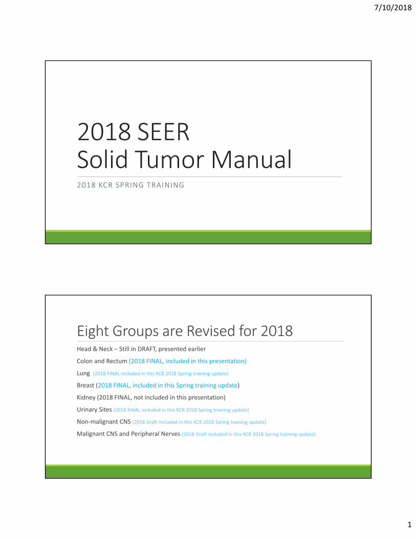
2018 SEER Solid Tumor Manual2018 KCR SPRING TRAINING
Eight Groups are Revised for 2018Head & Neck – Still in DRAFT, presented earlier
Colon and Rectum (2018 FINAL, included in this presentation)
Lung (2018 FINAL included in this KCR 2018 Spring training update)
Breast (2018 FINAL, included in this Spring training update)
Kidney (2018 FINAL, not included in this presentation)
Urinary Sites (2018 FINAL included in this KCR 2018 Spring training update)
Non‐malignant CNS (2018 Draft included in this KCR 2018 Spring training update)
Malignant CNS and Peripheral Nerves (2018 Draft included in this KCR 2018 Spring training update)
7/10/2018
2
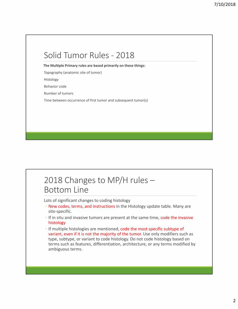
Solid Tumor Rules ‐ 2018The Multiple Primary rules are based primarily on these things:
Topography (anatomic site of tumor)
Histology
Behavior code
Number of tumors
Time between occurrence of first tumor and subsequent tumor(s)
2018 Changes to MP/H rules –Bottom LineLots of significant changes to coding histology◦ New codes, terms, and instructions in the Histology update table. Many are site‐specific.
◦ If in situ and invasive tumors are present at the same time, code the invasive histology
◦ If multiple histologies are mentioned, code the most specific subtype of variant, even if it is not the majority of the tumor. Use only modifiers such as type, subtype, or variant to code histology. Do not code histology based on terms such as features, differentiation, architecture, or any terms modified by ambiguous terms.
7/10/2018
3
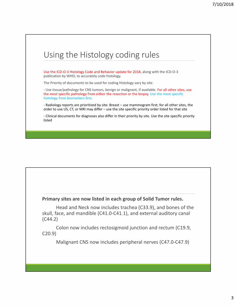
Using the Histology coding rules
Use the ICD‐O‐3 Histology Code and Behavior update for 2018, along with the ICD‐O‐3 publication by WHO, to accurately code histology.
The Priority of documents to be used for coding Histology vary by site:
‐ Use tissue/pathology for CNS tumors, benign or malignant, if available. For all other sites, use the most specific pathology from either the resection or the biopsy. Use the most specific histology from biomarkers first.
‐ Radiology reports are prioritized by site: Breast – use mammogram first; for all other sites, the order to use US, CT, or MRI may differ – use the site specific priority order listed for that site
‐ Clinical documents for diagnoses also differ in their priority by site. Use the site specific priority listed
Primary sites are now listed in each group of Solid Tumor rules.
Head and Neck now includes trachea (C33.9), and bones of the skull, face, and mandible (C41.0‐C41.1), and external auditory canal (C44.2)
Colon now includes rectosigmoid junction and rectum (C19.9, C20.9)
Malignant CNS now includes peripheral nerves (C47.0‐C47.9)

7/10/2018
4
Very few changes to the Multiple Primary Rules themselves:
◦ New instructions regarding ‘recurrence’ of colorectal tumors at the anastomotic site. Previously, all of these were considered new primaries.
◦ Glioblastoma multiforme is now considered a new primary, if it occurs after an astrocytoma or glial tumor. Previously this was NOT a new primary, but progression of disease.
◦ Added a new rule for meningiomas, to disregard laterality. Previously, they were being over‐counted.
Timing rule clarification◦ Time interval between subsequent tumor means after a disease free interval – if a recurrence occurs within the time interval, then the ‘clock starts over’
◦No other timing rules changed

7/10/2018
5
Solid Tumor RulesWhat we will cover:
◦ New Lung Multiple Primary and Histology coding rules
◦ New Urinary system MP/H rules
◦ New MP/H rules for non‐malignant CNS tumors
◦ New MP/H rules for malignant tumors of the CNS and Peripheral Nerves
Remember: The malignant and non‐malignant CNS tumors are currently in draft form and may change slightly in the final version!
Lung: 2018 Solid Tumor
Rules
7/10/2018
6
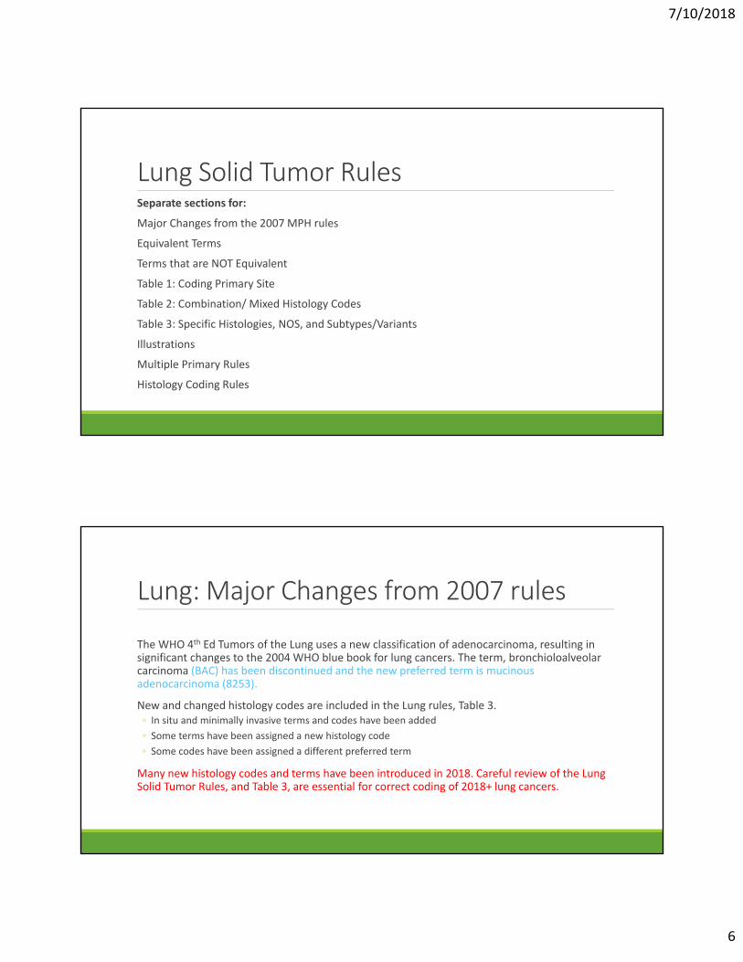
Lung Solid Tumor RulesSeparate sections for:
Major Changes from the 2007 MPH rules
Equivalent Terms
Terms that are NOT Equivalent
Table 1: Coding Primary Site
Table 2: Combination/ Mixed Histology Codes
Table 3: Specific Histologies, NOS, and Subtypes/Variants
Illustrations
Multiple Primary Rules
Histology Coding Rules
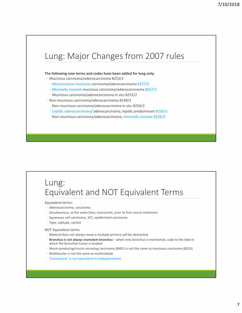
Lung: Major Changes from 2007 rules
The WHO 4th Ed Tumors of the Lung uses a new classification of adenocarcinoma, resulting in significant changes to the 2004 WHO blue book for lung cancers. The term, bronchioloalveolarcarcinoma (BAC) has been discontinued and the new preferred term is mucinous adenocarcinoma (8253).
New and changed histology codes are included in the Lung rules, Table 3.◦ In situ and minimally invasive terms and codes have been added
◦ Some terms have been assigned a new histology code
◦ Some codes have been assigned a different preferred term
Many new histology codes and terms have been introduced in 2018. Careful review of the Lung Solid Tumor Rules, and Table 3, are essential for correct coding of 2018+ lung cancers.
7/10/2018
7
Lung: Major Changes from 2007 rules
The following new terms and codes have been added for lung only:
◦ Mucinous carcinoma/adenocarcinoma 8253/3
◦ Microinvasive mucinous carcinoma/adenocarcinoma 8257/3
◦ Minimally invasive mucinous carcinoma/adenocarcinoma 8257/3
◦ Mucinous carcinoma/adenocarcinoma in situ 8253/2
◦ Non‐mucinous carcinoma/adenocarcinoma 8140/3
◦ Non‐mucinous carcinoma/adenocarcinoma in situ 8250/2
◦ Lepidic adenocarcinoma/ adenocarcinoma, lepidic predominant 8250/3
◦ Non‐mucinous carcinoma/adenocarcinoma, minimally invasive 8256/3
Lung:Equivalent and NOT Equivalent TermsEquivalent terms:◦ Adenocarcinoma, carcinoma
◦ Simultaneous, at the same time, concurrent, prior to first course treatment
◦ Squamous cell carcinoma, SCC, epidermoid carcinoma
◦ Type, subtype, variant
NOT Equivalent terms◦ Bilateral does not always mean a multiple primary will be abstracted
◦ Bronchus is not always mainstem bronchus – when only bronchus is mentioned, code to the lobe in which the bronchial tumor is located
◦ Mucin‐producing/mucin‐secreting carcinoma (8481) is not the same as mucinous carcinoma (8253)
◦ Multilocular is not the same as multinodular
◦ ‘Component’ is not equivalent to subtype/variant

7/10/2018
8
Lung: Coding Primary SiteTerminology Laterality Topography and code
Apex, Pancoast tumor, superior lobe or bronchus, upper lobe Either Upper lobe C34.1
Lung base, lower lobe, lower lobe bronchus Either Lower lobe C34.3
Bronchus, NOS, bronchogenic, pulmonary, Lung NOS Either Lung, NOS C34.9
Bronchus intermedius, Carina, Hilus, perihilar Either Mainstem bronchus, C34.0
Lingula of lung Left only Upper lobe C34.1
Lobar bronchus Either Use designated lobe or C34.9, if unknown
Middle lobe, Middle lobe bronchi Right only Middle lobe C34.2
Overlapping lesion of lung Either Overlapping lesion C34.8
Lung: Combination/Mixed HistologiesTable 2
Review the terms used in the diagnosis; if the terms match those listed in column 1, then use the combination code listed in column 2
Do NOT use Table 2 when:◦ Tumors are both invasive and in situ; Use the invasive histology code
◦ One of the histologies is described as ‘differentiation’ or ‘features’
◦ One of the histologies is an NOS term and another is a subtype/variant; Use the histology coding rules here instead of Table 2

7/10/2018
9
Lung: Combination/Mixed histologiesExamples from Table 2
Required ICD‐O Terms Combination Histology and Code
Adenocarcinoma, NOS ANDSquamous cell carcinoma, NOS(cannot be any subtypes or variants of adeno or SCC)
Adenosquamous carcinoma 8560
Giant cell carcinoma ANDSpindle cell carcinoma
Sarcomatoid carcinoma 8033
Epithelial carcinoma ANDMyoepithelial carcinoma
Epithelial‐myoepithelial carcinoma 8562
Lung: Histologies and Subtypes/VariantsExample from Table 3
Histology Term and Code(may be specific term or NOS term)
Synonyms for Histology Term
Subtypes/ variants and Histology code
Squamous cell carcinoma 8070 Epidermoid carcinomaSquamous carcinomaSquamous cell epithelioma
Basaloid carcinoma/basaloid squamous cell carcinoma 8083Keratinizing squamous cell carcinoma 8071Non‐keratinizing squamous cell carcinoma 8072

7/10/2018
10
Lung – MP RulesM1. Unknown if Single or Multiple Tumors – Abstract as Single Primary
M2. Single Tumor – A single tumor is always a Single Primary
Multiple Tumors –◦ M3. Multiple primary if separate tumors are present in topography codes that differ at the second or third character (example: C34.0 and C33.9)
◦ M4. Multiple primaries if diagnosed more than 3 years apart◦ NOTE: The time frame means clinically disease free for more than 3 years. If a patient has a recurrence within the 3 years, the ‘clock’ starts over, and the 3 year interval is computed from the date of last known recurrence. If recurrence is unknown, compute time from date of diagnosis.
◦ M5. Multiple primaries when there is at least one tumor that is small cell carcinoma, or a small cell variant, and another tumor that is non‐small cell carcinoma, or a non‐small cell carcinoma variant.
Lung – MP RulesMultiple Tumors – (cont.)◦ M6. Multiple primaries if separate tumors are different subtypes in column 3 of Table 3. The tumors may be subtypes of the same or different NOS histologies.
◦ M7. Single primary if multiple tumors are in the same row in Table 3, but they must be the same behavior.
◦ M8. Multiple primaries if separate tumors are described in different rows of Table 3, first or second column.
◦ M9. Single primary when there are simultaneously:
◦ A single tumor in one lung and multiple tumors in the other lung
◦ Multiple tumors in both lungs
◦ Multiple tumors in the same lung [Exception: this could be multiple primaries if pathology proves the tumors are of different histologies or the physician state unequivocally that the tumors are different primaries.]
7/10/2018
11
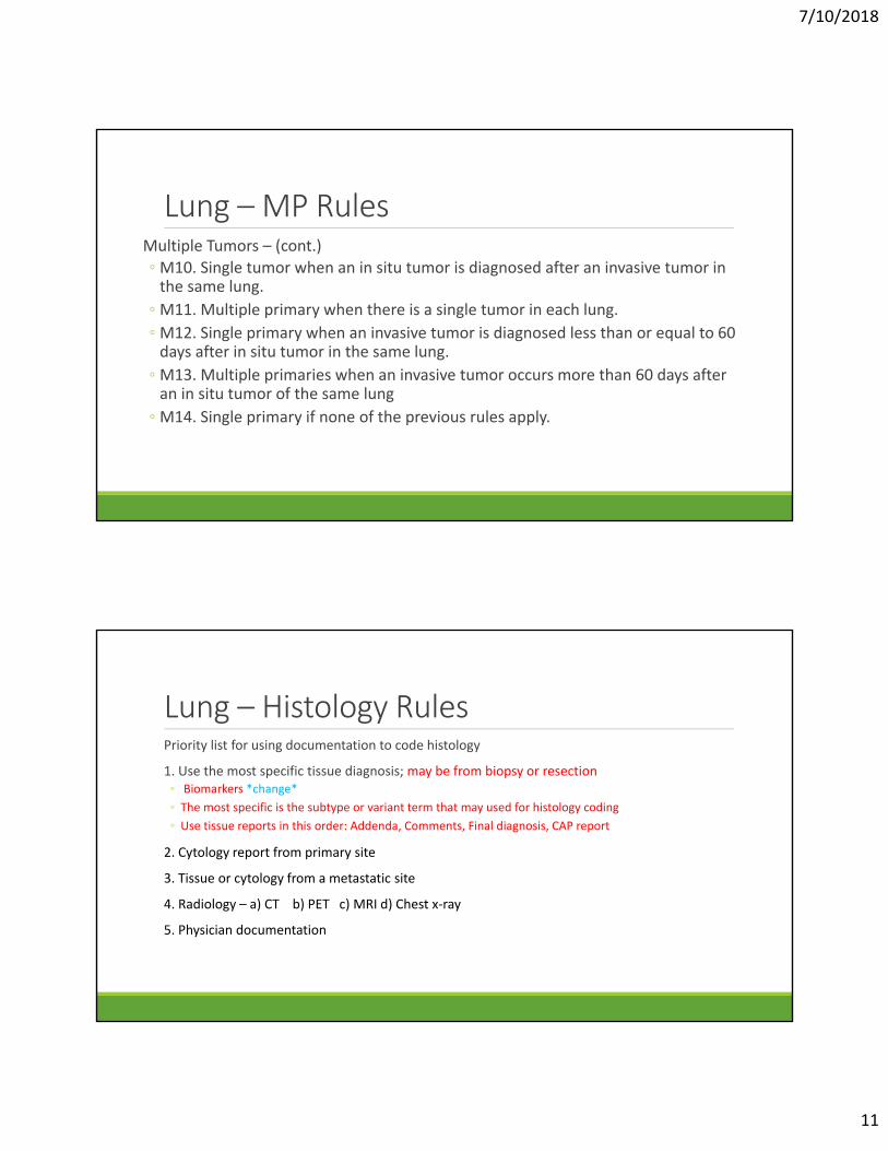
Lung – MP RulesMultiple Tumors – (cont.)◦M10. Single tumor when an in situ tumor is diagnosed after an invasive tumor in the same lung.
◦M11. Multiple primary when there is a single tumor in each lung.
◦M12. Single primary when an invasive tumor is diagnosed less than or equal to 60 days after in situ tumor in the same lung.
◦M13. Multiple primaries when an invasive tumor occurs more than 60 days after an in situ tumor of the same lung
◦M14. Single primary if none of the previous rules apply.
Lung – Histology RulesPriority list for using documentation to code histology
1. Use the most specific tissue diagnosis; may be from biopsy or resection ◦ Biomarkers *change*
◦ The most specific is the subtype or variant term that may used for histology coding
◦ Use tissue reports in this order: Addenda, Comments, Final diagnosis, CAP report
2. Cytology report from primary site
3. Tissue or cytology from a metastatic site
4. Radiology – a) CT b) PET c) MRI d) Chest x‐ray
5. Physician documentation

7/10/2018
12
Lung – Histology RulesTerminology to determine subtypes and variants:
DO USE:
Subtype
Type
Variant
DO NOT USE: NOS code, or specific histology or subtype/variant if described as:
Architecture
Differentiation
Features of
Foci, focus, focal
Majority – Use H instructions, most specific histology, even if not the majority of the tumor
Predominantly
Pattern
Any subtype or variant modified by an ambiguous term
Lung – Histology RulesSingle tumor –
H1. Code mucinous adenocarcinoma as follows: ◦ 8253/3 when behavior is invasive or unknown
◦ 8257/3 when microinvasive or minimally invasive
◦ 8253/2 when pre‐invasive or in situ
H2. Code non‐mucinous adenocarcinoma as follows:◦ 8250/3 when invasive, lepidic or behavior is unknown
◦ 8256/3 when microinvasive or minimally invasive
◦ 8250/2 when pre‐invasive or in situ

7/10/2018
13
Lung – Histology RulesSingle tumor (cont.) –
H3. Code the histology using Tables 3 when only 1 histologic type is identified
H4. Code the invasive histology when both invasive and in situ elements are present
H5. Code the subtype or variant when both a subtype and an NOS histology are identified
H6. Code the combination code when the combination is listed in Table 2
H7. Code adenocarcinoma with mixed subtypes (8255) ONLY when there is no combination code for the histologies within the tumor, or there are 2 or more subtypes of the same variant
Lung – Histology RulesMultiple tumors –
H8. Code mucinous adenocarcinoma as follows: ◦ 8253/3 when behavior is invasive or unknown
◦ 8257/3 when microinvasive or minimally invasive
◦ 8253/2 when pre‐invasive or in situ
H9. Code non‐mucinous adenocarcinoma as follows:◦ 8250/3 when invasive, lepidic or behavior is unknown
◦ 8256/3 when microinvasive or minimally invasive
◦ 8250/2 when pre‐invasive or in situ

7/10/2018
14
Lung – Histology RulesSingle tumor (cont.) –
H10. Code the histology using Tables 3 when only 1 histologic type is identified
H11. Code the invasive histology when both invasive and in situ elements are present
H12. Code the subtype or variant when both a subtype and an NOS histology are identified
H13. Code the combination code when the combination is listed in Table 2
H7. Code adenocarcinoma with mixed subtypes (8255) ONLY when there is no combination code for the histologies within the tumor, or there are 2 or more subtypes of the same variant
Urinary Sites: 2018 Solid Tumor
Rules

7/10/2018
15
Urinary Sites Solid Tumor RulesSeparate sections for:
Multifocal Tumors of Urinary Sites
Major Changes from 2007 MP/H rules
Equivalent Terms
Terms that are NOT Equivalent
Primary Site Coding for Urinary Organs and Urothelial Carcinoma
Table 1: Primary site/Topography codes
Table 2: Specific histologies, NOS Terms and Variants and Subtypes
Table 3. Non‐reportable urinary tumors
Illustrations
Multiple Primary Rules
Histology Coding Rules
Urinary Sites Solid Tumor RulesIntroduction◦ Multifocality in urothelial carcinomas is a common finding. The origin of the field effect concept has not been conclusively proven.
Major Changes from 2007 MP/H rules ‐ No significant new histology terms or codes for 2018
Priority for coding Primary Site, when multiple sites are involved◦ Non‐invasive or in situ urothelial carcinoma or subtypes ONLY
◦ Code Bladder, overlapping lesion (C67.8) when the bladder and one or both ureters ONLY are involved by a single tumor
◦ Code Bladder, NOS (C67.9) when multiple tumors are present in the bladder or both bladder and ureter(s)
◦ Code Urothelial system, NOS (C68.9) when multiple organs of the urinary system are involved
◦ Invasive urothelial carcinoma◦ Code Bladder, overlapping lesion (C67.8) when a single tumor overlaps sub‐sites of the bladder
◦ Code Bladder, NOS (C67.9) when multiple tumors are present in the bladder and the sub‐site is unknown
◦ Code Urothelial system, NOS (C68.9) when multiple organs of the urinary system are involved

7/10/2018
16
Urinary Sites Solid Tumor RulesTable 1 Lists the Topography codes with the preferred site description and synonyms
Table 2 Contains specific Histology and NOS terms, as well as Subtypes and Variants
Histology , Specific term or NOS term and Code
Synonyms for Histology Term Subtypes/ variants and Histology code
Urothelial carcinoma 8120
Clear cell urothelial carcinoma Infiltrating urothelial carcinoma with divergent differentiation Infiltrating urothelial carcinoma with glandular differentiation Infiltrating urothelial carcinoma with squamous differentiation Infiltrating urothelial carcinoma with trophoblastic differentiation Lipid‐rich urothelial carcinomaNested urothelial carcinomaPlasmacytoid urothelial carcinoma
Giant cell urothelial carcinoma 8031/3 Lymphoepithelioma‐like urothelial carcinoma 8082/3 Micropapillary urothelial carcinoma 8131/3 Papillary urothelial carcinomain situ 8130/2invasive 8130/3
Poorly differentiated carcinoma 8020/3 Sarcomatoid urothelial carcinoma 8122/3
Urinary Table 3. Neoplasms NOT ReportableHistology Synonyms
Benign perivascular epitheloid tumor 8714/0 Benign PEComa
Papillary urothelial neoplasm of low malignant potential 8130/1
Paraganglioma 8693/1 Extra‐adrenal pheochromocytoma
Urothelial dysplasia
Urothelial papilloma 8120/0

7/10/2018
17
Urinary – MP RulesM1. Unknown if Single or Multiple Tumors – Abstract as Single Primary
M2. Single Tumor – A single tumor is always a Single Primary
Multiple tumors
M3. Multiple primaries when there are tumors in both the left and the right renal pelvis and no other urinary sites are involved
M4. Multiple primaries when there are tumors in both the left and the right ureter and no other urinary sites are involved
M5. Multiple primaries when there are separate tumors with different subtypes or variants in Column 3 of Table 2
M6. Single primary when there are separate tumors are in the same row in Table 2
M7. Multiple primaries when there are multiple tumors with histology codes that are on different rows of Table 2
Urinary – MP RulesMultiple tumors – (cont.)
M8. Single primary when patient has a recurrence of in situ urothelial carcinoma
M9. Single primary when a non‐invasive urothelial tumors are diagnosed in the bladder and one or both ureters.
M10. Single primary when an in situ tumor is diagnosed after an invasive in the same urinary sites.
M11. Single tumor when an invasive tumor of the same histology occurs within 60 days of an in situ tumor in the same site
M12. Multiple primaries when an invasive tumor occurs more than 60 days after an in situ tumor of the same histology
M13. Single primary when there is an NOS histology and a subtype/variant of that histology

7/10/2018
18
Urinary – MP RulesM14. Single primary when a patient has multiple occurrences of invasive urothelial carcinoma of the bladder
M15. Multiple primaries if diagnosed more than 3 years apart◦ NOTE: The time frame means clinically disease free for more than 3 years. If a patient has a recurrence within the 3 years, the ‘clock’ starts over, and the 3 year interval is computed from the date of last known recurrence. If recurrence is unknown, compute time from date of diagnosis.
M16. Single primary when multifocal urothelial carcinomas are diagnosed simultaneously in two or more of these sites:◦ Renal pelvis◦ Ureter◦ Bladder◦ Urethra/prostatic urethra
M17. Abstract as a single primary when tumors do not meet any of the criteria above
Urinary– Histology RulesPriority list for using documentation to code histology
1. Use the most specific tissue diagnosis; may be from biopsy or resection ◦ Biomarkers
◦ The most specific is the subtype or variant term that may used for histology coding
◦ Use tissue reports in this order: Addenda, Comments, Final diagnosis, CAP report
2. Cytology report from primary site
3. Tissue or cytology from a metastatic site
4. Physician documentation
5. Radiology – CT, MRI

7/10/2018
19
Urinary – Histology RulesSingle tumor –
H1. Code the histology using Table 1 (or ICD‐O‐3) when only 1 histologic type is identified
H2. Code the invasive histology when in situ and invasive histologies occur in the same tumor
H3. Code the subtype/variant when an NOS and a subtype/variant of the NOS term is present
H4. Code mixed small cell carcinoma 8045 when a single tumor contains small cell neuroendocrine carcinoma, or one of its subtypes, and any other type of carcinoma
Urinary – Histology RulesSingle tumor –
H5. Code a mixture of urothelial carcinoma and another type of carcinoma as follows:◦ Adenocarcinoma and urothelial – code 8120 – adenocarcinoma is coded for a urinary site only when it is purely adenocarcinoma and not mixed with any other type
◦ Squamous cell carcinoma – code 8120 – Squamous cell carcinoma is coded for a urinary site only when it is purely squamous cell carcinoma and not mixed with any other type
◦ Clear cell (8310), Endometriod (8380), Sarcoma (8800), or any specific sarcoma –code to the non‐urothelial histology

7/10/2018
20
Urinary – Histology RulesMultiple tumors –
H6. Code the histology using Table 2 (or ICD‐O‐3) when only 1 histologic type is identified
H7. Code the invasive histology when there are invasive and in situ tumors
H8. Code the subtype/variant when an NOS and a single subtype/variant of that NOS is present
H9. Code mixed small cell carcinoma 8045 when a the diagnosis is small cell neuroendocrine carcinoma, or one of its subtypes, and any other type of carcinoma
Malignant CNS and Peripheral Nerves:
2018 Solid Tumor Rules

7/10/2018
21
Malignant CNS and Peripheral Nerves Solid Tumor RulesSeparate sections for:
Introduction
Changes from 2007 MP/H rules
Reportability Criteria
Section 1. Behavior Code includes◦ Instructions for identifying and assigning behavior code
◦ Tables 1: WHO Grades for select CNS neoplasms
Section 2. Reportable Primary Sites and Histologies◦ Reportable site terms
◦ Table 2: Reportable primary site terms and ICD‐O site codes
◦ Table 3: Reportable specific and NOS histologies, synonyms, subtypes/variants, and ICD‐O codes
◦ Table 4: Coding primary site for malignant tumors of cranial and peripheral nerves
Malignant CNS and Peripheral Nerves Solid Tumor Rules
Section 3. Additional information, including◦ Table 5: Paired sites
◦ Table 6: Non‐malignant CNS tumors with potential to transform to malignant behavior
Illustrations
Multiple Primary Rules
Histology Coding Rules

7/10/2018
22
Malignant CNS and Peripheral NervesIntroduction
Central nervous system (CNS) includes the following primary sites: Peripheral nerves, cerebral meninges; spinal meninges; meninges NOS; brain; spinal cord; cauda equina; olfactory nerve; optic nerve; acoustic nerve; cranial nerve NOS; overlapping lesion of brain and central nervous system; nervous system, nervous system NOS; pituitary gland, craniopharyngeal duct and pineal gland.
Pilocytic juvenile astrocytoma is reportable in the US as a malignant neoplasm 9421/3. It is reportable in Canada as non‐malignant 9421/1.
Juvenile pilocytic astrocytoma was malignant in ICD‐O‐1 and ICD‐O‐2 with the histology and behavior 9421/3. The behavior code was changed to borderline (9421/1) with ICD‐O‐3.
Intraosseous meningiomas and meningiomas of the cavernous sinus and sphenoid wing are reportable.
Malignant CNS and Peripheral NervesChanges from the 2007 MPH rules
2016 CNS WHO presents major restructuring of the diffuse gliomas, medulloblastomas and other embryonal tumors, and incorporates new entities that are defined by both histology and molecular features, including glioblastoma, IDH‐wildtype and glioblastoma, IDH‐mutant; diffuse midline glioma, H3 K27M‐mutant; RELA fusion‐positive ependymoma; medulloblastoma, WNT‐activated and medulloblastoma, SHH‐activated; and embryonal tumor with multilayered rosettes, C19MC‐altered.
Rule change: The 2007 rules said a glioblastoma multiforme (GBM) following an astrocytic or glial tumor was a single primary (recurrence).
• In the 2018 Solid Tumor rules, GBM subsequent to an astrocytic or glial tumor is a new (multiple) primary

7/10/2018
23
Malignant CNS and Peripheral NervesReportability criteria
Malignant CNS neoplasms must meet three criteria/conditions to be reportable:
1. The behavior must be reportable AND
a. Malignant, invasive, /3 OR
b. WHO Grade 3 or 4 (See Section 1, Table 1) ◦ Note 1: Always code the behavior as designated by the pathologist
◦ Note 2: Never report a malignant (/3) behavior code for a meningioma based on tumor extension to brain, skin of scalp, or other regional organs/tissue. Non‐malignant CNS tumors can extend to the regional tissue and bone.
2. The primary site must be reportable (See Section 2, Table 2) AND
3. The histology must be reportable (See Section 2, Table 3)
Malignant CNS and Peripheral Nerves
Section 1. Behavior code
Instructions for using source documentation to determine behavior are in priority order, with 1 having the highest priority.
Priority Order for Using Information to Assign Behavior
1. Pathology: Tissue from a resection ◦ A. Use the pathologist’s description of malignant behavior ◦ B. Cases are reportable as malignant when pathology states a WHO Grade 3 or 4 ◦ C. Never change behavior described by pathologist ◦ D. When there are discrepancies in behavior, use the documents in the following priority order: i. Addendum or comments on pathology report , ii. Final diagnosis, iii. CAP report
2. Pathology: Tissue from a biopsy
3. Cytology (usually cerebrospinal fluid)
4. Physician’s documentation
5. Scans: MRI, CT, PET, Angiogram
6. If none of the above are available, use Table 1. WHO Grade for select CNS neoplasms

7/10/2018
24
Malignant CNS: Table 1. WHO Grade 1. WHO does not provide Grades for all CNS and peripheral nerve neoplasms.
2. WHO Grade 3 and 4 neoplasms are always malignant
Table 1 contains histology terms and the WHO Grade based on molecular features, e.g.,
Histology WHO grade
Anaplastic meningioma 3
Anaplastic astrocytoma, IDH‐mutant 3
Angiocentric glioma 1
Atypical teratoid/rhabdoid tumor 4
Central neurocytoma 2
Choroid plexus carcinoma 3
Malignant CNS: Section 2. Reportable sites and HistologiesMajor groups of reportable sites are:
Intracranial (within the skull/cranium) ◦ Cerebral meninges (C70.0)
◦ Brain (C71.0‐C71.9)
◦ Cranial nerves (C72.0‐C72.9)
◦ Intracranial glands (craniopharyngeal duct C75.2, pineal gland C75.3, pituitary gland C75.1)
Spinal sites (spinal meninges and sites within the spinal meninges) ◦ Spinal meninges C70.1
◦ Spinal nerve roots C47.0, C47.3, C72.0, C72.1)
Peripheral nerves (extracranial and extraspinal nerves) (C47.0‐C47.9)

7/10/2018
25
Malignant CNS: Section 2. Reportable sites and HistologiesTable 2 displays each of the reportable topographies from the previous slide with their terms and codes.
Table 3 includes reportable specific and NOS histologies, synonyms, and subtypes/variants, e.g.,
Specific or NOS Histology Synonyms Subtypes/Variants
Anaplastic ganglioma 9505
Angiosarcoma 9120
Astroblastoma 9430
Astrocytoma, NOS 9400 Diffuse astrocytoma IDH –mutantDiffuse astrocytoma NOSDiffuse astrocytoma IDH –wild type
Anaplastic astrocytoma IDH‐mutant 9401Gemistocytic astrocytoma IDH‐mutant 9411Anaplastic astrocytoma IDH –wildtype 9401
Malignant CNS: Section 2. Reportable sites and HistologiesTable 4 displays each of the reportable topographies for cranial and peripheral nerves and their topography codes.
Name and CN# Exits cranium through Site code – cranial nerve Site code – peripheral nerve
Olfactory CN1 Cribriform plate Surface of the brain C72.2 Originates in mucosa of nasal cavity, travels to ethmoid bone C47.0
Optic CN2 Optic canal Intradural, all portions are reportable C72.3
Oculomotor CN3 Superior orbital fissure Originates in the midbrain C72.5
After exiting fissure, enters the orbit C47.0

7/10/2018
26
Malignant CNS: Section 3. Additional instructionsTable 5 identifies paired sites for which laterality must be coded.
Paired Sites and Codes
Acoustic nerve C724 Cerebral meninges C700
Cerebrum C710 Cranial nerves C725
Frontal lobe C711 Occipital lobe C714
Olfactory nerve C722 Optic nerve C723
Parietal lobe C713 Temporal lobe C712
Note: Midline tumors are limited to glioblastoma multiforme, meningiomas, lymphomas, and epidermoid cysts.
Table 6. Non‐malignant tumors that may transform to malignant (new primary)
Original histology Transformed histology
Chondroma 9220/0 Chondrosarcoma 9220/3
Ganglioglioma 9505/1 Anaplastic Ganglioglioma 9505/3
Hemangioma 9120/0 Angiosarcoma 9120/3
Hemangiopericytoma 9150/1 Anaplastic Hemangiopericytoma 9150/3
Leiomyoma 8890/0 Leiomyosarcoma 8890/3
Lipoma 8850/0 Liposarcoma 8850/3
Osteoma 9180/0 Osteosarcoma 9180/3
Perineurioma 9571/0 Malignant perineurioma 9571/3
Rhabdomyoma 8900/0 Rhabdomyosarcoma 8900/3
Teratoma 9080/1 Immature Teratoma 9080/3
Teratoma, mature 9080/0 Immature Teratoma 9080/3

7/10/2018
27
Malignant CNS – MP RulesM1. Unknown if Single or Multiple Tumors – Abstract as Single Primary
M2. Single Tumor – A single tumor is always a Single Primary
M3. Single primary when original diagnosis is oligodendroglioma and subsequently recurs with different features (path may state ‘looks like GBM’)
M4. Single primary when a single tumor is diagnosed clinically or radiographically or by stereotactic biopsy as non‐malignant and no resection is performed as first course treatment; then a resection is done when symptoms appear and the pathology from the resection is malignant. Change the behavior code on the original abstract to /3. There is no time limit for this rule. This is another example of a single tumor is always a single primary.
Malignant CNS – MP RulesMultiple tumors
M5. Multiple primaries when there is an invasive tumor and a non‐malignant tumor
M6. Multiple primaries when there are tumors in both ◦ Brain and any other part of the CNS
◦ Cerebral meninges and spinal meninges
◦ Cranial nerves and any other part of the CNS
◦ Meninges of cranial or peripheral nerves and any other part of the CNS
◦ Peripheral nerves and any other part of the CNS
M7. Multiple primaries when there are multiple tumors with histology codes that are on different rows of Table 3, first, second or third column
M8. Multiple primaries when there are separate tumors with different subtypes or variants in Column 3 of Table 3

7/10/2018
28
Malignant CNS – MP RulesMultiple Tumors – (cont.)
◦ M9. Single primary when there are separate tumors in
◦ Same lobe
◦ Different lateralities
◦ Different lobes
Multiple site involvement implies metastases or multicentric disease in these histologies: glioblastoma multiforme, gliomtosis, PNET medulloblastoma, ependymoma, oligodendrglioma, and hereditary syndromes (NF1, NF2)
◦ M10. Multiple primaries when there are multiple tumors with histology codes that differ at the first, second, or third digit
◦ M11. Single primary if none of the previous rules apply.
Malignant CNS– Histology RulesPriority list for using documentation to code histology
1. Use the pathology from a resection. Use tissue reports in this order: Addenda, Comments, Final diagnosis, CAP report
Use the pathology from a biopsy. Use tissue reports in this order: Addenda, Comments, Final diagnosis, CAP report
2. Cytology report from primary site
3. Tissue or cytology from a metastatic site
4. Radiology – MRI, CT, PET
5. Physician documentation
7/10/2018
29
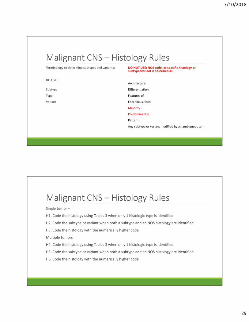
Malignant CNS – Histology RulesTerminology to determine subtypes and variants:
DO USE:
Subtype
Type
Variant
DO NOT USE: NOS code, or specific histology or subtype/variant if described as:
Architecture
Differentiation
Features of
Foci, focus, focal
Majority
Predominantly
Pattern
Any subtype or variant modified by an ambiguous term
Malignant CNS – Histology RulesSingle tumor –
H1. Code the histology using Tables 3 when only 1 histologic type is identified
H2. Code the subtype or variant when both a subtype and an NOS histology are identified
H3. Code the histology with the numerically higher code
Multiple tumors
H4. Code the histology using Tables 3 when only 1 histologic type is identified
H5. Code the subtype or variant when both a subtype and an NOS histology are identified
H6. Code the histology with the numerically higher code
7/10/2018
30
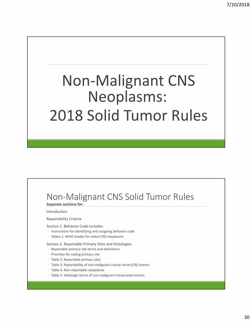
Non‐Malignant CNS Neoplasms:
2018 Solid Tumor Rules
Non‐Malignant CNS Solid Tumor RulesSeparate sections for:
Introduction
Reportability Criteria
Section 1. Behavior Code includes◦ Instructions for identifying and assigning behavior code
◦ Tables 1: WHO Grades for select CNS neoplasms
Section 2. Reportable Primary Sites and Histologies◦ Reportable primary site terms and definitions
◦ Priorities for coding primary site
◦ Table 2: Reportable primary sites
◦ Table 3: Reportability of non‐malignant cranial nerve (CN) tumors
◦ Table 4: Non‐reportable neoplasms
◦ Table 5: Histologic terms of non‐malignant intracranial tumors

7/10/2018
31
Non‐Malignant CNS Solid Tumor Rules
Section 3. Additional information, including◦ Conflicting information on path report
◦ Table 6: Specific histologies, NOS terms and synonyms, and subtypes/variants
◦ Table 7: Paired sites
◦ Table 8: Non‐malignant CNS tumors with potential to transform to malignant behavior
Illustrations
Multiple Primary Rules
Histology Coding Rules
Non‐Malignant CNS Section 1. Behavior code
Instructions for using source documentation to determine behavior are in priority order, with 1 having the highest priority.
Priority Order for Using Information to Assign Behavior
1. Pathology: Tissue from a biopsy or resection ◦ A. Use the tissue report with the highest grade (biopsy or resection)◦ B. Use the pathologist’s description of borderline or benign behavior ◦ C. Cases may be reportable when pathology states a WHO Grade 1◦ D. Never change behavior described by pathologist ◦ E. When there are discrepancies in behavior, use the documents in the following priority order: i. Addendum or comments on pathology report , ii. Final diagnosis, iii. CAP report
2. Cytology (usually cerebrospinal fluid)
3. Physician’s documentation
4. Scans: MRI, CT, PET, Angiogram
5. If none of the above are available, use Table 1. WHO Grade for select CNS neoplasms

7/10/2018
32
Non‐Malignant CNS: Table 1. WHO Grade 1. Use non‐malignant CNS rules for all WHO Grade 1 (always non‐malignant)
2. WHO Grade 3 and 4 neoplasms are always malignant; go to the malignant CNS rules
Table 1 contains histology terms and the WHO Grade based on molecular features, e.g.,
Histology WHO grade
Anaplastic meningioma 3
Anaplastic astrocytoma, IDH‐mutant 3
Angiocentric glioma 1
Atypical teratoid/rhabdoid tumor 4
Central neurocytoma 2
Choroid plexus carcinoma 3
Non‐Malignant CNS: Table 2. Reportable primary sites C700, C701, C709, C710‐C719, C720‐C725, C728, C729, C751‐C753
Note 1: Sphenoid wing meningiomas are reportable – code C70.0
Note 2: Cavernous sinus neoplasms are reportable; they arise in either the meninges or the cranial nerves (there is no site code for the cavernous sinus).
Major groups of reportable sites are:
Intracranial (within the skull/cranium) ◦ Cerebral meninges (C70.0)
◦ Brain (C71.0‐C71.9)
◦ Cranial nerves (C72.0‐C72.9)
◦ Intracranial glands (craniopharyngeal duct C75.2, pineal gland C75.3, pituitary gland C75.1)
Spinal sites (spinal meninges and sites within the spinal meninges) ◦ Spinal meninges (C70.1, C70.9)
◦ Spinal cord, Cauda equina (C72.0, C72.1)

7/10/2018
33
Non‐Malignant CNS: Table 2. Reportable primary sites
Olfactory nerve (C72.2)
Optic nerve (C72.3)
Acoustic nerve (C72.4)
Cranial nerves (C72.5)
Other parts of the CNS
Nervous system, NOS (C72.9)
Overlapping lesion of brain and CNS (C72.8)
Non‐Malignant CNS: Table 3: Reportable sites and HistologiesTable 3 displays each of the reportable topographies for non‐malignant cranial nerves
Name and CN# Exits cranium through Reportable portion –cranial nerve
Non‐reportable portion –cranial nerve
Olfactory CN1 Cribriform plate Surface of the brain C72.2 Originates in olfactory mucosa of nasal cavity, travels to ethmoid bone C47.0
Optic CN2 Optic canal Intradural, all portions are reportable C72.3
Oculomotor CN3 Superior orbital fissure Originates in the dorsalbrain stem C72.5
After exiting fissure, enters the orbit C47.0

7/10/2018
34
Non‐Malignant CNS: Table 4: Non‐reportable neoplasmsNon‐reportable histology Histology code Definition and sites
Carcinomas 8010‐8060, 8071‐8671, 8940‐8941 Brain C71.0‐C71.9Site/histology edit carcinoma/brain
Glomus tympanicum,Glomus jugulare
8690/1 Occur in inner ear, aortic body, or other paraganglia; non‐reportable sites
Hypothalamic hamartoma No code Occurs in hypothalamus
Neurofibromatosis, type 1 (NF1)
No code Genetic disease that produces non—malignant tumors in many sites. The brain and CNS tumors spawned by NF1 are reportable but the genetic disease is not.
Non‐Malignant CNS: Table 5. Histologic types 1. Use Table 5 to code primary site
2. Sites listed are the most common sites for that specific histology
Histology and code Most common site
Angiocentric glioma 9431/1 Cerebrum C71.0
Choroid plexus papilloma 9390/0 Intraventricular site (C71.3, C71.5, C71.7)
Craniopharyngioma 9350/1 Pituitary gland 75.1
Dermoid cyst 9084/0 Pineal gland C75.3 orsuprasellar C75.2
Pineocytoma 9361/1 Pineal gland C75.3
Subependymoma 9383/1 Lateral ventricle C71.5

7/10/2018
35
Non‐Malignant CNS: Table 6. Specific Histologies, Synonyms, Subtypes/VariantsTable 6 includes reportable specific and NOS histologies, synonyms, and subtypes/variants, e.g.,
Specific or NOS Histology Synonyms Subtypes/Variants
Angiocentric glioma 9431/1 Angiocentric neuroepithelialtumorMonomorphous angiocentricglioma
Central neurocytoma 9506/1 Cerebellar liponeuroneurocytomaExtraventricular neurocytoma
Chondroma 9220/0
Choroid plexus papilloma 9390/0 Atypical choroid plexus papilloma 9390/1
Non‐Malignant CNS: Table 7. Paired sitesTable 7 identifies paired sites for which laterality must be coded.
Paired Sites and Codes
Acoustic nerve C724 Cerebral meninges C700
Cerebrum C710 Cranial nerves C725
Frontal lobe C711 Occipital lobe C714
Olfactory nerve C722 Optic nerve C723
Parietal lobe C713 Temporal lobe C712
Note: Cerebellum C71.6 – laterality may be coded but is not required

7/10/2018
36
Table 8. Non‐malignant tumors that may transform to malignant (new primary)
Original histology Transformed histology
Chondroma 9220/0 Chondrosarcoma 9220/3
Ganglioglioma 9505/1 Anaplastic Ganglioglioma 9505/3
Hemangioma 9120/0 Angiosarcoma 9120/3
Hemangiopericytoma 9150/1 Anaplastic Hemangiopericytoma 9150/3
Leiomyoma 8890/0 Leiomyosarcoma 8890/3
Lipoma 8850/0 Liposarcoma 8850/3
Osteoma 9180/0 Osteosarcoma 9180/3
Perineurioma 9571/0 Malignant perineurioma 9571/3
Rhabdomyoma 8900/0 Rhabdomyosarcoma 8900/3
Teratoma 9080/1 Immature Teratoma 9080/3
Teratoma, mature 9080/0 Immature Teratoma 9080/3
Non‐Malignant CNS – MP RulesM1. Unknown if Single or Multiple Tumors – Abstract as Single Primary
M2. Single Tumor – A single tumor is always a Single Primary
M3. Single primary when a single tumor is diagnosed clinically or radiographically or by stereotactic biopsy as non‐malignant and no resection is performed as first course treatment. Then a resection is done when symptoms appear and the pathology from the resection is malignant. Change the behavior code on the original abstract to /3.
There is no time limit for this rule. This is another example of a single tumor is always a single primary.
M4. Single primary when a benign tumor transforms to a borderline (/1) tumor with the same histology as the original tumor, or a subtype of variant of that histology.

7/10/2018
37
Non‐Malignant CNS – MP RulesMultiple tumors
M5. Multiple primaries when a non‐malignant tumor transforms into a malignant tumor AND the patient had a resection, or it is unknown if a resection occurred.
M6. Single primary when the patient has bilateral acoustic neuromas, vestibular schwannomasor optic nerve gliomas.
M7. Multiple primaries when separate tumors are described in different rows of Table 6, first or second column.
M8. Multiple primaries when separate tumors are described as different subtypes in Column 3 of Table 6.
M9. Single primary when an NOS tumor and a subtype/variant of the NOS is in the same CNS site code (same second, third and fourth character). See Table 6.
Non‐Malignant CNS – MP RulesMultiple tumors (con’t.)
M10. Single primary when two or more separate meningiomas arise in the cranial meninges (laterality is irrelevant).
M11. Multiple primaries when there are tumors in both ◦ Brain and any other part of the CNS
◦ Cerebral meninges and spinal meninges
◦ Cranial nerves and any other part of the CNS
◦ Meninges of cranial or peripheral nerves and any other part of the CNS
M12. Single primary when there are multiple tumors in the brain
M13. Multiple primaries when there are multiple tumors with histology codes that are different at the first, second or third digit
M14. Single primary when tumors do not meet any of the above criteria

7/10/2018
38
Non‐Malignant CNS– Histology RulesPriority list for using documentation to code histology
1. Use the pathology from a resection. Use tissue reports in this order: Addenda, Comments, Final diagnosis, CAP report
Use the pathology from a biopsy. Use tissue reports in this order: Addenda, Comments, Final diagnosis, CAP report
2. Cytology report from primary site
3. Tissue or cytology from a metastatic site
4. Radiology – MRI, CT, PET
5. Physician documentation
Non‐Malignant CNS – Histology RulesTerminology to determine subtypes and variants:
DO USE:
Subtype
Type
Variant
DO NOT USE: NOS code, or specific histology or subtype/variant if described as:
Architecture
Differentiation
Features of
Foci, focus, focal
Majority
Predominantly
Pattern
Any subtype or variant modified by an ambiguous term

7/10/2018
39
Non‐Malignant CNS – Histology RulesSingle tumor –
H1. Code meningioma (9530/0) when the diagnosis is Meningioma, NOS, Metaplastic meningioma, Microcystic meningioma, Secretory meningioma, or Lymphoplasmacyte‐rich meningioma
H2. Code the reportable CNS tumor if the patient has a tumor along with NF1, NF2, or schwannomatosis (e.g., NF1: plexiform neurofibroma). Do not code the NF1 or NF2; only the tumors that result from the neurofibromatosis.
H3. Code the histology when only 1 histologic type is identified
H4. Code the subtype or variant when both a subtype and an NOS histology are identified
H5. Code the histology with the numerically higher code
Non‐Malignant CNS – Histology RulesMultiple tumors –
H6. Code meningioma (9530/1) when there are single or multiple Meningiomas of uncertain behavior
H7. Code meningioma (9530/0) when the diagnosis is Meningioma, NOS, Metaplastic meningioma, Microcystic meningioma, Secretory meningioma, or Lymphoplasmacyte‐rich meningioma
H8. Code the reportable CNS tumor if the patient has a tumor along with NF1, NF2, or schwannomatosis (e.g., NF1: plexiform neurofibroma). Do not code the NF1 or NF2; only the tumors that result from the neurofibromatosis.
H9. Code the histology when only 1 histologic type is identified
H10. Code the subtype or variant when both a subtype and an NOS histology are identified
H11. Code the histology with the numerically higher code