2015 hrs/ehra/aphrs/solaece expert …...2015 hrs/ehra/aphrs/solaece expert consensus statement on...
TRANSCRIPT
2015 HRS/EHRA/APHRS/SOLAECE expert consensusstatement on optimal implantablecardioverter-defibrillator programming and testingBruce L. Wilkoff, MD, FHRS, CCDS, (Chair),1 Laurent Fauchier, MD, PhD, (Co-Chair),2*Martin K. Stiles, MBCHB, PhD, (Co-Chair),3‡ Carlos A. Morillo, MD, FRCPC, FHRS, (Co-Chair),4††
Sana M. Al-Khatib, MD, MHSc, FHRS, CCDS,5 Jesús Almendral, MD, PhD, FESC,6*
Luis Aguinaga, MD, PhD, FACC, FESC,7†† Ronald D. Berger, MD, PhD, FHRS,8
Alejandro Cuesta, MD, PhD, FESC,9†† James P. Daubert, MD, FHRS,5
Sergio Dubner, MD, FACC,10†† Kenneth A. Ellenbogen, MD, FHRS,11
N.A. Mark Estes III, MD,12§ Guilherme Fenelon, MD, PhD,13†† Fermin C. Garcia, MD,14††
Maurizio Gasparini, MD,15* David E. Haines, MD, FHRS,16
Jeff S. Healey, MD, MSc, FRCPC, FHRS,4 Jodie L. Hurtwitz, MD,17† Roberto Keegan, MD,18††
Christof Kolb, MD,19* Karl-Heinz Kuck, MD, FHRS,20* Germanas Marinskis, MD, FESC,21*
Martino Martinelli, MD, PhD,22 Mark McGuire, MBBS, PhD,23‡ Luis G. Molina, MD, DSc,24††
Ken Okumura, MD, PhD,25‡ Alessandro Proclemer, MD,26* Andrea M. Russo, MD, FHRS,27
Jagmeet P. Singh, MD, DPhil, FHRS,28 Charles D. Swerdlow, MD, FHRS,29
Wee Siong Teo, MBBS, FHRS,30‡ William Uribe, MD, FHRS,31†† Sami Viskin, MD,32*
Chun-Chieh Wang, MD,33‡ Shu Zhang, MD34‡
Document Reviewers: Giuseppe Boriani, MD, PhD (Italy); Michele Brignole, MD, FESC (Italy);Alan Cheng, MD, FHRS (USA); Thomas C. Crawford, MD, FACC, FHRS (USA);Luigi Di Biase, MD, PhD, FACC, FHRS (USA); Kevin Donahue, MD (USA);Andrew E. Epstein, MD, FAHA, FACC, FHRS (USA); Michael E. Field, MD, FACC, FHRS (USA);Bulent Gorenek, MD, FACC, FESC (Turkey); Jin-Long Huang, MD, PhD (China);Julia H. Indik, MD, PhD, FACC, FAHA, FHRS (USA); Carsten W. Israel, MD (Germany);Mariell L. Jessup MD, FACC, FAHA, FESC (USA); Christophe Leclercq, MD, PhD (France);Robert J. MacFadyen, MD, PhD (UK); Christopher Madias, MD, FHRS (USA);Manlio F. Marquez, MD, FACC (Mexico); Brian Olshansky, MD, FACC, FAHA, FHRS (USA); KristenK. Patton, MD (USA); Marwan M. Refaat, MD, mMBA, FACC, FAHA, FHRS, FASE, FESC, FACP,FAAMA (USA); Cynthia M. Tracy, MD, FACC, FAHA (USA);Gaurav A. Upadhyay, MD (USA); Diego Vanegas, MD, FHRS (Colombia);Paul J. Wang, MD, FHRS, CCDS (USA)
From the 1Cleveland Clinic, Cleveland, Ohio, 2Centre Hospitalier Universitaire Trousseau, Tours, France,3Waikato Hospital, Hamilton, New Zealand, 4Department of Medicine, Cardiology Division, McMasterUniversity-Population Health Research Institute, Hamilton, Canada, 5Duke University Medical Center,Durham, North Carolina, 6Grupo HM Hospitales, Universidad CEU San Pablo, Madrid, Spain, 7CentroPrivado De Cardiologia, Tucuman, Argentina, 8Johns Hopkins University, Baltimore, Maryland, 9Servicio deArritmias, Instituto de Cardiologia Infantil, Montevideo, Uruguay, 10Clinica y Maternidad Suizo Argentina
*Representative of the European Heart Rhythm Association (EHRA)†Representative of the Sociedad Latinoamericana de Estimulacion Cardiaca y Electrofisiologia (SOLAECE)‡Representative of the Asia-Pacific Heart Rhythm Society (APHRS)††Representative of the American College of Cardiology (ACC)§Representative of the American Heart Association (AHA)
1547-5271/$-see front matter B 2016 © Heart Rhythm Society, European Heart Rhythm Association, aregistered branch of the European Society of Cardiology, the Asia Pacific Heart Rhythm Society,and the Sociedad Latinoamericana de Estimulación Cardíaca y Electrofisiología (SOLAECE) http://dx.doi.org/10.1016/j.hrthm.2015.11.018

e51Wilkoff et al Consensus Statement on Optimal ICD Programming and Testing
and De Los Arcos Sanatorio, Buenos Aires, Argentina, 11Virginia Commonwealth University Medical Center,Richmond, VA, 12New England Medical Center, Boston, Massachusetts, 13Federal University of São Paulo,São Paulo, Brazil, 14Hospital of the University of Pennsylvania, Philadelphia, Pennsylvania, 15HumanitasResearch Hospital, Milan, Italy, 16William Beaumont Hospital Division of Cardiology, Royal Oak, Michigan,17North Texas Heart Center, Dallas, Texas, 18Hospital Privado del Sur, Bahia Blanca, Argentina, 19DeutschesHerzzentrum Munchen, Munich, Germany, 20Allgemeines Krankenhaus St. Georg, Hamburg, Germany,21Vilnius University, Clinic of Cardiac and Vascular Diseases, Lithuania, 22Instituto do Coração,Universidade de São Paulo, São Paulo, Brazil, 23Royal Prince Alfred Hospital, Sydney, Australia, 24Mexico’sNational University, Mexico’s General Hospital, Mexico City, Mexico, 25Hirosaki University Graduate Schoolof Medicine, Hirosaki, Aomori, Japan, 26Azienda Ospedaliero Universitaria S. Maria della Misericordia–Udine, Udine, Italy, 27Cooper University Hospital, Camden, New Jersey, 28Massachusetts General Hospital,Harvard Medical School, Boston, Massachusetts, 29Cedars-Sinai Medical Center, Beverly Hills, California,30National Heart Centre Singapore, Singapore, Singapore, 31CES Cardiología and Centros EspecializadosSan Vicente Fundación, Medellín y Rionegro, Colombia, 32Tel Aviv Sourasky Medical Center and SacklerSchool of Medicine, Tel Aviv University, Tel Aviv, Israel, 33Chang Gung Memorial Hospital, Taipei, Taiwan,and 34National Center for Cardiovascular Disease and Beijing Fu Wai Hospital, Peking Union MedicalCollege and China Academy of Medical Sciences, Beijing, China.
TABLE OF CONTENTS
Introduction ............................................ e52
KEYWOrate; TPrograABBREAF =intervaCRT-DtestingfailureLV= leinfarctCardiovOR = oventricSCD=sdefibriattack;Rhythm
DeRhythm(APHRElectroand El(ACC)Society(CASSSenertDC 20
Bradycardia Mode and Rate
RDachmmVIAatril;= ca; E; Hft vionascddulauddllatoVF20
veloA
S),fisiectr, A(C
A).h, H005
Programming .......................................
e52Programming of Rate Modulation .......... e53 Sinus Node Disease ................................. e53S Implantable cardioverter-defibrillator; Bradycardia mode andycardia detection; Tachycardia therapy; Defibrillation testing;ingTIONS aCRT = adaptive cardiac resynchronization therapy;al fibrillation; ATP = antitachycardia pacing; CI = confidenceCL = cycle length; CRT = cardiac resynchronization therapy;rdiac resynchronization therapy–defibrillator; DT= defibrillationEG = electroencephalography; EGM = electrogram; HF = heartR = hazard ratio; ICD = implantable cardioverter-defibrillator;entricle; LVEF= left ventricular ejection fraction; MI=myocardial; MVP = managed ventricular pacing; NCDR = Nationalular Data Registry; NYHA = New York Heart Association;s ratio; PEA = peak endocardial acceleration; PVC = prematurer contraction; RCT=randomized clinical trial; RV=right ventricle;en cardiac death; S-ICD=subcutaneous implantable cardioverter-r; SVT = supraventricular tachycardia; TIA = transient ischemic= ventricular fibrillation; VT = ventricular tachycardia (Heart16;13:e50–e86)
ped in partnership with and endorsed by the European Heartssociation (EHRA), the Asia Pacific Heart Rhythm Societyand the Sociedad Latinoamericana de Estimulacion Cardiaca yologia (SOLAECE)-Latin American Society of Cardiac Pacingophysiology. Endorsed by the American College of Cardiologymerican Heart Association (AHA), Canadian Heart RhythmHRS), and Cardiac Arrhythmia Society of Southern AfricaAddress reprint requests and correspondence: Ms. Emilyeart Rhythm Society, 1325 G Street NW, Suite 400, Washington,. E-mail address: [email protected].
Atrial Fibrillation and AtrioventricularBlock .......................................................
e54Intact Atrioventricular Conduction .... e54
Non-CRT Devices: Algorithms to Reduce Right Ventricular Stimulation ................. e54Cardiac Resynchronization Therapy: Consistent Delivery of VentricularPacing ............................................................... e56Tachycardia Detection
Programming ........................................... e57Duration Criteria for the Detection of
Ventricular Arrhythmia ........................... e57Limitations of Data on the Duration of Tachycardia Required for Detection ....... e59Rate Criteria for the Detection of Ventricular Arrhythmia ........................... e59Single- or Multi-Zone Detection ............. e60 Discrimination Between Supraventricular and Ventricular Arrhythmia .................... e61SVT-VT Discriminator
Components ............................................ e61Rejection of Sinus Tachycardia by Onset ................................................... e61SVT-VT Discrimination Algorithms ............................................... e62Assessing Clinical Benefits and Risks .... e62 Additional Considerations .................. e62Programming to Reduce T-Wave
Oversensing ............................................. e63Lead-Related Oversensing ....................... e63 The Subcutaneous Defibrillator (S-ICD) e64 Integrating Tachycardia Detection Data Into ProgrammingRecommendations ................................... e64Tachycardia Therapy Programming .... e66

Heart Rhythm, Vol 13, No 2, February 2016e52
Benefits and Risks ................................... e66
Classification of Therapy ........................ e66Appropriate ......................................... e67
Inappropriate ...................................... e67 Avoidable ............................................ e67 Phantom .............................................. e67 Unintended Consequences of ICD Therapy and ICD TherapyProgramming ....................................... e67Intraprocedural Testing of Defibrillation Efficacy ......................... e68Periprocedural Mortality ......................... e71
DT-Related Complications ..................... e71 Contraindications to Defibrillation Threshold Testing .................................... e73S-ICD ...................................................... e73 Conclusion ............................................... e73Appendix Disclosures ......................................... e73
IntroductionImplantable cardioverter-defibrillator (ICD) therapy isclearly an effective therapy for selected patients in definablepopulations. The benefits and risks of ICD therapy aredirectly impacted by programming and surgical decisions.This flexibility is both a great strength and a weakness, forwhich there has been no prior official discussion or guidance.It is the consensus of the 4 continental electrophysiologysocieties that there are 4 important clinical issues for whichthere are sufficient ICD clinical and trial data to provideevidence-based expert guidance. This document systemati-cally describes the greater than 80% (83%–100%, mean96%) required consensus achieved for each recommendationby official balloting in regard to the programming of (1)bradycardia mode and rate, (2) tachycardia detection, (3)tachycardia therapy, and (4) the intraprocedural testing ofdefibrillation efficacy. Representatives nominated by theHeart Rhythm Society (HRS), European Heart RhythmAssociation (EHRA), Asian Pacific Heart Rhythm Society(APHRS), and the Sociedad Latinoamericana de Estimula-cion Cardiaca y Electrofisiologia (SOLAECE-Latin Amer-ican Society of Cardiac Pacing and Electrophysiology)participated in the project definition, the literature review,the recommendation development, the writing of the docu-ment, and its approval. The 32 recommendations wereballoted by the 35 writing committee members and wereapproved by an average of 96%.
The classification of the recommendations and the level ofevidence follow the recently updated ACC/AHA standard.1,2
Class I is a strong recommendation, denoting a benefitgreatly exceeding risk. Class IIa is a somewhat weakerrecommendation, with a benefit probably exceeding risk, andClass IIb denotes a benefit equivalent to or possibly exceed-ing risk. Class III is a recommendation against a specifictreatment because either there is no net benefit or there is net
harm. Level of Evidence A denotes the highest level ofevidence from more than 1 high-quality randomized clinicaltrial (RCT), a meta-analysis of high-quality RCTs, or RCTscorroborated by high-quality registry studies. Level ofevidence B indicates moderate-quality evidence from eitherRCTs with a meta-analysis (B-R) or well-executed non-randomized trials with a meta-analysis (B-NR). Level ofevidence C indicates randomized or nonrandomized obser-vational or registry studies with limited data (C-LD) or fromexpert opinions (C-EO) based on clinical experience in theabsence of credible published evidence. These recommen-dations were also subject to a 1-month public commentperiod. Each society then officially reviewed, commented,edited, and endorsed the final document and recommenda-tions. All author and peer reviewer disclosure information isprovided in Appendix A.
The care of individual patients must be provided incontext of their specific clinical condition and the dataavailable on that patient. Although the recommendations inthis document provide guidance for a strategic approach toICD programming, as an individual patient’s conditionchanges or progresses and additional clinical considerationsbecome apparent, the programming of their ICDs mustreflect those changes. Remote and in-person interrogationsof the ICD and clinical monitoring must continue to informthe programming choices made for each patient. Therecommendations in this document specifically target adultpatients and might not be applicable to pediatric patients,particularly when programming rate criteria.
Please consider that each ICD has specific programmableoptions that might not be specifically addressed by the 32distinctive recommendations in this document. Appendix B,published online (http://www.hrsonline.org/appendix-b),contains the writing committee’s translations specific to eachmanufacturer and is intended to best approximate therecommended behaviors for each available ICD model.
Bradycardia Mode and Rate ProgrammingSingle- or Dual-Chamber Pacing ModeEvidence. Because the ICD is primarily indicated fortachycardia therapy, there might be some uncertainty regard-ing optimal bradycardia management for ICD patients. Datafrom clinical studies adequately address only the pro-grammed mode rather than the number of leads implanted,the number of chambers stimulated, or how frequently thepatients required bradycardia support. It is of note that mostinformation on pacing modes has been collected frompacemaker patients, and these patients are clinically distinctfrom ICD recipients. Dual-chamber pacing (atrial andventricular) has been compared with single-chamber pacing(atrial or ventricular) in patients with bradycardia in 5multicenter, parallel, randomized trials, in 1 meta-analysisof randomized trials, and in 1 systematic review that alsoincluded 30 randomized crossover comparisons and 4economic analyses.3–9 Meta analyses comparing dual-chamber to single-chamber ICDs did not evaluatepacing modes.10,11 Compared with single-chamber pacing,

e53Wilkoff et al Consensus Statement on Optimal ICD Programming and Testing
dual-chamber pacing results in small but potentially signifi-cant benefits in patients with sinus node disease and/oratrioventricular block. No difference in mortality has beenobserved between ventricular pacing modes and dual-chamber pacing modes. Dual-chamber pacing was associ-ated with a lower rate of atrial fibrillation (AF) and stroke.12
The benefit in terms of AF prevention was more marked intrials comprised of patients with sinus node disease.Although trends in favor of dual-chamber pacing have beenobserved in some trials, there was no benefit in terms of heartfailure (HF). In patients without symptomatic bradycardia,however, the Dual Chamber and VVI Implantable Defib-rillator (DAVID) trial in ICD recipients showed that onespecific choice of dual-chamber rate-responsive (DDDR)programming parameters led to poorer outcomes than VVIbackup pacing, most likely secondary to unnecessary rightventricular (RV) pacing. The fact that RV stimulation wasresponsible was reinforced in the DAVID II trial, in whichAAI pacing was demonstrated to be noninferior to VVIbackup pacing.13
Approximately a quarter of patients with either sinus nodedisease or atrioventricular block develop “pacemaker syn-drome” with VVI pacing usually associated with retrograde(ventricular to atrial) conduction, which in turn is associatedwith a reduction in the quality of life.14 In crossover trials,symptoms of pacemaker syndrome (dyspnea, dizziness,palpitations, pulsations, and chest pain) were reduced byreprogramming to a dual-chamber mode.14 Dual-chamberpacing is associated with better exercise performance com-pared with single-chamber VVI pacing without rate adapta-tion, but produces similar exercise performance whencompared with rate-responsive VVIR pacing. Because ofthe additional lead, dual-chamber devices involve longerimplantation times, have a higher risk of complications, andare more expensive. However, because of the additionalclinical consequences of pacemaker syndrome and AF (andits sequelae), the overall cost difference between single- anddual-pacing systems is moderated.In patients with persistent sinus bradycardia, atrial rather
than ventricular dual-chamber pacing is the pacing mode ofchoice. There is evidence for superiority of atrial-basedpacing over ventricular pacing for patients who requirepacing for a significant proportion of the day. The evidenceis stronger for patients with sinus node disease, in whomdual-chamber pacing confers a modest reduction in AF andstroke, but not in hospitalization for HF or death comparedwith ventricular pacing. In patients with acquired atrioven-tricular block, large randomized parallel trials were unable todemonstrate the superiority of dual-chamber pacing overventricular pacing with regard to hard clinical endpoints ofmortality and morbidity.4,6–8 The benefit of dual-chamberover ventricular pacing is primarily due to the avoidance ofpacemaker syndrome and to improved exercise capacity.14
Even if it is a softer endpoint, pacemaker syndrome isassociated with a reduction in quality of life that justifies thepreference for dual-chamber pacing when reasonable; thus,there is strong evidence for the superiority of dual-chamber
pacing over ventricular pacing that is limited to symptomimprovement. Conversely, there is strong evidence of non-superiority with regard to survival and morbidity. The netresult is that the indications for programming the dual-chamber modes are weaker and the choice regarding thepacing mode should be individualized, taking into consid-eration the increased complication risk and costs of dual-chamber devices. Because ICD patients usually do notrequire bradycardia support, with the exception of patientswho require cardiac resynchronization, programmingchoices should avoid pacing and in particular avoid singleventricular pacing, if possible.15,16
Programming of Rate ModulationThe benefit of rate response programming has been eval-uated in patients with bradycardia in 5 multicenter, random-ized trials and in 1 systematic review that also included 7single-center studies.17–22 Most of these data were obtainedfrom pacemaker studies and must be interpreted in that light.
Although there is evidence of the superiority of VVIRpacing compared with VVI pacing in improving quality oflife and exercise capacity, improvements in exercise capacitywith DDDR compared with DDD have been inconsistent. In2 small studies on patients with chronotropic incompetencecomparing DDD and DDDR pacing, the latter had improvedquality of life and exercise capacity; however, a larger,multicenter randomized trial (Advanced Elements of PacingRandomized Controlled Trial [ADEPT]) failed to show adifference in patients with a modest blunted heart rateresponse to exercise.17–19 In addition, DDDR programmingin cardiac resynchronization therapy (CRT) patients has thepotential to impair AV synchrony and timing. It should benoted that trials evaluating CRT generally did not use rate-responsive pacing, and many in fact avoided atrial stimula-tion using atrial sensed and ventricular paced pacing modeswith a lower base rate. However, the Pacing Evaluation-Atrial Support Study in Cardiac Resynchronization Therapy(PEGASUS CRT) trial is the exception and did not demon-strate adverse impact on mortality and HF events.23
Sinus Node DiseaseIn patients with persistent or intermittent sinus node dys-function or chronotropic incompetence, the first choice isDDDR with algorithms responding to intermittent atrioven-tricular conduction. There is sufficient evidence for thesuperiority of VVIR compared with VVI in improvingquality of life and exercise capacity. The evidence is muchweaker in dual-chamber pacing (DDDR vs DDD).
Although only an issue when there is some concomitantAV block, the upper rate limit should be programmed higherthan the fastest spontaneous sinus rhythm to avoid upper ratelimit behavior. To avoid symptomatic bradycardia, the lowerrate should be programmed on an individual basis, accordingto the clinical characteristics and the underlying cardiacsubstrate of the patient.

Heart Rhythm, Vol 13, No 2, February 2016e54
Atrial Fibrillation and Atrioventricular BlockPatients with permanent AF and either spontaneous or AVjunctional ablation-induced high-degree atrioventricularblock have little to no chronotropic response to exercise;thus, VVIR pacing is associated with better exercise per-formance, improved daily activities, improved quality of life,and decreased symptoms of shortness of breath, chest pain,and heart palpitations, compared with VVI.20–22,24–26 There-fore, rate-adaptive pacing is the first choice of pacing mode;fixed-rate VVI pacing should be abandoned in patients withpermanent AF and atrioventricular block. It is the experts’opinion that the minimum rate can be programmed higher(e.g., 70 bpm) than for sinus rhythm patients, in an attempt tocompensate for the loss of active atrial filling. In addition, themaximum sensor rate should be programmed restrictively(e.g., 110–120 bpm) to avoid “overpacing” (i.e., pacing witha heart rate faster than necessary), which can be sympto-matic, particularly in patients with coronary artery disease. Ina small study, however, it was found that rate-responsivepacing could be safe and effective in patients with anginapectoris, without an increase in subjective or objective signsof ischemia.25 The lower rate should be programmed on anindividual basis, according to the clinical characteristics andthe underlying cardiac substrate of the patient. The clinicalbenefit of programming a lower resting rate at night based oninternal clocks has not been evaluated in ICD patients. Thereis some concern that atrioventricular junction ablation andpermanent ventricular pacing might predispose the patient toan increased risk of sudden cardiac death (SCD) related to abradycardia-dependent prolongation of the QT interval. Thisrisk might be overcome by setting the ventricular pacing rateto a minimum of 80 or 90 bpm for the first 1–2 monthsfollowing the atrioventricular junction ablation, then reduc-ing it to a conventional 60–70 bpm.27,28 Not all patients withAF and milder forms of atrioventricular block will require ahigh percentage of ventricular pacing or have a wide QRS.Physicians should consider the risk of increasing preexistingleft ventricular (LV) dysfunction with RV pacing vsimproved chronotropic responsiveness and the potentialvalue of CRT.
Intact Atrioventricular ConductionRight Ventricular PacingThe results of a number of large-scale, prospective random-ized trials demonstrated a significant reduction in AF inpacemaker patients with atrial-based pacing (AAI or DDD)compared with patients with ventricular-based pacing.4,8,29
In the Mode Selection Trial, which enrolled 2010 patientswith sick sinus syndrome, the risk of AF increased linearlywith the increasing percentage of RV pacing.30 At the sametime, deleterious effects of RV pacing in patients with LVdysfunction (left ventricular ejection fraction [LVEF]r40%) implanted with dual-chamber ICD systems wereobserved in the Dual Chamber and VVI ImplantableDefibrillator (DAVID) trial, which included 506 ICDpatients without indications for bradycardia pacing. Patients
within the DDDR-70 group (with paced and sensed atrio-ventricular delays of 170 and 150 ms, respectively, in mostof the DDDR group patients) showed a trend toward highermortality and an increased incidence of HF compared withthe patients programmed to ventricular backup pacing—theVVI-40 group. Within the DDDR-70 group, there were morecardiac events when the percentage of ventricular pacingexceeded 40% (P¼ .09) compared with patients witho40%of RV pacing, although almost all the patients had 495%RV stimulation (DDDR-70) or o5% RV stimulation (VVI-40).31,32 However, a more detailed post hoc analysis of theInhibition of Unnecessary RV Pacing With Atrial-Ventricular Search Hysteresis in ICDs (INTRINSIC RV)trial revealed that the most favorable clinical results were notin the VVI groups with the least percentage of RV pacing butin the subgroup that had DDD pacing with longer atrioven-tricular delays and 11%–19% of ventricular pacing. Thisparameter selection probably helped patients to avoidexceedingly low heart rates while preserving intrinsicatrioventricular conduction most of the time.31,33 In theSecond Multicenter Automated Defibrillator ImplantationTrial (MADIT II), a higher risk of HF was observed inpatients who had a greater than 50% burden of RV pacing.34
In another large observational study of 456 ICD patientswithout HF at baseline, a high RV pacing burden (RV pacingmore than 50% of the time) was associated with an increasedrisk of HF events and appropriate ICD shocks.35 Optimally,RV stimulation should be avoided, but the precise tradeoffbetween the percentage of ventricular pacing and atrioven-tricular timing is unclear in non-CRT patients.
Non-CRT Devices: Algorithms to Reduce RightVentricular StimulationThe importance of reducing or avoiding RV pacing in ICDpatients with LV dysfunction was illustrated in the DAVIDtrial.31 The feasibility of algorithms designed to decrease theburden of unnecessary ventricular pacing has been demon-strated in patients with dual-chamber pacemakers.36–38 Thesealgorithms usually provide functional AAI pacing withmonitoring of atrioventricular conduction and an automaticmode switch from AAI to DDD during episodes of atrioven-tricular block. Some studies directly compared variousalgorithms to decrease ventricular pacing, showing that a“managed ventricular pacing” (MVP) algorithm resulted ingreater ventricular pacing reduction than an “atrioventricularsearch” algorithm39,40: however, no randomized studiescomparing these two algorithms with respect to importantcardiovascular endpoints (e.g., HF, cardiac death) have beenperformed. The results of the studies on these pacingalgorithms are summarized in Table 1.
Unnecessary RV pacing should be minimized by usingspecific algorithms or programming longer atrioventriculardelays, and this process is more important for patients with ahigher risk of AF or who already have poorer LV function.49
Patients with longer baseline PR intervals have a higher riskof AF regardless of the percentage of ventricular pacing or
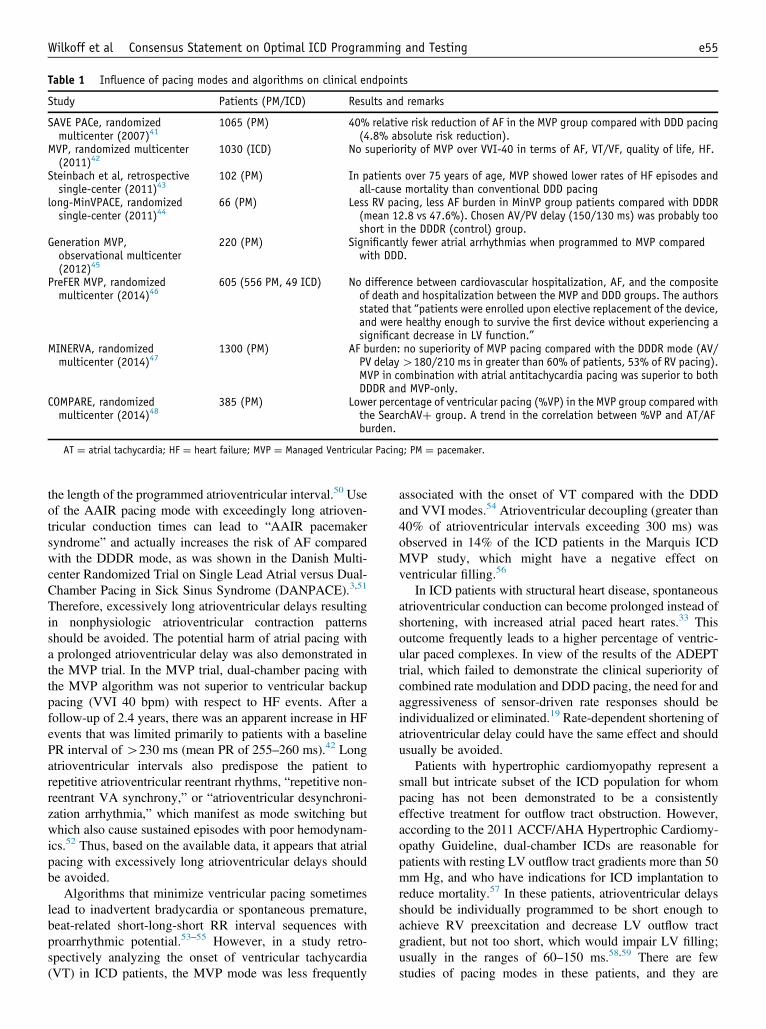
Table 1 Influence of pacing modes and algorithms on clinical endpoints
Study Patients (PM/ICD) Results and remarks
SAVE PACe, randomizedmulticenter (2007)41
1065 (PM) 40% relative risk reduction of AF in the MVP group compared with DDD pacing(4.8% absolute risk reduction).
MVP, randomized multicenter(2011)42
1030 (ICD) No superiority of MVP over VVI-40 in terms of AF, VT/VF, quality of life, HF.
Steinbach et al, retrospectivesingle-center (2011)43
102 (PM) In patients over 75 years of age, MVP showed lower rates of HF episodes andall-cause mortality than conventional DDD pacing
long-MinVPACE, randomizedsingle-center (2011)44
66 (PM) Less RV pacing, less AF burden in MinVP group patients compared with DDDR(mean 12.8 vs 47.6%). Chosen AV/PV delay (150/130 ms) was probably tooshort in the DDDR (control) group.
Generation MVP,observational multicenter(2012)45
220 (PM) Significantly fewer atrial arrhythmias when programmed to MVP comparedwith DDD.
PreFER MVP, randomizedmulticenter (2014)46
605 (556 PM, 49 ICD) No difference between cardiovascular hospitalization, AF, and the compositeof death and hospitalization between the MVP and DDD groups. The authorsstated that “patients were enrolled upon elective replacement of the device,and were healthy enough to survive the first device without experiencing asignificant decrease in LV function.”
MINERVA, randomizedmulticenter (2014)47
1300 (PM) AF burden: no superiority of MVP pacing compared with the DDDR mode (AV/PV delay4180/210 ms in greater than 60% of patients, 53% of RV pacing).MVP in combination with atrial antitachycardia pacing was superior to bothDDDR and MVP-only.
COMPARE, randomizedmulticenter (2014)48
385 (PM) Lower percentage of ventricular pacing (%VP) in the MVP group compared withthe SearchAVþ group. A trend in the correlation between %VP and AT/AFburden.
AT ¼ atrial tachycardia; HF ¼ heart failure; MVP ¼ Managed Ventricular Pacing; PM ¼ pacemaker.
e55Wilkoff et al Consensus Statement on Optimal ICD Programming and Testing
the length of the programmed atrioventricular interval.50 Useof the AAIR pacing mode with exceedingly long atrioven-tricular conduction times can lead to “AAIR pacemakersyndrome” and actually increases the risk of AF comparedwith the DDDR mode, as was shown in the Danish Multi-center Randomized Trial on Single Lead Atrial versus Dual-Chamber Pacing in Sick Sinus Syndrome (DANPACE).3,51
Therefore, excessively long atrioventricular delays resultingin nonphysiologic atrioventricular contraction patternsshould be avoided. The potential harm of atrial pacing witha prolonged atrioventricular delay was also demonstrated inthe MVP trial. In the MVP trial, dual-chamber pacing withthe MVP algorithm was not superior to ventricular backuppacing (VVI 40 bpm) with respect to HF events. After afollow-up of 2.4 years, there was an apparent increase in HFevents that was limited primarily to patients with a baselinePR interval of 4230 ms (mean PR of 255–260 ms).42 Longatrioventricular intervals also predispose the patient torepetitive atrioventricular reentrant rhythms, “repetitive non-reentrant VA synchrony,” or “atrioventricular desynchroni-zation arrhythmia,” which manifest as mode switching butwhich also cause sustained episodes with poor hemodynam-ics.52 Thus, based on the available data, it appears that atrialpacing with excessively long atrioventricular delays shouldbe avoided.
Algorithms that minimize ventricular pacing sometimeslead to inadvertent bradycardia or spontaneous premature,beat-related short-long-short RR interval sequences withproarrhythmic potential.53–55 However, in a study retro-spectively analyzing the onset of ventricular tachycardia(VT) in ICD patients, the MVP mode was less frequently
associated with the onset of VT compared with the DDDand VVI modes.54 Atrioventricular decoupling (greater than40% of atrioventricular intervals exceeding 300 ms) wasobserved in 14% of the ICD patients in the Marquis ICDMVP study, which might have a negative effect onventricular filling.56
In ICD patients with structural heart disease, spontaneousatrioventricular conduction can become prolonged instead ofshortening, with increased atrial paced heart rates.33 Thisoutcome frequently leads to a higher percentage of ventric-ular paced complexes. In view of the results of the ADEPTtrial, which failed to demonstrate the clinical superiority ofcombined rate modulation and DDD pacing, the need for andaggressiveness of sensor-driven rate responses should beindividualized or eliminated.19 Rate-dependent shortening ofatrioventricular delay could have the same effect and shouldusually be avoided.
Patients with hypertrophic cardiomyopathy represent asmall but intricate subset of the ICD population for whompacing has not been demonstrated to be a consistentlyeffective treatment for outflow tract obstruction. However,according to the 2011 ACCF/AHA Hypertrophic Cardiomy-opathy Guideline, dual-chamber ICDs are reasonable forpatients with resting LV outflow tract gradients more than 50mm Hg, and who have indications for ICD implantation toreduce mortality.57 In these patients, atrioventricular delaysshould be individually programmed to be short enough toachieve RV preexcitation and decrease LV outflow tractgradient, but not too short, which would impair LV filling;usually in the ranges of 60–150 ms.58,59 There are fewstudies of pacing modes in these patients, and they are

Heart Rhythm, Vol 13, No 2, February 2016e56
limited by small numbers and the failure to quantifyimportant cardiac outcomes.
In conclusion, atrioventricular interval programming andchoosing between DDDR and MVP or other atrioventricularinterval management modes should be performed on anindividual basis. The goal is to minimize the percentage ofRV pacing and to avoid atrial-based pacing with atrioven-tricular intervals exceeding 250–300 ms leading to atrioven-tricular uncoupling. In patients with prolonged PR intervalsand impaired LV function, biventricular pacing can beconsidered.
Cardiac Resynchronization Therapy: ConsistentDelivery of Ventricular PacingCRT in combination with a defibrillator device (CRT-D)improves survival and cardiac function in patients with LVsystolic dysfunction, prolonged QRS duration, and mild-to-severe HF.60–62 The beneficial effect of CRT-D comparedwith ICD is likely to be derived from biventricular pacing,with a decrease in dyssynchrony and an improvement incardiac function. The percentage of biventricular pacingcapture in the ventricles can be negatively influenced by anumber of factors, including atrial tachyarrhythmias, pre-mature ventricular complexes, and programming of theatrioventricular delay, giving way to the intrinsic conductionof the patient and a reduced percentage of biventricularpacing. Some large observational studies have investigatedthe optimal level of biventricular pacing percentage andfound a higher percentage to be associated with morepronounced CRT benefits. An optimal CRT benefit wasobserved with a biventricular pacing percentage as close to100% as possible.63–66
In the analysis of the left bundle branch block populationin the MADIT-CRT trial, those patients with less than 90%biventricular pacing had similar rates of HF and deathcompared with the patients randomized to no CRT. Bycontrast, biventricular pacing exceeding 90% was associatedwith a benefit of CRT-D in terms of HF or death whencompared with ICD patients and no CRT. Biventricularpacing 97% and greater was associated with a furtherreduction in HF or death and a significant reduction in deathalone. Consistently, every 1% increase in biventricularpacing percentage was associated with a 6% risk reductionin HF or death, a 10% risk reduction in death alone, and an
increase in LV reverse remodeling.67 Therefore, in ICDpatients with biventricular pacing, it can be beneficial toadjust the therapy to produce the highest achievable per-centage of ventricular pacing, preferably above 98%, toimprove survival and reduce HF hospitalization. Approachesto increasing the percentage of biventricular pacing includeprogramming shorter but hemodynamically appropriateatrioventricular delays and minimizing atrial and ventricularectopic activity and tachyarrhythmias.
Optimizing the location of ventricular pacing sites and thetiming of the pacing pulses can significantly improve cardiachemodynamics in CRT patients. Echocardiographic optimi-zation of atrioventricular delays in CRT patients can alleviateHF symptoms and increase exercise capacity compared withnominal programming, particularly when approaching non-responding populations.68 However, echocardiographic opti-mization in the PROSPECT study did not support thisapproach in a randomized trial, and the Frequent Optimiza-tion Study Using the QuickOpt Method (FREEDOM) trialsfailed to provide evidence supporting the benefit of CRToptimization and did not demonstrate superiority of therespective algorithms over nominal or empiric program-ming.69–71 There are limited data supporting the use of LV-only stimulation in a small subset of patients who fail torespond to biventricular stimulation.72 Adaptive CRT(aCRT) is an algorithm that periodically measures intrinsicconduction and dynamically adjusts CRT pacing parameters.The algorithm withholds RV pacing when intrinsic electricalconduction to the RV is normal and provides adjustment ofCRT pacing parameters based on electrical conduction. Aprospective, multicenter, randomized, double-blind clinicaltrial demonstrated the safety and efficacy of the aCRTalgorithm.73 This algorithm can increase the longevity ofthe implantable device and replace a manual device opti-mization process with an automatic ambulatory algorithm,although echo optimization might still be needed, at least innonresponders. The Clinical Evaluation on AdvancedResynchronization (CLEAR) study assessed the effects ofCRT with automatically optimized atrioventricular andinterventricular delays, based on a peak endocardial accel-eration (PEA) signal system. PEA-based optimization ofCRT in patients with HF significantly increased the propor-tion of patients who improved with therapy during follow-up, mainly through an improved New York Heart Associa-tion (NYHA) class.74

e57Wilkoff et al Consensus Statement on Optimal ICD Programming and Testing
Bradycardia Mode and Rate Programming Recommendations
Class ofRecommendationLevel ofEvidence
In ICD patients who also have sinus node disease and guideline-supported indicationsfor a bradycardia pacemaker, it is beneficial to provide dual-chamber pacing to reducethe risk of AF and stroke, to avoid pacemaker syndrome, and to improve quality of life.
I
B-RIn single- or dual-chamber ICD patients without guideline-supported indications forbradycardia pacing, adjusting the pacing parameters is recommended so thatventricular stimulation is minimized to improve survival and reduce HFhospitalization.
I
B-RIn ICD patients who have sinus rhythm, no or only mild LV dysfunction, andatrioventricular block where ventricular pacing is expected, it is reasonable to providedual-chamber pacing in preference to single-chamber ventricular pacing to avoidpacemaker syndrome and to improve quality of life.
IIa
B-RIn ICD patients who have sinus rhythm, mild-to-moderate LV dysfunction, andatrioventricular block where ventricular pacing is expected, it is reasonable to provideCRT in preference to dual-chamber ventricular pacing to improve the combination ofHF hospitalization, LV enlargement, and death.
IIa
B-RIn ICD patients who have chronotropic incompetence, it can be beneficial to program theICD to provide sensor-augmented rate response, especially if the patient is young andphysically active.
IIa
B-NRIn dual-chamber ICD patients with native PR intervals of 230 ms or less, it can bebeneficial to program the mode, automatic mode change, and rate response so thatthe patient’s native atrioventricular conduction minimizes ventricular pacing.
IIa
B-RIn biventricular pacing ICD patients, it can be beneficial to adjust the therapy to producethe highest achievable percentage of ventricular pacing, preferably above 98%, toimprove survival and reduce HF hospitalization.
IIa
B-NRIn biventricular pacing ICD patients, it can be reasonable to activate the algorithmsproviding automatic adjustment of atrioventricular delay and/or LV-RV offset toobtain a high percentage of synchronized pacing and reduce the incidence of clinicalevents.
IIb
B-RTachycardia Detection ProgrammingFollowing significant technological changes in ICDs inrecent years, the concept of optimal ICD programming haschanged dramatically. From the dawn of this therapy in theearly 1980s to the first decade of the 21st century, the rapiddetection and treatment of VT and ventricular fibrillation(VF) have been stressed. The argument for rapid detection ofVT and VF derived from a number of factors. Initialskepticism regarding the feasibility of sudden death preven-tion with ICDs, the fact that early ICD patients had allsurvived one or more cardiac arrests, concern for under-sensing and underdetection (of VF in particular), demon-stration of an increasing defibrillation threshold withprolonged VF duration, and the increased energy require-ment of monophasic defibrillation all created a culture ofprogramming for rapid tachycardia detection and the shortestpossible time to initial therapy.75–77 The initial generations ofICDs did not record and save electrograms (EGMs), leadingto a reduced appreciation for the frequency and impact ofinappropriate shocks. With the advent and then dominanceof primary prevention indications, avoidable shocks assumed
a relatively larger proportion of total therapy.78–83 Gradually,publications have increased awareness of the frequency andthe diverse range of adverse outcomes associated withavoidable ICD therapy, and have demonstrated that avoid-able ICD shocks can be reduced by evidence-based pro-gramming of the detection rate, detection duration,antitachycardia pacing (ATP), algorithms that discriminatesupraventricular tachycardia (SVT) from VT, and specificprogramming to minimize the sensing of noise.81–92
Duration Criteria for the Detectionof Ventricular ArrhythmiaUntil recently, default device programming used short-duration “detection” criteria that varied by manufacturerand a tachycardia rate of approximately 2.8 to 5 secondsbefore either ATP or charging (including detection time plusduration or number of intervals).82,93 With increased aware-ness of the potential harm from inappropriate shocks and therealization from stored pacemaker EGMs that even longepisodes of VT can self-terminate, a strategy of prolonged

Heart Rhythm, Vol 13, No 2, February 2016e58
detection settings has been explored. This strategy allowsepisodes to self-terminate without requiring device inter-vention and reduces inappropriate therapy for nonmalignantarrhythmias. The benefit of programming a prolongeddetection duration (30 of 40 beats) was first reported in thePrevention Parameters Evaluation (PREPARE) study onexclusively primary prevention subjects (n = 700), andcompared outcomes to a historical ICD cohort programmedat “conventional detection delays” with about half pro-grammed to 12 of 16 intervals within the programmeddetection zone and half to 18 of 24 intervals.94 Theprogramming in PREPARE demonstrated a significantreduction in inappropriate shocks for supraventriculararrhythmia and in avoidable shocks for VT. In addition, acomposite endpoint was reduced as well: the morbidityindex, which consists of shocks, syncope, and untreatedsustained VT. Within the limitations of a nonrandomizedstudy, it was concluded that extending detection timesreduces shocks without increasing serious adverse sequelae.
In 2009, the Role of Long-Detection Window Program-ming in Patients with Left Ventricular Dysfunction, Non-Ischemic Etiology in Primary Prevention Treated with aBiventricular ICD (RELEVANT) study confirmed andexpanded the results of the PREPARE trial in a cohort of324 primary prevention CRT-D patients with nonischemiccardiomyopathy.95 The subjects were treated with simplifiedVT management, which implies much longer detection forVF episodes (30 of 40) compared with the control group (12of 16) and a monitor-only window for VT. As in PREPARE,the RELEVANT study group experienced a significantlyreduced burden of ICD interventions (81% reduction) with-out increasing the incidence of syncope. Fewer inappropriateshocks and HF hospitalizations were reported in the REL-EVANT study group compared with the control group.
The Multicenter Automatic Defibrillator ImplantationTrial: Reduce Inappropriate Therapy (MADIT-RIT), a 3-arm study, compared a conventional programming strategy(a 1-second delay for VF [equivalent to approximately 12intervals including detection plus delay] and a 2.5-seconddelay for VT detection [equivalent to approximately 16intervals including detection plus delay]) (Arm A) to both ahigh-rate cutoff with a VF zone starting at 200 bpm (Arm B)(discussed in section Rate Criteria for the Detection ofVentricular Arrhythmia and discussed as referenced inreference96.) and to a delayed therapy strategy with a 60-second delay for rates between 170 and 199 bpm, a 12-second delay at 200 to 249 bpm, and a 2.5-second delay at250 bpm (Arm C).96 The MADIT-RIT population wasexclusively primary prevention and included approximatelyan equal proportion of nonischemic and ischemic cardiomy-opathy patients. All the patients were implanted with either adual-chamber ICD or a CRT-D programmed to deliver ATPbefore charging. After a mean 1.4-year follow-up, theprolonged detection group (Arm C) was associated with areduction in treated VT/VF leading to a 76% reduction inthe primary endpoint of the first inappropriate therapy(P o.001), as well as a significant reduction in the first
appropriate therapy, appropriate ATP, and inappropriateATP, but not in appropriate or inappropriate shock.
The Avoid Delivering Therapies for Non-SustainedArrhythmias in ICD Patients III (ADVANCE III) trialreported that a long detection was associated with a highlysignificant reduction of overall therapies (appropriate andinappropriate ATP and/or shocks), inappropriate shocks, andall-cause hospitalizations.97 Importantly, like PREPARE,RELEVANT, and MADIT-RIT, the extended detectionduration used in the ADVANCE III trial (30 of 40) did notnegatively impact the rate of syncopal events. There was nosignificant difference in mortality between the optimal andthe conventional programming groups. Compared with theMADIT-RIT trial, the ADVANCE III control group had alonger detection duration (primarily in the VF zone), andenrolled a larger cohort of subjects covering all ICD types(single, dual, and CRT with ATP delivered during charging)for both primary and secondary prevention indications.Finally, the Programming Implantable Cardioverter-Defibrillators in Patients With Primary Prevention Indication(PROVIDE) trial randomized 1670 patients to conventionalprogramming (12-beat detection in each of 2 zones) orexperimental programming (2 VT and 1 VF zone requiring25-, 18-, and 12-beat detection, respectively).98 PROVIDEobserved a significant 36% reduction in the 2-year all-causeshock rate and an improved survival (hazard ratio [HR] 0.7;95% confidence interval [CI] 0.50–0.98; P ¼ .036).
Whereas PREPARE, RELEVANT, MADIT-RIT, andPROVIDE only enrolled primary prevention patients, asubset of the ADVANCE III study evaluated the efficacyand safety of a long-detection approach in secondaryprevention patients who have a known higher burden ofarrhythmic episodes. In this particular subset of 25% of theenrolled patients, ADVANCE III reported that a longdetection duration reduced the overall therapies delivered,primarily due to a significant 36% reduction in appropriateshocks.99 Syncopal episodes related to arrhythmic events anddeaths were similar between the 2 groups.
Following shortly on the heels of these trials, 2 meta-analyses including the above studies were published in2014. Tan et al presented the data from the RELEVANT,PREPARE, MADIT-RIT, ADVANCE III, PROVIDE, andEMPIRIC trials.100,101 A 30% reduction in the risk of deathwas found in the therapy reduction group when including all6 studies; however, similar results were observed whenseparately considering the 4 randomized trials and the 2observational studies. Data on the appropriateness of shockswere available only for RELEVANT, MADIT-RIT,ADVANCE III, and PROVIDE, and a 50% reduction ininappropriate shock was observed without an increased riskof syncope and appropriate shock.
A meta-analysis evaluated the impact of a prolongedarrhythmia detection duration on outcome102—thus exclud-ing the EMPIRIC trial (which used 18 of 24 intervals for VFdetection), the PREPARE trial (which used a historicalcontrol group), and the high-rate therapy arm of theMADIT-RIT. Analyzing the cohort of patients enrolled in

e59Wilkoff et al Consensus Statement on Optimal ICD Programming and Testing
RELEVANT, Arm C of MADIT-RIT, ADVANCE III, andPROVIDE, the meta-analysis reported a reduction of overallburden of therapies, driven by the greater than 50% reductionin appropriate and inappropriate ATP and the 50% reductionin inappropriate shocks. A reduction in all-cause mortalitywas observed without an increase in the risk of syncope.
All the reports above clearly stress the necessity toconsider a long detection window setting as a “default”strategy for ICD programming. Moreover, they underline theimportance of choosing to reprogram the ICD rather thanusing the manufacturers’ out-of-the-box settings. A sum-mary of the large comparative datasets of tachycardiadetection is presented in Table 2.
Limitations of Data on the Duration ofTachycardia Required for DetectionAlthough the findings on the effect of tachycardia detectionduration are based on roughly 7000 patients, there arelimitations. Data on secondary prevention patients arelimited to 25% of the 1902 patients enrolled in theADVANCE III trial (n ¼ 477). Although this proportion isa fair representation of the real-world population receivingan ICD, more data are needed to fully understand the impactof a long-detection strategy in this subgroup of patients.MADIT-RIT and RELEVANT did not include single-chamber ICDs, and MADIT-RIT excluded patients withpermanent AF. The PROVIDE and MADIT-RIT trialswere designed to assess the time to first therapy and notthe overall rate of therapies. MADIT-RIT, ADVANCE III,RELEVANT, and PROVIDE used devices from 3 differentmanufacturers with detection strategies leading to differentdetection times, intervals, and definitions. Some manufac-turers of ICDs are not represented at all in these trials.Programming in the trial control groups was highly
Table 2 Tachycardia detection evidence
Study Participants (N)Short detectioncontrols
PREPARE 1391 12 of 16 (58%)Nonrandomized 18 of 24 (42%)Primary prevention
RELEVANT 324 12 of 16NonrandomizedPrimary prevention
MADIT-RIT 1500 2.5 s (170–199 bpm)Randomized 1 s (Z200 bpm)Primary prevention
ADVANCE-III 1902 18 of 24RandomizedPrimary & secondaryprevention
PROVIDE 1670 12 beatsRandomizedPrimary prevention
heterogeneous, with time until ATP or charging for VF asvaried as about 11–12 intervals (approximately 3.4 secondsat 200 bpm) in MADIT-RIT and PROVIDE and 18 intervals(approximately 5.4 seconds) in ADVANCE III. An approx-imate translation of the impact of the number of intervals todetection and tachycardia cycle length (CL) are listed inTable 3. A further limitation is the relatively short durationand lack of inclusion of the patients with the most severeillness receiving an ICD. This limitation minimizes theexposure to relatively rare events that might occur innonclinical trial, “real-world” patients. Lastly, as ICDbatteries deplete, the charge time lengthens. The effect ofsuch a delay to shock therapy in addition to prolongeddetection times has not been studied.
Rate Criteria for the Detection ofVentricular ArrhythmiaVentricular tachyarrhythmia detection by implantable devi-ces is primarily based on heart rate. Heart rates can beextremely rapid during ventricular tachyarrhythmias, and itis less likely that such rates are achieved during supra-ventricular tachyarrhythmias—thus making rate a powerfulcomponent of arrhythmia discrimination. However, VT canalso present slower rates in the range of those of supra-ventricular tachyarrhythmias or even of sinus tachycardia.Therefore, any rate cutoff will always imply a tradeoffbetween maximizing sensitivity for ventricular tachyarrhyth-mia detection at the expense of inappropriate detection of fastsupraventricular tachyarrhythmias and maximizing specific-ity at the expense of some slow VTs going undetected.103
Because ICD therapy was initially employed in secondaryprevention patients, the cutoff rate was usually tailored to arate slightly below that of the observed VT. With thedevelopment of ICD use in primary prevention, the detection
Prolonged detectionintervention Findings
30 of 40 Reduction in inappropriate shocks(SVT), avoidable shocks (VT),and ”morbidity index”
30 of 40 Reduction in inappropriate shocks(SVT), avoidable shocks (VT),and HF hospitalizations
60 s (170–199 bpm) Reduction in first inappropriatetherapy, first appropriatetherapy, appropriate ATP,and inappropriate ATP;improved survival
12 s (200–249 bpm)2.5 s (Z250 bpm)
30 of 40 Reduction in overall therapies,inappropriate shocks, andall-cause hospitalizations
25 beats (180–214 bpm) Reduction in all-cause shock rate;improved survival18 beats (214–250 bpm)
12 beats (4250 bpm)
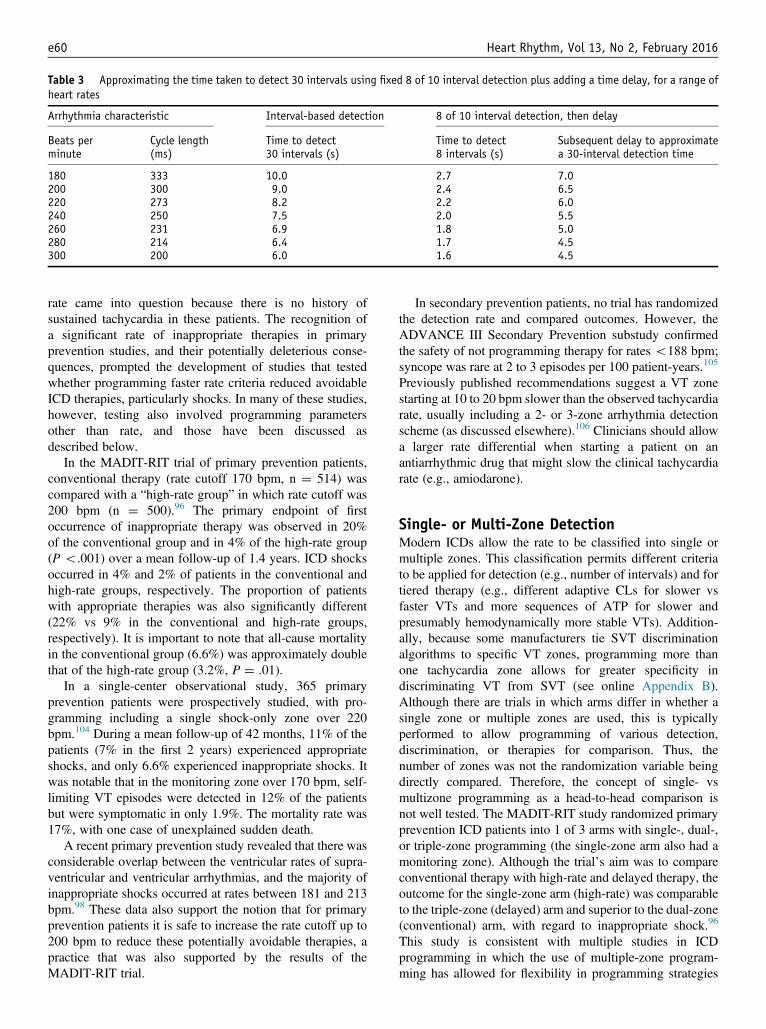
Table 3 Approximating the time taken to detect 30 intervals using fixed 8 of 10 interval detection plus adding a time delay, for a range ofheart rates
Arrhythmia characteristic Interval-based detection 8 of 10 interval detection, then delay
Beats perminute
Cycle length(ms)
Time to detect30 intervals (s)
Time to detect8 intervals (s)
Subsequent delay to approximatea 30-interval detection time
180 333 10.0 2.7 7.0200 300 9.0 2.4 6.5220 273 8.2 2.2 6.0240 250 7.5 2.0 5.5260 231 6.9 1.8 5.0280 214 6.4 1.7 4.5300 200 6.0 1.6 4.5
Heart Rhythm, Vol 13, No 2, February 2016e60
rate came into question because there is no history ofsustained tachycardia in these patients. The recognition ofa significant rate of inappropriate therapies in primaryprevention studies, and their potentially deleterious conse-quences, prompted the development of studies that testedwhether programming faster rate criteria reduced avoidableICD therapies, particularly shocks. In many of these studies,however, testing also involved programming parametersother than rate, and those have been discussed asdescribed below.
In the MADIT-RIT trial of primary prevention patients,conventional therapy (rate cutoff 170 bpm, n ¼ 514) wascompared with a “high-rate group” in which rate cutoff was200 bpm (n ¼ 500).96 The primary endpoint of firstoccurrence of inappropriate therapy was observed in 20%of the conventional group and in 4% of the high-rate group(P o.001) over a mean follow-up of 1.4 years. ICD shocksoccurred in 4% and 2% of patients in the conventional andhigh-rate groups, respectively. The proportion of patientswith appropriate therapies was also significantly different(22% vs 9% in the conventional and high-rate groups,respectively). It is important to note that all-cause mortalityin the conventional group (6.6%) was approximately doublethat of the high-rate group (3.2%, P ¼ .01).
In a single-center observational study, 365 primaryprevention patients were prospectively studied, with pro-gramming including a single shock-only zone over 220bpm.104 During a mean follow-up of 42 months, 11% of thepatients (7% in the first 2 years) experienced appropriateshocks, and only 6.6% experienced inappropriate shocks. Itwas notable that in the monitoring zone over 170 bpm, self-limiting VT episodes were detected in 12% of the patientsbut were symptomatic in only 1.9%. The mortality rate was17%, with one case of unexplained sudden death.
A recent primary prevention study revealed that there wasconsiderable overlap between the ventricular rates of supra-ventricular and ventricular arrhythmias, and the majority ofinappropriate shocks occurred at rates between 181 and 213bpm.98 These data also support the notion that for primaryprevention patients it is safe to increase the rate cutoff up to200 bpm to reduce these potentially avoidable therapies, apractice that was also supported by the results of theMADIT-RIT trial.
In secondary prevention patients, no trial has randomizedthe detection rate and compared outcomes. However, theADVANCE III Secondary Prevention substudy confirmedthe safety of not programming therapy for rates o188 bpm;syncope was rare at 2 to 3 episodes per 100 patient-years.105
Previously published recommendations suggest a VT zonestarting at 10 to 20 bpm slower than the observed tachycardiarate, usually including a 2- or 3-zone arrhythmia detectionscheme (as discussed elsewhere).106 Clinicians should allowa larger rate differential when starting a patient on anantiarrhythmic drug that might slow the clinical tachycardiarate (e.g., amiodarone).
Single- or Multi-Zone DetectionModern ICDs allow the rate to be classified into single ormultiple zones. This classification permits different criteriato be applied for detection (e.g., number of intervals) and fortiered therapy (e.g., different adaptive CLs for slower vsfaster VTs and more sequences of ATP for slower andpresumably hemodynamically more stable VTs). Addition-ally, because some manufacturers tie SVT discriminationalgorithms to specific VT zones, programming more thanone tachycardia zone allows for greater specificity indiscriminating VT from SVT (see online Appendix B).Although there are trials in which arms differ in whether asingle zone or multiple zones are used, this is typicallyperformed to allow programming of various detection,discrimination, or therapies for comparison. Thus, thenumber of zones was not the randomization variable beingdirectly compared. Therefore, the concept of single- vsmultizone programming as a head-to-head comparison isnot well tested. The MADIT-RIT study randomized primaryprevention ICD patients into 1 of 3 arms with single-, dual-,or triple-zone programming (the single-zone arm also had amonitoring zone). Although the trial’s aim was to compareconventional therapy with high-rate and delayed therapy, theoutcome for the single-zone arm (high-rate) was comparableto the triple-zone (delayed) arm and superior to the dual-zone(conventional) arm, with regard to inappropriate shock.96
This study is consistent with multiple studies in ICDprogramming in which the use of multiple-zone program-ming has allowed for flexibility in programming strategies

e61Wilkoff et al Consensus Statement on Optimal ICD Programming and Testing
with regard to detection, discrimination, and therapy. Addi-tionally, there are observational data from the ALTITUDEReal World Evaluation of Dual-Zone ICD and CRT-DProgramming Compared to Single-Zone Programming(ALTITUDE REDUCES) study that show that dual-zoneprogramming is associated with fewer shocks than single-zone programming, at least for rates o200 bpm.64 There-fore, the authors conclude that using more than 1 detectionzone can be useful for modern ICD programming. It shouldbe noted that ATP before or during charging was used in themajority of studies described in both the tachycardiadetection and therapy sections and thus is recommendedfor longer detection.
Discrimination Between Supraventricularand Ventricular ArrhythmiaThe SVT-VT discrimination process classifies a sequence ofsensed EGMs that satisfies rate and duration criteria as eitherSVT (therapy withheld) or VT/VF (therapy given). Discrim-inators are individual algorithm components that provide apartial rhythm classification or a definitive classification for asubset of rhythms. Discrimination algorithms combineindividual component discriminators to produce a finalrhythm classification. Discrimination algorithms vary amongmanufacturers and between individual ICD models (seeonline Appendix B). The final rhythm classification candiffer depending on the technical details of how eachindividual discriminator is calculated, the nominal or pro-grammed threshold for each discriminator, the order inwhich discriminator components are applied, and the logicalconnections between them (e.g., “and” vs “or”). In someICDs, rhythms classified as VT/VF undergo a subsequentsensing-verification step to confirm that EGMs represent truecardiac activation.
SVT-VT Discriminator ComponentsIndividual discriminators can be considered in relation to theEGMs analyzed as ventricular-only or both atrial andventricular, by the rhythm that they identify (e.g., AF, sinustachycardia, VT), or by the type of EGM informationanalyzed (intervals vs morphology). Note that ventricularrate alone is a mandatory discriminator, as discussed in thesection above. We summarize the most commonly useddiscriminators. More comprehensive discussions are avail-able in the literature.107–111
Rejection of Sinus Tachycardia by OnsetSeveral interval-based discriminators focus on differences inthe onset of sinus tachycardia (gradual and parallel accel-eration of atrial and conducted ventricular intervals) com-pared with VT (typically abrupt, with at least transientatrioventricular dissociation). Sudden (abrupt) onset wasone of the first single-chamber, interval-based discrimina-tors. It withholds therapy if acceleration across the sinus-VTrate boundary is gradual. Because onset discriminatorsclassify the rhythm only once, and thus cannot correct
misclassifications, they are now used infrequently and onlywith an override feature and/or other discriminators.112–115
Chamber of onset is a related, interval-based, dual-chamberdiscriminator that classifies a 1:1 tachycardia as SVT if theatrial rhythm accelerates at the device-defined onset. Arelated “Sinus Tachycardias” discriminator classifies atachycardia as VT if either the RR or the PR intervalsdeviate sufficiently from the range of the immediatelypreceding sinus intervals.116
Rejection of AF by Ventricular Interval RegularityVentricular interval regularity (interval stability) is anexplicit single-chamber, interval-based discriminator thatclassifies the rhythm as AF if the ventricular intervals aresufficiently irregular. Because interval variability in con-ducted AF decreases at faster rates, stability becomesunreliable in discriminating VT from conducted AF atventricular rates greater than 170 bpm.112,115 Interval stabil-ity can also fail if drugs (e.g., amiodarone) cause mono-morphic VT to become irregular or induce polymorphic VTto slow into the SVT-VT discrimination zone.114,117
Diagnosis of VT by Dual-Chamber Components: Atrial vsVentricular Rate and Atrioventricular AssociationIn contrast to the single-chamber discrimination algorithmsabove that diagnose SVT when their criteria are fulfilled, 2separate, interval-based, dual-chamber discrimination algo-rithms diagnose VT. First, atrial rate vs ventricular ratediagnoses VT if the ventricular rate exceeds the atrial rate.118
Second, atrioventricular dissociation identifies isorhythmicVT during sinus tachycardia. Inversely, the atrioventricularassociation discriminator diagnoses SVT in the presence ofN:1 (e.g., 2:1, 4:1) atrioventricular association consistentwith atrial flutter at a fixed conduction ratio.
The Ventricular Electrogram Morphology DiscriminatorThis versatile, single-chamber discriminator is the onlyalgorithm component that does not rely on inter-EGMintervals. It classifies tachycardias as SVT if the morphology(shape) of the ventricular EGM is sufficiently similar to themorphology during a conducted baseline rhythm. It canpotentially discriminate any SVT from VT, including SVTsthat challenge other discriminators, such as abrupt-onset 1:1SVTs and irregular VT during AF. Contemporary ICDs(including subcutaneous ICD [S-ICD]) analyze EGMs fromthe shock electrodes, which record a larger field of view thanEGMs from pace-sense electrodes.119 They operate using acommon series of steps and are susceptible to commonfailure modes.111,120–123 The first common step is acquisitionof a baseline rhythm template by mathematically extractingEGM features and storing them. Both the acquisition of theinitial template and the subsequent template updating areautomated in most ICDs. Nevertheless, physicians shouldconfirm that the conducted baseline beats match the templateboth at implant and during follow-up. For CRT patients, thetemplate must be manually collected. If the wavelet signal

Heart Rhythm, Vol 13, No 2, February 2016e62
during template acquisition appears clipped, adjustmentsspecific to the manufacturer might be necessary.
SVT-VT Discrimination AlgorithmsDiscrimination algorithms combine component discrimina-tors to provide a final rhythm classification of VT/VF orSVT. The morphology discriminator frequently forms theprimary component of single-chamber algorithms withstability playing a secondary role and sudden onset usedsparingly. By contrast, the cornerstone of most dual-chamberalgorithms is explicit or implicit comparison of atrial vsventricular rates. Because the ventricular rate is greater thanthe atrial rate in more than 80% of VTs, algorithms thatcompare atrial and ventricular rates as their first step applyadditional SVT discriminators to fewer than 20% of VTs,reducing the risk that they will misclassify VT as SVT.124,125
Most dual-chamber algorithms further restrict single-chamber discriminators to tachycardias for which they offerthe greatest benefit; thus, stability is applied only if AF isconfirmed by direct calculation of the atrial rate or the atrialrate is greater than the ventricular rate. Similarly, suddenonset, chamber of onset, or 1:1 atrioventricular associationare applied only if the atrial rate equals the ventricular rate.The use of discriminators in redetection varies amongmanufacturers and has not been systematically studied.
Assessing Clinical Benefits and RisksWhat Evidence Supports a Benefit?
1.
The annual rate of inappropriate shocks has fallendramatically from 37%–50% for SVT alone in earlystudies to 1%–5% for all causes in modern clinicaltrials.97,118,126–128 This decrease is likely due to differ-ences in both clinical populations and the programming ofmultiple ICD parameters, including longer detection timeand higher rate cutoffs. Thus, it is difficult to isolate thedifferential effect of SVT-VT discrimination algorithmsusing clinical data. These studies have programmeddiscrimination algorithms to ON, however, so it seemsreasonable to use them.2.
Although clinical trials that reported dramatic reductionsin shocks for SVT programmed discrimination algorithmsconsistently, they have been programmed inconsistently inclinical practice, and the rate of inappropriate shocks forSVT has been higher in observational studies of remote-monitoring ICD databases. In the ALTITUDE REDUCESstudy on 15,991 patients in the Latitudes database, SVTwas the most common cause of shocks when the detectionrate wasr180 bpm.129 For detection ratesr170 bpm, therate of inappropriate shocks at 1 year was significantlylower with dual-zone programming, which permits SVT-VT discrimination, than single-zone programming, whichdoes not (9.6% vs 4.3%). Similarly, Fischer et al130analyzed shocks in 106,513 patients in the CareLinks
database; programming SVT-VT discrimination ON wasassociated with a 17% reduction in all-cause shocks.
3.
Sophisticated simulations indicate that SVT-VT discrim-ination algorithms have substantial benefit. For example,the SCD-HeFT study on primary prevention patients didnot use discriminators. A validated Monte Carlo simu-lation predicted that use of single- or dual-chamber SVT-VT discriminators alone would have reduced inappropri-ate shocks for SVT by 75.5% and 78.8%, respectively.131Which Patients are Most Likely to Benefit, and Which are LeastLikely to Benefit?Despite limited direct evidence, it seems clear that patientswill benefit most if the rates of their VTs and SVTs overlap.This includes patients with slower monomorphic VT, thoseat risk for AF with rapid ventricular rates, or those capable ofexercising to sinus rates in the VT zone.103,132 In secondaryprevention patients with slower VT, older discriminationalgorithms reduced shocks for SVT compared with rate-onlydetection. The benefit is less for primary prevention patients,secondary prevention patients at risk only for VF, and thosewho cannot sustain rapid atrioventricular conduction.Patients with permanent complete atrioventricular block donot benefit.
What are the Risks?The risk of the misclassification of either VT or VF as SVTby the discrimination algorithms can either prevent VTdetection or delay the time to therapy (underdetection), asdocumented in clinically significant situations.112,113,115,125
When modern algorithms are programmed to recommendedparameters, clinically significant underdetection is rare.Large clinical trials on multiple shock-reduction strategies(including SVT-VT discrimination) report no or minimal andstatistically insignificant increases in syncope.95,97,126,133
Most reports do not include the causes of syncope and thusdo not permit identification of whether discriminationalgorithms contributed to any of the syncopal episodes byprolonging detection. However, in the PREPARE study, nosyncopal episode was caused by untreated tachycardia.133 Ingeneral, discriminators that re-evaluate the rhythm classifi-cation during ongoing tachycardia reduce the risk of under-detection compared with those that withhold therapy if therhythm is misclassified by the initial evaluation (e.g., onset,chamber of origin algorithms).
Additional ConsiderationsSVT LimitSVT-VT discrimination applies from the VT detection rate tothe SVT limit rate, which is programmable independently ofthe VT/VF therapy zones with some manufacturers (pref-erable), but which might be linked to one of the zoneboundaries in others. The minimum CL for SVT-VTdiscrimination should be set to prevent clinically significantdelays in the detection of hemodynamically unstable VT.PREPARE, EMPIRIC, and MADIT-RIT all support thesafety of empirical programming at 200 bpm.96,101,134 In

e63Wilkoff et al Consensus Statement on Optimal ICD Programming and Testing
MADIT-II, approximately 50% of SVT episodes were fasterthan 170 bpm, and a few were as fast as 250 bpm.82 InINTRINSIC RV, SVT comprised 19% of episodes, withrates between 200 and 250 bpm.135 More limited andpreliminary data from PainFree SST support programmingup to 222–230 bpm.116,136 We suggest the SVT limit notexceed 230 bpm in adults without a patient-specific indica-tion, based on the low incidence of SVTs in this rate rangeamong ICD patients and the potential—however small—formisclassifying hemodynamically unstable VT.
Duration-Based “Safety-Net” Features to OverrideDiscriminatorsThese features deliver VT/VF therapy if a tachycardiasatisfies the ventricular rate criterion for a sufficient duration,even if the discrimination algorithm indicates SVT. Thepremise is that the ventricular rate during transient sinustachycardia or AF will decrease to below the VT rateboundary before the override duration is exceeded. In onestudy, an override duration of 3 minutes delivered inappro-priate therapy to 10% of SVTs.112 Because SVT is muchmore common than VT, programming an override durationof less than 5–10 minutes results primarily or solely ininappropriate SVT therapy.122 Although more data would beuseful, in the absence of a documented benefit, we recom-mend programming this feature OFF or long (minutes)without a patient-specific or device-specific indication.
Dual-Chamber vs Single-Chamber AlgorithmsClinical trials and simulated testing of induced arrhythmiasthat compared single- vs dual-chamber discriminators havereported inconsistent results.10,33,137–139 Two meta-analysesfound no superiority of dual-chamber ICDs in terms ofmortality or inappropriate therapies.11,140 Any benefit ofdual-chamber discrimination is likely restricted to specificpatient groups.103,138 For example, the Dual Chamber andAtrial Tachyarrhythmias Adverse Events (DATAS) trial ofpredominantly secondary prevention patients with slowerVTs reported modest benefit from dual-chamber discrim-ination, while the recent Reduction and Prevention ofTachyarrhythmias and Shocks Using Reduced VentricularPacing with Atrial Algorithms (RAPTURE) trial of primaryprevention patients programmed to a fast detection rate(4182 bpm) and long detection duration (30/40 intervals)did not.103,138,139 Inappropriate therapy for SVT occurred inonly 2% of the patients in each group. Recent data fromPainFree SST notes very low rates of inappropriate shocks(3.7% for single chamber; 2.8% for dual and triple chamberafter 2 years). The choice of device was not randomized,suggesting that when physicians chose a dual- or triple-chamber device (perhaps due to known atrial arrhythmia orbradycardia), inappropriate shock rates were minimized.136
The Optimal Anti-Tachycardia Therapy in ImplantableCardioverter-Defibrillator Patients Without Pacing Indica-tions (OPTION) trial randomized 462 patients to single- ordual-chamber programming and noted inappropriate shock
rates of 10.3% for single chamber vs 4.3% for dual chamberafter 27 months (P¼ .015). Atrial lead-related complicationswere 1.3%, therapy was delivered from 170 bpm (VT) and200 bpm (VF), and no difference in ventricular pacingpercentage was noted.141 Dual-chamber algorithms probablyreduce the risk of underdetection compared with single-chamber algorithms because more than 80% of VTs with aventricular rate greater than the atrial rate undergo no furtheranalysis.103,124,125 However, the rate of clinically significantunderdetection with modern programming is so low that thisdifference is rarely of clinical significance. In most patients,improved SVT-VT discrimination should not be consideredan indication for a dual- vs single-chamber ICD. Even if adual-chamber ICD is implanted, dual-chamber discrimina-tion should be programmed only if the atrial lead becomeschronic or if atrial sensing is unreliable. Accurate sensing ofatrial EGMs is essential for dual-chamber SVT-VT discrim-ination. Atrial lead dislodgments, oversensing of far-field Rwaves, or undersensing due to low-amplitude atrial signalscan cause misclassification of VT/SVT. On implant, it isimportant to position the atrial lead to minimize far-fieldR waves.
Ventricular OversensingExcluding recalled leads, ventricular oversensing accountsfor less than 10% of inappropriate shocks, but it often resultsin repetitive shocks and severe symptoms.82,142–144 Recentlyintroduced features reduce inappropriate therapies fromoversensing of physiologic T waves and nonphysiologicsignals related to pace-sense lead failures as discussed below.
Programming to Reduce T-Wave OversensingThe problem of T-wave oversensing relates to the basicrequirement that ICDs reliably sense VF, which is charac-terized by RR intervals shorter than the normal QT intervaland some EGMs with low amplitudes and slew rates.Approaches to minimizing T-wave oversensing includereprogramming ventricular sensitivity, altering sensingbandwidth, and changing the sensing bipole.109,123,145 Onemanufacturer provides an algorithm that withholds therapyafter rate and duration criteria for VT/VF are fulfilled if aspecific pattern of T-wave oversensing is identified.146
T-wave oversensing rates vary based on device design;using an appropriate high band-pass filter results in verylow rates of T-wave oversensing.142 Because T-wave over-sensing is unpredictable, features that minimize T-waveoversensing should be enabled proactively at implant,providing they do not cause undersensing in VF.146
Lead-Related OversensingOversensed signals caused by pace-sense lead failure havespecific interval patterns and EGM characteristics.145,147,148
Present algorithms identify three features: (1) intervals canbe too short to represent successive ventricular activations;(2) such short intervals are often transient and can berepetitive; and (3) in true bipolar leads, oversensed signals

Heart Rhythm, Vol 13, No 2, February 2016e64
are absent on the shock EGM. Algorithms can providewarning alerts, withhold shocks after spurious detection ofVT/VF, or both. All 3 criteria can provide alerts, but only thethird is applied to withhold shocks. The present algorithmswere developed to identify impending lead failures onrecalled leads, notably the Sprint Fidelis. These algorithmsmight not be appropriate for detecting failures in otherleads.144 There is a high false-positive rate when using thesealgorithms, and caregivers must carefully review the devicedata that caused the alert to ensure the lead experienced a truefailure.145
Alerts that combine both oversensing and abrupt changesin impedance trends provide earlier warning of lead failurethan a fixed impedance threshold.144,145,149 Such alerts canbe delivered via wireless remote monitoring and/or bynotifying the patient via vibration or an audible tone.Caregivers must respond rapidly to alerts to minimizeinappropriate shocks.144,149 Wireless remote monitoringhas been reported to reduce response time.150 The principaldisadvantage of lead alerts is false-positive triggers. Theprincipal risk of shock-withholding algorithms is a failure toshock VF, which is extremely rare.151 In addition toalgorithmic approaches, oversensing due to failure of thecable leading to the ring electrode can be prevented bychanging the programming of the sensing configuration fromtrue bipolar to integrated bipolar. This approach is appro-priate prophylactically or as temporary programming after aring electrode cable failure; it is not a permanent solution,however, because increased rates of high-voltage cablefractures have been documented after sensing cablefractures.152
The Subcutaneous Defibrillator (S-ICD)The novel S-ICD follows many of the same principles asintravascular ICDs but is considered here separately forduration criteria, rate criteria, and discrimination algorithms.Candidates for the S-ICD must initially be screened with amodified tri-channel surface electrocardiogram that mimicsthe sensing vectors of the S-ICD system. This test is designedto assess the R-wave to T-wave ratio for appropriate signalcharacteristics and relationships. If the screening is notsatisfactory for at least 1 of the 3 vectors supine and standing,an S-ICD should not be implanted. On implant, the S-ICDautomatically analyzes and selects the optimal sensingvector.
Detection of VT or VF by the S-ICD is programmableusing a single or dual zone. In the single-zone configuration,shocks are delivered for detected heart rates above theprogrammed rate threshold: the “shock zone.”134 In thedual-zone configuration, arrhythmia discrimination algo-rithms are active from the lower rate: the “conditional shockzone.” In this latter zone, a unique discrimination algorithmis used to classify rhythms as either shockable or non-shockable. If they are classified as supraventricular arrhyth-mias or nonarrhythmic oversensing, therapy is withheld.
With dual-zone programming, the shock zone uses rate asthe sole method for rhythm analysis. In contrast, the condi-tional shock zone uses a stepwise discrimination algorithmto distinguish shockable from nonshockable rhythms. Theconditional shock zone has a morphology analysis processbased on a normal rhythm transthoracic QRS:T wavetemplate. The template uses up to 41 fiduciary points toreconstruct morphology for the template as well as theprogrammed targeted heart rate zones. The comparison ofthe template to the high-rate rhythm electrocardiogram fordiscrimination constitutes the static waveform analysis. Agood template match designates a sensed beat as supra-ventricular, thereby preventing a shock. A poor match to thestatic QRS:T morphology template moves the algorithm to adynamic waveform analysis that compares single-beat mor-phologies in groups of 4 beats for uniformity. A consistentdynamic waveform match adjusts the sensing to evaluateQRS width. If a tachycardia has a prolonged QRS widthcompared with the template width (420 ms) and is ofsufficient duration, it will lead to a shock.
The system uses an initial 18 of 24 duration criteria(nonprogrammable) prior to initiating capacitor charging;however, this duration is automatically extended followingnonsustained ventricular tachyarrhythmia events. A confir-mation algorithm is also used at the end of capacitor chargingto ensure persistence of the ventricular arrhythmia prior toshock delivery. Shocks for spontaneous (noninduced) epi-sodes are delivered at a nonprogrammable 80 J regardless ofthe therapy zone of origination.
When programmed to include a conditional shock zone,the S-ICD VT detection algorithm has been demonstrated tobe more effective than transvenous ICD systems programmedat nominal settings to prevent the detection of inducedsupraventricular arrhythmias.153 Furthermore, in the clinicalevaluation of the conditional shock zone, the S-ICD systemwas strongly associated with a reduction in inappropriateshocks from supraventricular arrhythmias and did not resultin prolongation of detection times or increased syncope.154
Integrating Tachycardia Detection Data IntoProgramming RecommendationsWhen taking data from specific single-manufacturer studiesand producing generic guidelines applicable across all ICDs,some compromises and potential pitfalls have been encoun-tered. Nevertheless, it is our intention to convey the generalprinciples of good quality evidence (e.g., extending detectiontime) to apply to ICD programming in general. Thus,attempts have been made to translate interval-based detectionto time-based detection and to provide a range of reasonableheart rate cutoffs that are inclusive of those proven in good-quality trials. We encourage programming ICDs tomanufacturer-specific therapies of proven benefit; however,when evidence is lacking, the guidelines provide a frame-work for programming within the evidence base. See onlineAppendix B for manufacturer-specific examples of optimalICD programming.

e65Wilkoff et al Consensus Statement on Optimal ICD Programming and Testing
Tachycardia Detection Programming Recommendations
Class ofRecommendationLevel ofEvidence
For primary prevention ICD patients, tachyarrhythmia detection duration criteria should be programmedto require the tachycardia to continue for at least 6–12 seconds* or for 30 intervals beforecompleting detection, to reduce total therapies.
I
A*Tachyarrhythmia detection duration is directly related to the tachyarrhythmia rate. Direct evidence tosupport a delay42.5 seconds for rates over 250 bpm is not available, but can be inferred from evidencethat 30 detection intervals are safe at that rate.
For primary prevention ICD patients, the slowest tachycardia therapy zone limit should be programmedbetween 185 and 200bpm*, to reduce total therapies.
I
A*Higher minimum rates for detection might be appropriate for young patients or for those in whom SVT-VTdiscriminators cannot reliably distinguish SVT from VT, provided there is no clinical VT below this rate.
For secondary prevention ICD patients, tachyarrhythmia detection duration criteria should beprogrammed to require the tachycardia to continue for at least 6–12 seconds* or for 30 intervalsbefore completing detection, to reduce total therapies.
I
B-R*Tachyarrhythmia detection duration is directly related to the tachyarrhythmia rate. Direct evidence tosupport a delay42.5 seconds for rates over 250 bpm is not available, but can be inferred from evidencethat 30 detection intervals are safe at that rate.
Discrimination algorithms to distinguish SVT from VT should be programmed to include rhythms withrates faster than 200 bpm and potentially up to 230 bpm (unless contraindicated*) to reduceinappropriate therapies.
I
B-R*Discrimination algorithms and/or their individual components are contraindicated in patients withcomplete heart block or if the algorithm/component is known to be unreliable in an individual patient.Dual-chamber discriminators that misclassify VT as SVT if the atrial lead dislodges are discouraged in theperioperative period. Dual-chamber discriminators are contraindicated in patients with known atrial leaddislodgment, atrial undersensing or oversensing of far field R waves, and in those with permanent AF.
It is recommended to activate lead-failure alerts to detect potential lead problems.
I B-NR For secondary prevention ICD patients for whom the clinical VT rate is known, it is reasonable toprogram the slowest tachycardia therapy zone at least 10 bpm below the documented tachycardiarate but not faster than 200bpm*, to reduce total therapies.IIa
C-EO*Higher minimum rates for detection might be appropriate for young patients or for those in whom SVT-VTdiscriminators cannot reliably distinguish SVT from VT, provided there is no clinical VT below this rate.
It can be useful to program more than one tachycardia detection zone to allow effective use of tieredtherapy and/or SVT-VT discriminators and allow for a shorter delay in time-based detectionprogramming for faster arrhythmias.
IIa
B-RWhen a morphology discriminator is activated, it is reasonable to reacquire the morphology templatewhen the morphology match is unsatisfactory, to improve the accuracy of the morphologydiscriminator.
IIa
C-LDIt is reasonable to choose single-chamber ICD therapy in preference to dual-chamber ICD therapy if thesole reason for the atrial lead is SVT discrimination, unless a known SVT exists that may enter the VTtreatment zone, to reduce both lead-related complications and the cost of ICD therapy.
IIa
B-NRFor the S-ICD, it is reasonable to program 2 tachycardia detection zones: 1 zone with tachycardiadiscrimination algorithms from a rate r200 bpm and a second zone without tachycardiadiscrimination algorithms from a rate Z230 bpm, to reduce avoidable shocks.
IIa
B-NRProgramming a nontherapy zone for tachycardia monitoring might be considered to alert clinicians tountreated arrhythmias.
IIb
B-NRIt may be reasonable to disable the SVT discriminator timeout function, to reduce inappropriatetherapies.
IIb
C-EOIt may be reasonable to activate lead “noise” algorithms that withhold shocks when detected VT/VF isnot confirmed on a shock or other far-field channel to avoid therapies for nonphysiologic signals.
IIb
C-EOIt may be reasonable to activate T-wave oversensing algorithms, to reduce inappropriate therapies.
IIb C-LD It may be reasonable to program the sensing vector from bipolar to integrated-bipolar in true-bipolarleads at risk for failure of the cable to the ring electrode to reduce inappropriate therapies.*IIb
C-EO*This is not intended as a long-term solution when a cable fracture has been identified.

Heart Rhythm, Vol 13, No 2, February 2016e66
Tachycardia Therapy Programming
Although therapies delivered by the ICD can abort SCD,appropriate and inappropriate ICD shocks have been asso-ciated with a considerable increase in the risk of mortal-ity.82,83,155–158 In the Sudden Cardiac Death in Heart FailureTrial (SCD-HeFT), the risk of mortality was 5-fold higher inpatients who received appropriate ICD shocks and 2-foldhigher in patients who received inappropriate shocks.83Similarly, pooling data from 4 studies of 2135 ICD patients,shocked VT was associated with a 32% increase in the risk ofmortality. In that analysis, shocked patients had poorersurvival than patients treated with ATP only.155 ICD shocksare likely a marker of more advanced heart disease andsubsequent death, but defibrillation therapies have beenassociated with troponin release and increased LV dysfunc-tion with the potential of further mortality risk.
The incidence of appropriate and inappropriate ICDshocks depends on the patient’s characteristics, includingthe indication for the device, concomitant medical therapiesincluding antiarrhythmic medications, programming of theICD, and the duration of follow-up. With regard to ICDprogramming, faster VT/VF detection rates, longer detectiondurations, use of a single zone, use of SVT discriminators,and delivery of ATP have been shown to reduce bothappropriate and inappropriate shocks and to improve qualityof life.91,101,126,129,130,133,159,160 This programming mightimprove survival.126 Indeed, several studies have shown thatATP is effective at terminating slow and fast VT withexceedingly low rates of adverse events like syn-cope.93,135,161–165 The initial bias of the ICD communitywas to reserve ATP therapy for those patients in whom thetherapy was demonstrated to be effective, usually during anelectrophysiologic study. However, the approach ofphysician-directed programming based on the knowledgeof induced arrhythmias was found to be significantly inferiorto the routine strategic (EMPIRIC) programming of ATP. Itis not reflective of the arrhythmias experienced outside theelectrophysiology laboratory for primary and secondaryprevention patients with ischemic and nonischemic sub-strates.101,166 Although the ideal number of ATP bursts hasnot been definitively determined, current data support the useof up to 2 ATP attempts, given additional attempts yield verylittle additional efficacy.93,135,161–165,167,168 In one study, upto 5 attempts were found to be safe.168 The most effectiveATP duration is likewise uncertain; however, in the ATPDelivery for Painless ICD Therapy (ADVANCE-D) trial—aprospective RCT of 925 patients—8-pulse ATP was aseffective and safe as 15-pulse ATP.169 The PITAGORAICD clinical trial randomized 206 patients with an ICD to 2ATP strategies: an 88% coupling interval burst vs a 91%coupling interval ramp. The results of the trial showed thatover a median follow-up of 36 months and compared withramp pacing, burst pacing was more effective for terminatingfast VT episodes (between CL 240 and 320 ms).170 In aprospective study of 602 patients, a strategy of tiered ATPand low-energy shock was efficacious and safe in patientswith VT CL greater than 250 ms, with extremely low
syncope rates.171 However, a “real-world” retrospectivestudy on 2000 patients with 5279 shock episodes from theLATITUDE remote monitoring system showed that thesuccess rate of first shock as first therapy was approximately90%, but the success rate was lower after failed ATP.Therefore, that study recommended programming a higherlevel of energy after ATP.172 Finally, a substudy of theEffectiveness and Cost of ICD Follow-Up Schedule withTelecardiology (ECOST) study, which randomly assigned433 patients to remote monitoring (n¼ 221; active group) vsambulatory follow-up (n ¼ 212; control group), showed thatremote monitoring was highly effective in the long-termprevention of inappropriate ICD shocks through earlydetection and prevention of AF with a rapid ventricular rate,nonsustained VT, or diverted VT episodes.173
Benefits and RisksThe goal of ICD therapy is to prolong life while causing aslittle morbidity as possible. Although survival is quantifiablyobjective, morbidity is more subjective and includes bothphysical and emotional components. Clearly, shocks areusually painful to the patient, whereas ATP is typically notuncomfortable. However, there can be other morbiditiesrelated to both therapies, including mild to extreme emo-tional distress, syncope, palpitations, and proarrhythmiayielding more therapies and occasionally leading to death.Paradoxically, the need for life-saving therapies, includingshocks and potentially ATP, might also be associated withincreased mortality; however, the causal relationships areunclear. Also, the prevalence of tachycardia amenable toATP or hemodynamic significance varies with the mecha-nism of the risk (e.g., long QT vs ischemic cardiomyopathy).In addition, although the risk of having a hemodynamicallyimportant or life-threatening arrhythmia can vary frompatient group to patient group, the largest proportion ofpatients in whom ICD therapy is applied has yet to have apreviously recorded arrhythmia, and we must thereforestrategically choose on the basis of other factors how wewill treat the first event and subsequent events.
Classification of TherapyThe literature uses definitions of therapies that differ fromeach other and that impact their results and conclusions. Theoccurrence rates of these events not only are dependent ontheir definition but also are highly dependent on theprogramming of the defibrillation system. Both shock andnonshock therapies can be categorized as being appropriate,inappropriate, and avoidable. Whereas appropriate andinappropriate therapies refer to therapies that were actuallydelivered, avoidable therapies are theoretical events in thefuture. These potential future tachycardia therapies, deliv-ered for either appropriately or inappropriately detectedevents, can frequently be avoided by establishing program-ming to either prevent the initiation of the arrhythmia or toallow the condition to pass without therapy.

e67Wilkoff et al Consensus Statement on Optimal ICD Programming and Testing
AppropriateA response to a sustained ventricular arrhythmia (VT, VF) orhemodynamically poorly tolerated arrhythmias (e.g., asso-ciated with syncope, rate over 200 bpm, or hemodynamicallycompromising supraventricular arrhythmias).
InappropriateA response to signals generated by something other thansustained ventricular arrhythmias or hemodynamicallypoorly tolerated arrhythmias. Possible signals include supra-ventricular rhythms such as sinus tachycardia, AF, atrialflutter, reentrant SVT, atrial tachycardia, or instances ofsignal misinterpretation. Signal misinterpretation includesmultiple counting of single events (e.g., atrial, T-wave or R-wave), environmental signals such as electromagnetic inter-ference, frequent premature ventricular contractions (PVCs)and nonsustained ventricular arrhythmias, extracardiac phys-iologic signals (e.g., diaphragmatic or pectoral myopoten-tials), other implantable electronic devices (e.g., pacemakers,LV assist devices, nerve stimulators), inappropriate leadplacement or dislodgment, conductor or insulation failures,header connection instability, and pulse generator failure.
AvoidableProgramming of detection and therapy parameters andalgorithms so that shock or ATP therapy is withheld fromarrhythmias that would be expected to be hemodynamicallytolerated. Examples include self-terminating ventriculararrhythmias, ATP-susceptible ventricular arrhythmias, andoverdrive suppression responsive rhythms. Many appropri-ate and most inappropriate therapies are also potentiallyavoidable.
PhantomThese are not true therapies; however, there is the patient’sperception that a therapy was delivered. Interrogation of theICD and/or coincident rhythm monitoring does not identify atachycardia or therapy.
Unintended Consequences of ICD Therapy and ICDTherapy ProgrammingIn the SCD-HeFT and MADIT II trials, inappropriate shocksmore than doubled the risk of death. Mortality rates weresubstantially higher after shocks: 10% within days after thefirst shock, 25% within 1 year, and 40% by 2 years. Theleading cause of death was progressive HF. In an analysis ofthe MADIT-CRT trial, the patients with appropriate shocksexperienced increased mortality when compared with thepatients without ICD shocks, after accounting for mechanicalremodeling effects; this was not the case for patients whoreceived appropriate ATP only.156 ICD shocks have alsobeen associated with independent predictors of mortality inthe large ALTITUDE registry of 3809 ICD recipients and ina meta-analysis of ICD trials in which ATP wasapplied.155,157 Emotional morbidities associated with ICDshocks are well recognized and include anxiety, depression,
and posttraumatic stress disorders.174–176 Phantom shockscan result from fear and/or anxiety and have a reportedincidence of 5% in a European study of ICD recipients over35 months of follow-up.177 If possible, and when safe, it isbest to avoid both the discomfort and psychological impactof shocks for ventricular arrhythmias, supraventriculararrhythmias, noise events including lead failures, and forself-terminating arrhythmias, as is discussed in the section ontachycardia detection. The 1500-patient MADIT-RIT studydemonstrated a mortality reduction by changing both tachy-cardia detection criteria and tachycardia therapy (shocks andATP). Therefore, it is difficult to assign the outcome result toATP, shocks, or both when compared with older, moreconventional programming.126 In addition, in a randomizedstudy of remote follow-up of ICDs, home monitoringshowed an incidence of 52% fewer inappropriate shocks,72% fewer hospitalizations due to inappropriate shocks, 76%fewer capacitor charges, and a significant positive impact onbattery longevity.178
ATPSeveral large clinical trials have established the safety andefficacy of ATP as a first-line therapy to treat even very fastVTs.93,95,101,133 The use of first-line ATP involving VT atrates between 188 and 250 bpm in the PainFREE Rx II trialresulted in a 71% relative shock reduction.93 In the PRE-PARE study, a primary prevention cohort of 700 patientswas programmed with 30 of 40 detection intervals with ATP-first for VT between 182 and 250 bpm with SVT discrim-inators active up to 200 bpm. The results demonstrated arobust absolute risk reduction for shocks at 1 year from 17%to 9% without an increase in arrhythmic syncope whencompared with historical controls.133 Similar findings werenoted in the RELEVANT study, which evaluated a cohort ofpatients with nonischemic heart disease and cardiac resynch-ronization defibrillators.95 In the earlier EMPIRIC study,standardized VT detection and ATP therapy parametersdemonstrated a reduction in shocks when compared withphysician-tailored treatment in a randomized assessment of900 primary prevention patients.101 The use of ATP duringICD capacitor charging has been clinically validated as safeand effective.163 It is important to recognize that inappro-priate therapies including inappropriate ATP, deliveredprimarily in the setting of supraventricular arrhythmias, havebeen associated with increased mortality in the MADIT-RITand MADIT-CRT trials.156,179 However, the overall safetyof ATP and its role as a contributor to improved survival arewell established, particularly in terms of preventing avoid-able ICD shocks.
Customized vs Strategic ProgrammingBecause primary prevention patients have no prior ventric-ular arrhythmias, programming individual devices onimplant is largely empiric. There are more data for secondaryprevention patients, but how the patient will behave in thefuture is still uncertain. The ability to individualize the

Heart Rhythm, Vol 13, No 2, February 2016e68
antitachycardia programming for patients with both primaryand secondary prevention indications was tested in theEMPIRIC trial and found to be an inferior approach toprevent these therapy events.101 The application of stand-ardized programming and borrowing data from the Pain-FREE Rx II and PREPARE studies resulted in acomprehensive review of programming and its applicationacross manufacturers.
Secondary PreventionFor the secondary prevention ICD patient, specific knowl-edge of the patient’s arrhythmia history facilitates thecreation of an effective antitachycardia programming strat-egy. Using what is known about the ventricular arrhythmia,including any electrocardiograms, available telemetry strips,and EMS recordings, provides insight into the arrhythmiamechanism. In cases of monomorphic VT, discerning therate (CL) and the hemodynamic impact is useful in making
choices, particularly for detection at a minimum; the devicemust be programmed with active VT detection zonessufficient to cover the clinical arrhythmia. Slower, mono-morphic VT that is better tolerated hemodynamically favorsa robust approach using ATP termination with at least 2–3sequences and at least 8 pulses. The use of a second burst ofATP has also been shown to increase effectiveness from64% to 83% in the fast VT range of 188 to 250 bpm.167
Although a second burst has clear value, value beyond 2bursts is limited, except in rare situations.101 The use ofICDs in patients with implanted LV assist devices allowsprolongation of detection times and programming of multi-ple ATP attempts without significant risk to the patient, andit reduces the opportunity for shock therapies. Adjunctmedications and ablation of VT (or SVT) might also beconsidered for cases in which slow VT occurs or if there isan overlap between the SVT and VT rates, leading to ICDtherapies.
Tachycardia Therapy Programming Recommendations
Class ofRecommendationLevel ofEvidence
It is recommended in all patients with structural heart disease and ATP-capable ICD therapydevices that ATP therapy be active for all ventricular tachyarrhythmia detection zones toinclude arrhythmias up to 230 bpm, to reduce total shocks except when ATP is documentedto be ineffective or proarrhythmic.
I
AIt is recommended in all patients with structural heart disease and ATP-capable ICD therapydevices that ATP therapy be programmed to deliver at least 1 ATP attempt with a minimumof 8 stimuli and a cycle length of 84%–88% of the tachycardia cycle length for ventriculartachyarrhythmias to reduce total shocks, except when ATP is documented to be ineffectiveor proarrhythmic.
I
AIt is indicated to program burst ATP therapy in preference to ramp ATP therapy, to improve thetermination rate of treated ventricular tachyarrhythmias.
I
B-RIt is reasonable to activate shock therapy to be available in all* ventricular tachyarrhythmiatherapy zones, to improve the termination rate of ventricular tachyarrhythmias.
IIa
C-EO*Rarely, to limit patient discomfort and anxiety, hemodynamically stable slow VT can be treatedwithout programming a backup shock.
It is reasonable to program the initial shock energy to the maximum available energy in thehighest rate detection zone to improve the first shock termination of ventricular arrhythmiasunless specific defibrillation testing demonstrates efficacy at lower energies.
IIa
C-LDIntraprocedural Testing of Defibrillation EfficacyThe efficacy of the ICD for the primary and secondaryprevention of SCD has been well established in severallandmark clinical trials.180–185 Most of these trials haverequired induction, detection, and termination of VF atthe time of implantation as a measure of defibrillationefficacy and as a surrogate of the ICD’s ability to preventSCD. Testing defibrillation efficacy has been consideredan integral part of ICD implantation for many years, and itis performed to establish the appropriate connection ofhigh-voltage electrodes and to test the ability of the ICDto detect and terminate VF with a shock. However,identifying system failures or high defibrillation
thresholds is difficult, mainly due to the low prevalence,which also depends on the definition employed, about 5%combined. Significant improvements over the past 2decades have reduced energy requirements for defibrilla-tion.186–189 Similarly, current transvenous ICD technol-ogy is capable of delivering energies of 35–40 J, raisingthe question of the value of routine defibrillation testing(DT). Physicians have therefore gravitated to implantingICDs with minimum or no DT with wide variability inpractice, despite a paucity of rigorous data. DT iscurrently being performed during ICD implant in onlyabout half the procedures.190–195 Studies evaluating DTare summarized in Table 4.

Table 4 Defibrillation testing
Study (n) Patients (DT/no DT) Results and remarks
CREDIT193 64%/36% More frequent DT for new implants vs generator replacements (71% vs 32%, P ¼ .0001), DT forprimary and secondary prevention indications (64% vs 63%, P ¼ NS).(361)
Prospective multicenter registry Reasons for no DT were as follows: unnecessary (44%); persistent atrial fibrillation (37%); noanesthetist (20%); and patient or physician preference (6%).
DT was not performed in a third of ICD implants, usually due to a perceived lack of need orrelative contraindication.
Nonconsecutive patients, single manufacturer.Ontario DT Registry225 PP: 65%/45% Multivariate predictors for DT included new ICD implant (OR 13.9; P o.0001), DCM (OR 1.8; P
o.0001), amiodarone (OR 1.5; P ¼ .004), and LVEF 420% (OR 1.3; P ¼ .05). History of AF(OR 0.58; P¼ .0001) or OAC use (OR: 0.75; P¼ .03) was associated with a lower likelihood ofhaving DT. Complications, including death, were similar: DT 8.7% vs no DT 8.3% (P ¼ .7).
(2173) SP: 67%/43%Prospective multicenter registry GR: 24%/ 76%
All consecutive implants at 10 centers in OntarioNCDR223 71%/29% No DT; older, higher incidence of HF, lower LVEF, atrial arrhythmias, and a primary prevention
indication; hospital adverse events; DT 2.56% vs. 3.58% no DT (Po.001). Death or any no DTcomplication (OR [95%CI] 1.46 [1.33–1.61]; P o.001), DT is not performed on many (29%)patients in clinical practice.
(64,277)Prospective multicenter registry
Generator replacement excluded.Israel DFT Registry226 17%/83% Variables associated with ICD testing: implantation for secondary prevention (relative risk [RR]
1.87), prior ventricular arrhythmias (RR 1.81), use of AADs (RR 1.59), and sinus rhythm (RR2.05). No significant differences in the incidence of mortality, malignant ventriculararrhythmias, or inappropriate ICD discharges were observed between patients who underwentDT compared with those who were not tested.
(3596)Prospective multicenter registry
All consecutive implants during 1 year at 22 centers: HOCM: 6.2% DT, 6.3% no DT; ARVC: 0.6%DT, 0.5% no DT; congenital heart disease: 0.8% DT, 2.1% no DT; Long QT: 1.2% DT, 0.26%no DT; Brugada syndrome: 0.3% DT, 0.44% no DT; family history cardiac death: 5.3% DT, 4.7%no DT.
SAFE-ICD210 836 DT Followed up for 24 months. Primary endpoint was composite of severe implant complications,sudden cardiac death, or resuscitation at 2 years.2120 1284 no DT
Prospective observational study Primary endpoint: Of 34 patients, 12 intraoperative complications (8 in DT; 4 in no DT) and 22during follow-up (10 in DT; 12 in no DT). Estimated yearly incidence: DT 1.15% (0.73 to 1.83)and no DT 0.68% (0.42 to 1.12); no difference.
In 41 Italian centers. The only exclusion criterion was refusal to provide consent. Other ICDindications: 15% DT, 12% no DT.
Healey JS, et al212 75 DT All patients in DT arm achieved a successful DT (r25 J); 96% without requiring any systemmodification. No patient experienced perioperative stroke, myocardial infarction, HF,intubation, or unplanned ICU stay. The composite of HF hospitalization or all-cause mortalityoccurred in 10% of no DT vs. 19% of the DT arm (HR: 0.53; 95% CI 0.21–1.31; P ¼ .14).
(145) 70 no DTRandomized Multicenter subgroup study
Conclusions: Perioperative complications, failed appropriate shocks, and arrhythmic death areuncommon regardless of DT. There was a nonsignificant increase in the risk of death or HFhospitalization with DT.
Excluded: intracardiac thrombus, persistent or permanent AF without appropriateanticoagulation, right-sided implant, or felt ineligible for DT.
e69Wilkoff
etal
ConsensusStatem
enton
OptimalICD
Programming
andTesting

SIMPLE215 1253 DT Primary outcome: arrhythmic death or failed appropriate shock occurred in fewer patients (90[7% per year]) in no DT vs DT (104 [8% per year]; HR 0.86; 95% CI 0.65–1.14; P noninferiorityo.0001). The first safety composite outcome occurred in 69 of 1236 patients (5.6%) with noDT and in 81 of 1242 patients (6.5%) with DT (P ¼ .33). The second safety compositeoutcome, including only events most likely to be directly caused by DT, occurred in 3.2% ofpatients without DT vs 4.5% with DT (P ¼ .08).
Routine DT at the time of ICD implantation is generally well tolerated but does not improveshock efficacy or reduce arrhythmic death.
Single manufacturer, excluded patients on active transplantation list, ICD expected to be right-sided implant. HOCM: 4.2% DT, 3.4% no DT; long QT, Brugada syndrome, or catecholaminergicpolymorphic VT: 2.3% DT, 1.9% no DT.
2500 1247 no DTRandomized multicenter trial
NORDIC ICD216 540 DT ICD shocks were programmed to 40 J in all patients. Primary endpoint: first shock efficacy for alltrue VT and fibrillation episodes during 22.8 months of follow-up. Noninferior with or withoutDT. First shock efficacy 3.0% in favor of no DT. A total of 112 procedure-related seriousadverse events occurred within 30 days in 94 DT patients (17.6%) and 89 events in 74 no-DTpatients (13.9%).
1077 537 no DTRandomized multicenter trial
Excluded were the following: survived an episode of VF due to acute ischemia or potentiallyreversible causes, listed for heart transplant, life expectancy less than the study duration dueto malignant conditions, terminal renal insufficiency, any conditions precluding DT (e.g., leftatrial or ventricular thrombus), preexisting or previous ICD or CRT-D, or if the device wasintended to be implanted on the right side.
AAD ¼ antiarrhythmic drug; ARVC ¼ arrhythmogenic right ventricular cardiomyopathy; CAD ¼ cpronary heart disease; HOCM ¼ hypertrophic obstructive cardiomyopathy; ICU ¼ intensive care unit; OAC ¼ oralanticoagulant
Intraprocedural Testing of Defibrillation Efficacy Recommendations Class of Recommendation Level of Evidence
Defibrillation efficacy testing is recommended in patients undergoing a subcutaneous ICD implantation. I C-LDIt is reasonable to omit defibrillation efficacy testing in patients undergoing initial left pectoral transvenous ICD implantationprocedures where appropriate sensing, pacing, and impedance values are obtained with fluoroscopically well-positioned RVleads.
IIa B-R
Defibrillation efficacy testing is reasonable in patients undergoing a right pectoral transvenous ICD implantation or ICD pulsegenerator changes.
IIa B-NR
Defibrillation efficacy testing at the time of implantation of a transvenous ICD should not be performed on patients with adocumented nonchronic cardiac thrombus, atrial fibrillation or atrial flutter without adequate systemic anticoagulation, criticalaortic stenosis, unstable CAD, recent stroke or TIA, hemodynamic instability, or other known morbidities associated with pooroutcomes.
III (Harm) C-LD
Heart
Rhythm,Vol13,
No2,
February2016
e70

e71Wilkoff et al Consensus Statement on Optimal ICD Programming and Testing
One of the most important reasons to avoid DT at the time
of ICD implantation is that testing might result in compli-cations or even death. The risks of DT include (1) thoserelated to VF itself, which can lead to circulatory arrest andhypoperfusion, (2) risks related to the shocks delivered toterminate VT, and (3) risks related to anesthetic drugs thatare required for heavier sedation, which are used to providepatient comfort during testing.Periprocedural MortalityAlthough improved ICD technology has led to the need forfewer inductions of VF at the time of implantation testing,procedure-related mortality has not been completely elimi-nated. Using modern ICD technology with transvenoussystems and biphasic waveforms, the perioperative mortalityrate within 30 days of implantation is reported to be 0.2% to0.4%.191,196 Recent data from the National CardiovascularData Registry (NCDR) demonstrated an in-hospital mortalityof 0.03% following ICD implantation, with death occurringin the laboratory in 0.02%.196 A Canadian report from 21implanting centers estimates that 3 of 19,067 deaths(0.016%) are related to DT.
DT-Related ComplicationsComplications occurring during ICD implantation proce-dures are infrequent, and many can be directly or indirectlyrelated to DT. Adverse effects related to DT includemyocardial injury, depression of contractile function leadingto worsening of HF, persistent hypotension, central nervoussystem injury, thromboembolic events, or respiratorydepression.
Transient central nervous system hypoperfusion andcerebral ischemic changes can be demonstrated duringintraoperative electroencephalographic (EEG) monitoringat the time of DT. However, EEG recovery occurs withinless than 30 seconds, with a slightly longer time to the returnof middle cerebral blood flow.197–199 However, the clinicalrelevance of this transient finding is unclear because DT doesnot appear to cause cognitive dysfunction 24–48 hoursfollowing ICD implantation.200,201 Although an increase inbiochemical markers of myocardial injury can be observedduring ICD implantation or after spontaneous clinicalshocks, true intraoperative myocardial infarction (MI) israre, even when extensive DT is performed.202–205 In 2recent studies using transvenous ICDs and a more abbre-viated testing protocol, there was no significant increase inCK, CK-MB, myoglobin, and NT-proBNP before and afterDT, whereas elevated levels of high-sensitive troponin Twere observed after DT.206,207 In the NCDR ICD Registry,the incidence of MI during ICD implantation was reported tobe 0.02%.196
Defibrillator shocks and VF transiently depress contractilefunction, although fatal pulseless electrical activity is rare atthe time of ICD implantation.202,206,208–210 Refractory VFhas been reported to occur during DT, but this is alsouncommon, particularly with contemporary devices. One
study reported that all tested ICD shocks failed and at least 3external rescue shocks were required in 0.5% of patients.203
A Canadian study reported that 27 of 19,067 implants(0.14%) required prolonged resuscitations during DT.211
Thromboembolic complications can occur during DT inthe presence of intracardiac thrombus or when there are lessthan 3 weeks of therapeutic and uninterrupted anticoagula-tion in the setting of AF. Stroke or transient ischemic attack(TIA) is reported to occur in 0.026%–0.05% of cases.204,211
Multiple strategies have been employed, but none weredocumented to reduce the incidence of thromboembolism,including the avoidance of DT. These include preproceduretransesophageal echocardiography to exclude left atrialappendage thrombus and deferring testing when a thrombusis identified, or using transthoracic echocardiography todetect LV thrombi.
Anesthetic agents can contribute to complications relatedto a depressant effect on myocardial contractility or can leadto respiratory depression if oversedation occurs. Heaviersedation is typically used in patients undergoing DT.Although patients with underlying chronic obstructive pul-monary disease or sleep apnea might be at increased risk,oversedation and respiratory depression could occur in anypatient. Randomized trial data can help to identify whichadverse events are directly (or indirectly) related to DT. Forexample, stroke or TIA might be “directly” related to DT dueto dislodgment of intracardiac thrombus during conversionof AF in the absence of therapeutic anticoagulation, and anepisode of prolonged hypotension could result in reducedcerebral perfusion. Respiratory depression, respiratory fail-ure requiring intubation, or hypotension might be directresults of DT or might be due to the drugs required toperform testing. Pulseless electrical activity or even deathcan occur with hemodynamic complications related toinduction of VF or multiple external shocks. In contrast,DT can indirectly increase the risk for pneumothorax,perforation, tamponade, lead dislodgment, or infection asmore leads are inserted, or the procedure might be prolongeddue to the system modifications required to improve defib-rillation efficacy; however, all these complications can alsooccur in the absence of DT. In addition, due to the rates andtypes of adverse events reported in the literature, it appearsthat overall complication rates are primarily driven bymechanical complications or infection, most of which arenot related to DT.
In a substudy of the Resynchronization for AmbulatoryHeart Failure Trial (RAFT), in which 145 patients wererandomized to DT compared with no DT at the time of initialICD implantation, the risk of perioperative complicationswas extremely low, regardless of DT performance.212 Therewas, however, a nonsignificant increase in the risk of deathor HF hospitalization in the group that underwent DT.Likewise, no significant difference in implant-related com-plications was demonstrated in DT compared with thegroups without DT in the Safety of Two Strategies of ICDManagement at Implantation (SAFE-ICD) study, a prospec-tive observational study of 2120 patients performed at 41

Heart Rhythm, Vol 13, No 2, February 2016e72
centers.213 Similar findings were observed in the prospectiverandomized Test-No Test Implantable Cardioverter Defib-rillator (TNT-ICD) pilot study on 66 patients, in which therewas no difference in adverse events between patients whounderwent testing compared with those who did not.214
The Shockless Implant Evaluation (SIMPLE) trial is thelargest randomized study assessing the effect of DT onclinical outcomes.215 This large-scale study randomized2500 patients to DT or not at the time of ICD implantation;1253 patients were randomly assigned to DT and 1247 wereassigned to no-testing, and were followed for a mean of 3.1years (SD 1.0). The primary outcome of arrhythmic death orfailed appropriate shock was noninferior (90 [7% per year])in the no-testing group compared with patients undergoingDT (104 [8% per year]; HR 0.86; 95% CI 0.65–1.14; Pnoninferiority o.0001). The first safety composite outcomeoccurred in 69 of 1236 patients (5.6%) with no testing and in81 of 1242 patients (6.5%) with DT (P ¼ .33). The second,prespecified safety composite outcome, which included onlyevents most likely to be directly caused by testing, occurredin 3.2% of patients with no testing and in 4.5% with DT (P¼.08). Heart failure needing intravenous treatment withinotropes or diuretics was the most common adverse event(in 20 of 1236 patients [2%] in the no-testing group vs 28 of1242 patients [2%] in the testing group, P ¼ .25). Insummary, routine DT at the time of ICD implantation isgenerally well tolerated without a statistically significantlyincreased rate of complications, but it also does not improveshock efficacy or reduce arrhythmic death.
Finally, the No Regular Defibrillation Testing In Cardi-overter Defibrillator Implantation (NORDIC-ICD) trial,another prospective randomized parallel group multicenternoninferiority trial conducted in 48 centers in Europe,assessed the effects of DT at the time of ICD implantationon first shock efficacy.216 The primary endpoint was differ-ent from the SIMPLE trial and assessed the average first-shock efficacy for all true VT and VF episodes occurring inany patient during follow-up. NORDIC-ICD randomized540 patients to DT and 537 to no DT at the time of ICDimplantation. During a median follow-up of 22.8 months, thefirst shock efficacy was demonstrated to be noninferior in thepatients undergoing ICD implantation without DT, with adifference in first shock efficacy of 3.0% in favor of the no-DT test group (95% CI 3.0%–9.0%; P noninferiorityo.001). Overall, 112 procedure-related serious adverseevents were reported within 30 days of ICD implantationin 94 patients (17.6%) undergoing DT compared with 74patients (13.9%) not undergoing DT (P¼ .095). The authorsconcluded that defibrillation efficacy without DT was non-inferior to ICD implantation with DT in left-sided ICDimplants. Because no major benefit or harm associated withDT was detected, in patients with a left-sided pectoralimplantation it is reasonable to omit routine VF inductionand DT during ICD implantation, assuming stable ICD leadposition and good sensing and capture function.217–220 Thisapproach is particularly applicable to patients with ischemicand idiopathic dilated cardiomyopathy, given these entities
were well represented in the studied cohort. Patients wellrepresented within the cohort included those with implanta-tion in the left pectoral location, those indicated for primaryand secondary prevention of SCD, and patients withischemic and nonischemic cardiomyopathies. Fewer dataare available regarding other cardiomyopathies, such aspatients with hypertrophic obstructive cardiomyopathy,congenital channelopathies, patients undergoing generatorreplacement, and procedures in the right pectoral location. Inthese instances, and when there is any question of theadequacy of the lead position or function, DT is reasonable.It is worth emphasizing that a nontesting strategy requires ananatomically well-positioned defibrillation lead in the RVwith adequate sensing of intrinsic R waves (45–7 mV),adequate pacing thresholds, and a thorough verification ofproper lead connection.
Other important considerations include the use of alter-native RV defibrillation lead sites such as the mid-septum.Pooled data from 2 randomized studies do not indicate aclinically relevant elevation of energy required for defib-rillation with mid-septal sites. Positioning of the RVdefibrillation lead in other positions such as the RV outflowtract has not been systematically addressed.221
The SIMPLE trial data were consistent between sub-groups, both from patients with single- or dual-coil ICDleads and with or without the use of amiodarone. Morerecently, the Multicenter Comparison of Shock EfficacyUsing Single vs Dual-Coil Lead Systems and Anodal vsCathodal Polarity Defibrillation in Patients UndergoingTransvenous Cardioverter-Defibrillator Implantation(MODALITY) study was reported.222 This was a multicenterregistry that prospectively followed 469 consecutive patientsundergoing DT at the time of implant; 158 (34%) had dual-coil and 311 (66%) had single-coil lead systems configu-ration, 254 (54%) received anodal shock, and 215 (46%)received cathodal shock. In 35 patients (7.4%), the shockwas unsuccessful. No significant differences in the outcomeof DT using a single- vs dual-coil lead were observed, but themultivariate analysis showed an increased risk of shockfailure using cathodal shock polarity (odds ratio [OR] 2.37;95% CI 1.12–5.03). These and other registry data support theuse of either single- or dual-coil leads, preferably pro-grammed to deliver anodal shocks.211,213,223
Performing DT has not been determined to be harmful orinappropriate. One reason to perform DT in specific pop-ulations is that high defibrillation thresholds have beenreported in 2.2% to 12% of subjects undergoing DT. Theprobabilistic nature of DT with the failure of a single shock10 J below the maximum ICD output does not necessarilyimply long-term ICD failure. Determinations of DT usingmultiple shock protocols have reported that a safety marginof only 5.2 � 1.1 J has a 97.3% rate of successful VT/VFconversion224; however, the inability to convert VF atmaximum output occurs in approximately 1% of proceduresduring DT. The long-term outcomes of these patients havenot been evaluated without modification of the lead system.Further supporters of DT suggest that routine testing is

e73Wilkoff et al Consensus Statement on Optimal ICD Programming and Testing
necessary to identify system integrity and sensing failures.R-wave amplitude r5–7 mV at implant almost invariablyreliably sense VF.190,221 Failure to sense and some innerinsulation failures might only be detected by DT. Thissituation has not been systematically evaluated.
Contraindications to DefibrillationThreshold TestingA great paucity of systematic data limits the assessment ofthe literature regarding contraindications to DT. Mostimplanters tend to avoid DT in patients perceived to be athigh risk. Information derived from an NCDR-ICD registryidentified advanced age, impaired LVEF, NYHA Class IVHF, atrial fibrillation/flutter, need to withhold warfarin, andseveral other factors as high-risk situations. Unfortunately,the strength of these associations was weak, given the ORswere under 2.223 Other registries have identified patients withbroader QRS durations, advanced NYHA class, and CRT asreasons for not performing DT.213 There are no convincingdata to identify high-risk patients, and clinical judgment haslikely kept the highest-risk patients, particularly those whowere hemodynamically unstable, from being tested in thecurrent literature.
S-ICDPatients receiving a nontransvenous ICD system shouldroutinely undergo DT, given there are no current dataregarding the safety and efficacy of not performing DT withthis lead configuration and device.
ConclusionIn providing focused recommendations for ICD program-ming and DT of patients implanted with a device we haveintentionally left many questions unanswered. There arehundreds of choices for which there are inadequate data toprovide evidence or consensus-based recommendations.This document is a long overdue effort to provide analysisand guidance to the clinician as to how to make strategicprogramming choices in the implementation of ICD therapy.The four continental electrophysiology societies limited thediscussion and recommendations to four areas for whichthere was sufficient consensus and data. In the reviewprocess, clearly articulated opinions pointed out that addi-tional recommendations are desirable. However, there is aninformation gap of insufficient data filled with opinions andlogical arguments. Generalizations and inferences weremade from the existing data, e.g., taking data from pace-maker trials and applying to this to ICD bradycardiaprogramming, logical arguments bridging the differencesbetween primary and secondary prevention patients fortachycardia detection and therapy, and the use of non-inferiority data to make decisions about DT. This documentis a beginning, necessary because there are now sufficientdata to support recommendations that improve the safety,morbidity, and mortality of patients with ICDs.
Appendix DisclosuresTables A1, A2
References1. Jacobs AK, Anderson JL, Halperin JL, et al. The evolution and future of ACC/
AHA clinical practice guidelines: a 30-year journey: a report of the AmericanCollege of Cardiology/American Heart Association Task Force on practiceguidelines. Circulation Sep 30 2014;130(14):1208–1217.
2. Anderson JL. Evolution of the ACC/AHA Clinical Practice Guidelines inPerspective. J Am Coll Cardiol 2015;65(25):2735–2738.
3. Nielsen JC, Thomsen PEB, Hojberg S, et al. A comparison of single-lead atrialpacing with dual-chamber pacing in sick sinus syndrome. Eur Heart J 2011;32(6):686–696.
4. Connolly SJ, Kerr CR, Gent M, et al. Effects of Physiologic Pacing versusVentricular Pacing on the Risk of Stroke and Death Due to CardiovascularCauses. N Engl J Med 2000;342(19):1385–1391.
5. Lamas GA, Lee KL, Sweeney MO, et al. Ventricular Pacing or Dual-ChamberPacing for Sinus-Node Dysfunction. N Engl J Med 2002;346(24):1854–1862.
6. Lamas GA, Orav EJ, Stambler BS, et al. Quality of Life and Clinical Outcomes inElderly Patients Treated with Ventricular Pacing as Compared with Dual-Chamber Pacing. N Engl J Med 1998;338(16):1097–1104.
7. Toff WD, Camm AJ, Skehan JD. Single-Chamber versus Dual-Chamber Pacingfor High-Grade Atrioventricular Block. N Engl J Med 2005;353(2):145–155.
8. Healey JS. Cardiovascular Outcomes With Atrial-Based Pacing Compared WithVentricular Pacing: Meta-Analysis of Randomized Trials, Using IndividualPatient Data. Circulation 2006;114(1):11–17.
9. Castelnuovo E, Stein K, Pitt M, Garside R, Payne E. The effectiveness and cost-effectiveness of dual-chamber pacemakers compared with single-chamber pace-makers for bradycardia due to atrioventricular block or sick sinus syndrome:systematic review and economic evaluation. Health Technol. Assess Nov 2005;9(43):1–246. iii, xi-xiii.
10. Theuns DA, Klootwijk AP, Goedhart DM, Jordaens LJ. Prevention ofinappropriate therapy in implantable cardioverter-defibrillators: results of aprospective, randomized study of tachyarrhythmia detection algorithms. J AmColl Cardiol Dec 21 2004;44(12):2362–2367.
11. Chen BW, Liu Q, Wang X, Dang AM. Are dual-chamber implantablecardioverter-defibrillators really better than single-chamber ones? A systematicreview and meta-analysis. J Interv Card Electrophysiol Apr 2014;39(3):273–280.
12. Wilkoff BL, Cook JR, Epstein AE, et al. Dual-chamber pacing or ventricularbackup pacing in patients with an implantable defibrillator: the Dual Chamberand VVI Implantable Defibrillator (DAVID) Trial. JAMA Dec 25 2002;288(24):3115–3123.
13. Wilkoff BL, Kudenchuk PJ, Buxton AE, et al. The DAVID (Dual Chamber andVVI Implantable Defibrillator) II trial. J Am Coll Cardiol Mar 10 2009;53(10):872–880.
14. Castelnuovo E, Stein K, Pitt M, Garside R, Payne E. The effectiveness and cost-effectiveness of dual-chamber pacemakers compared with single-chamberpacemakers for bradycardia due to atrioventricular block or sick sinussyndrome: systematic review and economic evaluation. Health Technol. Assess.2005/11 2005;9(43).
15. Magrì D, Corrà U, Di Lenarda A, et al. Cardiovascular mortality andchronotropic incompetence in systolic heart failure: the importance of areappraisal of current cut-off criteria. Eur J Heart Fail 2013/2017;16(2):201–209.
16. Sims DB, Mignatti A, Colombo PC, et al. Rate responsive pacing using cardiacresynchronization therapy in patients with chronotropic incompetence andchronic heart failure. Europace 2011;13(10):1459–1463.
17. Padeletti L, Pieragnoli P, Di Biase L, et al. Is a Dual-Sensor PacemakerAppropriate in Patients with Sino-Atrial Disease? Results from the DUSISLOGStudy. Pacing Clin Electrophysiol 2006;29(1):34–40.
18. Sulke N, Chambers J, Dritsas A, Sowton E. A randomized double-blindcrossover comparison of four rate-responsive pacing modes. J Am Coll Cardiol1991;17(3):696–706.
19. Lamas GA, Knight JD, Sweeney MO, et al. Impact of rate-modulated pacing onquality of life and exercise capacity—Evidence from the Advanced Elements ofPacing Randomized Controlled Trial (ADEPT). Heart Rhythm 2007;4(9):1125–1132.
20. Lau CP, Rushby J, Leigh-Jones M, et al. Symptomatology and quality of life inpatients with rate-responsive pacemakers: A double-blind, randomized, cross-over study. Clin Cardiol 1989;12(9):505–512.
21. Oto MA, Muderrisoglu H, Ozin MB, et al. Quality of Life in Patients with RateResponsive Pacemakers: A Randomized, Cross-Over Study. Pacing ClinElectrophysiol 1991;14(5):800–806.

Heart Rhythm, Vol 13, No 2, February 2016e74
22. Dell’Orto S, Valli P, Greco EM. Sensors for rate responsive pacing. IndianPacing Electrophysiol J 2004;4(3):137–145.
23. Martin DO, Day JD, Lai PY, et al. Atrial support pacing in heart failure: resultsfrom the multicenter PEGASUS CRT trial. J Cardiovasc Electrophysiol Dec2012;23(12):1317–1325.
24. Leung S-K, Lau C-P. Developments in sensor-driven pacing. Cardiol Clin2000;18(1):133–155.
25. Van Campen LCMC De Cock CC, Visser FC, Visser CA. The effect of rateresponsive pacing in patients with angina pectoris on the extent of ischemia on201-thallium exercise scintigraphy. Pacing Clin Electrophysiol 2002;25(4):430–434.
26. Brignole M, Auricchio A, Baron-Esquivias G, et al. 2013 ESC Guidelines oncardiac pacing and cardiac resynchronization therapy: The Task Force oncardiac pacing and resynchronization therapy of the European Society ofCardiology (ESC). Developed in collaboration with the European Heart RhythmAssociation (EHRA). Eur Heart J 2013;34(29):2281–2329.
27. Geelen P, Brugada J, Andries E, Brugada P. Ventricular Fibrillation and SuddenDeath After Radiofrequency Catheter Ablation of the Atrioventricular Junction.Pacing Clin Electrophysiol 1997;20(2):343–348.
28. Nowinski K, Gadler F, Jensen-Urstad M, Bergfeldt L. Transient proarrhythmicstate following atrioventricular junction radiofrequency ablation: pathophysio-logic mechanisms and recommendations for management. The AmericanJournal of Medicine 2002;113(7):596–602.
29. Andersen HR, Nielsen JC, Thomsen PEB, et al. Long-term follow-up of patientsfrom a randomised trial of atrial versus ventricular pacing for sick-sinussyndrome. The Lancet 1997;350(9086):1210–1216.
30. Sweeney MO. Adverse Effect of Ventricular Pacing on Heart Failure and AtrialFibrillation Among Patients With Normal Baseline QRS Duration in a ClinicalTrial of Pacemaker Therapy for Sinus Node Dysfunction. Circulation 2003;107(23):2932–2937.
31. The DTI. Dual-Chamber Pacing or Ventricular Backup Pacing in Patients Withan Implantable Defibrillator. JAMA 2002;288(24):3115.
32. Sharma AD, Rizo-Patron C, Hallstrom AP, et al. Percent right ventricular pacingpredicts outcomes in the DAVID trial. Heart Rhythm 2005;2(8):830–834.
33. Olshansky B, Day JD, Moore S, et al. Is dual-chamber programming inferior tosingle-chamber programming in an implantable cardioverter-defibrillator?Results of the INTRINSIC RV (Inhibition of Unnecessary RV Pacing WithAVSH in ICDs) study. Circulation Jan 2 2007;115(1):9–16.
34. Steinberg JS, Fischer AVI, Wang P, et al. The Clinical Implications ofCumulative Right Ventricular Pacing in the Multicenter Automatic DefibrillatorTrial II. J Cardiovasc Electrophysiol 2005;16(4):359–365.
35. Smit MD, Van Dessel PFHM, Nieuwland W, et al. Right ventricular pacing andthe risk of heart failure in implantable cardioverter-defibrillator patients. HeartRhythm 2006;3(12):1397–1403.
36. Gillis AM, Purerfellner H, Israel CW, et al. Reducing Unnecessary RightVentricular Pacing with the Managed Ventricular Pacing Mode in Patients withSinus Node Disease and AV Block. Pacing Clin Electrophysiol 2006;29(7):697–705.
37. Kolb C, Schmidt R, Dietl JU, et al. Reduction of Right Ventricular Pacing withAdvanced Atrioventricular Search Hysteresis: Results of the PREVENT Study.Pacing Clin Electrophysiol 2011;34(8):975–983.
38. Akerström F, Arias MA, Pachón M, Puchol A, Jiménez-López J, Rodríguez-Padial L. The reverse mode switch algorithm: How well does it work? HeartRhythm 2013;10(8):1146–1152.
39. PüRerfellner H, Brandt J, Israel C, et al. Comparison of Two Strategies toReduce Ventricular Pacing in Pacemaker Patients. Pacing Clin Electrophysiol2008;31(2):167–176.
40. Murakami Y, Tsuboi N, Inden Y, et al. Difference in percentage of ventricularpacing between two algorithms for minimizing ventricular pacing: results of theIDEAL RVP (Identify the Best Algorithm for Reducing Unnecessary RightVentricular Pacing) study. Europace 2009;12(1):96–102.
41. Sweeney MO, Bank AJ, Nsah E, et al. Minimizing Ventricular Pacing to ReduceAtrial Fibrillation in Sinus-Node Disease. N Engl J Med 2007;357(10):1000–1008.
42. Sweeney MO, Ellenbogen KA, Tang ASL, et al. Atrial pacing or ventricularbackup–only pacing in implantable cardioverter-defibrillator patients. HeartRhythm 2010;7(11):1152–1560.
43. Steinbach M, Douchet M-P, Bakouboula B, Bronner F, Chauvin M. Outcome ofpatients aged over 75 years who received a pacemaker to treat sinus nodedysfunction. Arch Cardiovasc Dis 2011;104(2):89–96.
44. Veasey RA, Arya A, Silberbauer J, et al. The relationship between rightventricular pacing and atrial fibrillation burden and disease progression inpatients with paroxysmal atrial fibrillation: the long-MinVPACE study. Euro-pace 2011;13(6):815–820.
45. Rey J-L, Quenum S, Hero M. Effect of a Pacing Mode Preserving SpontaneousAV Conduction on Ventricular Pacing Burden and Atrial Arrhythmias. PacingClin Electrophysiol 2012;35(5):580–585.
46. L. Botto G, Ricci RP, Bénézet JM, et al. Managed ventricular pacing comparedwith conventional dual-chamber pacing for elective replacement in chronicallypaced patients: Results of the Prefer for Elective Replacement ManagedVentricular Pacing randomized study. Heart Rhythm 2014;11(6):992–1000.
47. Boriani G, Tukkie R, Manolis AS, et al. Atrial antitachycardia pacing andmanaged ventricular pacing in bradycardia patients with paroxysmal orpersistent atrial tachyarrhythmias: the MINERVA randomized multicentreinternational trial. Eur Heart J 2014;35(35):2352–2362.
48. Chen S, Chen K, Tao Q, et al. Reduction of unnecessary right ventricular pacingby managed ventricular pacing and search AVþ algorithms in pacemakerpatients: 12-month follow-up results of a randomized study. Europace 2014;16(11):1595–1602.
49. Gillis AM, Russo AM, Ellenbogen KA, et al. HRS/ACCF Expert ConsensusStatement on Pacemaker Device and Mode Selection. Heart Rhythm 2012;9(8):1344–1365.
50. Nielsen JC, Thomsen PEB, Hojberg S, et al. Atrial fibrillation in patients withsick sinus syndrome: the association with PQ-interval and percentage ofventricular pacing. Europace 2011;14(5):682–689.
51. Den Dulk K, Lindemans FW, Brugada P, Smeets JLRM, Wellens HJJ.Pacemaker Syndrome with AAI Rate Variable Pacing: Importance of Atrioven-tricular Conduction Properties, Medication, and Pacemaker Programmability.Pacing Clin Electrophysiol 1998;11(11):1226–1233.
52. Sweeney MO. Novel Cause of Spurious Mode Switching in Dual-ChamberPacemakers: Atrioventricular Desynchronization Arrhythmia. J CardiovascElectrophysiol 2002;13(6):616–619.
53. Pascale P, Pruvot E, Graf D. Pacemaker Syndrome During Managed VentricularPacing Mode: What is the Mechanism? J Cardiovasc Electrophysiol 2009;20(5):574–576.
54. Sweeney MO, Ruetz LL, Belk P, Mullen TJ, Johnson JW, Sheldon T.Bradycardia Pacing-Induced Short-Long-Short Sequences at the Onset ofVentricular Tachyarrhythmias. J Am Coll Cardiol 2007;50(7):614–622.
55. Vavasis C, Slotwiner DJ, Goldner BG, Cheung JW. Frequent RecurrentPolymorphic Ventricular Tachycardia during Sleep Due to Managed VentricularPacing. Pacing Clin Electrophysiol 2009;33(5):641–644.
56. Sweeney MO, Ellenbogen KA, Tang ASL, Johnson J, Belk P, Sheldon T.Severe Atrioventricular Decoupling, Uncoupling, and Ventriculoatrial CouplingDuring Enhanced Atrial Pacing: Incidence, Mechanisms, and Implications forMinimizing Right Ventricular Pacing in ICD Patients. J Cardiovasc Electro-physiol 2008;19(11):1175–1180.
57. Gersh BJ, Maron BJ, Bonow RO, et al. 2011 ACCF/AHA Guideline for theDiagnosis and Treatment of Hypertrophic Cardiomyopathy: Executive Summary: AReport of the American College of Cardiology Foundation/American HeartAssociation Task Force on Practice Guidelines. Circulation 2011;124(24):2761–2796.
58. Nishimura RA, Hayes DL, Ilstrup DM, Holmes DR, Jamil Tajik A. Effect ofdual-chamber pacing on systolic and diastolic function in patients with hyper-trophic cardiomyopathy Acute doppler echocardiographic and catheterizationhemodynamic study. J Am Coll Cardiol 1996;27(2):421–430.
59. Topilski I, Sherez J, Keren G, Copperman I. Long-Term Effects of Dual-Chamber Pacing With Periodic Echocardiographic Evaluation of OptimalAtrioventricular Delay in Patients With Hypertrophic Cardiomyopathy 450Years of Age. The American Journal of Cardiology 2006;97(12):1769–1775.
60. Moss AJ, Hall WJ, Cannom DS, et al. Cardiac-Resynchronization Therapy forthe Prevention of Heart-Failure Events. N Engl J Med 2009;361(14):1329–1338.
61. Tang ASL, Wells GA, Talajic M, et al. Cardiac-Resynchronization Therapy forMild-to-Moderate Heart Failure. N Engl J Med 2010;363(25):2385–2395.
62. Chen S, Ling Z, Kiuchi MG, Yin Y, Krucoff MW. The efficacy and safety ofcardiac resynchronization therapy combined with implantable cardioverter defib-rillator for heart failure: a meta-analysis of 5674 patients. Europace 2013;15(7):992–1001.
63. Koplan BA, Kaplan AJ, Weiner S, Jones PW, Seth M, Christman SA. HeartFailure Decompensation and All-Cause Mortality in Relation to PercentBiventricular Pacing in Patients With Heart Failure. J Am Coll Cardiol2009;53(4):355–360.
64. Hayes DL, Boehmer JP, Day JD, et al. Cardiac resynchronization therapy andthe relationship of percent biventricular pacing to symptoms and survival. HeartRhythm 2011;8(9):1469–1475.
65. Gasparini M, Auricchio A, Regoli F, et al. Four-Year Efficacy of CardiacResynchronization Therapy on Exercise Tolerance and Disease Progression.J Am Coll Cardiol 2006;48(4):734–743.

e75Wilkoff et al Consensus Statement on Optimal ICD Programming and Testing
66. Ousdigian KT, Borek PP, Koehler JL, Heywood JT, Ziegler PD, Wilkoff BL.The Epidemic of Inadequate Biventricular Pacing in Patients With Persistent orPermanent Atrial Fibrillation and Its Association With Mortality. Circulation:Arrhythmia and Electrophysiology 2014;7(3):370–376.
67. Ruwald AC, Kutyifa V, Ruwald MH, et al. The association betweenbiventricular pacing and cardiac resynchronization therapy-defibrillator efficacywhen compared with implantable cardioverter defibrillator on outcomes andreverse remodelling. Eur Heart J 2014;36(7):440–448.
68. Mullens W, Grimm RA, Verga T, et al. Insights From a Cardiac Resynchro-nization Optimization Clinic as Part of a Heart Failure Disease ManagementProgram. J Am Coll Cardiol 2009;53(9):765–773.
69. Ellenbogen KA, Gold MR, Meyer TE, et al. Primary Results From theSmartDelay Determined AV Optimization: A Comparison to Other AV DelayMethods Used in Cardiac Resynchronization Therapy (SMART-AV) Trial: ARandomized Trial Comparing Empirical, Echocardiography-Guided, and Algo-rithmic Atrioventricular Delay Programming in Cardiac ResynchronizationTherapy. Circulation 2010;122(25):2660–2668.
70. Abraham WT, Gras D, Yu CM, Guzzo L, Gupta MS. Rationale and design of arandomized clinical trial to assess the safety and efficacy of frequentoptimization of cardiac resynchronization therapy: The Frequent OptimizationStudy Using the QuickOpt Method (FREEDOM) trial. Am Heart J 2010;159(6):944–948.
71. Bax JJ, Gorcsan J. Echocardiography and Noninvasive Imaging in CardiacResynchronization Therapy. J Am Coll Cardiol 2009;53(21):1933–1943.
72. Thibault B, Ducharme A, Harel F, et al. Left ventricular versus simultaneousbiventricular pacing in patients with heart failure and a QRS complex4/¼ 120milliseconds. Circulation Dec 20 2011;124(25):2874–2881.
73. Martin DO, Lemke B, Birnie D, et al. Investigation of a novel algorithm forsynchronized left-ventricular pacing and ambulatory optimization of cardiacresynchronization therapy: Results of the adaptive CRT trial. Heart Rhythm2012;9(11):1807–1814.
74. Ritter P, Delnoy PPH, Padeletti L, et al. A randomized pilot study ofoptimization of cardiac resynchronization therapy in sinus rhythm patientsusing a peak endocardial acceleration sensor vs. standard methods. Europace2012;14(9):1324–1333.
75. Lown B. Implanted Standby Defibrillators. Circ 1972;46:637.76. Mirowski M, Reid PR, Mower MM, et al. Termination of malignant ventricular
arrhythmias with an implanted automatic defibrillator in human beings. N Engl JMed Aug 7 1980;303(6):322–324.
77. Gradaus R, Bode-Schnurbus L, Weber M, et al. Effect of ventricular fibrillationduration on the defibrillation threshold in humans. Pacing Clin Electrophysiol2002;25(1):14–19.
78. Moss AJ, Zareba W, Hall WJ, et al. Prophylactic implantation of a defibrillatorin patients with myocardial infarction and reduced ejection fraction. N Engl JMed Mar 21 2002;346(12):877–883.
79. Moss AJ, Hall WJ, Cannom DS, et al. Improved Survival with an ImplantedDefibrillator in Patients with Coronary Disease at High Risk for VentricularArrhythmia. N Engl J Med 1996;335(26):1933–1940.
80. Bardy GH, Lee KL, Mark DB, et al. Amiodarone or an ImplantableCardioverter–Defibrillator for Congestive Heart Failure. N Engl J Med2005;352(3):225–237.
81. Klein RC, Raitt MH, Wilkoff BL, et al. Analysis of implantable cardioverterdefibrillator therapy in the Antiarrhythmics Versus Implantable Defibrillators(AVID) Trial. J. Cardiovasc Electrophysiol Sep 2003;14(9):940–948.
82. Daubert JP, Zareba W, Cannom DS, et al. Inappropriate implantablecardioverter-defibrillator shocks in MADIT II: frequency, mechanisms, pre-dictors, and survival impact. J Am Coll Cardiol Apr 8 2008;51(14):1357–1365.
83. Poole JE, Johnson GW, Hellkamp AS, et al. Prognostic importance ofdefibrillator shocks in patients with heart failure. N Engl J Med Sep 42008;359(10):1009–1017.
84. Grimm W, Flores BT, Marchlinski FE. Shock occurrence and survival in 241patients with implantable cardioverter-defibrillator therapy. Circulation Jun1993;87(6):1880–1888.
85. Luderitz B, Jung W, Deister A, Marneros A, Manz M. Patient acceptance of theimplantable cardioverter defibrillator in ventricular tachyarrhythmias. PacingClin Electrophysiol 1993;16(9):1815–1821.
86. Rosenqvist M, Beyer T, Block M, den Dulk K, Minten J, Lindemans F. AdverseEvents With Transvenous Implantable Cardioverter-Defibrillators: A Prospec-tive Multicenter Study. Circ 1998;98(7):663–670.
87. Gold MR, Peters RW, Johnson JW, Shorofsky SR. Complications Associatedwith Pectoral Implantation of Cardioverter Defibrillators. Pacing Clin Electro-physiol 1997;20(1):208–211.
88. Kron J. Clinical significance of device-related complications in clinical trialsand implications for future trials: Insights from the Antiarrhythmics Versus
Implantable Defibrillators (AVID) trial. Card Electrophysiol Rev CardiovascMed 2003;7:473–478.
89. Schron EB, Exner DV, Yao Q, et al. Quality of life in the antiarrhythmics versusimplantable defibrillators trial: impact of therapy and influence of adversesymptoms and defibrillator shocks. Circulation Feb 5 2002;105(5):589–594.
90. Vollmann D, Luthje L, Vonhof S, Unterberg C. Inappropriate therapy and fatalproarrhythmia by an implantable cardioverter-defibrillator. Heart Rhythm Mar2005;2(3):307–309.
91. Sweeney MO, Wathen MS, Volosin K, et al. Appropriate and InappropriateVentricular Therapies, Quality of Life, and Mortality Among Primary andSecondary Prevention Implantable Cardioverter Defibrillator Patients: ResultsFrom the Pacing Fast VT REduces Shock ThErapies (PainFREE Rx II) Trial.Circ 2005;111(22):2898–2905.
92. Pinski SL, Fahy GJ. The proarrhythmic potential of implantable cardioverter-defibrillators. Circ 1995;92(6):1651–1664.
93. Wathen MS, DeGroot PJ, Sweeney MO, et al. Prospective randomized multi-center trial of empirical antitachycardia pacing versus shocks for spontaneousrapid ventricular tachycardia in patients with implantable cardioverter-defib-rillators: Pacing Fast Ventricular Tachycardia Reduces Shock Therapies (Pain-FREE Rx II) trial results. Circulation Oct 26 2004;110(17):2591–2596.
94. Wilkoff BL, Williamson BD, Stern RS, et al. Strategic programming ofdetection and therapy parameters in implantable cardioverter-defibrillatorsreduces shocks in primary prevention patients: results from the PREPARE(Primary Prevention Parameters Evaluation) study. J Am Coll Cardiol Aug 122008;52(7):541–550.
95. Gasparini M, Menozzi C, Proclemer A, et al. A simplified biventriculardefibrillator with fixed long detection intervals reduces implantable cardioverterdefibrillator (ICD) interventions and heart failure hospitalizations in patientswith non-ischaemic cardiomyopathy implanted for primary prevention: theRELEVANT [Role of long dEtection window programming in patients withLEft VentriculAr dysfunction, Non-ischemic eTiology in primary preventiontreated with a biventricular ICD] study. Eur Heart J 2009;30(22):2758–2767.
96. Moss AJ, Schuger C, Beck CA, et al. Reduction in Inappropriate Therapy andMortality through ICD Programming. New England Journal of Medicine2012;367(24):2275–2283.
97. Gasparini M, Proclemer A, Klersy C, et al. Effect of long-detection interval vsstandard-detection interval for implantable cardioverter-defibrillators on anti-tachycardia pacing and shock delivery: the ADVANCE III randomized clinicaltrial. JAMA May 8 2013;309(18):1903–1911.
98. Saeed M, Hanna I, Robotis D, et al. Programming implantable cardioverter-defibrillators in patients with primary prevention indication to prolong time tofirst shock: results from the PROVIDE study. J Cardiovasc Electrophysiol Jan2014;25(1):52–59.
99. Kloppe A, Proclemer A, Arenal A, et al. Efficacy of long detection interval ICDsettings in secondary prevention population: Data from the Advance III trial.Circulation 2014;130:308–314.
100. Tan VH, Wilton SB, Kuriachan V, Sumner GL, Exner DV. Impact ofProgramming Strategies Aimed at Reducing Nonessential Implantable Cardi-overter Defibrillator Therapies on Mortality: A Systematic Review and Meta-Analysis. Circulation: Arrhythmia and Electrophysiology 2014;7(1):164–170.
101. Wilkoff BL, Ousdigian KT, Sterns LD, et al. A comparison of empiric tophysician-tailored programming of implantable cardioverter-defibrillators:results from the prospective randomized multicenter EMPIRIC trial. J Am CollCardiol Jul 18 2006;48(2):330–339.
102. Scott PA, Silberbauer J, McDonagh TA, Murgatroyd FD. The Impact ofProlonged ICD Arrhythmia Detection Times On Outcomes: A Meta-analysis.Heart Rhythm 2014;11:828–835.
103. Bansch D, Steffgen F, Gronefeld G, et al. The 1þ1 trial: a prospective trial of adual- versus a single-chamber implantable defibrillator in patients with slowventricular tachycardias. Circulation Aug 31 2004;110(9):1022–1029.
104. Clementy N, Pierre B, Lallemand B, et al. Long-term follow-up on high-rate cut-off programming for implantable cardioverter defibrillators in primary preven-tion patients with left ventricular systolic dysfunction. Europace: Europeanpacing, arrhythmias, and cardiac electrophysiology: journal of the workinggroups on cardiac pacing, arrhythmias, and cardiac cellular electrophysiology ofthe European Society of Cardiology Jul 2012;14(7):968–974.
105. Kloppe A, Proclemer A, Arenal A, et al. Efficacy of long detection intervalimplantable cardioverter-defibrillator settings in secondary prevention popula-tion: data from the Avoid Delivering Therapies for Nonsustained Arrhythmiasin ICD Patients III (ADVANCE III) trial. Circulation Jul 22 2014;130(4):308–314.
106. Webber MR, Stiles MK. Recommendations for the programming of implantablecardioverter-defibrillators in New Zealand. Heart Lung Circ Dec 2012;21(12):765–777.

Heart Rhythm, Vol 13, No 2, February 2016e76
107. Gard JJ, Friedman PA. Strategies to Reduce ICD Shocks: The Role ofSupraventricular Tachycardia–Ventricular Tachycardia Discriminators. CardElectrophysiol Clin 2011;3(3):373–387.
108. Aliot E, Nitzsche R, Ripart A. Arrhythmia detection by dual-chamberimplantable cardioverter defibrillators. A review of current algorithms. Euro-pace Jul 2004;6(4):273–286.
109. Koneru JN, Swerdlow CD, Wood MA, Ellenbogen KA. Minimizing inappro-priate or “unnecessary” implantable cardioverter-defibrillator shocks: appropri-ate programming. Circ Arrhythm Electrophysiol Oct 2011;4(5):778–790.
110. Mansour F, Khairy P. Programming ICDs in the Modern Era beyond Out-of-theBox Settings. Pacing Clin Electrophysiol Apr 2011;34(4):506–520.
111. Swerdlow C, Gillberg J, Khairy P. Sensing and detection. In: Ellenbogen K, KayG, Lau C, Wilkoff B, eds. Clinical Cardiac Pacing, Defibrillation andResynchronization Therapy, 4th ed. Philadelphia: W.B. Saunders Company;2011:56–126.
112. Brugada J, Mont L, Figueiredo M, Valentino M, Matas M, Navarro-Lopez F.Enhanced detection criteria in implantable defibrillators. J Cardiovasc Electro-physiol Mar 1998;9(3):261–268.
113. Weber M, Bocker D, Bansch D, et al. Efficacy and safety of the initial use ofstability and onset criteria in implantable cardioverter defibrillators. J Cardio-vasc Electrophysiol Feb 1999;10(2):145–153.
114. Swerdlow CD, Ahern T, Chen PS, et al. Underdetection of ventriculartachycardia by algorithms to enhance specificity in a tiered-therapy cardi-overter-defibrillator. J Am Coll Cardiol Aug 1994;24(2):416–424.
115. Swerdlow CD, Chen PS, Kass RM, Allard JR, Peter CT. Discrimination ofventricular tachycardia from sinus tachycardia and atrial fibrillation in a tiered-therapy cardioverter-defibrillator. J Am Coll Cardiol May 1994;23(6):1342–1355.
116. Stadler RW, Gunderson BD, Gillberg JM. An adaptive interval-based algorithmfor withholding ICD therapy during sinus tachycardia. Pacing Clin Electro-physiol May 2003;26(5):1189–1201.
117. Neuzner J, Pitschner HF, Schlepper M. Programmable VT detection enhance-ments in implantable cardioverter defibrillator therapy. Pacing Clin. Electro-physiol Mar 1995;18(3 Pt 2):539–547.
118. Dorian P, Philippon F, Thibault B, et al. Randomized controlled study ofdetection enhancements versus rate-only detection to prevent inappropriatetherapy in a dual-chamber implantable cardioverter-defibrillator. Heart RhythmNov 2004;1(5):540–547.
119. Requena-Carrion J, Vaisanen J, Alonso-Atienza F, Garcia-Alberola A, Ramos-Lopez FJ, Rojo-Alvarez JL. Sensitivity and spatial resolution of transvenousleads in implantable cardioverter defibrillator. IEEE Trans Biomed Eng Dec2009;56(12):2773–2781.
120. Swerdlow CD, Brown ML, Lurie K, et al. Discrimination of ventriculartachycardia from supraventricular tachycardia by a downloaded wavelet-transform morphology algorithm: a paradigm for development of implantablecardioverter defibrillator detection algorithms. J Cardiovasc Electrophysiol May2002;13(5):432–441.
121. Theuns DA, Rivero-Ayerza M, Goedhart DM, van der Perk R, Jordaens LJ.Evaluation of morphology discrimination for ventricular tachycardia diagnosisin implantable cardioverter-defibrillators. Heart Rhythm Nov 2006;3(11):1332–1338.
122. Klein GJ, Gillberg JM, Tang A, et al. Improving SVT discrimination in single-chamber ICDs: A new electrogram morphology-based algorithm. J CardiovascElectrophysiol 2006;17(12):1310–1319.
123. Swerdlow CD, Friedman PA. Advanced ICD troubleshooting: Part I. PacingClin. Electrophysiol Dec 2005;28(12):1322–1346.
124. Wilkoff BL, Kuhlkamp V, Volosin K, et al. Critical analysis of dual-chamberimplantable cardioverter-defibrillator arrhythmia detection: results and technicalconsiderations. Circulation 2001;103(3):381–386.
125. Glikson M, Swerdlow CD, Gurevitz OT, et al. Optimal combination ofdiscriminators for differentiating ventricular from supraventricular tachycardiaby dual-chamber defibrillators. J Cardiovasc Electrophysiol Jul 2005;16(7):732–739.
126. Moss AJ, Schuger C, Beck CA, et al. Reduction in inappropriate therapy andmortality through ICD programming. N Engl J Med Dec 13 2012;367(24):2275–2283.
127. Meijer A, Auricchio A, Kurita T, et al. Inappropriate shock rates in patients withsingle chamber ICDs using a novel suite of detection algorithms. Europace.152013: ii116-ii117.
128. Grimm W, Flores BF, Marchlinski FE. Electrocardiographically documentedunnecessary, spontaneous shocks in 241 patients with implantable cardioverterdefibrillators. Pacing Clin Electrophysiol 1992;15(11):1667–1673.
129. Gilliam FR, Hayes DL, Boehmer JP, et al. Real world evaluation of dual-zoneICD and CRT-D programming compared to single-zone programming: the
ALTITUDE REDUCES study. J Cardiovasc Electrophysiol Sep 2011;22(9):1023–1029.
130. Fischer A, Ousdigian KT, Johnson JW, Gillberg JM, Wilkoff BL. The impact ofatrial fibrillation with rapid ventricular rates and device programming on shocksin 106,513 ICD and CRT-D patients. Heart Rhythm Jan 2012;9(1):24–31.
131. Volosin KJ, Exner DV, Wathen MS, Sherfesee L, Scinicariello AP, Gillberg JM.Combining shock reduction strategies to enhance ICD therapy: a role forcomputer modeling. J Cardiovasc Electrophysiol Mar 2011;22(3):280–289.
132. Nanthakumar K, Dorian P, Paquette M, et al. Is inappropriate implantabledefibrillator shock therapy predictable? J Interv Card Electrophysiol Jun 2003;8(3):215–220.
133. Wilkoff BL, Williamson BD, Stern RS, et al. Strategic programming ofdetection and therapy parameters in implantable cardioverter-defibrillatorsreduces shocks in primary prevention patients: results from the PREPARE(Primary Prevention Parameters Evaluation) study. J Am Coll Cardiol Aug 122008;52(7):541–550.
134. Weiss R, Knight BP, Gold MR, et al. Safety and efficacy of a totallysubcutaneous implantable-cardioverter defibrillator. Circulation Aug 272013;128(9):944–953.
135. Sullivan RM, Russo AM, Berg KC, et al. Arrhythmia rate distribution andtachyarrhythmia therapy in an ICD population: results from the INTRINSIC RVtrial. Heart Rhythm Mar 2012;9(3):351–358.
136. Auricchio A, Schloss EJ, Kurita T, et al. Low inappropriate shock rates inpatients with single- and dual/triple-chamber implantable cardioverter-defibrillators using a novel suite of detection algorithms: PainFree SST trialprimary results. Heart Rhythm May 2015;12(5):926–936.
137. Friedman PA, McClelland RL, Bamlet WR, et al. Dual-Chamber Versus Single-Chamber Detection Enhancements for Implantable Defibrillator RhythmDiagnosis: The Detect Supraventricular Tachycardia Study. Circulation June27, 2006 2006;113(25):2871–2879.
138. Almendral J, Arribas F, Wolpert C, et al. Dual-chamber defibrillators reduceclinically significant adverse events compared with single-chamber devices:results from the DATAS (Dual chamber and Atrial Tachyarrhythmias Adverseevents Study) trial. Europace: European pacing, arrhythmias, and cardiacelectrophysiology: journal of the working groups on cardiac pacing, arrhyth-mias, and cardiac cellular electrophysiology of the European Society ofCardiology May 2008;10(5):528–535.
139. Friedman PA, Slusser J, Hodge DO, et al. Prospective Randomized Trial OfDual Chamber Vs. Single Chamber ICDs To Minimize Shocks In OptimallyProgrammed Devices. Heart Rhythm 2012;9(9):1583.
140. Goncalves J, Pereira T. Inappropriate shocks in patients with ICDs: singlechamber versus dual chamber. Arq Bras Cardiol Aug 2013;101(2):141–148.
141. Kolb C, Sturmer M, Sick P, et al. Reduced risk for inappropriate implantablecardioverter-defibrillator shocks with dual-chamber therapy compared withsingle-chamber therapy: results of the randomized OPTION study. JACC HeartFail Dec 2014;2(6):611–619.
142. Powell BD, Asirvatham SJ, Perschbacher DL, et al. Noise, Artifact, andOversensing Related Inappropriate ICD Shock Evaluation: ALTITUDE NOISEStudy. Pacing Clin Electrophysiol 2012;35(7):863–869.
143. Sears SF, Hauf JD, Kirian K, Hazelton G, Conti JB. Posttraumatic stress and theimplantable cardioverter-defibrillator patient: what the electrophysiologist needsto know. Circ Arrhythm Electrophysiol Apr 2011;4(2):242–250.
144. Swerdlow CD, Gunderson BD, Ousdigian KT, et al. Downloadable algorithm toreduce inappropriate shocks caused by fractures of implantable cardioverter-defibrillator leads. Circulation Nov 18 2008;118(21):2122–2129.
145. Ellenbogen KA, Gunderson BD, Stromberg KD, Swerdlow CD. Performance ofLead Integrity Alert to assist in the clinical diagnosis of implantable cardioverterdefibrillator lead failures: analysis of different implantable cardioverter defib-rillator leads. Circ Arrhythm Electrophysiol Dec 2013;6(6):1169–1177.
146. Cao J, Gillberg JM, Swerdlow CD. A fully automatic, implantable cardioverter-defibrillator algorithm to prevent inappropriate detection of ventricular tachy-cardia or fibrillation due to T-wave oversensing in spontaneous rhythm. HeartRhythm Apr 2012;9(4):522–530.
147. Swerdlow CD, Sachanandani H, Gunderson BD, Ousdigian KT, Hjelle M,Ellenbogen KA. Preventing overdiagnosis of implantable cardioverter-defibrillator lead fractures using device diagnostics. J Am Coll Cardiol Jun 72011;57(23):2330–2339.
148. Swerdlow CD, Ellenbogen KA. Implantable cardioverter-defibrillator leads:design, diagnostics, and management. Circulation Oct 29 2013;128(18):2062–2071, 2061-2069.
149. Swerdlow CD, Gunderson BD, Ousdigian KT, Abeyratne A, Sachanandani H,Ellenbogen KA. Downloadable software algorithm reduces inappropriateshocks caused by implantable cardioverter-defibrillator lead fractures: aprospective study. Circulation Oct 12 2010;122(15):1449–1455.

e77Wilkoff et al Consensus Statement on Optimal ICD Programming and Testing
150. Blanck Z, Axtell K, Brodhagen K, et al. Inappropriate shocks in patients withFidelis(R) lead fractures: impact of remote monitoring and the lead integrityalgorithm. J Cardiovasc Electrophysiol Oct 2011;22(10):1107–1114.
151. Gunderson BD, Gillberg JM, Wood MA, Vijayaraman P, Shepard RK,Ellenbogen KA. Development and testing of an algorithm to detect implantablecardioverter-defibrillator lead failure. Heart Rhythm Feb 2006;3(2):155–162.
152. Sprint Fideliss Lead Patient Management Recommendations Update: Models6949, 6948, 6931, 6930. 2011; http://www.medtronic.com/wcm/groups/mdtcom_sg/@mdt/documents/documents/fidelis-phys-ltr-2011-04.pdf. Accessed4/21/2013, 2013.
153. Gold MR, Theuns DA, Knight BP, et al. Head-to-head comparison ofarrhythmia discrimination performance of subcutaneous and transvenous ICDarrhythmia detection algorithms: the START study. J Cardiovasc ElectrophysiolApr 2012;23(4):359–366.
154. Gold MR, Weiss R, Theuns DA, et al. Use of a discrimination algorithm toreduce inappropriate shocks with a subcutaneous implantable cardioverter-defibrillator. Heart Rhythm Aug 2014;11(8):1352–1358.
155. Sweeney MO, Sherfesee L, DeGroot PJ, Wathen MS, Wilkoff BL. Differencesin effects of electrical therapy type for ventricular arrhythmias on mortality inimplantable cardioverter-defibrillator patients. Heart Rhythm 2010;7(3):353–360.
156. Sood N, Ruwald ACH, Solomon S, et al. Association between myocardialsubstrate, implantable cardioverter defibrillator shocks and mortality in MADIT-CRT. Eur Heart J 2013;35(2):106–115.
157. Powell BD, Saxon LA, Boehmer JP, et al. Survival After Shock Therapy inImplantable Cardioverter-Defibrillator and Cardiac Resynchronization Therapy-Defibrillator Recipients According to Rhythm Shocked. J Am Coll Cardiol2013;62(18):1674–1679.
158. van Rees JB, Borleffs CJW, de Bie MK, et al. Inappropriate ImplantableCardioverter-Defibrillator Shocks. J Am Coll Cardiol 2011;57(5):556–562.
159. Gasparini M, Proclemer A, Klersy C, et al. Effect of Long-Detection Interval vsStandard-Detection Interval for Implantable Cardioverter-Defibrillators onAntitachycardia Pacing and Shock Delivery. JAMA 2013;309(18):1903.
160. González-Enríquez S, Rodríguez-Entem F, Expósito V, et al. Single-chamberICD, single-zone therapy in primary and secondary prevention patients: thesimpler the better? J Interv Card Electrophysiol 2012;35(3):343–349.
161. Saeed M, Neason CG, Razavi M, et al. Programming Antitachycardia Pacing forPrimary Prevention in Patients With Implantable Cardioverter Defibrillators:Results From the PROVE Trial. J Cardiovasc Electrophysiol 2010/12 2010;21(12):1349–1354.
162. Sivagangabalan G, Eshoo S, Eipper VE, Thiagalingam A, Kovoor P. Discrim-inatory Therapy for Very Fast Ventricular Tachycardia in Patients with ImplantableCardioverter Defibrillators. Pacing Clin Electrophysiol 2008;31(9):1095–1099.
163. Schoels W, Steinhaus D, Johnson WB, et al. Optimizing implantablecardioverter-defibrillator treatment of rapid ventricular tachycardia: Antitachy-cardia pacing therapy during charging. Heart Rhythm 2007;4(7):879–885.
164. Wathen MS, Sweeney MO, DeGroot PJ, et al. Shock Reduction UsingAntitachycardia Pacing for Spontaneous Rapid Ventricular Tachycardia inPatients With Coronary Artery Disease. Circulation 2001;104(7):796–801.
165. Gasparini M, Anselme F, Clementy J, et al. BIVentricular versus rightventricular antitachycardia pacing to terminate ventricular tachyarrhythmias inpatients receiving cardiac resynchronization therapy: The ADVANCE CRT-DTrial. Am Heart J 2010;159(6):1116–1123. e1112.
166. Yee R, Birgersdotter-Green U, Belk P, Jackson T, Christensen J, Wathen MS.The Relationship between Pacing Site and Induction or Termination ofSustained Monomorphic Ventricular Tachycardia by Antitachycardia Pacing.Pacing Clin Electrophysiol 2010;33(1):27–32.
167. Anguera I, Dallaglio P, SabatÉ X, et al. The Benefit of a Second BurstAntitachycardia Sequence for Fast Ventricular Tachycardia in Patients withImplantable Cardioverter Defibrillators. Pacing Clin Electrophysiol 2013;37(4):486–494.
168. Martins RP, Blangy H, Muresan L, et al. Safety and efficacy of programming ahigh number of antitachycardia pacing attempts for fast ventricular tachycardia:a prospective study. Europace 2012;14(10):1457–1464.
169. Santini M, Lunati M, Defaye P, et al. Prospective multicenter randomized trialof fast ventricular tachycardia termination by prolonged versus conventionalanti-tachyarrhythmia burst pacing in implantable cardioverter-defibrillatorpatients-Atp DeliVery for pAiNless ICD thErapy (ADVANCE-D) Trial results.J Interv Card Electrophysiol 2010;27(2):127–135.
170. Gulizia MM, Piraino L, Scherillo M, et al. A Randomized Study to CompareRamp Versus Burst Antitachycardia Pacing Therapies to Treat Fast VentricularTachyarrhythmias in Patients With Implantable Cardioverter Defibrillators: ThePITAGORA ICD Trial. Circulation: Arrhythmia and Electrophysiology 2009;2(2):146–153.
171. Sivagangabalan G, Chik W, Zaman S, et al. Antitachycardia Pacing for VeryFast Ventricular Tachycardia and Low-Energy Shock for Ventricular Arrhyth-mias in Patients With Implantable Defibrillators. The American Journal ofCardiology 2013;112(8):1153–1157.
172. Kanal E, Barkovich AJ, Bell C, et al. ACR guidance document on MR safepractices: 2013. J Magn Reson Imaging Mar 2013;37(3):501–530.
173. Guédon-Moreau L, Kouakam C, KlugD, et al. Decreased Delivery of InappropriateShocks Achieved by Remote Monitoring of ICD: A Substudy of the ECOST Trial.J Cardiovasc Electrophysiol 2014/04/10 2014;25(7):763–770.
174. Camm AJ, Sears SF, Todaro JF, Lewis TS, Sotile W, Conti JB. Examining thepsychosocial impact of implantable cardioverter defibrillators: A literaturereview. Clin Cardiol 1999;22(7):481–489.
175. Sears SF, Vazquez LD, Matchett M, Pitzalis M. State-of-the-art: anxietymanagement in patients with implantable cardioverter defibrillators. Stressand Health 2008;24(3):239–248.
176. Magyar-Russell G, Thombs BD, Cai JX, et al. The prevalence of anxiety anddepression in adults with implantable cardioverter defibrillators: A systematicreview. J Psychosom Res 2011;71(4):223–231.
177. Kraaier K, Starrenburg AH, Verheggen RM, van der Palen J, Scholten MF.Incidence and predictors of phantom shocks in implantable cardioverterdefibrillator recipients. Neth Heart J 2012;21(4):191–195.
178. Guédon-Moreau L, Lacroix D, Sadoul N, et al. A randomized study of remotefollow-up of implantable cardioverter defibrillators: safety and efficacy report ofthe ECOST trial. Eur Heart J 2013;34(8):605–614.
179. Ruwald AC, Schuger C, Moss AJ, et al. Mortality Reduction in Relation toImplantable Cardioverter Defibrillator Programming in the MulticenterAutomatic Defibrillator Implantation Trial-Reduce Inappropriate Therapy(MADIT-RIT). Circulation: Arrhythmia and Electrophysiology 2014;7(5):785–792.
180. A Comparison of Antiarrhythmic-Drug Therapy with Implantable Defibrillatorsin Patients Resuscitated from Near-Fatal Ventricular Arrhythmias. N Engl JMed 1997;337(22):1576–1584.
181. Connolly S. Meta-analysis of the implantable cardioverter defibrillator secon-dary prevention trials. Eur Heart J 2000;21(24):2071–2078.
182. Moss AJ, Cannom DS, Daubert JP, et al. Multicenter Automatic DefibrillatorImplantation Trial II (MADIT II): Design and Clinical Protocol. Ann Non-invasive Electrocardiol 1999;4(1):83–91.
183. Buxton AE, Lee KL, Fisher JD, Josephson ME, Prystowsky EN, Hafley G. ARandomized Study of the Prevention of Sudden Death in Patients with CoronaryArtery Disease. N Engl J Med 1999;341(25):1882–1890.
184. Kadish A, Dyer A, Daubert JP, et al. Prophylactic Defibrillator Implantation inPatients with Nonischemic Dilated Cardiomyopathy. N Engl J Med 2004;350(21):2151–2158.
185. Bardy GH, Lee KL, Mark DB, et al. Amiodarone or an ImplantableCardioverter-Defibrillator for Congestive Heart Failure. N Engl J Med January20, 2005;352(3):225–237.
186. Neuzner J, Pitschner HF, Huth C, Schlepper M. Effect of Biphasic WaveformPulse on Endocardial Defibrillation Efficacy in Humans. Pacing Clin Electro-physiol 1994;17(2):207–212.
187. Gold MR, Foster AH, Shorofsky SR. Effects of an active pectoral-pulsegenerator shell on defibrillation efficacy with a transvenous lead system. TheAmerican Journal of Cardiology 1996;78(5):540–543.
188. Bardy GH, Yee R, Jung W. Multicenter experience with a pectoral unipolarimplantable cardioverter-defibrillator. J Am Coll Cardiol 1996;28(2):400–410.
189. Natale A, Sra J, Axtell K, et al. Preliminary experience with a hybridnonthoracotomy defibrillating system that includes a biphasic device: Compar-ison with a standard monophasic device using the same lead system. J Am CollCardiol 1994;24(2):406–412.
190. Swerdlow CD. Implantation of Cardioverter Defibrillators Without Induction ofVentricular Fibrillation. Circulation 2001;103(17):2159–2164.
191. Russo AM, Sauer W, Gerstenfeld EP, et al. Defibrillation threshold testing: Is itreally necessary at the time of implantable cardioverter-defibrillator insertion?Heart Rhythm 2005;2(5):456–461.
192. Kolb C, Tzeis S, Zrenner B. Defibrillation Threshold Testing: Tradition orNecessity? Pacing Clin. Electrophysiol 2009;32(5):570–572.
193. Healey JS, Dorian P, Mitchell LB, et al. Canadian Registry of ICD ImplantTesting Procedures (CREDIT): Current Practice, Risks, and Costs of Intra-operative Defibrillation Testing. J Cardiovasc Electrophysiol 2010;21(2):177–182.
194. Vischer AS, Sticherling C, KÜHne MS, Osswald S, Schaer BA. Role ofDefibrillation Threshold Testing in the Contemporary Defibrillator PatientPopulation. J Cardiovasc Electrophysiol 2012;24(4):437–441.
195. Leong-Sit P, Gula LJ, Diamantouros P, et al. Effect of defibrillation testing onmanagement during implantable cardioverter-defibrillator implantation. AmHeart J 2006;152(6):1104–1108.

Heart Rhythm, Vol 13, No 2, February 2016e78
196. Kremers MS, Hammill SC, Berul CI, et al. The National ICD Registry Report:Version 2.1 including leads and pediatrics for years 2010 and 2011. HeartRhythm 2013;10(4):e59–e65.
197. de Vries JW, Bakker PFA, Visser GH, Diephuis JC, van Huffelen AC. Changesin Cerebral Oxygen Uptake and Cerebral Electrical Activity During Defibrilla-tion Threshold Testing. Anesth Analg 1998;87(1):16–20.
198. Singer I, Edmonds H. Changes in cerebral perfusion during third-generationimplantable cardioverter defibrillator testing. Am Heart J 1994;127(4):1052–1057.
199. Vroems EM, Bakker PFA, De Vries JW, Wieneke GH, Van Huffelen AC. Theimpact of repeated short episodes of circulatory arrest on cerebral function.Reassuring electroencephalographic (EEG) findings during defibrillation thresh-old testing at defibrillator implantation. Electroencephalogr Clin Neurophysiol1996;98(4):236–242.
200. da Silva MP, Rivetti LA, Mathias LAST, Cagno G, Matsui C. Impact of InducedCardiac Arrest on Cognitive Function after Implantation of a Cardioverter-Defibrillator. Brazilian Journal of Anesthesiology 2009;59(1):37–45.
201. Karaoguz R, Altln T, Atbasoglu EC, et al. Defibrillation Testing and EarlyNeurologic Outcome. Int Heart J 2008;49(5):553–563.
202. Schluter T, Baum H, Plewan A, Neumeier D. Effects of implantable cardioverterdefibrillator implantation and shock application on biochemical markers ofmyocardial damage. Clin Chem 2001;47:459–463.
203. Hasdemir CAN, Shah N, Rao AP, et al. Analysis of Troponin I Levels AfterSpontaneous Implantable Cardioverter Defibrillator Shocks. J CardiovascElectrophysiol 2002;13(2):144–150.
204. Hurst TM, Hinrichs M, Breidenbach C, Katz N, Waldecker B. Detection ofmyocardial injury during transvenous implantation of automatic cardioverter-defibrillators. J Am Coll Cardiol 1999;34(2):402–408.
205. Frame R, Brodman R, Furman S, et al. Clinical Evaluation of the Safety ofRepetitive Intraoperative Defibrillation Threshold Testing. Pacing Clin Electro-physiol 1992;15(6):870–877.
206. Toh N, Nishii N, Nakamura K, et al. Cardiac Dysfunction and ProlongedHemodynamic Deterioration After Implantable Cardioverter-DefibrillatorShock in Patients With Systolic Heart Failure. Circulation: Arrhythmia andElectrophysiology 2012;5(5):898–905.
207. Semmler V, Biermann J, Haller B, et al. ICD Shock, Not VentricularFibrillation, Causes Elevation of High Sensitive Troponin T after DefibrillationThreshold Testing—The Prospective, Randomized, Multicentre TropShock-Trial. PLoS One 2015;10(7):e0131570.
208. Mollerus M, Naslund L. Myocardial stunning following defibrillation thresholdtesting. J Interv Card Electrophysiol 2007;19(3):213–216.
209. Frame R, Brodman R, Gross JAY, et al. Initial Experience with TransvenousImplantable Cardioverter Defibrillator Lead Systems: Operative Morbidity andMortality. Pacing Clin Electrophysiol 1993;16(1):149–152.
210. Cardiac Arrhythmias. J Am Coll Cardiol 2008;51(10s1):A1–A34.211. Birnie D, Tung S, Simpson C, et al. Complications associated with defib-
rillation threshold testing: The Canadian experience. Heart Rhythm 2008;5(3):387–390.
212. Healey JS, Gula LJ, Birnie DH, et al. A Randomized-Controlled Pilot StudyComparing ICD Implantation with and Without Intraoperative DefibrillationTesting in Patients with Heart Failure and Severe Left Ventricular Dysfunction:
A Substudy of the RAFT Trial. J Cardiovasc Electrophysiol 2012;23(12):1313–1316.
213. Brignole M, Occhetta E, Bongiorni MG, et al. Clinical Evaluation ofDefibrillation Testing in an Unselected Population of 2,120 ConsecutivePatients Undergoing First Implantable Cardioverter-Defibrillator Implant. JAm Coll Cardiol 2012;60(11):981–987.
214. Russo A, Andriulli J, Ortman M, et al. Outcome following ICD implantationwith versus without defibrillation testing: preliminary results of the ProspectiveRandomized Test-No Test (TNT) Pilot Study. J Am Coll Cardiol 2014;63(12):A451.
215. Healey JS, Hohnloser SH, Glikson M, et al. Cardioverter defibrillatorimplantation without induction of ventricular fibrillation: a single-blind, non-inferiority, randomised controlled trial (SIMPLE). The Lancet 2015;385(9970):785–791.
216. Bansch D, Bonnemeier H, Brandt J, et al. Intra-operative defibrillation testingand clinical shock efficacy in patients with implantable cardioverter-defibrilla-tors: the NORDIC ICD randomized clinical trial. Eur Heart J 2015.
217. Swerdlow CD, Russo AM, Degroot PJ. The Dilemma of ICD Implant Testing.Pacing Clin Electrophysiol 2007;30(5):675–700.
218. Smits K, Virag N, Swerdlow CD. Impact of defibrillation testing on predictedICD shock efficacy: Implications for clinical practice. Heart Rhythm 2013;10(5):709–717.
219. Healey JS, Brambatti M. Is Defibrillation Testing Necessary for ImplantableTransvenous Defibrillators?: Defibrillation Testing Should Not Be RoutinelyPerformed at the Time of Implantable Cardioverter Defibrillator Implantation.Circulation: Arrhythmia and Electrophysiology 2014;7(2):347–351.
220. Blatt JA, Poole JE, Johnson GW, et al. No Benefit From DefibrillationThreshold Testing in the SCD-HeFT (Sudden Cardiac Death in Heart FailureTrial). J Am Coll Cardiol 2008;52(7):551–556.
221. Ellenbogen KA, Wood MA, Stambler BS, Welch WJ, Damiano RJ. Measure-ment of ventricular electrogram amplitude during intraoperative induction ofventricular tachyarrhythmias. The American Journal of Cardiology 1992;70(11):1017–1022.
222. Baccillieri MS, Gasparini G, Benacchio L, et al. Multicentre comparison Ofshock efficacy using single-vs. Dual-coil lead systems and Anodal vs. cathodaLpolarITY defibrillation in patients undergoing transvenous cardioverter-defibrillator implantation. The MODALITY study. J Interv Card Electrophysiol2015;43(1):45–54.
223. Russo AM, Wang Y, Al-Khatib SM, Curtis JP, Lampert R. Patient,Physician, and Procedural Factors Influencing the Use of DefibrillationTesting during Initial Implantable Cardioverter Defibrillator Insertion:Findings from the NCDRs. Pacing Clin Electrophysiol 2013;36(12):1522–1531.
224. Gold MR. Efficacy and Temporal Stability of Reduced Safety Margins forVentricular Defibrillation: Primary Results From the Low Energy Safety Study(LESS). Circulation 2002;105(17):2043–2048.
225. Healey JS, Birnie DH, Lee DS, et al. Defibrillation Testing at the Time of ICDInsertion: An Analysis From the Ontario ICD Registry. J Cardiovasc Electro-physiol 2010;21(12):1344–1348.
226. Arnson Y, Suleiman M, Glikson M, et al. Role of defibrillation threshold testingduring implantable cardioverter-defibrillator placement: Data from the IsraeliICD Registry. Heart Rhythm 2014;11(5):814–821.

Table A1 Author Disclosure Table
Writing GroupMember Institution
Consultant/AdvisoryBoard/Honoraria Speakers’ Bureau Research Grant Fellowship Support
Stock Options/Partner Board Mbs/Other
Bruce L. Wilkoff, MD,FHRS, CCDS (Chair)
Cleveland Clinic,Cleveland, OH
1: St. Jude Medical,Boston ScientificCorp.
None None None None Equity interests: 1CardioMEMS;Royalty Income:2Medtronic, Inc.2: Spectranetics
Corporation,Medtronic, Inc.
Laurent Fauchier,MD, PhD (Co-Chair)
Centre HospitalierUniversitaireTrousseau, Tours,France
1: Bayer HealthCare,LLC; BoehringerIngelheim; BostonScientific Corp.;Brital MeyersSquibb;Medtronic, Inc.;NovartisPharmaceuticalsCorp; SanofiAventis; Daiichi;Sorin Group
None None None None None
Martin K. Stiles,MBCHB, PhD(Co-Chair)
Waikato Hospital,Cardiology,Hamilton, NewZealand
1: Medtronic, Inc.;Boston ScientificCorp.; Biotronik
None None 2: Medtronic, Inc. 3:Biosense Webster,Inc. St. JudeMedical
None None
Carlos A. Morillo,MD, FRCPC, FHRS(Co-Chair)
McMaster University,Hamilton, Canada
1: Sanofi Aventis;Biotronik; 2:BoehringerIngelheim; MarckPharmaceuticals
1: BoehringherIngelheim; SanofiAventis; 2: MerckPharmaceuticals
3: St. Jude Medical;4: Medtronic, Inc.;Boston ScientificCorp.
None None None
Sana Al-Khatib, MD,MHSc, FHRS, CCDS
Duke UniversityMedical Center,Durham, NC
None None None None None None
Jesús Almendral,MD, PhD, FESC
Hospital GeneralAlicante, Alicante,Spain
1: Boston Scientific,St. Jude Medical,Medtronic
None None 2: St. Jude Medical None None
Luis Aguinaga, MD,PhD, FACC, FESC
Centro Privado DeCardiologia,Tucuman,Argentina
None None None None None None
Ronald D. Berger,MD, PhD, FHRS
Johns HopkinsUniversity,Baltimore,Maryland
2: Boston ScientificCorp;
None None 3: Medtronic, Inc; 4:St. Jude Medical
None Royalty Income:3:Zoll MedicalCorporation
Alejandro Cuesta,MD, PhD, FESC
Montevideo,Uruguay
None None None None None None
e79Wilkoff
etal
ConsensusStatem
enton
OptimalICD
Programming
andTesting

Table A1 (continued )
Writing GroupMember Institution
Consultant/AdvisoryBoard/Honoraria Speakers’ Bureau Research Grant Fellowship Support
Stock Options/Partner Board Mbs/Other
James P. Daubert,MD, FHRS
Duke UniversityMedical Center,Durham, NorthCarolina
1: Medtronic, Inc.;St. Jude Medical;Boston ScientificCorp.; SorinGroup;CardioFocus, Inc.;Gilead Sciences,Inc.; BiosenseWebster, Inc.;Biotronik; SanofiAventis
None 5: Boston ScientificCorp.; BiosenseWebster, Inc.;Medtronic, Inc.;Gilead SciencesInc.
3: Medtronic, Inc.;Boston ScientificCorp.; Biotronik;St. Jude Medical;Biosense Webster,Inc.; BardElectrophysiology
None None
Sergio Dubner, MD,FACC
Clinica y MaternidadSuizo Argentinaand De Los ArcosSanatorio, BuenosAires, Argentina
1: BoehringerIngelheim
None None None None 0; Officer, trustee,director or anyother fiduciaryrole: CardiologyJournal
Kenneth A.Ellenbogen, MD,FHRS
VirginiaCommonwealthUniversity MedicalCenter, Richmond,VA
1: Biotronik; St.Jude Medical;Cameron HealthCorp.; AmericanCollege ofCardiology;American HeartAssociation;CardioNet, Inc.;Daiichi; JanssenPharmaceuticals;Biosense Webster,Inc.; AtriCure,Inc.; Pfizer, Inc. 2:Boston ScientificCorp.; Medtronic,Inc.; Sentreheart;Endosense;Elsevier
None 1: Nationalinstitutes forHealth; St. JudeMedical; 2:Biosense Webster,Inc. 3: Medtronic,Inc.; BostonScientific Corp.
2: Medtronic, Inc. 3:Boston ScientificCorp; BiosenseWebster, Inc.
None None
N.A. Mark Estes III,MD
New EnglandMedical Center,Boston,Massachusetts
1: Medtronic, Inc.;St. Jude Medical;2: BostonScientific Corp.
None 4: Boston ScientificCorp.
4: Medtronic, Inc.;Boston ScientificCorp.; St. JudeMedical
None None
Guilherme Fenelon,MD, PhD
Federal University ofSão Paulo, SãoPaulo, Brazil
None None 2: Biosense Webster,Inc.
None None None
Heart
Rhythm,Vol13,
No2,
February2016
e80

Table A1 (continued )
Writing GroupMember Institution
Consultant/AdvisoryBoard/Honoraria Speakers’ Bureau Research Grant Fellowship Support
Stock Options/Partner Board Mbs/Other
Fermin C. Garcia, MD Hospital of theUniversity ofPennsylvania,Philadelphia,Pennsylvania
None 1: St. Jude Medical;Biotronik; 2:Boston ScientificCorp.
1: Biosense Webster,Inc.
None None None
Maurizio Gasparini,MD
Humanitas ResearchHospital, Milan,Italy
None 1: Boston ScientificCorp.; Medtronic,Inc.
None None None None
David E. Haines, MD,FHRS
William BeaumontHospital Divisionof Cardiology,Royal Oak,Michigan
None None None None 1: nContact, Inc. None
Jeff S. Healey, MD,MSc, FRCPC, FHRS
Population HealthResearchInstitute,McMasterUniversity,Hamilton, Canada
1: BoehringerIngelheim; St.Jude Medical;Bayer HealthCare,LLC
None 4: Bristol MeyersSquibb; 5: BostonScientific Corp.;St. Jude Medical;BoehringerIngelheim;Medtronic, Inc.;Bristol MeyersSquibb; Pfizer,Inc.
None None None
Jodie L. Hurtwitz,MD
North Texas HeartCenter, Dallas,Texas
1: Biosense Webster,Inc.
1: BoehringerIngelheim;Medtronic, Inc.;St. Jude Medical
None None None None
Roberto Keegan, MD Hospital Privado delSur, Bahia Blanca,Argentina
None None None None None None
Christof Kolb, MD DeutschesHerzzentrumMunchen, Munich,Germany
1: Biotronik; BostonScientific;Medtronic; SorinGroup; St. JudeMedical; GermanCardiac Society
None 1: Biotronik, SorinGroup; St. JudeMedical
None None None
Karl-Heinz Kuck, MD,FHRS
AllgemeinesKrankenhaus St.Georg, Hamburg,Germany
1: Biotronik; St.Jude Medical;Stereotaxis, Inc.;Biosense Webster,Inc.
None 1: Biotronik; St.Jude Medical;Biosense Webster,Inc.; Stereotaxis,Inc.
None 1: Endosense None
e81Wilkoff
etal
ConsensusStatem
enton
OptimalICD
Programming
andTesting

Table A1 (continued )
Writing GroupMember Institution
Consultant/AdvisoryBoard/Honoraria Speakers’ Bureau Research Grant Fellowship Support
Stock Options/Partner Board Mbs/Other
Germanas Marinskis,MD, FESC
Vilnius University,Clinic of Cardiacand VascularDiseases,Lithuania
None None None None None None
Martino Martinelli,MD, PhD
Instituto doCoração,Universidade deSão Paulo, SãoPaulo, Brazil
1: Biotronik None 1: St. Jude Medical None None None
Mark McGuire, MBBS,PhD
Royal Prince AlfredHospital, Sydney,Australia
None 1: Medtronic, Inc.;St. Jude Medical;Astra ZenecaPharmaceuticals;Sanofi Aventis
None None None None
Luis G. Molina, MD,DSc
Mexico’s NationalUniversity,Mexico’s GeneralHospital, MexicoCity, Mexico
3: Medtronic, Inc. None None None None None
Ken Okumura, MD,PhD
Hirosaki UniversityGraduate Schoolof Medicine,Hirosaki, Aomori,Japan
1: Bayer/ScheringPharma;BoehringerIngelheim; BristolMeyers Squibb;Daiichi; BiosenseWebster, Inc.
None 2: Biosense Webster,Inc.
None None None
AlessandroProclemer, MD
Azienda OspedalieroUniversitaria S.Maria dellaMisericordia–Udine, Udine,Italy
1: Medtronic, Inc.;Boston ScientificCorp.
None 1: Medtronic, Inc.;Sorin Group;Boston ScientificCorp.
None None None
Andrea M. Russo,MD, FHRS
Cooper UniversityHospital, Camden,New Jersey
1: Medtronic, Inc.;Boston ScientificCorp.; St. JudeMedical; Biotronik
None 1: Boston ScientificCorp.; Biotronik;2: Medtronic, Inc.;Cameron HealthCorp.
2: Medtronic, Inc.;Boston ScientificCorp.
None None
Jagmeet P. Singh,MD, DPhil, FHRS
MassachusettsGeneral Hospital,Harvard MedicalSchool, Boston,Massachusetts
1: Boston ScientificCorp.; Biotronik;Medtronic, Inc.;CardioInsightTechnologies; St.Jude Medical;Respicardia; SorinGroup; GuidepointGlobal Advisors
None 4: Boston ScientificCorp.; Medtronic,Inc.; 5: Biotronik;St. Jude Medical
None None None
Heart
Rhythm,Vol13,
No2,
February2016
e82

Table A1 (continued )
Writing GroupMember Institution
Consultant/AdvisoryBoard/Honoraria Speakers’ Bureau Research Grant Fellowship Support
Stock Options/Partner Board Mbs/Other
Charles D. Swerdlow,MD, FHRS
Cedars-Sinai MedicalCenter, BeverlyHills, California
1: St. Jude Medical;Sorin Group; 2:Medtronic, Inc.
2: Medtronic, Inc. None None None Intellectual PropertyRights: 3:Medtronic, Inc.
Wee Siong Teo,MBBS, FHRS
National HeartCentre Singapore,Singapore,Singapore;
1: Biosense Webster,Inc.; SanofiAventis;Medtronic, Inc.;Bayer HealthCare,LLC
1: Biosense Webster,Inc.; BoehringerIngelheim; St.Jude Medical
None None None None
William Uribe, MD Cardiologia Ces YCentrosEspecializados SanVicenteFundacion,Medellin yRionegro,Colombia
1: St. Jude; Bayer;Sanofi; BoehringerIngelheim
None None None None None
Sami Viskin, MD Tel Aviv SouraskyMedical Centerand Sackler Schoolof Medicine, TelAviv University,Tel Aviv, Israel
0: Boston ScientificCorp.
None None None None None
Chun-Chieh Wang,MD
Chang GungMemorialHospital, Taipei,Taiwan
None None None None None None
Shu Zhang, MD National Center forCardiovascularDisease andBeijing Fu WaiHospital, PekingUnion MedicalCollege and ChinaAcademy ofMedical Sciences,Beijing, China
1: Medtronic, Inc.;Johnson andJohnson; St. JudeMedical; BostonScientific Corp.
None None None None None
Number Value: 0 ¼ $0; 1 ¼ o$10,000; 2 ¼ 4$10,000 to o$25,000; 3 ¼ 4$25,000 to o$50,000; 4 ¼ 4$50,000 to o$100,000; 5 ¼ 4$100,000.
e83Wilkoff
etal
ConsensusStatem
enton
OptimalICD
Programming
andTesting

Table A2 Reviewer Disclosure Table
Peer Reviewer InstitutionConsultant/AdvisoryBoard/Honoraria Speakers’ Bureau Research Grant Fellowship Support
Stock Options/Partner Board Mbs/Other
Giuseppe Boriani,MD, PhD
University ofBologna, Italy
1: Medtronic, Inc.;Boston ScientificCorp.; St. JudeMedical
None None None None None
Michele Brignole,MD, FESC
Ospedali delTigullio, Lavagna,Italy; Universityof Wisconsin,Madison, WI
None None None None None None
Alan Cheng, MD,FHRS
Johns HopkinsUniversity Schoolof Medicine,Baltimore, MD
1: Boston ScientificCorp.; Medtronic,Inc.; St. JudeMedical
None None None None None
Thomas C. Crawford,MD, FACC, FHRS
The University ofMichigan HealthSystem, AnnArbor, MI
None None None None None None
Luigi Di Biase, MD,PhD, FACC, FHRS
Montefiore-EinsteinCenter for Heart &Vascular Care,Bronx, NY
1: Stereotaxis; St.Jude Medical;Boston ScientificCorp.; Medtronic,Inc.; EpiEP, Inc.;Janssen; Pfizer
None None None None None
2: Biosense Webster,Inc.; Biotronik
Kevin Donahue, MD University ofMassachusettsMedical School,Worcester, MA
None None 0: Medtronic, Inc.;Boston ScientificCorp.; St. JudeMedical
None None 1: NIH (salary)
Andrew E. Epstein,MD, FAHA, FACC,FHRS
Philadelphia VAMedical Center,Philadelphia, PA
1: Medtronic, Inc. None 4: Boston ScientificCorp.; Biotronik,Medtronic, Inc.;St. Jude Medical
4: Boston ScientificCorp.; Biotronik;Medtronic; St.Jude Medical
None None2: Boston ScientificCorp.; St. JudeMedical
Michael E. Field, MD,FACC, FHRS
University ofWisconsin Schoolof Medicine andPublic Health,Madison, WI
None None None None None None
Bulent Gorenek, MD,FACC, FESC
Eskisehir OsmangaziUniversity,Eskisehir, Turkey
None None None None None None
Heart
Rhythm,Vol13,
No2,
February2016
e84

Table A2 (continued )
Peer Reviewer InstitutionConsultant/AdvisoryBoard/Honoraria Speakers’ Bureau Research Grant Fellowship Support
Stock Options/Partner Board Mbs/Other
Jin-Long Huang,MD, PhD
School of Medicine,National Yang-Ming University,Taichung City,Taiwan
1: Pfizer; Sanofi-Aventis;AstraZeneca;BoehringerIngelheim
None None None None None
Julia H. Indik, MD,PhD, FACC, FAHA,FHRS
University ofArizona, SarverHeart Center,Tucson, AZ
None None None None None None
Carsten W. Israel,MD
Chefarzt der Klinikfür Innere Medizin- Kardiologie,Diabetologie undNephrologie desEv. KrankenhausesBielefeldPrivatdozent derJ.W. Goethe-UniversitätFrankfurt
0: European Societyof Cardiology
1: Biotronik; BostonScientific Corp.;Medtronic, Inc.;Sorin Group; St.Jude Medical
1: Medtronic, Inc.;Sorin Group; St.Jude Medical
None None None
1: Biotronik; BostonScientific Corp.;Medtronic, Inc.;Sorin Group; St.Jude Medical
Mariell L. Jessup,MD, FACC, FAHA,FESC
University ofPennsylvaniaSchool ofMedicine,Philadelphia, PA
None None None None None None
Christophe Leclercq,MD, PhD
CHU Pontchaillou,Rennes, France
1: Biotronik;Medtronic, Inc.;Sorin Group;Boston ScientificCorp.; Bayer;Bristol MeyersSquibb
None None None None None
2: St. Jude MedicalRobert J.MacFadyen, MD,PhD
Ballarat Health andUniversity ofMelbourne,Victoria, Australia
None None None None None None
Christopher Madias,MD, FHRS
Rush UniversityMedical Center,Chicago, IL
None None None None None None
Manlio F. Marquez,MD, FACC
Sociedad Mexicanade Electrofisio-logía y Estimula-ción Cardíaca(SOMEEC), MexicoCity, Mexico
None None None None None None
e85Wilkoff
etal
ConsensusStatem
enton
OptimalICD
Programming
andTesting

Table A2 (continued )
Peer Reviewer InstitutionConsultant/AdvisoryBoard/Honoraria Speakers’ Bureau Research Grant Fellowship Support
Stock Options/Partner Board Mbs/Other
Brian Olshansky, MD,FACC, FAHA, FHRS
University of IowaCarver College ofMedicine, IowaCity, IA
1: Daiichi Sankyo;Boston ScientificCorp.; Medtronic,Inc.; Biotronik;BioControl;Amarin; On-X;BoehringerIngelheim;Lundbeck
1: Daiichi Sankyo None None None None
Kristen K. Patton,MD
University ofWashington,Seattle, WA
None None None None None 0: American Collegeof Cardiology;American HeartAssociation
Marwan M. Refaat,MD, mMBA, FACC,FAHA, FHRS, FASE,FESC, FACP,FAAMA
American Universityof Beirut Facultyof Medicine andMedical Center,Beirut, Lebanon
None None None None None None
Cynthia M. Tracy,MD, FACC, FAHA
George WashingtonUniversity,Washington, DC
None None None None None None
Gaurav A. Upadhyay,MD
University ofChicago Medicineand BiologicalSciences, Chicago,IL
None None 4: Medtronic, Inc. None None None
Diego Vanegas, MD,FHRS
ElectrophysiologyUnit. HospitalMilitar Central,Bogotá, Colombia
None None None None None None
Paul J. Wang, MD,FHRS, CCDS
Stanford School ofMedicine,Stanford, CA
1: Janssen,Medtronic, Inc.
None 3: Biosense Webster;Johnson &Johnson; St. JudeMedical;Medtronic, Inc;Boston ScientificCorp.
None 1: VytronUS None
Number Value: 0 ¼ $0; 1 ¼ o$10,000; 2 ¼ 4$10,000 to o$25,000; 3 ¼ 4$25,000 to o$50,000; 4 ¼ 4$50,000 to o$100,000; 5 ¼ 4 $100,000.
Heart
Rhythm,Vol13,
No2,
February2016
e86