2.0 korea · dlk1 inhibited activin a-induced apoptosis in steatotic hepatocyte by ampk activation...
TRANSCRIPT

Attribution-NonCommercial-NoDerivs 2.0 KOREA
You are free to :
Share — copy and redistribute the material in any medium or format
Under the follwing terms :
Attribution — You must give appropriate credit, provide a link to the license, and
indicate if changes were made. You may do so in any reasonable manner, but
not in any way that suggests the licensor endorses you or your use.
NonCommercial — You may not use the material for commercial purposes.
NoDerivatives — If you remix, transform, or build upon the material, you may
not distribute the modified material.
You do not have to comply with the license for elements of the material in the public domain or where your use
is permitted by an applicable exception or limitation.
This is a human-readable summary of (and not a substitute for) the license.
Disclaimer
Effects of exogenous DLK1
administration on the hepatic steatosis
and fibrosis in two different murine
models
Hyun Min Kim
Department of Medicine
The Graduate School, Yonsei University
Effects of exogenous DLK1
administration on the hepatic steatosis
and fibrosis in two different murine
models
Directed by Professor Bong Soo Cha
The Doctoral Dissertation submitted to the Department of Medicine,the Graduate School of Yonsei University
in partial fulfillment of the requirements for the degree of Doctor of Philosophy
Hyun Min Kim
June 2016
This certifies that the Doctoral Dissertation of Hyun Min Kim is
approved.
------------------------------------ Thesis Supervisor: Bong Soo Cha
------------------------------------Thesis Committee Member#1: Sahng Wook Park
------------------------------------Thesis Committee Member#2: Jae-woo Kim
------------------------------------Thesis Committee Member#3: Eun-Jung Rhee
------------------------------------Thesis Committee Member#4: Yong-ho Lee
The Graduate School Yonsei University
June 2016

ACKNOWLEDGEMENTS
During my doctor’s course, I have desperately felt I am not
good enough to do it. Thanks to many people’s helps and
supports, I can complete it.
Most of all, I sincerely appreciate my academic advisor,
Professor Bong Soo Cha. He led me from beginning to end of
this research, encouraged me, endured me who is not good
enough and gave me opportunities.
I would like to say a word of gratitude to the degree
committee, Professor Sahng Wook Park, Jae-woo Kim,
Eun-Jung Rhee, and Yong-ho Lee, too. Their practical
advices and helps led me complete this study well. I
appreciate Mi-Ra Yun and Eugene Shin who gave a great
help in the experiment process. Also, especially, I thank
Professor Jaetaek Kim from Chung-Ang University.
Thank God and lastly I would like to thank all my loving
family. I sincerely appreciate my parents and parents in law
who always pray for me and give me great helps and loves.
Thank you and love you my dear husband, Heetae Yu, and
my dearest Hyun Kyu and Hyun Ji.
June 2016
Hyun Min Kim
<TABLE OF CONTENTS>
ABSTRACT ···································································1
I. INTRODUCTION ··························································3
II. MATERIALS AND METHODS ········································5
1. Development of soluble DLK1 protein································5
2. Animal procedure ························································5
3. Biochemical analysis ····················································6
4. Cell culture and treatment···············································6
5. Protein extraction and immunoblotting································6
6. RNA isolation and real-time PCR······································7
7. Hepatic triglyceride measurement and Oil Red O staining··········8
8. Hepatic hydroxyproline content ········································8
9. Histological analysis·····················································8
10. In situ apoptosis detection ·············································8
11. Cell viability assay ·····················································9
12. Statistical analysis ······················································9
III. RESULTS ································································11
1. DLK1 ameliorated hepatic steatosis in db/db mice.··················11
2. DLK1 reduced hepatic lipid accumulation via AMPK activation in
db/db mice and HepG2 cells ············································13
3. DLK1 ameliorated hepatic steatosis, fibrosis, and apoptosis in
MCD-fed mice···························································15
4. DLK1 ameliorated HSC activation and hepatic fibrosis by AMPK
activation. ································································16
5. DLK1 reduced activin A-induced cell death in steatotic hepatocyte
by AMPK activation. ····················································20
IV. DISCUSSION ···························································21
V. CONCLUSION ···························································25

REFERENCES ·······························································26
ABSTRACT(IN KOREAN) ················································30
LIST OF FIGURES
Figure 1. Schematic of the structure of DLK1. ·················11
Figure 2. DLK1 did not affect body weight and food intake in
db/db mice.··························································12
Figure 3. DLK1 ameliorated hepatic steatosis in db/db mice.12
Figure 4. DLK1 stimulated AMPK activation followed by
induction of genes related to fatty acid oxidation in db/db
mice.··································································13
Figure 5. DLK1 stimulated AMPK activation in a dose-
dependent manner in HepG2 cells.······························14
Figure 6. DLK1 inhibited lipid accumulation by activation of
AMPK.·······························································15
Figure 7. DLK1 ameliorated hepatic steatosis, fibrosis, and
apoptosis in MCD-fed mice..·····································17
Figure 8. DLK1 decreased HSCs activation and fibrosis in
KLA-induced LX-2 cells.·········································19
Figure 9. DLK1 inhibited TGF-β-induced Smad2/3
phosphorylation in LX-2 cells via AMPK activation.········19
Figure 10. DLK1 inhibited activin A-induced apoptosis in
steatotic hepatocyte by AMPK activation in HepG2 cells.. ·20
1
ABSTRACT
Effects of exogenous DLK1 administration on the hepatic steatosis and fibrosis in two different murine models
Hyun Min Kim
Department of MedicineThe Graduate School, Yonsei University
(Directed by Professor Bong Soo Cha)
Notch signaling activation is involved in development and progression
of lipogenesis and fibrosis in liver, and results in various spectrums of
non-alcoholic fatty liver disease (NAFLD). One of the endogenous
inhibitors of Notch signaling, Delta-like 1 homolog (DLK1), is widely
expressed in developing tissues. Recent finding showed the effect of
DLK1 on modulating adipogenesis and muscle development. Therefore,
we investigated the effect of exogenous DLK1 in vitro and in vivo on the
development or progression of NAFLD.
A soluble DLK1 peptide was generated with fusion between a human
Fc fragment and extracellular domain of DLK1. DLK1 were treated in
the db/db mice and methionine and choline deficient (MCD) diet–fed
mice for 4 weeks. HepG2 and LX-2 cells were used for in vitro
experiments.
After exogenous DLK1 administration, hepatic triglyceride content
and lipid droplets in liver tissues, as well as serum levels of liver
enzymes, were markedly decreased in db/db mice. DLK1 treatment
induced phosphorylation of AMP-activated protein kinase (AMPK) in
hepatocytes and increasing fatty acid oxidation in liver. Furthermore,
2
DLK1-treated MCD diet-fed mice showed significantly lower levels
steatosis and fibrosis, with decreased hepatocyte apoptosis compared
with the vehicle-treated group. In activated LX-2 cells, DLK1 treatment
increased AMPK activation, reduced TGF-β/Smad2/3 phosphorylation,
and decreased fibrogenic gene expression. Furthermore, DLK1 treatment
prevented hepatocyte apoptosis by AMPK activation in HepG2 cells.
In conclusion, this study demonstrated that exogenous administration
of DLK1 reduced hepatic steatosis, fibrosis, and apoptosis by AMPK
activation. These results suggest that DLK1 may be a novel therapeutic
approach for treating NAFLD.
----------------------------------------------------------------------------------------
Key words: Delta-like 1 homolog, non-alcoholic fatty liver disease,
hepatic fibrosis, AMP-activated protein kinase
3
Effects of exogenous DLK1 administration on the hepatic steatosis and
fibrosis in two different murine models
Hyun Min Kim
Department of MedicineThe Graduate School, Yonsei University
(Directed by Professor Bong Soo Cha)
I. INTRODUCTION
Non-alcoholic fatty liver disease (NAFLD), the hepatic manifestation of
metabolic syndrome, is currently a significant health concern globally1,2. A
disease spectrum of NAFLD ranges from simple steatosis to steatosis with liver
inflammation and fibrosis, referred to as non-alcoholic steatohepatitis (NASH)3.
Though simple hepatic steatosis is considered benign, NAFLD with the
presence of histologic changes consistent with NASH significantly affects the
life expectancy. About ten to twenty percent of NAFLD patients have NASH,
and about ten percent of NASH patients are known to progresses into liver
cirrhosis. Patients with liver cirrhosis have higher risk for the development of
hepatocellular carcinoma1,4. Furthermore, obesity and type 2 diabetes are
closely linked to the progression of NAFLD5. However, despite the
development of several classes of therapeutic agents for treating type 2 diabetes
or obesity, there are no clinically available drugs to treat or prevent hepatic
steatosis, NASH, or progression to liver cirrhosis.
Notch pathway has an essential role in many fundamental processes during
early development and mature tissue homeostasis by regulating cell fate
determination, proliferation, differentiation and death6,7. Recently, Notch
4
signaling has been recognized to have a crucial role in the development of
NAFLD and diabetes8,9. Notch activation is known to enhance lipogenesis and
gluconeogenesis in hepatocytes, resulting in increased insulin resistance8,9. In
humans, Notch signaling is activated in patients with obesity-related liver
disease such as NAFLD, and diabetes, and may represent a therapeutic target
for these patients10. Also, Notch pathway interacts to control the fate of hepatic
satellite cells (HSC) involved in adult liver repair by modulating
epithelial-to-mesenchymal–like/mesenchymal-to- epithelial–like transitions11.
The canonical Notch signaling pathway is activated by direct interaction
between one of four Notch receptors (Notch1–4) and transmembrane Notch
ligands of the Jagged or Delta-like families on a neighboring cell, resulting in a
series of proteolytic cleavages that induce the transcription of Notch targets7,12.
Delta-like 1 homolog (DLK1) is known as a transmembrane protein belonging
to the epidermal growth factor-like repeat-containing family, which also
includes Notch receptors and their ligands13. Various evidences shows that
DLK1 interacts with Notch1 and functions as an inhibitory regulator of Notch
signaling14,15. Furthermore, the soluble extracellular domain of DLK1 produced
by a protease of tumor necrosis factor-α converting enzyme suppressed
adipogenesis in vitro and in vivo16. Based on these previous studies, DLK1 may
be a promising preventive or therapeutic target for hepatic steatosis and fibrosis
due to metabolic dysfunction by inhibiting Notch signaling.
Therefore we investigated the therapeutic effect of the recombinant DLK1
protein in two different murine models of hepatic steatosis and fibrosis.
5
II. MATERIALS AND METHODS
1. Soluble DLK1 protein
To produce a soluble form of DLK1 protein, the extracellular domain of
DLK1 (Glu25 to Gly302) was fused to a human Fc fragment. Recombinant
plasmids, pYK602-sDLK1, which contains a signal sequence for secretion and
the CMV promoter, were generated. Expression was performed as described
previously17. Purification was then conducted with using protein A-Sepharose
(GE Healthcare, Uppsala, Sweden). Soluble DLK1 protein was kindly given by
Dr. Y.W. Park from Korea Research Institute of Bioscience and Biotechnology.
2. Animal procedure
Seven-week-old db/db male mice and C57BL/6J mice were purchased from
Orient Bio (Sungnam, Korea). Mice were housed in an animal facility
maintained at a temperature of 23 ± 2 °C and a humidity of 55 ± 5%. The mice
were exposed to a 12-hr light, 12-hr dark cycle and fed an unrestricted diet with
monitoring for food intake and body weight twice a week. In db/db mice, the
DLK1-treated group (n = 12) received soluble DLK1 protein intraperitoneally
(25 mg/kg) twice a week, and the vehicle-treated group (n = 12) were given the
same volume of PBS instead of DLK1. C57BL/6J mice were divided into three
groups. The mice in the first group (n = 6) were fed a standard chow diet and
treated with vehicle as control, the second group (n = 6) were fed a diet
completely devoid of methionine and choline (MCD) and treated with vehicle.
The third group (n = 6) of mice was fed a MCD diet and administered soluble
DLK1 protein intraperitoneally (15 mg/kg) twice a week. The mice were
sacrificed after 4 weeks of treatment. While eating the experimental formulas,
all animals had free access to drinking water. Animals were killed after fasting
for 6 hr. All of the procedures were approved by the Institutional Animal Care
and Use Committee at the Yonsei University College of Medicine.
6
3. Biochemical analysis
Blood samples were obtained with heparinized syringes by puncture into the
inferior vena cava and immediately centrifuged at 5000 g for 15 min. Serum
levels of aspartate aminotransferase (AST) and alanine aminotransferase (ALT)
were measured by ELISA (BioAssay Systems, Hayward, CA, USA).
4. Cell culture and treatment
HepG2 cells were maintained in Dulbecco's Modified Eagle's medium
(DMEM; GE Healthcare Hyclone, Sungnam, Korea) supplemented with 25 mM
D-glucose, 10% fetal bovine serum, 100 U penicillin, and 100 μg streptomycin.
Hepatic stellate cells (HSCs), LX-2 cells were kindly obtained from Dr.
Friedman SL (Mount Sinai School of Medicine, NY, USA) and maintained in
DMEM, high glucose (25 mM D-glucose), GlutaMAX media (Gibco,
Invitrogen, Carlsbad, CA, USA) supplemented with 1% fetal bovine serum, 100
U penicillin, and 100 μg streptomycin. To stimulate HSCs activation, 1 ng/mL
of Kdo2-lipid A (KLA) (Avanti Polar Lipids, AL, USA) or 5 ng/mL of
transforming growth factor beta 1 (TGF-β1, R&D Systems, Minneapolis, MN,
USA) was treated in LX-2 cells for 24 hrs. KLA is an active component of
lipopolysaccharide with an analogous response. To induce apoptosis in
hepatocyte, 200 μM of palmitate and 10 ng/mL TGF-β1 or 10 ng/mL activin A
(R&D Systems, Minneapolis, MN, USA) were treated in HepG2 for 24 hrs.
Compound C (Sigma-Aldrich, St Louis, MO, USA) was used to block
AMP-activated protein kinase (AMPK) activation, and WY14643
(Sigma-Aldrich, St Louis, MO, USA) was used as a positive control.
5. Protein extraction and immunoblotting
Mouse livers, HepG2 cells, and LX-2 cells were lysed in RIPA buffer (Cell
Signaling Technology, Danvers, MA, USA), and the protein content was
measured using the Bradford assay (Bio-Rad, 162-0115, Hercules, CA, USA).
7
Nuclear and cytosolic proteins were extracted using the NE-PER kit (Pierce
Biotechnology, Rockford, IL, USA) according to the manufacturer’s
instructions. Equal amounts of protein (30 μg) were heat-denatured in 4 ×
sample buffer (2% sodium dodecyl sulfate, 62.5 mM Tris (pH 6.8), 0.01%
bromophenol blue, 1.43 mM β-mercaptoethanol and 0.1% glycerol), separated
on 10 or 12% sodium dodecyl sulfate-polyacrylamide gels and electro-
transferred onto nitrocellulose membrane (Bio-Rad). Membranes were
subsequently blotted with antibodies against the following proteins: AMPK
(cat#2603, Cell Signaling Technology), pAMPK (cat#2535, Cell Signaling
Technology), Smad2 (cat#5339S, Cell Signaling Technology), Smad3
(cat#9523S, Cell Signaling Technology), pSmad2 (Ser465/467) (cat#3101S,
Cell Signaling Technology), p-Smad3 (Ser423/425) (cat#9520S, Cell Signaling
Technology), and GAPDH (cat#sc-25778, Santa Cruz).
6. RNA isolation and real-time PCR
Total RNA was extracted using Tirol reagent (Invitrogen, Grand Island, NY,
USA) according to the manufacturer’s instructions and subjected to reverse
transcription with the high capacity complementary DNA transcription kit
(Applied Biosystems, Foster City, CA, USA) followed by quantitative real-time
PCR using the ABI 7500 sequence detection system (Applied Biosystems). PCR
was conducted using the following primers (for SYBR Green): acyl-Coenzyme
A dehydrogenase (ACADM), forward (5′-TGA CGG AGC AGC CAA TGA-3′)
and reverse (5′-TCG TCA CCC TTC TTC TCT GCT T-3′); carnitine
palmitoyltransferase 1 (CPT1), forward (5′-GGG AGG ACA GAG ACT GTA
CGC TC-3′) and reverse (R 5′-TGT AGG AAA CAC CAT AGC CGT CAT-3′);
ACOX, forward (5′-GGG TGG TAT GCT GTC GTA C-3′) and reverse (5′-CAA
AGA CCT TAA CGG TCA CGT AGT G-3′); AMPK, forward (5′-TGA CGG
AGC AGC CAA TGA-3′) and reverse (5′-TCG TCA CCC TTC TTC TCT GCT
T-3′); alpha-smooth muscle actin (α-SMA), forward (5′-ACT GGG ACG ACA
8
TGG AAA AG-3′) and reverse (5′-CAT CTC CAG AGT CCA GCA -3′);
collagen, forward (5′-TGC CGT GAC CTC AAG ATG TG-3′) and reverse
(5′-CAC AAG CGT GCT GTA GGT GA-3′); TGF-β, forward (5′-CGG CAG
CTG TAC ATT GAC TT-3′); and reverse (5′-TCA GCT GCA CTT GCA GGA
GC-3′); and GAPDH, forward (5′-AAC TTT GGC ATT GTG GAA GG-3′) and
reverse (5′-TGT TCC TAC CCC CAA TGT GT-3′). Quantitative analyses were
performed using the ΔΔcycle threshold method and StepOne Software version
2.2.2 (Grand Island, NY, USA).
7. Hepatic triglyceride measurement and Oil Red O staining
After homogenization, triglyceride content in liver tissues was measured with
the Triglycerides Quantification Kit (K622, Biovision, Milipitas, CA, USA)
according to the manufacturer’s instructions. Lipid droplets in HepG2 cells
were visualized and subsequently quantified by Oil Red O staining after
treatment with palmitate and DLK1. To measure the quantification of lipid
accumulation, Oil Red O was eluted by adding 100% isopropanol and the
optical density was measured by spectrophotometry at 520 nm.
8. Hepatic hydroxyproline content
For quantification of fibrosis, hepatic hydroxyproline content was measured
using a hydroxyproline assay kit (BioVision, Mountain View, CA, USA).
Briefly, frozen liver tissue from mice were weighed and homogenized in
distilled H2O and hydrolyzed. Ten L of hydrolysate was transferred to a 96-well
plate and evaporated to dryness under vacuum; a standard curve was applied
following the kit protocol. Total hydroxyproline content was analyzed and data
presented per gram of wet weight liver tissue.
9. Histological analysis
The mice livers were fixed using 10% neutral-buffered formalin for 48 hr,
9
paraffin embedded and sectioned into 4-mm thick slices for hematoxylin and
eosin (H-E) stain or Masson's trichrome stain. After sealing the slides
containing the tissue slices with neutral gum, the stained tissue slices were
microscopically examined at 200× magnification. Histological images were
analyzed using ImageJ software (NIH Image, Bethesda, MA, USA).
10. In situ apoptosis detection
The terminal deoxynucleotidyl transferase (TdT) mediated digoxigenin-dUTP
nick end labeling (TUNEL) method was carried out using a commercially
available kit (Trevigen, Gaithersburg, MD, USA). Paraffin sections (4 μm) were
dewaxed, rehydrated, treated with protease K, and blocked with H2O2. After
labeling with TdT and biotin-labeled dNTP, the sections were incubated with
peroxidase conjugated streptavidin, followed by counter-staining with
hematoxylin. Labeled cells were then visualized under a light microscope. Cells
which stained brown by the TUNEL assay (TUNEL positive) were judged
apoptotic; non-apoptotic cells remained blue.
11. Cell viability assays
WST-8 Cell Counting Kit-8 (#CK04; Dojindo Laboratories, Kumamoto,
Japan) was used to evaluate the viability of HepG2 cells. Cells were grown in
96-well plates at 5,000 cells in 200 mL culture medium per well. Cells were
labeled with the Iron oxide-PLL complex overnight at 37℃ in a 5% CO2
atmosphere. Unlabeled cells which were kept under identical conditions served
as control cells. After overnight incubation, 20 mL of WST-8 solution per well
was added to the growing cells and were incubated for an additional 1-4 hr. The
absorbance of the formazan product was then measured at a wavelength of 450
nm subtracting absorbance at 600 nm or above according to manufacturer’s
instructions.
10
12. Statistical analysis
All statistical analyses were performed with SPSS software (version 18.0;
SPSS, Chicago, IL, USA) and graphs were plotted using GraphPad Prism
(Version 5.0, GraphPad, San Diego, CA, USA). Results are expressed as a mean
± SD. Statistical significance was calculated using Student’s t test to assess the
differences between the groups. Data with a p value < 0.05 were considered
significant.
11
III. RESULTS
1. DLK1 ameliorated hepatic steatosis in db/db mice.
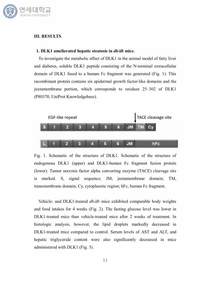
To investigate the metabolic effect of DLK1 in the animal model of fatty liver
and diabetes, soluble DLK1 peptide consisting of the N-terminal extracellular
domain of DLK1 fused to a human Fc fragment was generated (Fig. 1). This
recombinant protein contains six epidermal growth factor-like domains and the
juxtamembrane portion, which corresponds to residues 25–302 of DLK1
(P80370, UniProt Knowledgebase).
Fig. 1. Schematic of the structure of DLK1. Schematic of the structure of
endogenous DLK1 (upper) and DLK1-human Fc fragment fusion protein
(lower). Tumor necrosis factor alpha converting enzyme (TACE) cleavage site
is marked. S, signal sequence; JM, juxtamembrane domain; TM,
transmembrane domain; Cy, cytoplasmic region; hFc, human Fc fragment.
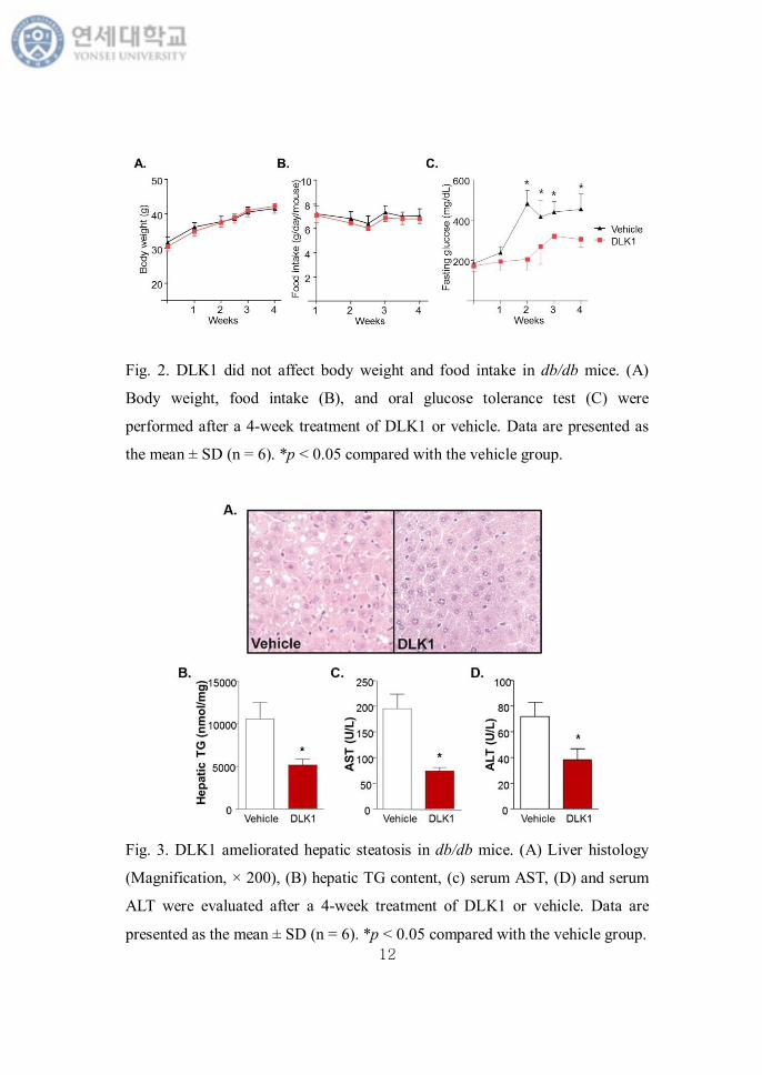
Vehicle- and DLK1-treated db/db mice exhibited comparable body weights
and food intakes for 4 weeks (Fig. 2). The fasting glucose level was lower in
DLK1-treated mice than vehicle-treated mice after 2 weeks of treatment. In
histologic analysis, however, the lipid droplets markedly decreased in
DLK1-treated mice compared to control. Serum levels of AST and ALT, and
hepatic triglyceride content were also significantly decreased in mice
administered with DLK1 (Fig. 3).
12
Fig. 2. DLK1 did not affect body weight and food intake in db/db mice. (A)
Body weight, food intake (B), and oral glucose tolerance test (C) were
performed after a 4-week treatment of DLK1 or vehicle. Data are presented as
the mean ± SD (n = 6). *p < 0.05 compared with the vehicle group.
Fig. 3. DLK1 ameliorated hepatic steatosis in db/db mice. (A) Liver histology
(Magnification, × 200), (B) hepatic TG content, (c) serum AST, (D) and serum
ALT were evaluated after a 4-week treatment of DLK1 or vehicle. Data are
presented as the mean ± SD (n = 6). *p < 0.05 compared with the vehicle group.
13
2. DLK1 reduced hepatic lipid accumulation via AMPK activation in
db/db mice and HepG2 cells.
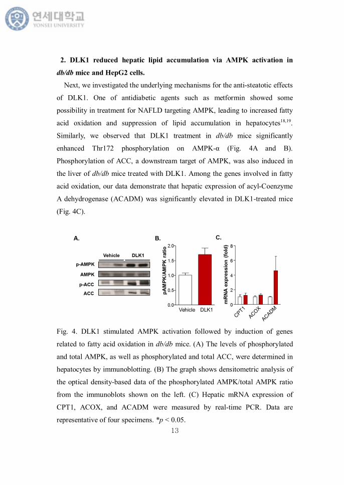
Next, we investigated the underlying mechanisms for the anti-steatotic effects
of DLK1. One of antidiabetic agents such as metformin showed some
possibility in treatment for NAFLD targeting AMPK, leading to increased fatty
acid oxidation and suppression of lipid accumulation in hepatocytes18,19.
Similarly, we observed that DLK1 treatment in db/db mice significantly
enhanced Thr172 phosphorylation on AMPK-α (Fig. 4A and B).
Phosphorylation of ACC, a downstream target of AMPK, was also induced in
the liver of db/db mice treated with DLK1. Among the genes involved in fatty
acid oxidation, our data demonstrate that hepatic expression of acyl-Coenzyme
A dehydrogenase (ACADM) was significantly elevated in DLK1-treated mice
(Fig. 4C).
Fig. 4. DLK1 stimulated AMPK activation followed by induction of genes
related to fatty acid oxidation in db/db mice. (A) The levels of phosphorylated
and total AMPK, as well as phosphorylated and total ACC, were determined in
hepatocytes by immunoblotting. (B) The graph shows densitometric analysis of
the optical density-based data of the phosphorylated AMPK/total AMPK ratio
from the immunoblots shown on the left. (C) Hepatic mRNA expression of
CPT1, ACOX, and ACADM were measured by real-time PCR. Data are
representative of four specimens. *p < 0.05.
14
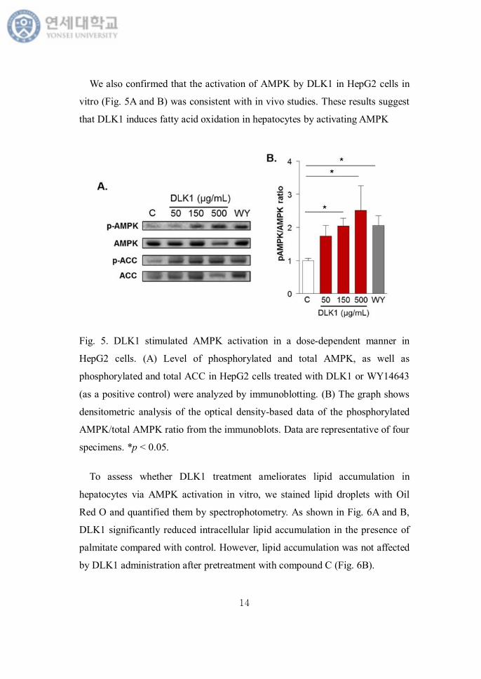
We also confirmed that the activation of AMPK by DLK1 in HepG2 cells in
vitro (Fig. 5A and B) was consistent with in vivo studies. These results suggest
that DLK1 induces fatty acid oxidation in hepatocytes by activating AMPK
Fig. 5. DLK1 stimulated AMPK activation in a dose-dependent manner in
HepG2 cells. (A) Level of phosphorylated and total AMPK, as well as
phosphorylated and total ACC in HepG2 cells treated with DLK1 or WY14643
(as a positive control) were analyzed by immunoblotting. (B) The graph shows
densitometric analysis of the optical density-based data of the phosphorylated
AMPK/total AMPK ratio from the immunoblots. Data are representative of four
specimens. *p < 0.05.
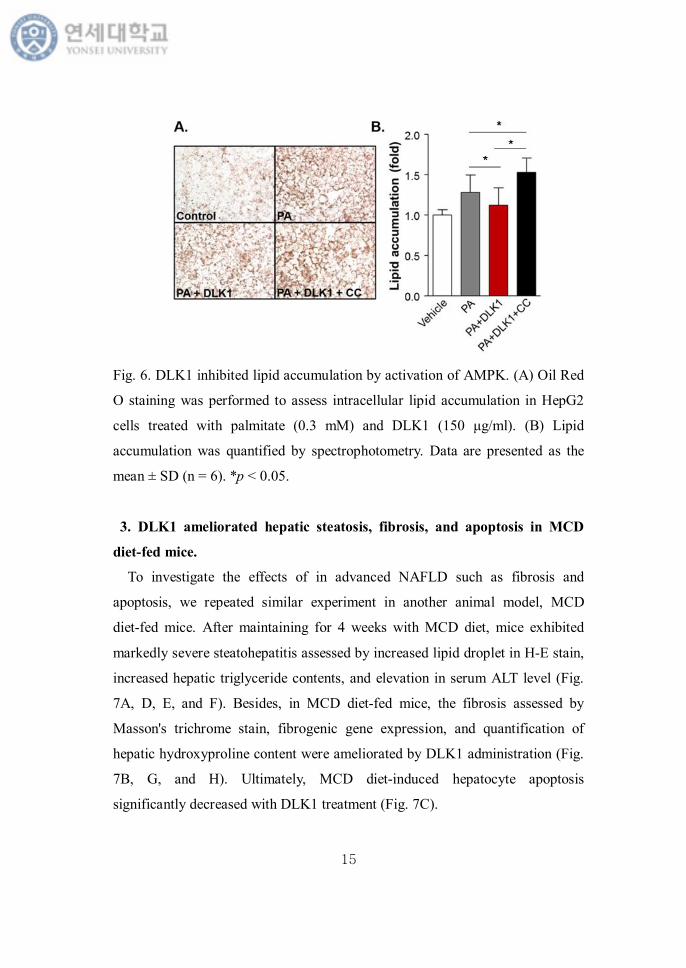
To assess whether DLK1 treatment ameliorates lipid accumulation in
hepatocytes via AMPK activation in vitro, we stained lipid droplets with Oil
Red O and quantified them by spectrophotometry. As shown in Fig. 6A and B,
DLK1 significantly reduced intracellular lipid accumulation in the presence of
palmitate compared with control. However, lipid accumulation was not affected
by DLK1 administration after pretreatment with compound C (Fig. 6B).
15
Fig. 6. DLK1 inhibited lipid accumulation by activation of AMPK. (A) Oil Red
O staining was performed to assess intracellular lipid accumulation in HepG2
cells treated with palmitate (0.3 mM) and DLK1 (150 μg/ml). (B) Lipid
accumulation was quantified by spectrophotometry. Data are presented as the
mean ± SD (n = 6). *p < 0.05.
3. DLK1 ameliorated hepatic steatosis, fibrosis, and apoptosis in MCD
diet-fed mice.
To investigate the effects of in advanced NAFLD such as fibrosis and
apoptosis, we repeated similar experiment in another animal model, MCD
diet-fed mice. After maintaining for 4 weeks with MCD diet, mice exhibited
markedly severe steatohepatitis assessed by increased lipid droplet in H-E stain,
increased hepatic triglyceride contents, and elevation in serum ALT level (Fig.
7A, D, E, and F). Besides, in MCD diet-fed mice, the fibrosis assessed by
Masson's trichrome stain, fibrogenic gene expression, and quantification of
hepatic hydroxyproline content were ameliorated by DLK1 administration (Fig.
7B, G, and H). Ultimately, MCD diet-induced hepatocyte apoptosis
significantly decreased with DLK1 treatment (Fig. 7C).
16
4. DLK1 ameliorated HSC activation and hepatic fibrosis by AMPK
activation.
In the setting of chronic liver disease including NAFLD, HSCs are the
primary target of fibrogenic stimuli in the diseased liver. TGF-β signals through
the sequential phosphorylation of Smad2 or Smad3 are considered one of the
major underlying mechanisms in the process of HSCs activation. So we
investigate these pathways in MCD-diet fed mice. To stimulate HSCs, we used
KLA induced LX-2 cells. KLA increased mRNA expression of TGF-β in a dose
dependent manner (Fig. 8A). Then we tested the anti-fibrotic effect of DLK1
treatment. After a dose of 1 ng/mL of KLA treatment for 24 hours, TGF-β, the
marker of HSCs activation, and fibrogenic gene expression such as α-SMA and
collagen were much increased. DLK1 pretreatment recovered this change (Fig.
8B-D).
17
18
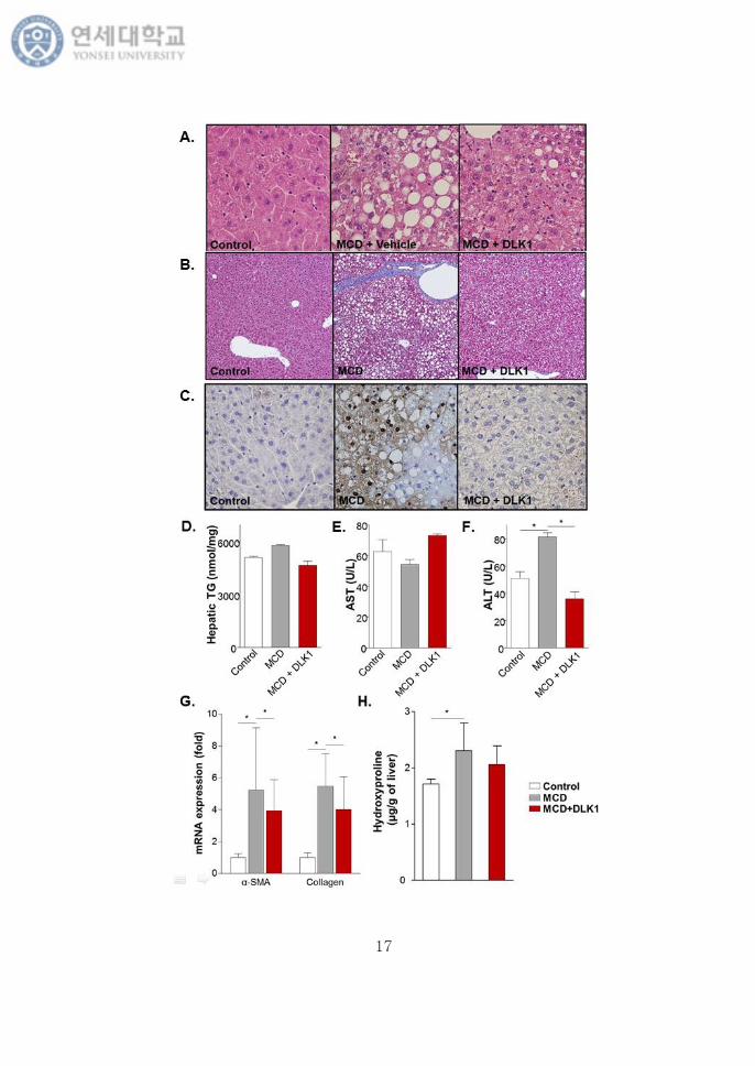
Fig 7. DLK1 ameliorated hepatic steatosis, fibrosis, and apoptosis in MCD
diet-fed mice. (A) Liver histology (Magnification, × 200), (B) Masson's
trichrome stain (Magnification, × 200), and (C) TUNEL stain were performed in
MCD diet-fed mice with DLK1 treatment for 4 weeks. (D) Hepatic TG content,
(E) serum AST, (F) and serum ALT were evaluated. (G) Hepatic mRNA
expression of α-SMA and collagen were measured by real-time PCR. (H)
Hydroxyproline content was quantified. Data are representative of four
specimens. *p < 0.05.
To investigate the underlying mechanism with DLK1 treatment, we
stimulated LX-2 cells with 5 ng/mL of TGF-β. Consistent with the previous
reports, TGF-β treatment increased phosphorylated form of Smad2/3 in LX-2
cells, however, DLK1 treatment reduced pSmad2/3 expression. Furthermore, 10
μM of compound C treatment blocked this effect of DLK1 on TGF-β/Smad
signaling activation (Fig. 9).
19
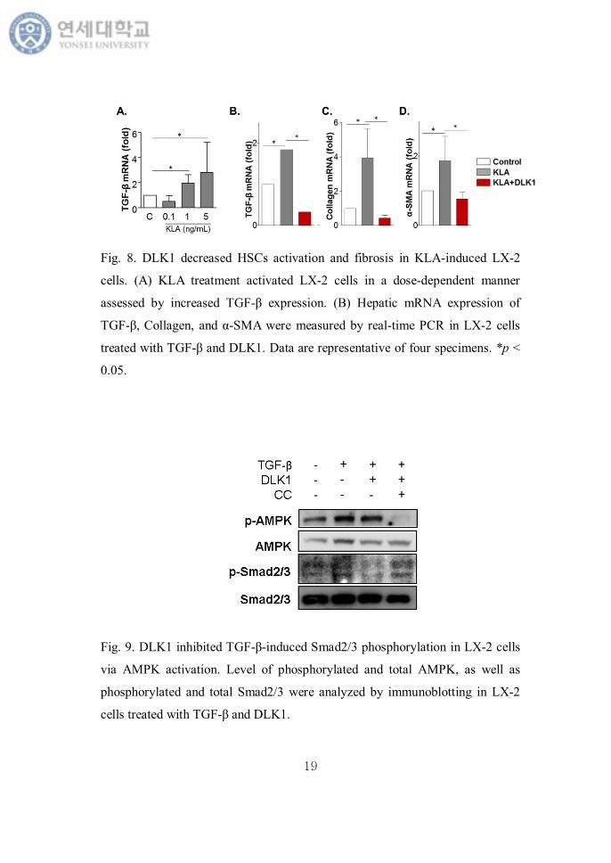
Fig. 8. DLK1 decreased HSCs activation and fibrosis in KLA-induced LX-2
cells. (A) KLA treatment activated LX-2 cells in a dose-dependent manner
assessed by increased TGF-β expression. (B) Hepatic mRNA expression of
TGF-β, Collagen, and α-SMA were measured by real-time PCR in LX-2 cells
treated with TGF-β and DLK1. Data are representative of four specimens. *p <
0.05.
Fig. 9. DLK1 inhibited TGF-β-induced Smad2/3 phosphorylation in LX-2 cells
via AMPK activation. Level of phosphorylated and total AMPK, as well as
phosphorylated and total Smad2/3 were analyzed by immunoblotting in LX-2
cells treated with TGF-β and DLK1.
20
5. DLK1 reduced activin A-induced cell death in steatotic hepatocyte by
AMPK activation.
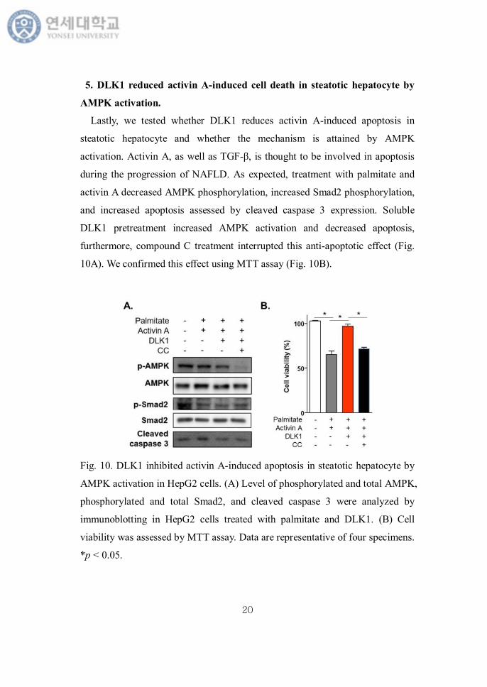
Lastly, we tested whether DLK1 reduces activin A-induced apoptosis in
steatotic hepatocyte and whether the mechanism is attained by AMPK
activation. Activin A, as well as TGF-β, is thought to be involved in apoptosis
during the progression of NAFLD. As expected, treatment with palmitate and
activin A decreased AMPK phosphorylation, increased Smad2 phosphorylation,
and increased apoptosis assessed by cleaved caspase 3 expression. Soluble
DLK1 pretreatment increased AMPK activation and decreased apoptosis,
furthermore, compound C treatment interrupted this anti-apoptotic effect (Fig.
10A). We confirmed this effect using MTT assay (Fig. 10B).
Fig. 10. DLK1 inhibited activin A-induced apoptosis in steatotic hepatocyte by
AMPK activation in HepG2 cells. (A) Level of phosphorylated and total AMPK,
phosphorylated and total Smad2, and cleaved caspase 3 were analyzed by
immunoblotting in HepG2 cells treated with palmitate and DLK1. (B) Cell
viability was assessed by MTT assay. Data are representative of four specimens.
*p < 0.05.
21
IV. DISCUSSION
The present study reports that exogenous DLK1 administration ameliorated
hepatic steatosis and fibrosis in two different murine models. By generating a
fusion protein containing the extracellular domain of DLK1 and the human Fc
region, which increases its stability, we found the unexpected action of DLK1
on prevention of steatosis by increased hepatic fatty acid oxidation via AMPK
activation in db/db mice, one of the typical animal models for obesity and
hepatic steatosis. This animal model, however, has a limitation that it represents
only hepatic lipid accumulation, not severe inflammation and cirrhosis as shown
in advanced form of NAFLD in human. So, we tried to investigate the effect of
exogenous DLK1 administration on the development and progression of
NAFLD to NASH in another model. In mice fed MCD diet, exogenous DLK1
administration significantly decreased hepatic steatosis, fibrosis, and finally
hepatocyte apoptosis.
NAFLD has become an important worldwide health problem due to its high
prevalence and close association with various metabolic diseases, including
obesity, type 2 diabetes, metabolic syndrome, and cardiovascular disease. The
major cause of the clinical significance of NAFLD is that it could progress from
simple steatosis to steatohepatitis, liver cirrhosis, and ultimately hepatocellular
carcinoma. Actually, NAFLD is projected to replace hepatitis C as the leading
cause for liver transplantation by 2020 in USA.20. In spite of this current
situation, however, it is still unclear what triggers the progression of NAFLD to
NASH accompanied by inflammation and fibrosis. A "multiple-hit" hypothesis
for the pathogenesis of NAFLD based on an animal model has been proposed,
however, the exact mechanism remains incomplete.
DLK1 was first known from a 3T3-L1 preadipocyte complementary DNA
library and has been linked to the inhibition of adipogenic differentiation21.
Some researches using DLK1-null mice and transgenic mice overexpressing
22
DLK1 in adipose tissue, demonstrated that DLK1 has an important role as a
negative regulator of adipogenesis. DLK1-null mice exhibited accelerated body
weight gain with increased mass of adipose tissues 22. These conflicting findings
may be due to the differences in the dosing of DLK1 and the timing of action.
This explanation is supported by a recent study showing that tightly regulated
dosage control of DLK1 is important for its regulatory function in
neurogenesis23. Furthermore, DLK1 has been identified as a dosage-critical
gene during development and growth24. So the effect of exogenous DLK1
administered in adult animal models may be different from that of previous
genetic animal models in which DLK1 expression during early life has been
manipulated. In contrast to previous findings from genetically engineered mice,
we observed that exogenous administration of soluble DLK1 did not affect the
development of adipose tissue.
AMPK acts as a metabolic master regulator by regulating cellular ATP
concentration and energy homeostasis25. Therefore AMPK activation could be
the treatment of diabetes, obesity, and NAFLD. In our study, DLK1 treatment
protected the mice or cell from hepatic steatosis in db/db mice by AMPK
activation. Activated AMPK has been known to be related with suppressing
biosynthetic pathways like fatty acid synthesis, and stimulating catabolic
processes such as fatty acid oxidation in the liver26.
Recently, Charalambous et al. demonstrated that DLK1 overexpression
ameliorated hepatic steatosis with reduced body weight and improved insulin
sensitivity in obese mice27 This might be consistent with our data on positive
metabolic effect and protection from hepatic steatosis. However, the
mechanisms of the metabolic effect are different, because the body weight,
adipose tissue mass as well as the food intake did not decrease with DLK1
treatment in the present study. So it is not a secondary effect with decreased
energy intake or body weight. Further study to clarify the direct function of
DLK1 administration on liver metabolism would be warranted.
23
We confirmed the anti-fibrotic and anti-apoptotic effect of exogenous DLK1
treatment in another animal model. These interesting effects were also attained
by activation of AMPK, which leads to inhibition of Smad signaling pathway.
There have been several reports of protective effect of AMPK activation from
hepatic fibrosis, however, the exact mechanisms remained unknown. Lim et al.
showed the inhibition of TGF-β-induced fibrosis in LX-2 cells by AMPK
agonist such as metformin and AICAR. AMPK activation, however, did not
affect TGF-β-stimulated phosphorylation of Smad2 or Smad3. AICAR or
metformin decreased Smad3 interaction with transcriptional coactivator p30028.
In contrast, another study with adiponectin-based short peptide, another AMPK
activator, demonstrated the association between AMPK activation and
TGF-β/Smad2 signaling in thioacetamide-induced hepatic fibrosis cell model29.
In addition, AMPK activation could inhibit hepatic fibrosis by targeting Smad
pathway by itself28.
Furthermore, our study suggests the ant-apoptotic role of DLK1 in
progression from NAFLD to NASH induced by activin signaling. Activins, one
of TGF-β superfamily members, have been known to be related with hepatic
fibrosis and apoptosis. It has been discovered that activin A plays as important
roles in liver regeneration and fibrosis among activins30. In this process, HSCs
were identified as the major source of activin A, and expression of activin A was
rapidly induced upon culturing of these cells31. Activin A acts in an autocrine
and paracrine manner on hepatocytes and activated HSCs30. In hepatic fibrosis,
activin A pathway involves Smad2 and Smad3 signaling through direct serine
phosphorylation after receptor binding32. Also, in hepatocytes with activin A
stimulation, Smad proteins are known to be involved in the apoptotic
process33,34. In our study, treatment with palmitate and activin A in HepG2 cells
reproduced the condition with apoptotic signal in steatotic hepatocyte in our
experiment. Co-treatment with palmitate and activin A markedly increased
hepatocyte lipoapoptosis, however, DLK1 pretreatment ameliorated the
24
apoptosis. When treated with compound C, an AMPK inhibitor, the
anti-apoptotic effect of DLK1 disappeared.
This study has some limitations. Further research will be required to better
determine the underlying molecular mechanism of DLK1 in AMPK activation,
and the direct relationship between AMPK activation and Smad2/3 pathway
inhibition in the process of hepatic fibrosis and apoptosis. In addition, the
effective dosage of soluble DLK1 protein to activate AMPK is high, at least 150
μg/mL in our study with HepG2 cells. Because there is few studies using
soluble DLK1 protein in animal study, we could not compare the dosage
directly. However, delicate pharmacokinetic and pharmacodynamics studies
should also be conducted to define the optimal therapeutic concentration of
DLK1 in the blood to avoid causing adverse effects. In spite of these, this study
is still meaningful. The strength of this study is it demonstrates that DLK1
administration showed beneficial effects on hepatic steatosis, fibrosis, and
apoptosis in murine models. These results provide novel evidence that DLK1
may be a good therapeutic option for treating various spectrums of
obesity-related chronic liver disease.
25
V. CONCLUSION
In conclusion, the present study demonstrated that exogenous administration
of DLK1 markedly reduced hepatic steatosis by activation of fatty acid
oxidation through increased AMPK phosphorylation. Furthermore, DLK1
significantly protected the progression of hepatic fibrosis and apoptosis in MCD
diet-fed mice by inhibition of Smad signaling pathway following AMPK
activation. Taken together, these findings suggest that DLK1 might be a novel
therapeutic strategy for the treatment of various stages of NAFLD from
steatosis to fibrosis.
26
REFERENCES
1. Day CP. Non-alcoholic fatty liver disease: a massive problem. Clin Med
(Lond) 2011;11:176-8.
2. Sanyal AJ, Brunt EM, Kleiner DE, Kowdley KV, Chalasani N, Lavine
JE, et al. Endpoints and clinical trial design for nonalcoholic
steatohepatitis. Hepatology 2011;54:344-53.
3. Tiniakos DG, Vos MB, Brunt EM. Nonalcoholic fatty liver disease:
pathology and pathogenesis. Annu Rev Pathol 2010;5:145-71.
4. Ratziu V, Bellentani S, Cortez-Pinto H, Day C, Marchesini G. A
position statement on NAFLD/NASH based on the EASL 2009 special
conference. J Hepatol 2010;53:372-84.
5. Loomba R, Abraham M, Unalp A, Wilson L, Lavine J, Doo E, et al.
Association between diabetes, family history of diabetes, and risk of
nonalcoholic steatohepatitis and fibrosis. Hepatology 2012;56:943-51.
6. Bolos V, Grego-Bessa J, de la Pompa JL. Notch signaling in
development and cancer. Endocr Rev 2007;28:339-63.
7. Kopan R, Ilagan MX. The canonical Notch signaling pathway:
unfolding the activation mechanism. Cell 2009;137:216-33.
8. Pajvani UB, Shawber CJ, Samuel VT, Birkenfeld AL, Shulman GI,
Kitajewski J, et al. Inhibition of Notch signaling ameliorates insulin
resistance in a FoxO1-dependent manner. Nat Med 2011;17:961-7.
9. Pajvani UB, Qiang L, Kangsamaksin T, Kitajewski J, Ginsberg HN,
Accili D. Inhibition of Notch uncouples Akt activation from hepatic
lipid accumulation by decreasing mTorc1 stability. Nat Med
2013;19:1054-60.
10. Valenti L, Mendoza RM, Rametta R, Maggioni M, Kitajewski C,
Shawber CJ, et al. Hepatic notch signaling correlates with insulin
resistance and nonalcoholic fatty liver disease. Diabetes
27
2013;62:4052-62.
11. Xie G, Karaca G, Swiderska-Syn M, Michelotti GA, Kruger L, Chen Y,
et al. Cross-talk between Notch and Hedgehog regulates hepatic stellate
cell fate in mice. Hepatology 2013;58:1801-13.
12. Dufraine J, Funahashi Y, Kitajewski J. Notch signaling regulates tumor
angiogenesis by diverse mechanisms. Oncogene 2008;27:5132-7.
13. Lai EC. Notch signaling: control of cell communication and cell fate.
Development 2004;131:965-73.
14. Baladron V, Ruiz-Hidalgo MJ, Nueda ML, Diaz-Guerra MJ,
Garcia-Ramirez JJ, Bonvini E, et al. dlk acts as a negative regulator of
Notch1 activation through interactions with specific EGF-like repeats.
Exp Cell Res 2005;303:343-59.
15. Nueda ML, Baladron V, Sanchez-Solana B, Ballesteros MA, Laborda J.
The EGF-like protein dlk1 inhibits notch signaling and potentiates
adipogenesis of mesenchymal cells. J Mol Biol 2007;367:1281-93.
16. Wang Y, Sul HS. Ectodomain shedding of preadipocyte factor 1 (Pref-1)
by tumor necrosis factor alpha converting enzyme (TACE) and
inhibition of adipocyte differentiation. Mol Cell Biol 2006;26:5421-35.
17. Backliwal G, Hildinger M, Chenuet S, Wulhfard S, De Jesus M, Wurm
FM. Rational vector design and multi-pathway modulation of HEK
293E cells yield recombinant antibody titers exceeding 1 g/l by
transient transfection under serum-free conditions. Nucleic Acids Res
2008;36:e96.
18. Pernicova I, Korbonits M. Metformin--mode of action and clinical
implications for diabetes and cancer. Nat Rev Endocrinol
2014;10:143-56.
19. Zhang BB, Zhou G, Li C. AMPK: an emerging drug target for diabetes
and the metabolic syndrome. Cell Metab 2009;9:407-16.
20. Kneeman JM, Misdraji J, Corey KE. Secondary causes of nonalcoholic
28
fatty liver disease. Therap Adv Gastroenterol 2012;5:199-207.
21. Smas CM, Sul HS. Pref-1, a protein containing EGF-like repeats,
inhibits adipocyte differentiation. Cell 1993;73:725-34.
22. Moon YS, Smas CM, Lee K, Villena JA, Kim KH, Yun EJ, et al. Mice
lacking paternally expressed Pref-1/Dlk1 display growth retardation and
accelerated adiposity. Mol Cell Biol 2002;22:5585-92.
23. Ferron SR, Charalambous M, Radford E, McEwen K, Wildner H, Hind
E, et al. Postnatal loss of Dlk1 imprinting in stem cells and niche
astrocytes regulates neurogenesis. Nature 2011;475:381-5.
24. da Rocha ST, Charalambous M, Lin SP, Gutteridge I, Ito Y, Gray D, et
al. Gene dosage effects of the imprinted delta-like homologue 1
(dlk1/pref1) in development: implications for the evolution of
imprinting. PLoS Genet 2009;5:e1000392.
25. Hardie DG. The AMP-activated protein kinase pathway--new players
upstream and downstream. J Cell Sci 2004;117:5479-87.
26. Hardie DG, Ross FA, Hawley SA. AMPK: a nutrient and energy sensor
that maintains energy homeostasis. Nat Rev Mol Cell Biol
2012;13:251-62.
27. Charalambous M, Da Rocha ST, Radford EJ, Medina-Gomez G, Curran
S, Pinnock SB, et al. DLK1/PREF1 regulates nutrient metabolism and
protects from steatosis. Proc Natl Acad Sci U S A 2014;111:16088-93.
28. Lim JY, Oh MA, Kim WH, Sohn HY, Park SI. AMP-activated protein
kinase inhibits TGF-beta-induced fibrogenic responses of hepatic
stellate cells by targeting transcriptional coactivator p300. J Cell
Physiol 2012;227:1081-9.
29. Wang H, Zhang H, Zhang Z, Huang B, Cheng X, Wang D, et al.
Adiponectin-derived active peptide ADP355 exerts anti-inflammatory
and anti-fibrotic activities in thioacetamide-induced liver injury. Sci
Rep 2016;6:19445.
29
30. Werner S, Alzheimer C. Roles of activin in tissue repair, fibrosis, and
inflammatory disease. Cytokine Growth Factor Rev 2006;17:157-71.
31. De Bleser PJ, Niki T, Xu G, Rogiers V, Geerts A. Localization and
cellular sources of activins in normal and fibrotic rat liver. Hepatology
1997;26:905-12.
32. Shi Y, Massague J. Mechanisms of TGF-beta signaling from cell
membrane to the nucleus. Cell 2003;113:685-700.
33. Chen W, Woodruff TK, Mayo KE. Activin A-induced HepG2 liver cell
apoptosis: involvement of activin receptors and smad proteins.
Endocrinology 2000;141:1263-72.
34. Yang L, Roh YS, Song J, Zhang B, Liu C, Loomba R, et al.
Transforming growth factor beta signaling in hepatocytes participates in
steatohepatitis through regulation of cell death and lipid metabolism in
mice. Hepatology 2014;59:483-95.
30
ABSTRACT(IN KOREAN)
DLK1 여가 지 간 생 에 미치는 과
<지도 수 차 수>
연 학 학원 학과
민
Notch 신 달체계 가 지 간 다양한 단계에
한다는 사실 알 다. Delta-like 1 homolog (DLK1) 는
Notch 신 달체계 내 억 , 근 보고에
DLK1 지 직 나 골격근 뿐 아니라
간 재생 과 에도 여한다고 한다. 라 본 연 에 는
DLK1 여가 지 간 생 나 지 에 어 한 역할 하는
지 알아보고 하 다.
합 DLK1 단 질 , DLK1 포 도 과 간 역
린 Fc 합하여 만들었다. 동물 실험 해 는
db/db 마우스 동물 과 티 닌-콜린 결핍 식 지한
C57BL/6 마우스 사 하 다. DLK1 4주동안 여하
다. HepG2 포주 간 간 상 포 LX-2 포주 하
여 포 실험 진행하 다.
합 DLK1 단 질 여는 db/db 마우스 동물 에 간
직내 지 함량 고 간 수치 시키는 등 지
간 진행 히 감 시켰다. 또한 DLK1 여는 간 직
31
에 AMP-activated protein kinase (AMPK) 산 진하 고,
지 산 산 담당하는 다. HepG2 포
에 도 마찬가지 결과 보 다. 티 닌-콜린 결핍 식 마우
스 에 는 DLK1 여가 지 간 진행 뿐 아니라 간
, 간 포 포 사 감 시켰다. LX-2 포에
DLK1 여는 간 상 포 억 하 고,
키는 감 시켰는 , 는 AMPK
통하여 TGF-β/Smad 신 달체계 억 함 나타났다. 또
한 DLK1 여는 간 포 지 사 는 결과
보 다.
결 , DLK1 여는 지 간 생과 진행 다양한
단계에 억 하 , 는 간 포 간 상 포에 AMPK
통하여 한다. 는 DLK1 지 간
치료에 어 새 운 약물 타겟 가능 시하 다.
----------------------------------------------------------------------------------------
핵심 는 말: Delta-like 1 homolog, 비알코 지 간, 간 ,
AMP-activated protein kinase
32
PUBLICATION LIST
Lee YH, Yun MR, Kim HM, Jeon BH, Park BC, Lee BW, et al.
Exogenous administration of DLK1 ameliorates hepatic
steatosis and regulates gluconeogenesis via activation of
AMPK. Int J Obes (Lond) 2016;40:356-65.