2 hyperpigmentation and melasma 2
TRANSCRIPT
2Hyperpigmentation and Melasma 2
2.1Hyperpigmentation
Almost all women, presumably up to 90%, and particularly those with dark hair and com-plexions, note some degree of hyperpigmentation during pregnancy. Patients are often deeply concerned by this condition, and may view these changes with uneasiness, yet hardly ever do they voluntarily express their concerns [21].
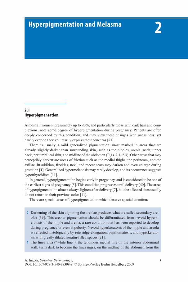
There is usually a mild generalized pigmentation, most marked in areas that are already slightly darker than surrounding skin, such as the nipples, areola, neck, upper back, periumbilical skin, and midline of the abdomen (Figs. 2.1–2.3). Other areas that may perceptibly darken are areas of friction such as the medial thighs, the perineum, and the axillae. In addition, freckles, nevi, and recent scars may darken and even enlarge during gestation [1]. Generalized hypermelanosis may rarely develop, and its occurrence suggests hyperthyroidism [11].
In general, hyperpigmentation begins early in pregnancy, and is considered to be one of the earliest signs of pregnancy [5]. This condition progresses until delivery [40]. The areas of hyperpigmentation almost always lighten after delivery [7], but the affected sites usually do not return to their previous color [11].
There are special areas of hyperpigmentation which deserve special attention:
› Darkening of the skin adjoining the areolae produces what are called secondary are-olae [39]. This areolar pigmentation should be differentiated from nevoid hyperk-eratosis of the nipple and areola, a rare condition that has been reported to develop during pregnancy or even at puberty. Nevoid hyperkeratosis of the nipple and areola is reflected histologically by rete ridge elongation, papillomatosis, and hyperkerato-sis with greatly dilated keratin-filled spaces [21].
› The linea alba (“white line”), the tendinous medial line on the anterior abdominal wall, turns dark to become the linea nigra, on the midline of the abdomen from the
A. Ingber, Obstetric Dermatology, 7DOI: 10.1007/978-3-540-88399-9, © Springer-Verlag Berlin Heidelberg 2009
8 2 Hyperpigmentation and Melasma
2
2.2Melasma
The term “melasma,” or facial hyperpigmentation, is derived from the Greek melas, mean-ing “black” [8]. It has been also termed “chloasma gravidarum” and “the mask of preg-nancy” [38] (Fig. 2.5). Its onset is usually during the second half of the gestational period, and it occurs in 45–75% of pregnancies [1, 11, 38]. It is said to be more common in dark-haired, brown-eyed, dark-complexion women [38, 40]. A review of major studies evaluating
umbilicus to the symphysis pubis (Fig. 2.4). It can extend superiorly to the xiphoid process. It appears during the first trimester of pregnancy and, like other areas of hyperpigmentation, is most pronounced in dark-haired, dark-complexioned women [40]. This condition is often accompanied by displacement of the umbilicus, at term, to the right side of the patient, the “ligamentum teres” sign. This shift persists post-partum until the abdominal muscles regain their normal tone [21]. This displacement is the consequence of the pressure of the uterus on the ligamentum teres and falci-form ligament [2]. The development of a darker areola and a linea nigra were used clinically in the past to establish parity [27].
› Group B natural pigmentary demarcation lines have been reported to occur on the lower limbs during pregnancy. Pigmentary demarcation lines are borders of abrupt transition between more deeply pigmented skin and that of lighter pigmentation. One report describes two women developing demarcation lines: one developed the condi-tion during her pregnancy, and the condition remained unchanged 8 months postpar-tum; the other developed the condition immediately postpartum [12]. Another report describes four cases of women developing demarcation lines; all of them developed the condition only during pregnancy, and it disappeared completely between the first and third months postpartum [37]. There were also reports of demarcation lines dur-ing pregnancy which were accompanied by erythema. While the erythema disap-peared quickly, the pigmentation subsided more slowly. The explanation suggestion for this phenomenon was compression of peripheral nerves by the enlarged uterus in the late period of pregnancy, thus influencing the innervated cutaneous microvascu-lature to induce neurogenic inflammation with resultant erythema and pigmentation [26]. Demarcation lines were also reported to occur during pregnancy on the medial aspect of the arms [15].
› Longitudinal melanonychia, which is commonly seen in black persons and orientals, but is rarely present in white women, was reported to develop in a white woman dur-ing pregnancy [9, 21].
› Dermal melanocytosis is a rare dermatosis, which was found to darken during preg-nancy. This dermatosis may also be triggered during pregnancy [31]. Reactivation of preexisting dormant dermal melanocytes by a variety of hormonal factors is thought to occur. Elevation in estrogen and progesterone levels and elevation in keratinocyte-derived endothelin 1 and melanocyte simulating hormone (MSH) levels upon sun exposure potentiate tyrosinase activity and thus stimulate melanogenesis [21].
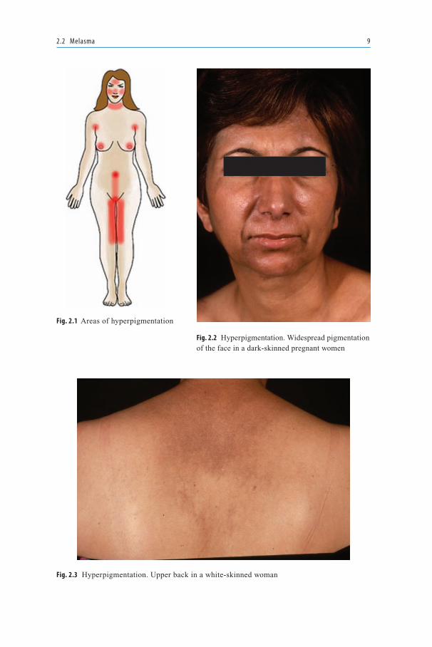
Fig. 2.1 Areas of hyperpigmentation
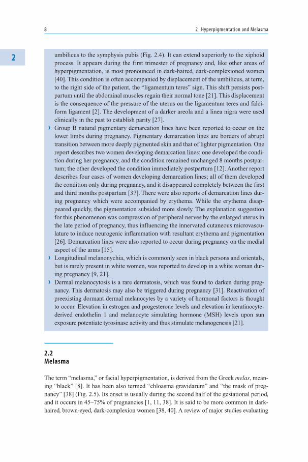
Fig. 2.2 Hyperpigmentation. Widespread pigmentation of the face in a dark-skinned pregnant women
2.2 Melasma 9
Fig. 2.3 Hyperpigmentation. Upper back in a white-skinned woman
10 2 Hyperpigmentation and Melasma
2
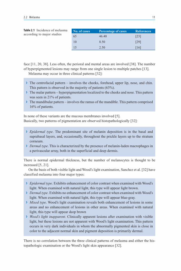
the incidence of melasma during pregnancy is summarized in Table 2.1. The major differ-ences in the incidence of melasma in the different studies were attributed to the fact that pigmentary changes are more discernible in fair-skinned individuals [16].
Superimposed on the general pigmentation induced by pregnancy, melasma may appear as blotchy, irregular, sharply demarcated, tan to dark brown plaques or patches of pigmen-tation in symmetrical distribution on the forehead, temples, cheeks, and central areas of the
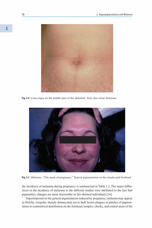
Fig. 2.5 Melasma. “The mask of pregnancy.” Typical pigmentation on the cheeks and forehead
Fig. 2.4 Linea nigra on the middle part of the abdomen. Note also striae distensae
2.2 Melasma 11
face [11, 20, 38]. Less often, the perioral and mental areas are involved [38]. The number of hyperpigmented lesions may range from one single lesion to multiple patches [13].
Melasma may occur in three clinical patterns [32]:
› The centrofacial pattern – involves the cheeks, forehead, upper lip, nose, and chin. This pattern is observed in the majority of patients (63%).
› The malar pattern – hyperpigmentation localized to the cheeks and nose. This pattern was seen in 21% of patients.
› The mandibular pattern – involves the ramus of the mandible. This pattern comprised 16% of patients.
In none of these variants are the mucous membranes involved [5].Basically, two patterns of pigmentation are observed histopathologically [32]:
› Epidermal type. The predominant site of melanin deposition is in the basal and suprabasal layers, and, occasionally, throughout the prickle layers up to the stratum corneum.
› Dermal type. This is characterized by the presence of melanin-laden macrophages in a perivascular array, both in the superficial and deep dermis.
There is normal epidermal thickness, but the number of melanocytes is thought to be increased [5, 21].
On the basis of both visible light and Wood’s light examination, Sanchez et al. [32] have classified melasma into four major types:
› Epidermal type. Exhibits enhancement of color contrast when examined with Wood’s light. When examined with natural light, this type will appear light brown.
› Dermal type. Exhibits no enhancement of color contrast when examined with Wood’s light. When examined with natural light, this type will appear blue-gray.
› Mixed type. Wood’s light examination reveals both enhancement of lesions in some areas and no enhancement of lesions in other areas. When examined with natural light, this type will appear deep brown
› Wood’s light inapparent. Clinically apparent lesions after examination with visible light, but these lesions are not apparent with Wood’s light examination. This pattern occurs in very dark individuals in whom the abnormally pigmented skin is close in color to the adjacent normal skin and pigment deposition is primarily dermal.
There is no correlation between the three clinical patterns of melasma and either the his-topathologic examination or the Wood’s light skin appearance [32].
No. of cases Percentage of cases References
65 46.40 [23]
10 8.50 [29]
15 2.50 [16]
Table 2.1 Incidence of melasma according to major studies
12 2 Hyperpigmentation and Melasma
2Oral contraceptives are also a main etiologic factor of melasma, and melasma and
pigmentation represent the most common cutaneous side effects of oral contraceptives: 5–34% of individuals are affected, the higher incidence being seen in the more deeply pigmented race. Hyperpigmentation of the face also occurs in some women during nor-mal menstrual periods. Melasma during pregnancy indicates susceptibility to increased pigmentation with the contraceptive pill and the drug may induce melasma when none existed in pregnancy [20, 38]. Melasma during menstrual periods and melasma induced by the contraceptive pill may be predictive of women who will have pigmentary changes during pregnancy [38].
The etiologic factors in melasma include, other than pregnancy and oral contraception, the following [32]:
› Cosmetics. In the past, cosmetics made of poorly refined ingredients, containing irri-tating or phosphosensitizing substances, and increased occupational exposure to crude tars and oils were a major cause for melanosis of the face. Other ingredients in cosmet-ics that were selectively implicated as causative factors for dermal melanoses include certain fatty acids, photoactive contaminants of mineral oils, petrolatum, beeswax, cer-tain dyes, para-phenylenediamine, and perfume ingredients.
› Genetic and racial predisposition. A genetic predisposition has been supported only by occasional reports of familial occurrence. There is a high occurrence of melasma in patients of Hispanic origin.
› Medications. A wide variety of medications have produced hyperpigmentation. These include metals such as arsenic, iron, copper, bismuth, silver, and gold and phototoxic and antiseizure drugs [5]. Hyperpigmentation has also followed the administration of organic compounds such as quinacrine and 5-ethyl-3-methyl-5-phenylhydantoin.
Other etiologic factors for the development of melasma are nutrition, liver disease, and parasitosis [5, 8]. A significant association was also found in nonpregnant women between thyroid autoimmunity and melasma, primarily in women whose condition developed during pregnancy or after the ingestion of oral contraceptive drugs [10].
Exposure to UV light radiation plays a significant etiologic role in the pathogenesis of melasma, and is felt to be necessary for the development of melasma [8]. The majority of patients with melasma observed the onset of their melasma during the summer months. Sun exposure is also a prominent cause of exacerbation of melasma, while during the win-ter months, melasma appears to be less noticeable [32]. The areas of the face that are most affected are those that have maximum exposure to the sun. Clearly, however, many women are exposed to sunlight during pregnancy without developing melasma [40].
In a study of 210 melasma patients, the incidence of various causative factors was found to be universally due to sunlight exposure (100%), and in descending order was due to pregnancy (27%), cosmetics (14%), familial factors (13%), and oral contraceptive use (6.3%) [13].
The course of melasma is progressive during pregnancy [38], but its intensity is not necessarily proportional to that of general melanosis. Unlike the persistent melasma associated with the use of oral contraceptives, the melasma of pregnancy usually fades within 1 year of delivery, but it may occasionally persist if the hyperpigmentation is deep
2.3 Pathogenesis of Pigmentation 13
[11, 20, 38]. The persistence incidence was reported to be less than 10%, although one study found persistence in 30% of cases after 10 years [1, 40]. If it is persistent postpartum, some women note a premenstrual hyperpigmented flare [5].
2.3 Pathogenesis of Pigmentation
The enzyme responsible for melanin production is tyrosinase, a copper-containing oxyge-nase enzyme, which mediates melanin production through the intermediate, L-dopa [27]. Tyrosinase activity may be regulated by many factors. For example, incubation of human melanocytes with 1,25-dihydroxyvitamin D
3, α-MSH, and β-estradiol caused an increase
in tyrosinase activity [30]. Nevertheless, no specific receptors have yet been demonstrated on the melanocyte [27].
Hyperpigmentation in pregnancy is attributed by some investigators to an increased out-put of some combination of placental, pituitary, and ovarian hormones [40]. An increased amount of MSH of the pituitary was postulated in the past to be the cause of hyperpig-mentation. It was even believed that the demonstration of MSH in urine could be used as a pregnancy test [4].
Shizume and Lerner [33] showed by analyzing the urine of 38 pregnant and postpartum women and the blood of 13 pregnant women using bioassay methods that after the second month of pregnancy MSH levels in the urine became elevated above normal levels. MSH levels continued to increase until delivery, and then rapidly decreased to normal within 5 days postpartum. The importance of MSH in pregnancy was also suggested by other investigators [4, 19].
Later, these bioassay methods were doubted, because they lacked real specificity; it has not been possible to identify the substances and it was not clear if they were of pituitary origin [36].
Since the major pituitary MSH in the human is thought to be β-MSH, Thody et al. [36] measured plasma β-MSH levels in pregnancy and postpartum using a specific radioimmu-noassay. They showed that plasma β-MSH levels in late pregnancy were within the normal range and were no different from the levels in women after parturition.
Later, the levels of circulating immunoreactive α-MSH were evaluated during preg-nancy. It was found that during the first trimester immunoreactive α-MSH levels were undetectable in plasma of most subjects. During late pregnancy, however, the levels of α-MSH were significantly higher than those of the control. It was concluded that α-MSH has no pigmentary role in pregnancy because the pigmentation begins early and is nor-mally focal, unlike that of Addison’s disease and Cushing’s syndrome [3].
Other factors which were suggested to be related to hyperpigmentation in pregnancy are progesterone and estrogen, the levels of both of which are increased during pregnancy [7]. Blood levels of progesterone increase throughout pregnancy, and estrogen production rises from the eighth week and begins to decrease after the 30th week of pregnancy. This pattern follows the progression of hyperpigmentation [18]. Using guinea pigs as a model and by utilizing controlled histochemical experiments, Snell et al. [34] showed that estrogen,
14 2 Hyperpigmentation and Melasma
2when given alone in small doses, increased the output of melanin by the melanocytes and exerted its greatest influence on sexual skin – the areola. When the dose was raised, the effect was increased and was accompanied by a rise in the melanocyte count in the skin of the anterior abdominal wall. The effect of small doses of estrogen was shown to be aug-mented by giving progesterone at the same time. These hormones were thus suggested to be strong stimulants of melanogenesis.
Thus, hyperpigmentation of the face occurs in women taking oral contraceptives or dur-ing the menstrual cycle, supporting the idea that estrogen and progesterone are involved [27]. In addition, hyperpigmentation of the face is associated with disorders involving the function of the uterus and ovaries, a condition termed “chloasma uterinum” [24].
A small study found that nevi from patients who are pregnant or 1 month postpartum have increased numbers of estrogen and progesterone binding sites. The time of onset of increased hormonal binding of nevi in pregnancy could not be determined in this study, but it was speculated to be prior to 5 months’ gestation [6].
It should be noted that although progesterone, estrogen, and MSH were implicated as causative factors in melasma of pregnancy, they have not been found to be consistently ele-vated in melasma in general [32]. Generally, the hormone levels with oral contraceptives are not though to be high enough to initiate melasma [22]. A study evaluating MSH levels in nine women with idiopathic melasma (nonpregnancy or oral-contraceptive-associated melasma) found no elevation of MSH levels compared with levels in controls [28].
Recently, investigators found that a lipid extraction from the placenta had a pigment-inducing activity both in vivo and in vitro. The placenta was found to be rich in bioactive sphingolipids, which were found to induce melanogenesis by upregulating the expression of various melanogenic enzymes – tyrosinase and tyrosinase-related proteins 1 and 2 at the translational and transcriptional levels [17].
Wade et al. [38] have suggested that the reason why only certain body areas, but not the entire body, are affected by hyperpigmentation is that melanocytes in the areas affected are more sensitive to hormonal stimulation. Another explanation may be the greater population of melanocytes in the affected sites [40]. One investigation showed that there is a greater population of melanocytes in the skin of the face and forehead, with the population density of melanocytes being 2–4 times greater than in the skin of the thigh and arms [35].
2.4Treatment
Treatment of pigmentary alteration in pregnancy is unsatisfactory and preventive measures or treatment for hyperpigmentation in pregnancy is limited [40]. For generalized hyper-pigmentation, many physicians do not recommend therapy other than sunscreens for sun-exposed areas [41].
The treatment of melasma is usually postponed until after delivery for apparent rea-sons: (1) the hormonal cause for melasma persists throughout pregnancy, making melasma more resistant to treatment; (2) most women have a significant improvement in melasma after parturition, making therapy unnecessary; (3) the mainstay of therapy for melasma is relatively contraindicated during pregnancy [25].
2.4 Treatment 15
During pregnancy, women should avoid heavy suntanning and should use broad-spectrum (UVA and UVB) sunscreens and appropriate clothing [22, 40]. In addition, patients are encouraged to use opaque sunscreens to diminish all UV and visible light in order to minimize further pigment production [22]. It has been demonstrated that a correlation exists between the use of sunscreen and the result of melasma treatment [13]. Sunbathing is absolutely contraindicated, as a few minutes of sunbathing can reverse the benefit of months of therapy [13].
Avoidance of trauma, especially if the patient is prone to easy scarring (scars may hyperpigment) is recommended, and patients are cautioned to avoid rubbing or irritation [22]. Cosmetics on the face, especially perfumed preparations, should be avoided; how-ever, the patients may choose to use nonallergenic, skin-colored, cover-up preparations. Nonhormonal methods of contraception should be considered.
Bleaching agents have been used with variable, but usually poor, results. Phenolic com-pounds were regarded as dangerous as they can cause considerable skin irritation, contact dermatitis, and dyschromias. However, two trials have shown satisfactory results without major side effects. The combination of 4-hydroxianisol (2%) and tretinoin (0.01%) and the use of n-acetyl-4-S-cysteaminylphenol in a 4% oil-in-water emulsion applied twice daily for 6 months were found to give good results [13]. Vitamin C was used systematically in mild forms of melasma, and vitamin E seems to act synergistically to vitamin C.
A report by Kligman and Willis [14] shows satisfactory results in 14 of 16 patients with melasma using a formulation of tretinoin (0.1%), hydroquinone (5%), and dexamethasone (0.1%). The vehicle was hydrophilic ointment or a solution of equal parts of ethanol and propylene glycol. The patients in this study were young, adult, white women taking con-traceptive pills. The regimen used was once-daily application. The depigmentation was of satisfactory magnitude by 5–7 weeks after the start of the treatment. No adverse effects were reported, and normal skin was only slightly, if at all, lightened.
The usefulness of depigmenting agents is dependent on the histologic classification and Wood’s light typing described by Sanchez et al. [32]. The predominantly epidermal type of melasma responds to formulations containing 2% hydroquinone plus 0.05% retinoic acid with nightly use. Melasma of mixed type with predominantly dermal deposition of melanin, however, responds poorly to this therapy [32]. Wong and Ellis [40] reported that their experience with 2–5% hydroquinone is only partially effective in some patients with the epidermal type of melasma, even after months of use.
A slight modification to the treatment suggested by Kligman and Willis was reported by Katsambas and Antoniou [13]. Their formulation includes 4% hydroquinone, 0.05% tretinoin, and 1%hydrocortisone acetate, which is applied twice a day until patients reach the desired degree of depigmentation, but never for more than 8–10 weeks [13]. Another treatment suggestion is the use of 0.05% tretinoin, 2% hydroquinone, and 0.1% betham-ethasone valerate cream [13].
Hydroquinone is in the US FDA pregnancy category C because studies have never been conducted to see whether hydroquinone causes fetal harm when applied topically [25]. Tretinoin is in US FDA pregnancy category C and in category D in the equivalent Austral-ian administration. There are several reports of fetal malformation when mothers used tretinoin during pregnancy; nonetheless, causation cannot be definitely determined [25].
The development of hypopigmented lesions is a potential side effect of the treatment with the combinations described above, but the risk can be minimized with proper patient
16 2 Hyperpigmentation and Melasma
2education and follow-up examination [39]. Other side effects include dermatitis and hyperpigmentation [40]. In addition, the prolonged and excessive use of preparations of hydroquinone higher than 3% can result in ochronosis, a blue-black postinflammatory discoloration [13].
Azelaic acid cream has been reported to be of benefit in the treatment of melasma when applied twice daily. Various study have shown good to excellent results in 63–80% of patients with epidermal or mixed type of melasma after 6 months. Since treatment of melasma with azelaic acid requires several months, a combination of azelaic acid with other drugs deserves consideration [13].
Medium-depth chemical peeling has been used alone in the treatment of melasma; however, sometimes this treatment may result in worsening of the hyperpigmented lesion. A combined treatment with 35% trichloracetic acid peel followed by 4% hydroquinone acetate cream and 0.05% tretinoin cream was reported to be effective for epidermal and mixed-type melasma in fair-skinned women. The response of melasma to chemical peel-ing is, however, unpredictable, and may be aggravated as a result of postinflammatory hyperpigmentation. In general, chemical peeling should not be used in dark-skinned individuals [13].
The treatment of melasma with various types of lasers has been tried, but without sig-nificant success. The use of a 510 nm dye laser, resulted in failure and sometimes even in hyperpigmentation. Use of Q-switched ruby lasers has good results initially, but a quick recurrence occurs when the treatment is stopped [13]. The copper vapor laser and the argon laser also failed to show improvement in patients with melasma [10]. The use of lasers during pregnancy has not been studied [25].
In conclusion, the important aspect of treatment is stressing the natural course of hyper-melanosis, since improvement is expected postpartum. Reassuring is thus a major con-tributor to treatment.
› Hyperpigmentation during pregnancy is a very common condition, affecting mainly the areolae, genital skin, and linea alba. This condition usually regresses after parturition.
› Melasma is hyperpigmentation of the face, which occurs in 45–75% of pregnant women. Melasma may also be seen with oral contraceptive pills. It may occur in three clinical patterns – centrofacial, malar, and mandibular – and histologically in two types – epidermal and dermal. UV light radiation plays a major etiologic role.
› Hyperpigmentation in pregnancy has been attributed to increased output of some combination of placental, pituitary, and ovarian hormones – namely, melanocyte-simulating hormone, estrogen, progesterone, and bioactive sphingolipids derived from the placenta.
› Treatment of pigmentary alterations during pregnancy consists mainly of reassur-ance, prevention of sun exposure, and the use of sunscreens.
Summary
References 17
References
1. Barankin B, Silver SG, Carruthers A (2002) The skin in pregnancy. J Cutan Med Surg 6:236–240 2. Beischer NA, Wein P (1996) Linea alba pigmentation and umbilical deviation in nulliparous
pregnancy: the ligamentum teres sign. Obstet Gynecol 87:254–256 3. Clark D, Thody AJ, Shuster S et al (1978) Immunoreactive alpha-MSH in human plasma in
pregnancy. Nature 273:163–164 4. Dahlberg BC (1961) Melanocyte stimulating substances in the urine of pregnant women. Acta
Endocrinol Suppl (Copenh) 38(Suppl 60):1–51 5. Elling SV, Powell FC (1997) Physiological changes in the skin during pregnancy. Clin Derma-
tol 15:35–43 6. Ellis DL, Wheeland RG (1986) Increased nevus estrogen and progesterone ligand binding
related to oral contraceptives or pregnancy. J Am Acad Dermatol 14:25–31 7. Errickson CV, Matus NR (1994) Skin disorders of pregnancy. Am Fam Physician 49:605–610 8. Eudy SF, Baker GF (1990) Dermatopathology for the obstetrician. Clin Obstet Gynecol
33:728–737 9. Fryer JM, Werth VP (1992) Pregnancy-associated hyperpigmentation: longitudinal melanony-
chia. J Am Acad Dermatol 26:493–49410. Grimes PE (1995) Melasma. Etiologic and therapeutic considerations. Arch Dermatol
131:1453–145711. Hellreich P (1974) The skin changes of pregnancy. Cutis 13:82–8612. James WD, Meltzer MS, Guill MA et al (1984) Pigmentary demarcation lines associated with
pregnancy. J Am Acad Dermatol 11:438–44013. Katsambas A, Antoniou C (1995) Melasma. Classification and treatment. J Eur Acad Dermatol
Venereol 4:21714. Kligman AM, Willis I (1975) A new formula for depigmenting human skin. Arch Dermatol
111:40–4815. Kumari R, Laxmisha C, Thappa DM (2006) Pigmentary demarcation lines associated with
pregnancy. J Cosmet Dermatol 5:169–17016. Kumari R, Jaisankar TJ, Thappa DM (2007) A clinical study of skin changes in pregnancy.
Indian J Dermatol Venereol Leprol 73:14117. Mallick S, Singh SK, Sarkar C et al (2005) Human placental lipid induces melanogenesis
by increasing the expression of tyrosinase and its related proteins in vitro. Pigment Cell Res 18:25–33
18. Martin AG, Leal-Khouri S (1992) Physiologic skin changes associated with pregnancy. Int J Dermatol 31:375–378
19. McGuinness B (1963) Melanocyte-stimulating hormone. A clinical and laboratory study. Ann N Y Acad Sci 100:640–657
20. McKenzie AW (1971) Skin disorders in pregnancy. Practitioner 206:773–78021. Muallem MM, Rubeiz NG (2006) Physiological and biological skin changes in pregnancy. Clin
Dermatol 24:80–8322. Murray JC (1990) Pregnancy and the skin. Dermatol Clin 8:327–33423. Muzaffar F, Hussain I, Haroon TS (1998) Physiologic skin changes during pregnancy: a study
of 140 cases. Int J Dermatol 37:429–43124. Newcomer VD, Lindberg MC, Sternberg TH (1961) A melanosis of the face (“chloasma”).
Arch Dermatol 83:284–29925. Nussbaum R, Benedetto AV (2006) Cosmetic aspects of pregnancy. Clin Dermatol 24:
133–141
18 2 Hyperpigmentation and Melasma
226. Ozawa H, Rokugo M, Aoyama H (1993) Pigmentary demarcation lines of pregnancy with
erythema. Dermatology 187:134–13627. Parmley T, O’Brien TJ (1990) Skin changes during pregnancy. Clin Obstet Gynecol 33:
713–71728. Perez M, Sanchez JL, Aguilo F (1983) Endocrinologic profile of patients with idiopathic
melasma. J Invest Dermatol 81:543–54529. Raj S, Khopkar U, Kapasi A et al (1992) Skin in pregnancy. Indian Dermatol Venereol Leprol
58:84–8830. Ranson M, Posen S, Mason RS (1988) Human melanocytes as a target tissue for hormones: in
vitro studies with 1 alpha-25, dihydroxyvitamin D3, alpha-melanocyte stimulating hormone, and beta-estradiol. J Invest Dermatol 91:593–598
31. Rubin AI, Laborde SV, Stiller MJ (2001) Acquired dermal melanocytosis: appearance during pregnancy. J Am Acad Dermatol 45:609–613
32. Sanchez NP, Pathak MA, Sato S et al (1981) Melasma: a clinical, light microscopic, ultrastruc-tural, and immunofluorescence study. J Am Acad Dermatol 4:698–710
33. Shizume K, Lerner AB (1954) Determination of melanocyte-stimulating hormone in urine and blood. J Clin Endocrinol Metab 14:1491–1510
34. Snell RS, Bischitz PG (1960) The effect of large doses of estrogen and estrogen and progester-one on melanin pigmentation. J Invest Dermatol 35:73–82
35. Szabo G (1954) The number of melanocytes in human epidermis. Br Med J 1:1016–101736. Thody AJ, Plummer NA, Burton JL et al (1974) Plasma beta-melanocyte-stimulating hormone
levels in pregnancy. J Obstet Gynaecol Br Commonw 81:875–87737. Vazquez M, Ibanez MI, Sanchez JL (1986) Pigmentary demarcation lines during pregnancy.
Cutis 38:263–26638. Wade TR, Wade SL, Jones HE (1978) Skin changes and diseases associated with pregnancy.
Obstet Gynecol 52:233–24239. Winton GB, Lewis CW (1982) Dermatoses of pregnancy. J Am Acad Dermatol 6:977–99840. Wong RC, Ellis CN (1984) Physiologic skin changes in pregnancy. J Am Acad Dermatol
10:929–94041. Wong RC, Ellis CN (1989) Physiologic skin changes in pregnancy. Semin Dermatol 8:7–11

http://www.springer.com/978-3-540-88398-2