19th international platelet immunology workshop of...
TRANSCRIPT
19th
International Platelet Immunology Workshop
of ISBT Organizing Labs Héma-Québec, Montreal (Quebec) CANADA Lucie Richard Canadian Blood Services, Winnipeg (Manitoba) CANADA Lynnette Beaudin
Lynne Meilleur Canadian Blood Services, Edmonton (Alberta) CANADA Dr. Gwen Clarke University of British Columbia, Vancouver (British Columbia) CANADA & Department of Blood Bank Services, Oman Shadhiya Al Khan

19th International Platelet Immunology Workshop of ISBT
2
19th International Platelet Immunology Workshop of ISBT
3
Table of Contents
Content Organizing Laboratories ........................................................................................................................ 4
Acknowledgments................................................................................................................................... 5
Participating Laboratories ...................................................................................................................... 7
Report for the 19th International Immunology Workshop of ISBT ................................................ 13
Exercise 1 (part 1) ................................................................................................................................ 15
Results for Exercise 1 (part 1) ............................................................................................................ 17
Exercise 1 (part 2) ................................................................................................................................ 24
Results for Exercise 1 (part 2) ............................................................................................................ 25
Exercise 2 .............................................................................................................................................. 54
Results for Exercise 2 .......................................................................................................................... 55
Exercise 3 .............................................................................................................................................. 63
Results for Exercise 3 .......................................................................................................................... 64
Exercise 4 .............................................................................................................................................. 75
Results for Exercise 4 .......................................................................................................................... 76
Exercise 5 .............................................................................................................................................. 91
Results for Exercise 5 .......................................................................................................................... 92
Exercise 6 .............................................................................................................................................. 99
Background: ........................................................................................................................................ 100
Results for Exercise 6 ........................................................................................................................ 101
Final Conclusions ............................................................................................................................... 108
19th International Platelet Immunology Workshop of ISBT
4
Organizing Laboratories (by alphabetical country order)
Héma-Québec Lucie Richard Reference and Stem Cell Laboratory 4045, boulevard Côte-Vertu Montréal, (Québec) H4R 2W7 CANADA
Héma-Québec Antoine Lewin Medical Affairs and Innovation 4045, boulevard Côte-Vertu Montréal, (Québec) H4R 2W7 CANADA
Canadian Blood Services Lynnette Beaudin Lynne Meilleur Platelet Immunology Laboratory 777 William Avenue Winnipeg (Manitoba) R3E 3R4 CANADA
Canadian Blood Services Dr. Gwen Clarke Canadian Blood Services Medical Affairs and Innovation 8249 – 114th Street Edmonton (Alberta) T6G 2R8 CANADA University of British Columbia & Department of Blood Bank Services, Oman Dr. Shadhiya Al Khan Blood Bank Services, Bausher PO Box 393; Postal Code 100 Muscat, Oman
19th International Platelet Immunology Workshop of ISBT
5
Acknowledgments
We would like to thank: Immucor for providing the PAKLx kit to each participating laboratory who was interested to perform the Exercise 4. Ulrich Sachs, Nelson Tsuno and Sentot Santoso for their invaluable advice during the preparation of the workshop materials. Gerald Bertrand, for his contribution of one DNA sample. All of those who made generous offers of serum and/or DNA. All of you, for your feedback and encouragement.

19th International Platelet Immunology Workshop of ISBT
6

19th International Platelet Immunology Workshop of ISBT
7
Participating Laboratories (by alphabetical country order)
Country City Participant Email Laboratory Phone
Australia Brisbane Gail Pahn Mark Burton
[email protected] [email protected]
Platelet & Neutrophil Reference Laboratory Australian Red Cross Blood Service PO BOX 1197 Stafford, QLD, 4053 Australia
61 7 3838 9173
Australia Victoria Grant Mraz [email protected]
Victorian Transplantation and Immunogenetics Service Australian Red Cross Blood Service 100-154 Batman Street West Melbourne 3003 Victoria Australia
61 3 9694 0153
Austria Vienna Simon Panzer [email protected]
Vienna General Hospital Medical University of Vienna Laboratory for Platelet Immunobiology and Function Leitstelle 4i Waehringer Guertel 18-20 1090 Vienna Austria
43 1 40400 5330
Brazil Sao Paulo Rita Fontão-Wendel [email protected]
Blood Bank Hospital Sírio Libanês R. D. Adma Jafet, 91 Hospital Sirio Libanês 3o sub-solo 01308-050 São Paulo - SP - Brazil
55 11 3394 5272
Canada Saint-Laurent Lucie Richard [email protected]
Stem cell and Reference Laboratory Platelet Immunology 4300 Rue Garand Saint-Laurent (Québec) H4R 2W7, Canada
(514) 832-5000
Canada Winnipeg Lynnette Beaudin [email protected]
Canadian Blood Services Platelet Immunology Laboratory 777 William Ave., Winnipeg, Manitoba R3E 3R4, Canada
(204) 789-1149
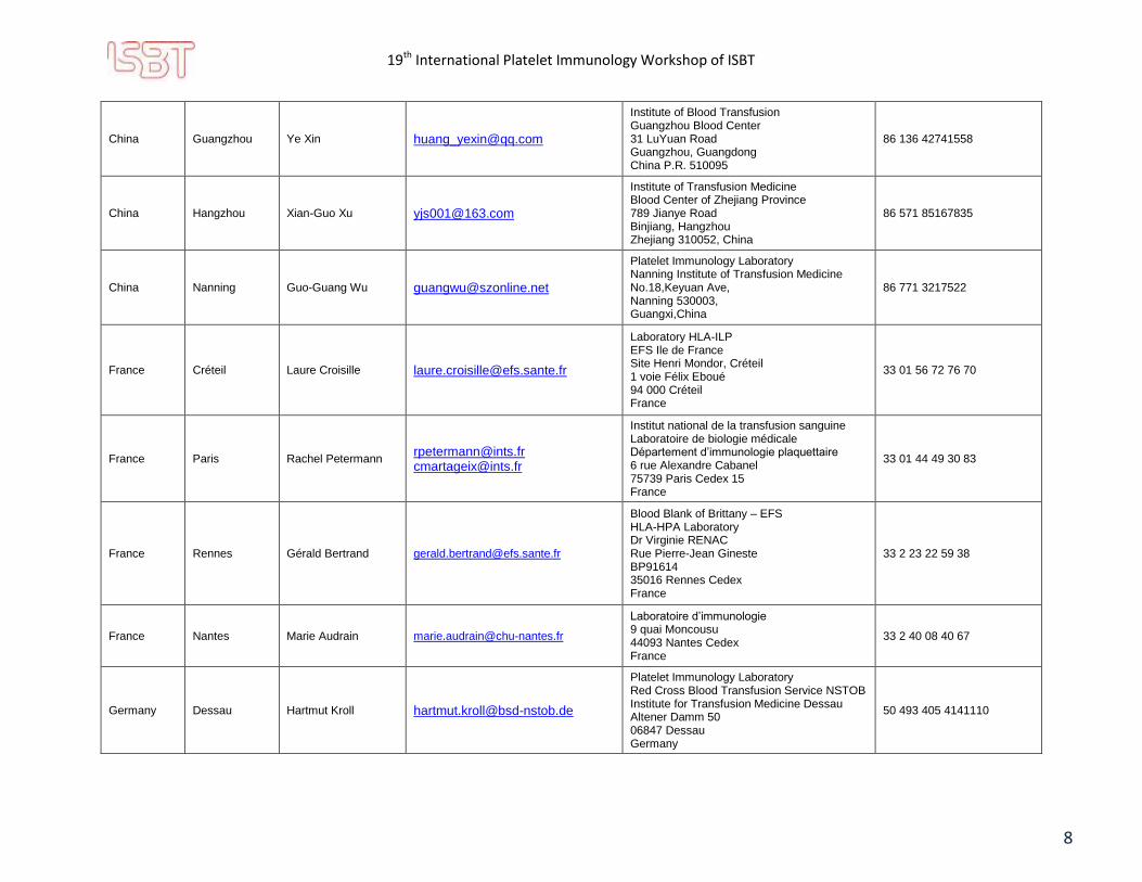
19th International Platelet Immunology Workshop of ISBT
8
China Guangzhou Ye Xin [email protected]
Institute of Blood Transfusion Guangzhou Blood Center 31 LuYuan Road Guangzhou, Guangdong China P.R. 510095
86 136 42741558
China Hangzhou Xian-Guo Xu [email protected]
Institute of Transfusion Medicine Blood Center of Zhejiang Province 789 Jianye Road Binjiang, Hangzhou Zhejiang 310052, China
86 571 85167835
China Nanning Guo-Guang Wu [email protected]
Platelet Immunology Laboratory Nanning Institute of Transfusion Medicine No.18,Keyuan Ave, Nanning 530003, Guangxi,China
86 771 3217522
France Créteil Laure Croisille [email protected]
Laboratory HLA-ILP EFS Ile de France Site Henri Mondor, Créteil 1 voie Félix Eboué 94 000 Créteil France
33 01 56 72 76 70
France Paris Rachel Petermann [email protected] [email protected]
Institut national de la transfusion sanguine Laboratoire de biologie médicale Département d’immunologie plaquettaire 6 rue Alexandre Cabanel 75739 Paris Cedex 15 France
33 01 44 49 30 83
France Rennes Gérald Bertrand [email protected]
Blood Blank of Brittany – EFS HLA-HPA Laboratory Dr Virginie RENAC Rue Pierre-Jean Gineste BP91614 35016 Rennes Cedex France
33 2 23 22 59 38
France Nantes Marie Audrain [email protected]
Laboratoire d’immunologie 9 quai Moncousu 44093 Nantes Cedex France
33 2 40 08 40 67
Germany Dessau Hartmut Kroll [email protected]
Platelet Immunology Laboratory Red Cross Blood Transfusion Service NSTOB Institute for Transfusion Medicine Dessau Altener Damm 50 06847 Dessau Germany
50 493 405 4141110
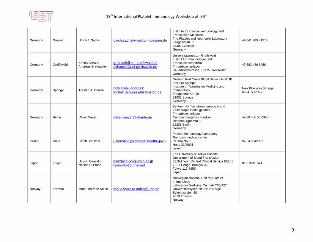
19th International Platelet Immunology Workshop of ISBT
9
Germany Giessen Ulrich J. Sachs [email protected]
Institute for Clinical Immunology and Transfusion Medicine The Platelet and Neutrophil Laboratory Langhansstr. 7 35392 Giessen Germany
49 641 985 41515
Germany Greifswald Karina Althaus Andreas Greinacher
[email protected] [email protected]
Universitatsmedizin Greifswald Institut fur Immunologie und Transfusionsmedizin Thrombozytenlabor Sauerbruchstrasse, 17475 Greifswald, Germany
49 383 486 5458
Germany Springe Torsten J Schulze new email address: [email protected]
German Red Cross Blood Service NSTOB Institute Springe Institute of Transfusion Medicine and Immunology Eldagsener Str. 38 31832 Springe Germany
New Phone in Springe: 05041/772-533
Germany Berlin Oliver Meyer [email protected]
Zentrum für Transfusionsmedizin und Zelltherapie Berlin gGmbH Thrombozytenlabor Campus Benjamin Franklin Hindenburgdamm 30 12203 Berlin Germany
49 30 450 553268
Israel Haifa Lilach Bonstein [email protected]
Platelet Immunology Laboratory Rambam medical center PO box 9602 Haifa 3109601 Israel
972 4 8542052
Japan Tokyo Hitoshi Okazaki Nelson H.Tsuno
[email protected] [email protected]
The University of Tokyo Hospital Department of Blood Transfusion 33,3rd floor, Central Clinical Service Bldg 1 7-3-1 Hongo, Bunkyo-ku, Tokyo 113-8655 Japan
81 3 3815 5411
Norway Tromsø Maria Therese Ahlen [email protected]
Norwegian National Unit for Platelet Immunology Laboratory Medicine, Trc. lab C00-427 Universitetssykehuset Nord-Norge Sykehusveien 38 9019 Tromsø Norway
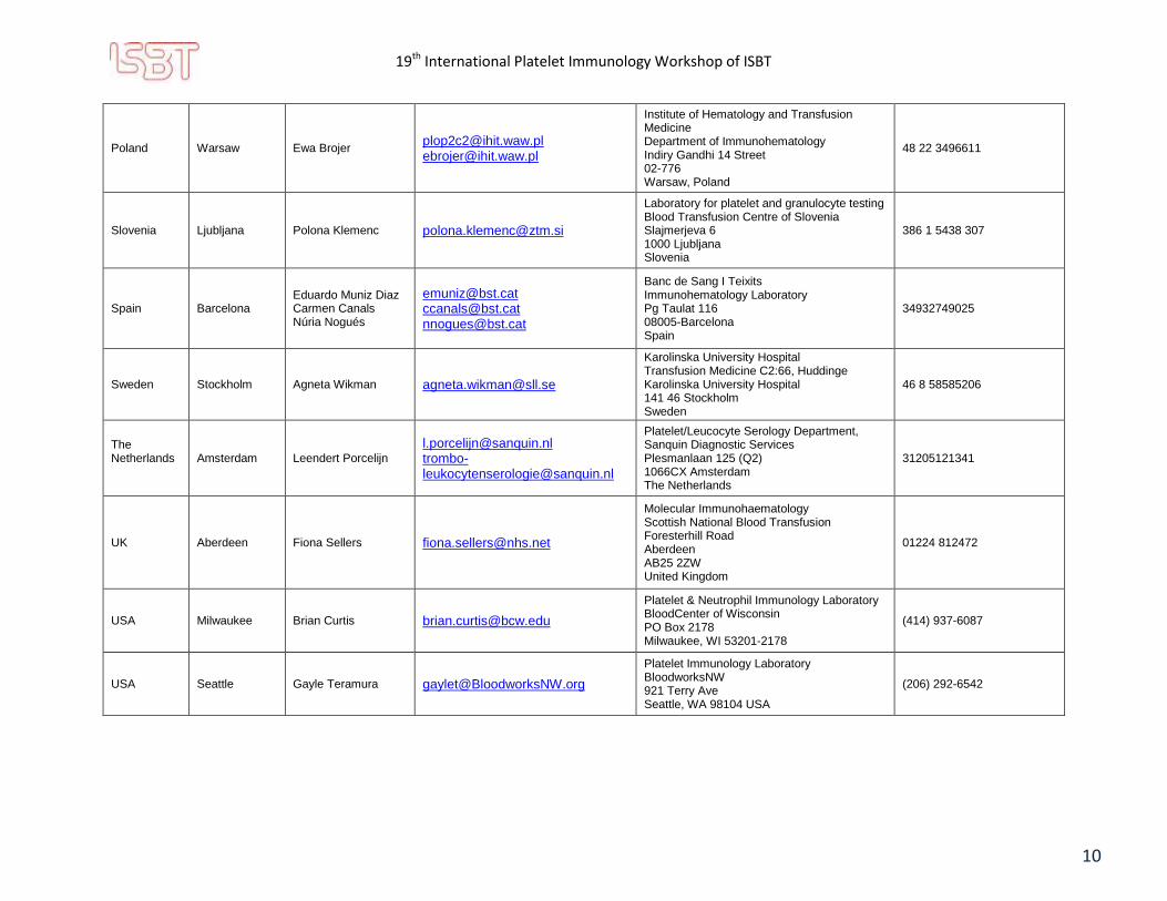
19th International Platelet Immunology Workshop of ISBT
10
Poland Warsaw Ewa Brojer [email protected] [email protected]
Institute of Hematology and Transfusion Medicine Department of Immunohematology Indiry Gandhi 14 Street 02-776 Warsaw, Poland
48 22 3496611
Slovenia Ljubljana Polona Klemenc [email protected]
Laboratory for platelet and granulocyte testing Blood Transfusion Centre of Slovenia Slajmerjeva 6 1000 Ljubljana Slovenia
386 1 5438 307
Spain Barcelona Eduardo Muniz Diaz Carmen Canals Núria Nogués
[email protected] [email protected] [email protected]
Banc de Sang I Teixits Immunohematology Laboratory Pg Taulat 116 08005-Barcelona Spain
34932749025
Sweden Stockholm Agneta Wikman [email protected]
Karolinska University Hospital Transfusion Medicine C2:66, Huddinge Karolinska University Hospital 141 46 Stockholm Sweden
46 8 58585206
The Netherlands
Amsterdam Leendert Porcelijn
[email protected] [email protected]
Platelet/Leucocyte Serology Department, Sanquin Diagnostic Services Plesmanlaan 125 (Q2) 1066CX Amsterdam The Netherlands
31205121341
UK Aberdeen Fiona Sellers [email protected]
Molecular Immunohaematology Scottish National Blood Transfusion Foresterhill Road Aberdeen AB25 2ZW United Kingdom
01224 812472
USA Milwaukee Brian Curtis [email protected]
Platelet & Neutrophil Immunology Laboratory BloodCenter of Wisconsin PO Box 2178 Milwaukee, WI 53201-2178
(414) 937-6087
USA Seattle Gayle Teramura [email protected]
Platelet Immunology Laboratory BloodworksNW 921 Terry Ave Seattle, WA 98104 USA
(206) 292-6542

19th International Platelet Immunology Workshop of ISBT
11
19th International Platelet Immunology Workshop of ISBT
12
19th International Platelet Immunology Workshop of ISBT
13
Report for the 19th International Immunology Workshop of ISBT
Introduction
The International Platelet Immunology Workshop aims to be a unique hands-on exercise which explores a variety of subjects in the field of clinical platelet immunology. Over the years, serology and molecular biology techniques as well as clinical practice have commensurately evolved. The past International Platelet Workshops have been a major witness of this evolution by addressing many clinical and laboratory aspects such as clinical management of NAIT, clinical and laboratory identification of ITP, new HPA antibody detection, development of control HPA positive cells, etc. The clinical aspects have been well covered; however, the laboratory aspects are still in need of development and standardization. The variability in antibody detection between labs, sensitivity of the techniques and standardization of results are a main preoccupation even today. Anti-HPA antibody identification is still a challenge today. The 19th Workshop was prepared to address these different points. For this, six exercises were proposed:
1) Serologic evaluation of 4 clinical cases and one donor case with a survey of laboratory practice for FNAIT diagnosis.
2) Special evaluation of one clinical case (Anti-HLA)
3) Genotyping of 5 DNA samples.
4) Assay on the PAKLx commercial kit.
5) Platelet preparation for detection of Anti-HPA-3 in MAIPA
6) MAIPA with focus to challenge a variety of Anti-CD109 monoclonals for the detection of Anti-HPA-15.
General Comments
There were 29 inscriptions, so 29 packages were sent. However, 28 laboratories participated in the majority of the proposed exercises and one laboratory desisted from participation.
Challenges
This workshop was ambitiously designed to address many subjects and problems; but by doing so, the amount of work that it necessitated was very high.
Lack of sample volume also compounded the challenge of participating labs to complete the workshop.
Also, we experienced problems with the shipping of material in some countries. Fortunately, these do not represent the majority, as shipping for the most part went as expected.
Regardless of the challenges, the majority of the labs participated in all exercises and performed all testing suggested.
19th International Platelet Immunology Workshop of ISBT
14
Observations
One main observation made throughout the workshop was the failure to strictly adhere to proper WHO nomenclature (WHO, 2017 at http://www.who.int/medicines/services/inn/en/) conventions at different levels.
1) Variation in reporting of nomenclature for HPA, monoclonal antibodies, glycoproteins, HLA.
2) Variation in answers in the survey.
3) Variation in MAIPA protocol and approach for case resolution.
4) Variation in reporting of genotyping results.
The saving and sending of the PAKLx data files were done successfully by the majority of the labs. Only two labs experienced technical issues, necessitating them to resend their files after reacquiring data.
Recommendations
For the next Workshop we may want to:
1) Focus on no more than two aspects or problems to be addressed.
2) Provide a maximum of 2 or 3 serum samples with rare antibody or perform one or two cases analysis with a rare or particular reactivity (including genotyping) instead of multiple cases with only regular reactivity.
3) Introduce new approaches or new techniques.
4) Work on a standardization of the MAIPA protocol and an optimization for enabling the use of small volume of sample.
5) Try new monoclonal antibodies and work on the optimization of the panel cells for the GPIV, HPA-3 and HPA-15 MAIPA.
19th International Platelet Immunology Workshop of ISBT
15
Exercise 1 (part 1) Characterization of Platelet-Specific Antibodies
Aim:
1) To determine the ability of the participant laboratories’ routine screening method to detect the presence of platelet-specific alloantibodies in ‘blind’ serum/plasma samples.
2) To identify the specificity of platelet-specific alloantibodies using MAIPA.
3) To compare performance in platelet-specific alloantibody detection and determine level of consensus for each antibody.
Materials Supplied:
Participating laboratories were provided with:
4 serum samples (S1, S2, S3, S4) containing between 1.0 mL and 0.5 mL each
1 plasma sample (S5) containing 0.3 mL
Methods:
The provided 5 samples were to be investigated for the presence of platelet-specific alloantibodies (see case description). Participating laboratories were to:
1) Test all samples using their routine screening method.
2) Test all samples using their regular MAIPA method.
3) Test all samples using any other detection/identification technique.
Note: S5 (plasma) was to be tested against all test methods indicated above.
Results:
Assay data and the identified specificity of platelet-specific alloantibodies were reported in the Excel answer grid provided.
19th International Platelet Immunology Workshop of ISBT
16
Exercise 1 (part 1 continued) Cases History
Case No.1 (S1):
This is a case of FNAIT. The mother is blood group AB Rh(D) positive and father is blood group A Rh(D) positive, both caucasians from Canada. It was the second pregnancy of the mother. The first pregnancy and delivery were unremarkable. The second baby had a platelet count of 70x109/L at delivery. He was transfused with HPA-1b/b platelet one day after delivery.The platelet count dropped to 30x109/L post transfusion. The hospital sent samples from the mother and father for investigation. The case was rushed because the baby had significant purpura and was at risk for intracranial hemorrhage.
Case No.2 (S2):
This is a case of FNAIT. The mother is Greek and the father is Algerian and both parents are blood group A Rh(D) positive. This was the first pregnancy of the mother. The baby was born with a platelet count of 18x109/L and an intracranial hemorrhage. He received crossmatched platelets until the antibody could be identified.
Case No.3 (S3):
This is a case of FNAIT. Both parents are Caucasians from Algeria. The mother has blood group O Rh(D) positive and the father has blood group A Rh(D) positive. Her first three pregnancies were unremarkable. She was then referred after her fourth pregnancy/delivery. The fourth newborn had a platelet count of 35x109/L but no antibody was identified at that time. She became pregnant again, for a 5th time, 19 years later. The case was referred to us again during the 12th week of pregnancy. The fifth child had a platelet count of 30x109/L at birth but demonstrated no complications.
Case No.4 (S4):
This is a female blood donor implicated in a transfusion reaction after her first donation. The transfusion was associated with pronounced thrombocytopenia in the recipient.
Case No.5 (S5):
This is a case of FNAIT. 37-year-old female G2P2 of Dutch ethnicity from Canada, with an uneventful 1st pregnancy in 2014, delivered a premature infant at 33.4 weeks of gestation due to fetal intracranial bleed and abnormal heart rate. At birth, baby had intracranial hemorrhage, bruising and thrombocytopenia (15 x109/L platelet count). Baby was treated with platelet transfusions initially followed by IVIG and antigen negative platelets transfusion.
19th International Platelet Immunology Workshop of ISBT
17
Results for Exercise 1 (part 1) Characterization of Platelet-Specific Antibodies
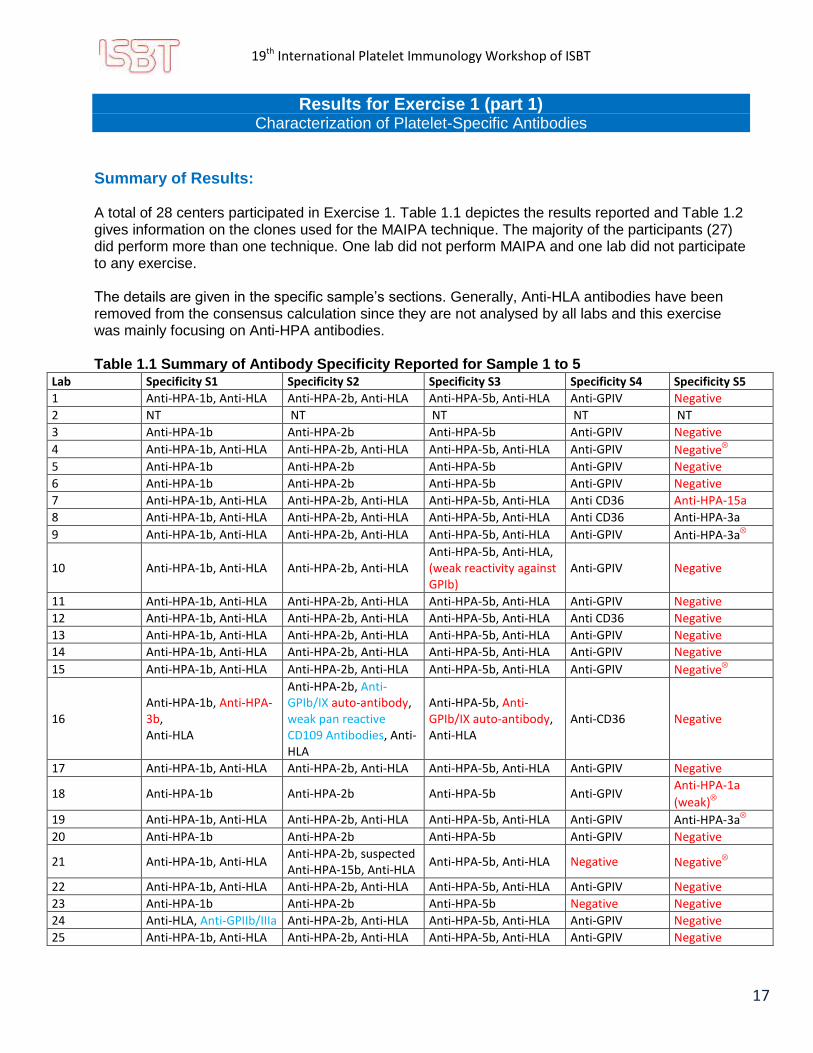
Summary of Results: A total of 28 centers participated in Exercise 1. Table 1.1 depictes the results reported and Table 1.2 gives information on the clones used for the MAIPA technique. The majority of the participants (27) did perform more than one technique. One lab did not perform MAIPA and one lab did not participate to any exercise. The details are given in the specific sample’s sections. Generally, Anti-HLA antibodies have been removed from the consensus calculation since they are not analysed by all labs and this exercise was mainly focusing on Anti-HPA antibodies. Table 1.1 Summary of Antibody Specificity Reported for Sample 1 to 5
Lab Specificity S1 Specificity S2 Specificity S3 Specificity S4 Specificity S5
1 Anti-HPA-1b, Anti-HLA Anti-HPA-2b, Anti-HLA Anti-HPA-5b, Anti-HLA Anti-GPIV Negative
2 NT NT NT NT NT
3 Anti-HPA-1b Anti-HPA-2b Anti-HPA-5b Anti-GPIV Negative
4 Anti-HPA-1b, Anti-HLA Anti-HPA-2b, Anti-HLA Anti-HPA-5b, Anti-HLA Anti-GPIV Negative
5 Anti-HPA-1b Anti-HPA-2b Anti-HPA-5b Anti-GPIV Negative
6 Anti-HPA-1b Anti-HPA-2b Anti-HPA-5b Anti-GPIV Negative
7 Anti-HPA-1b, Anti-HLA Anti-HPA-2b, Anti-HLA Anti-HPA-5b, Anti-HLA Anti CD36 Anti-HPA-15a
8 Anti-HPA-1b, Anti-HLA Anti-HPA-2b, Anti-HLA Anti-HPA-5b, Anti-HLA Anti CD36 Anti-HPA-3a
9 Anti-HPA-1b, Anti-HLA Anti-HPA-2b, Anti-HLA Anti-HPA-5b, Anti-HLA Anti-GPIV Anti-HPA-3a
10 Anti-HPA-1b, Anti-HLA Anti-HPA-2b, Anti-HLA Anti-HPA-5b, Anti-HLA, (weak reactivity against GPIb)
Anti-GPIV Negative
11 Anti-HPA-1b, Anti-HLA Anti-HPA-2b, Anti-HLA Anti-HPA-5b, Anti-HLA Anti-GPIV Negative
12 Anti-HPA-1b, Anti-HLA Anti-HPA-2b, Anti-HLA Anti-HPA-5b, Anti-HLA Anti CD36 Negative
13 Anti-HPA-1b, Anti-HLA Anti-HPA-2b, Anti-HLA Anti-HPA-5b, Anti-HLA Anti-GPIV Negative
14 Anti-HPA-1b, Anti-HLA Anti-HPA-2b, Anti-HLA Anti-HPA-5b, Anti-HLA Anti-GPIV Negative
15 Anti-HPA-1b, Anti-HLA Anti-HPA-2b, Anti-HLA Anti-HPA-5b, Anti-HLA Anti-GPIV Negative
16 Anti-HPA-1b, Anti-HPA-3b, Anti-HLA
Anti-HPA-2b, Anti-GPIb/IX auto-antibody, weak pan reactive CD109 Antibodies, Anti-HLA
Anti-HPA-5b, Anti-GPIb/IX auto-antibody, Anti-HLA
Anti-CD36 Negative
17 Anti-HPA-1b, Anti-HLA Anti-HPA-2b, Anti-HLA Anti-HPA-5b, Anti-HLA Anti-GPIV Negative
18 Anti-HPA-1b Anti-HPA-2b Anti-HPA-5b Anti-GPIV Anti-HPA-1a
(weak)
19 Anti-HPA-1b, Anti-HLA Anti-HPA-2b, Anti-HLA Anti-HPA-5b, Anti-HLA Anti-GPIV Anti-HPA-3a
20 Anti-HPA-1b Anti-HPA-2b Anti-HPA-5b Anti-GPIV Negative
21 Anti-HPA-1b, Anti-HLA Anti-HPA-2b, suspected Anti-HPA-15b, Anti-HLA
Anti-HPA-5b, Anti-HLA Negative Negative
22 Anti-HPA-1b, Anti-HLA Anti-HPA-2b, Anti-HLA Anti-HPA-5b, Anti-HLA Anti-GPIV Negative
23 Anti-HPA-1b Anti-HPA-2b Anti-HPA-5b Negative Negative
24 Anti-HLA, Anti-GPIIb/IIIa Anti-HPA-2b, Anti-HLA Anti-HPA-5b, Anti-HLA Anti-GPIV Negative
25 Anti-HPA-1b, Anti-HLA Anti-HPA-2b, Anti-HLA Anti-HPA-5b, Anti-HLA Anti-GPIV Negative
19th International Platelet Immunology Workshop of ISBT
18
Lab Specificity S1 Specificity S2 Specificity S3 Specificity S4 Specificity S5
26 Anti-HPA-1b, Anti-HLA Anti-HPA-2b, Anti-HLA, Anti-GPIIb/IIIa, Anti-GPIa/IIa
Anti-HPA-5b, Anti-HLA, Anti-GPIb/IX, Anti-GPIIb/IIIa
Anti-GPIV Anti-HPA-3a Anti-GPIIb/IIIa
27 Anti-HPA-1b, Anti-HLA Anti-HPA-2b, Anti-HLA Anti-HPA-5b, Anti-HLA Negative Negative
28 Anti-HPA-1b, Anti-HLA Anti-HPA-2b, Anti-HLA Anti-HPA-5b, Anti-HLA Anti-GPIV Negative
29 Anti-HPA-1b, Anti-HLA Anti-HPA-2b, Anti-HPA-15b, Anti-HLA
Anti-HPA-5b, Anti-HPA-15b, Anti-HLA
Anti-GPIV Negative
Expected results
Anti-HPA-1b, Anti-HLA Anti-HPA-2b, Anti-HPA-15b, Anti-HLA
Anti-HPA-5b, Anti-HPA-15b, Anti-HLA
Anti-GPIV Anti-HPA-3a
Expected concordance*
71.43% (20 Labs) 7.14% (2 Labs) 3.57% (1 Lab) 89.26% (25 Labs) 14.29% (4 Labs)
Consensus results
Anti-HPA-1b Anti-HPA-2b Anti-HPA-5b Anti-GPIV Negative
Consensus concordance
+
96.43% (27 Labs) 100.00% (28 Labs) 100.00% (28 Labs) 89.26% (25 Labs) 78.57% (22 Labs)
In red: Unlikely to be present; Discordant result In blue: Good system identified but lack of precision
Results from PAKLx were negative for Anti-HPA-3a via software assignment, however the MFI suggests antibody present. *Corresponds to the total concordance based on the expected result given the overall responses (i.e. center that completely found the expected results) +Corresponds to the total concordance based on the consensus results given the overall responses
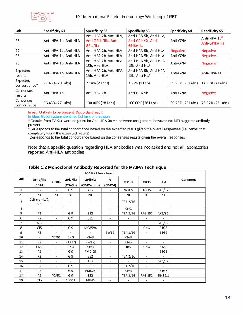
Note that a specific question regarding HLA antibodies was not asked and not all laboratories reported Anti-HLA antibodies. Table 1.2 Monoclonal Antibody Reported for the MAIPA Technique
Lab
MAIPA Monoclonals
GPllb/llla GPllla
GPla/lla GPlb/lX V CD109 CD36 HLA
Comment
(CD41) (CD49b) (CD42a or b) (CD42d)
1 P2 - Gi9 AK2 - W7C5 FA6-152 W6/32
2* NT NT NT NT - NT NT NT
3 CLB-tromb/7,
6C9 - - - - TEA 2/16 - -
4 - - - - - CNG - -
5 P2 - Gi9 SZ2 - TEA 2/16 FA6-152 W6/32
6 P2 - Gi9 SZ1 - - - -
7 AP2 - - - - - - W6/32
8 Gi5 - Gi9 MCA594 - - CNG B1G6
9 P2 - - - SW16 TEA 2/16 - B1G6
10 - Y2/51 CNG CNG - CNG
11 P2 - (AK7?) (SZ1?) - CNG - -
12 CNG - CNG CNG - IB3 CNG CNG
13 P2 - Gi9 FMC-25 - - - B1G6
14 P2 - Gi9 SZ2 - TEA 2/16 - -
15 P2 - - AK2 - - - W6/32
16 P2 - Gi9 GRP - TEA 2/16 - -
17 P2 - Gi9 FMC25 - CNG - B1G6
18 P2 Y2/51 Gi9 SZ2 - TEA 2/16 FA6-152 B9.12.1
19 C17 - 10G11 MB45 - - - -
19th International Platelet Immunology Workshop of ISBT
19
Lab
MAIPA Monoclonals
GPllb/llla GPllla
GPla/lla GPlb/lX V CD109 CD36 HLA
Comment
(CD41) (CD49b) (CD42a or b) (CD42d)
20 PAB 1 - P16 - - CNG - -
21 P2 - Gi9 CLB-MB45 - BD
(TEA 2/16) - W6/32
22 CNG - CNG CNG - CNG CNG CNG IIbIIIa+ chloro IbIX +
chloro
23 P2 - Gi9 FMC25 - TEA 2/16 - W6/32 PIFT-FFC Unt'd IgG
24 P2 - Gi9 SZ1; SZ2 - CNG - CNG
25 NT NT NT NT NT NT NT NT No MAIPA
26 CNG - CNG CNG - - - CNG "IIbIIIa
(PL246/PL164)"
27 P2 - Gi9 FMC25 - TEA 2/16 FA6-152 B1G6
28 P2 - Gi9 AK2 - TEA 2/16 FA6-152 -
29 6B9 - Gi9 IM0538 - TEA 2/16 - -
*Lab 2 did not participate. This laboratory was removed from all the following tables NA: Not attributable NT: Not tested CNG: Clone not given: center mentioned using one but did not give the name of the clone.
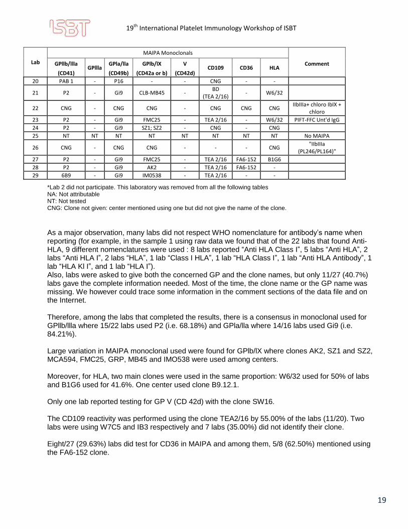
As a major observation, many labs did not respect WHO nomenclature for antibody’s name when reporting (for example, in the sample 1 using raw data we found that of the 22 labs that found Anti-HLA, 9 different nomenclatures were used : 8 labs reported “Anti HLA Class I”, 5 labs “Anti HLA”, 2 labs “Anti HLA I”, 2 labs “HLA”, 1 lab “Class I HLA”, 1 lab “HLA Class I”, 1 lab “Anti HLA Antibody”, 1 lab “HLA Kl I”, and 1 lab “HLA I”). Also, labs were asked to give both the concerned GP and the clone names, but only 11/27 (40.7%) labs gave the complete information needed. Most of the time, the clone name or the GP name was missing. We however could trace some information in the comment sections of the data file and on the Internet. Therefore, among the labs that completed the results, there is a consensus in monoclonal used for GPllb/llla where 15/22 labs used P2 (i.e. 68.18%) and GPla/lla where 14/16 labs used Gi9 (i.e. 84.21%). Large variation in MAIPA monoclonal used were found for GPlb/lX where clones AK2, SZ1 and SZ2, MCA594, FMC25, GRP, MB45 and IMO538 were used among centers. Moreover, for HLA, two main clones were used in the same proportion: W6/32 used for 50% of labs and B1G6 used for 41.6%. One center used clone B9.12.1. Only one lab reported testing for GP V (CD 42d) with the clone SW16. The CD109 reactivity was performed using the clone TEA2/16 by 55.00% of the labs (11/20). Two labs were using W7C5 and IB3 respectively and 7 labs (35.00%) did not identify their clone. Eight/27 (29.63%) labs did test for CD36 in MAIPA and among them, 5/8 (62.50%) mentioned using the FA6-152 clone.
19th International Platelet Immunology Workshop of ISBT
20
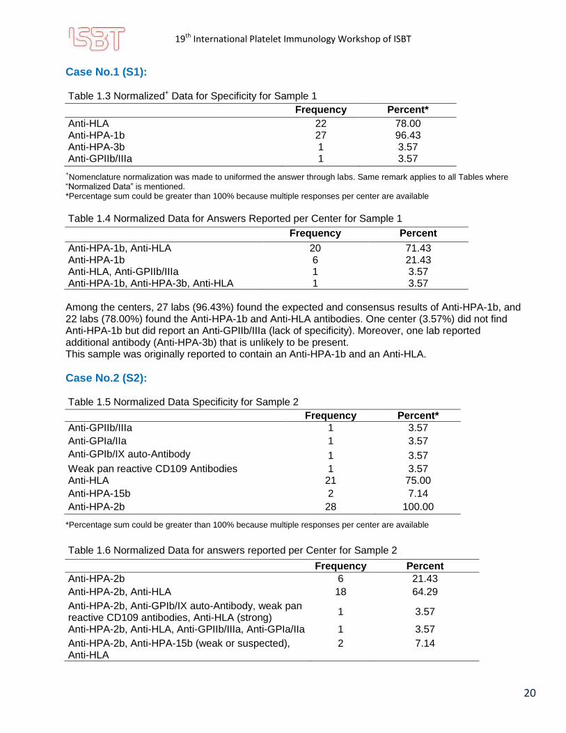
Case No.1 (S1): Table 1.3 Normalized+ Data for Specificity for Sample 1
Frequency Percent*
Anti-HLA 22 78.00 Anti-HPA-1b 27 96.43 Anti-HPA-3b 1 3.57 Anti-GPIIb/IIIa 1 3.57
+Nomenclature normalization was made to uniformed the answer through labs. Same remark applies to all Tables where
“Normalized Data” is mentioned. *Percentage sum could be greater than 100% because multiple responses per center are available
Table 1.4 Normalized Data for Answers Reported per Center for Sample 1
Frequency Percent
Anti-HPA-1b, Anti-HLA 20 71.43 Anti-HPA-1b 6 21.43 Anti-HLA, Anti-GPIIb/IIIa 1 3.57 Anti-HPA-1b, Anti-HPA-3b, Anti-HLA 1 3.57
Among the centers, 27 labs (96.43%) found the expected and consensus results of Anti-HPA-1b, and 22 labs (78.00%) found the Anti-HPA-1b and Anti-HLA antibodies. One center (3.57%) did not find Anti-HPA-1b but did report an Anti-GPIIb/IIIa (lack of specificity). Moreover, one lab reported additional antibody (Anti-HPA-3b) that is unlikely to be present. This sample was originally reported to contain an Anti-HPA-1b and an Anti-HLA.
Case No.2 (S2): Table 1.5 Normalized Data Specificity for Sample 2
Frequency Percent*
Anti-GPIIb/IIIa 1 3.57
Anti-GPIa/IIa 1 3.57
Anti-GPIb/IX auto-Antibody 1 3.57
Weak pan reactive CD109 Antibodies 1 3.57 Anti-HLA 21 75.00
Anti-HPA-15b 2 7.14
Anti-HPA-2b 28 100.00
*Percentage sum could be greater than 100% because multiple responses per center are available
Table 1.6 Normalized Data for answers reported per Center for Sample 2
Frequency Percent
Anti-HPA-2b 6 21.43
Anti-HPA-2b, Anti-HLA 18 64.29
Anti-HPA-2b, Anti-GPIb/IX auto-Antibody, weak pan reactive CD109 antibodies, Anti-HLA (strong)
1 3.57
Anti-HPA-2b, Anti-HLA, Anti-GPIIb/IIIa, Anti-GPIa/IIa 1 3.57
Anti-HPA-2b, Anti-HPA-15b (weak or suspected), Anti-HLA
2 7.14
19th International Platelet Immunology Workshop of ISBT
21
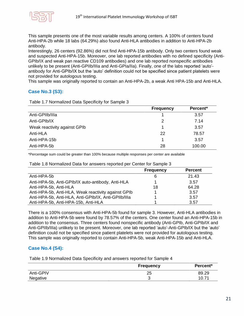
This sample presents one of the most variable results among centers. A 100% of centers found Anti-HPA-2b while 18 labs (64.29%) also found Anti-HLA antibodies in addition to Anti-HPA-2b antibody. Interestingly, 26 centers (92.86%) did not find Anti-HPA-15b antibody. Only two centers found weak and suspected Anti-HPA-15b. Moreover, one lab reported antibodies with no defined specificity (Anti-GPIb/IX and weak pan reactive CD109 antibodies) and one lab reported nonspecific antibodies unlikely to be present (Anti-GPIIb/IIIa and Anti-GPIa/IIa). Finally, one of the labs reported ‘auto’-antibody for Anti-GPIb/IX but the ‘auto’ definition could not be specified since patient platelets were not provided for autologous testing. This sample was originally reported to contain an Anti-HPA-2b, a weak Anti HPA-15b and Anti-HLA.
Case No.3 (S3): Table 1.7 Normalized Data Specificity for Sample 3
Frequency Percent*
Anti-GPIIb/IIIa 1 3.57
Anti-GPIb/IX 2 7.14
Weak reactivity against GPIb 1 3.57
Anti-HLA 22 78.57
Anti-HPA-15b 1 3.57
Anti-HPA-5b 28 100.00
*Percentage sum could be greater than 100% because multiple responses per center are available
Table 1.8 Normalized Data for answers reported per Center for Sample 3
Frequency Percent
Anti-HPA-5b 6 21.43
Anti-HPA-5b, Anti-GPIb/IX auto-antibody, Anti-HLA 1 3.57 Anti-HPA-5b, Anti-HLA 18 64.28 Anti-HPA-5b, Anti-HLA, Weak reactivity against GPIb 1 3.57 Anti-HPA-5b, Anti-HLA, Anti-GPIb/IX, Anti-GPIIb/IIIa 1 3.57 Anti-HPA-5b, Anti-HPA-15b, Anti-HLA 1 3.57
There is a 100% consensus with Anti-HPA-5b found for sample 3. However, Anti-HLA antibodies in addition to Anti-HPA-5b were found by 78.57% of the centers. One center found an Anti-HPA-15b in addition to the consensus. Three centers found nonspecific antibody (Anti-GPIb, Anti-GPIb/IX and Anti-GPIIb/IIIa) unlikely to be present. Moreover, one lab reported ‘auto’-Anti-GPIb/IX but the ‘auto’ definition could not be specified since patient platelets were not provided for autologous testing. This sample was originally reported to contain Anti-HPA-5b, weak Anti-HPA-15b and Anti-HLA.
Case No.4 (S4): Table 1.9 Normalized Data Specificity and answers reported for Sample 4
Frequency Percent*
Anti-GPIV 25 89.29 Negative 3 10.71
19th International Platelet Immunology Workshop of ISBT
22
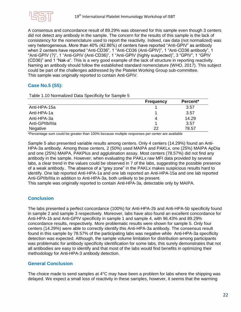
A consensus and concordance result of 89.29% was observed for this sample even though 3 centers did not detect any antibody in the sample. The concern for the results of this sample is the lack of consistency for the nomenclature used to report the reactivity. Indeed, raw data (not normalized) was very heterogeneous. More than 40% (42.86%) of centers have reported “Anti-GPIV” as antibody when 2 centers have reported “Anti-CD36”, 1 “Anti-CD36 (Anti-GPIV)”, 1 “Anti-CD36 antibody”, 1 “Anti-GPIV (?)”, 1 “Anti-GPIV (Anti-CD36)”, 1 “Anti-GPIV (highly suspected)”, 3 “GPIV”, 1 “GPIV (CD36)” and 1 “Nak-a”. This is a very good example of the lack of structure in reporting reactivity. Naming an antibody should follow the established standard nomenclature (WHO, 2017). This subject could be part of the challenges addressed by the Platelet Working Group sub-committee. This sample was originally reported to contain Anti-GPIV.
Case No.5 (S5): Table 1.10 Normalized Data Specificity for Sample 5
Frequency Percent*
Anti-HPA-15a 1 3.57
Anti-HPA-1a 1 3.57
Anti-HPA-3a 4 14.29 Anti-GPIIb/IIIa 1 3.57 Negative 22 78.57
*Percentage sum could be greater than 100% because multiple responses per center are available
Sample 5 also presented variable results among centers. Only 4 centers (14.29%) found an Anti-HPA-3a antibody. Among those centers, 2 (50%) used MAIPA and PAKLx, one (25%) MAIPA ApDia and one (25%) MAIPA, PAKPlus and agglutination assay. Most centers (78.57%) did not find any antibody in the sample. However, when evaluating the PAKLx raw MFI data provided by several labs, a clear trend in the values could be observed in 7 of the labs, suggesting the possible presence of a weak antibody.. The absence of a “grey zone” in the PAKLx makes suspicious results hard to identify. One lab reported Anti-HPA-1a and one lab reported an Anti-HPA-15a and one lab reported Anti-GPIIb/IIIa in addition to Anti-HPA-3a, both unlikely to be present. This sample was originally reported to contain Anti-HPA-3a, detectable only by MAIPA.
Conclusion The labs presented a perfect concordance (100%) for Anti-HPA-2b and Anti-HPA-5b specificity found in sample 2 and sample 3 respectively. Moreover, labs have also found an excellent concordance for Anti-HPA-1b and Anti-GPIV specificity in sample 1 and sample 4, with 96.43% and 89.29% concordance results, respectively. More problematic results were shown for sample 5. Only four centers (14.29%) were able to correctly identify this Anti-HPA-3a antibody. The consensus result found in this sample by 78.57% of the participating labs was negative while Anti-HPA-3a specificity detection was expected. Although, the sample volume limitation for distribution among participants was problematic for antibody specificity identification for some labs, this surely demonstrates that not all antibodies are easy to identify and that most of the labs would find benefits in optimizing their methodology for Anti-HPA-3 antibody detection.
General Conclusion
The choice made to send samples at 4°C may have been a problem for labs where the shipping was delayed. We expect a small loss of reactivity in these samples, however, it seems that the warming

19th International Platelet Immunology Workshop of ISBT
23
of the sample did not cause any loss in reactivity. Tracing back these labs, we can see that the results they obtained were comparable to the results from the labs who experienced no shipping problem. Another general observation would be that Anti-HPA-3 and Anti-HPA-15 are more prone to degrade with time since they were initially detected in the case study, but were not identified by the majority of the participants and could not reach the consensus in samples 2, 3 and 5.
19th International Platelet Immunology Workshop of ISBT
24
Exercise 1 (part 2) Survey on Lab Management of FNAIT
Aim:
1) To highlight the laboratory component of FNAIT management
2) To identify the spectrum of analysis performed and results reported by the majority of labs in FNAIT cases
3) To determine the proportion of labs performing antibody quantification
4) To evaluate the proportion of labs which are using antibody quantification to guide FNAIT management
5) To prepare for next Workshop’s exercises on quantification of antibodies
Materials Supplied:
Participating laboratories were provided with:
The link for participation in a digital survey (which was sent on 2017-10-02)
Section C of the survey was sent by email in the Excel answer grid
Methods:
The majority of the questions were multiple choices and some of them required short free text answers.
Section C of the survey required information on the respondents’ antibody quantification protocol and were answered electronically on the Excel answer grid.
19th International Platelet Immunology Workshop of ISBT
25
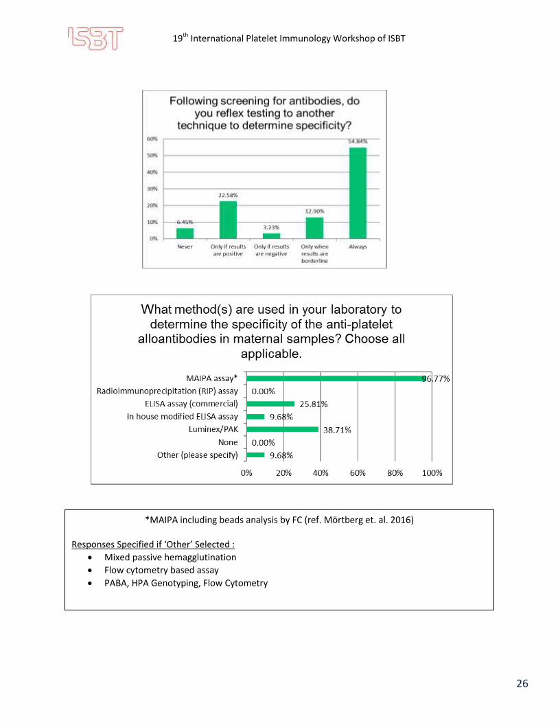
Responses Specified if ‘Other’ Selected : Luminex PAKLX if necessary, based on medical
decision such as "emergencies" or inconclusive MAIPA
MPHA (Mixed passive hemagglutination)
capture-P
Platelet immunofluorescence test
Results for Exercise 1 (part 2) Survey on Lab Management of FNAIT
Survey Results:
Responses Specified if ‘Other’ Selected : Anti-HPA-1 thru 5; GPIV; and occasionally HPA-15. All for both a and b alleles except HPA-4a only
any other based on HPA-15 genotypes and/or crossmatch results
HPA-1, 2, 3, 5, 15 and crossmatch maternal serum - paternal platelets
Anti-HPA 1a/b, 3a/b, 5a/b and others if negative (2 and 15)
CD36, and all rare and new HPA
Anti-HLA class I antibodies
*MAIPA including beads analysis by FC (ref. Mörtberg et. Al, 2016)
19th International Platelet Immunology Workshop of ISBT
26
*MAIPA including beads analysis by FC (ref. Mörtberg et. al. 2016) Responses Specified if ‘Other’ Selected :
Mixed passive hemagglutination
Flow cytometry based assay
PABA, HPA Genotyping, Flow Cytometry
19th International Platelet Immunology Workshop of ISBT
27
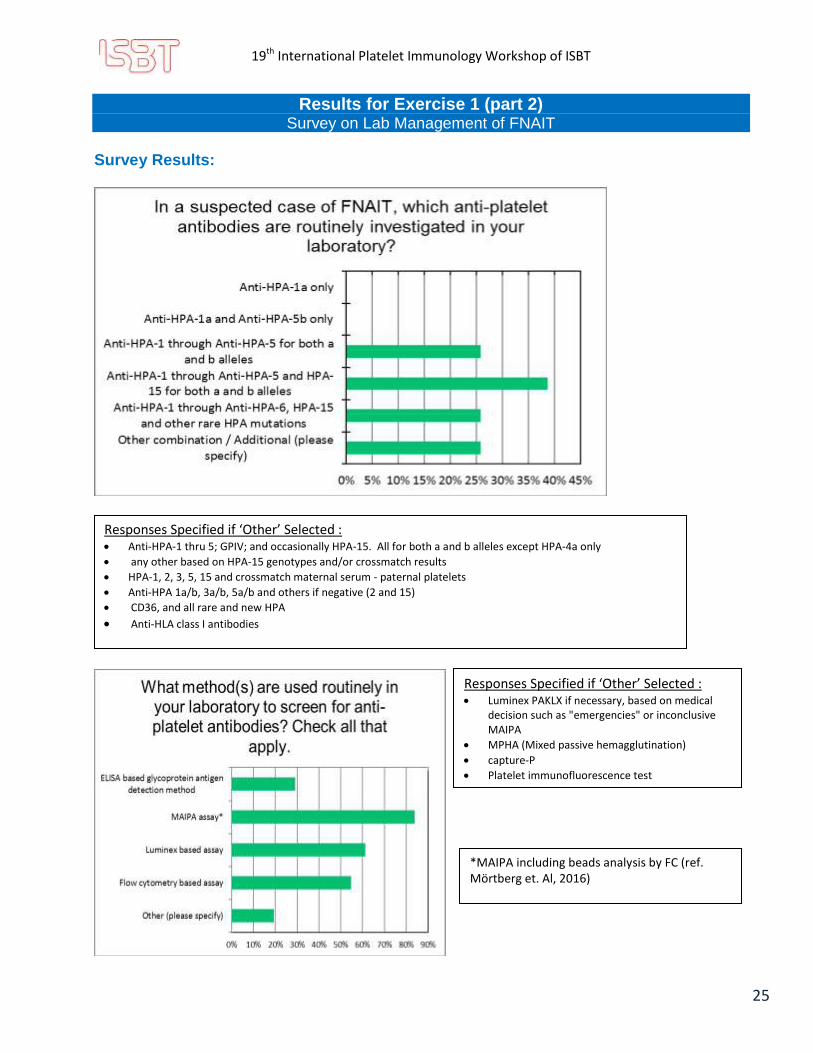
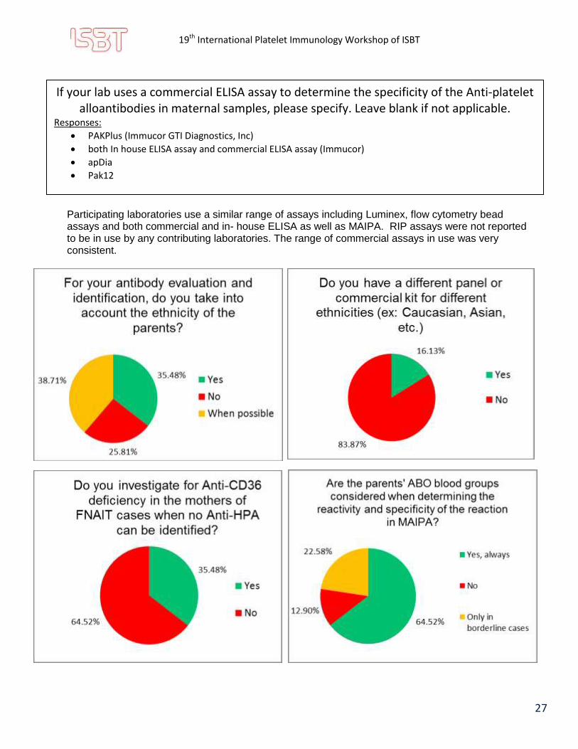
Participating laboratories use a similar range of assays including Luminex, flow cytometry bead assays and both commercial and in- house ELISA as well as MAIPA. RIP assays were not reported to be in use by any contributing laboratories. The range of commercial assays in use was very consistent.
If your lab uses a commercial ELISA assay to determine the specificity of the Anti-platelet alloantibodies in maternal samples, please specify. Leave blank if not applicable.
Responses:
PAKPlus (Immucor GTI Diagnostics, Inc)
both In house ELISA assay and commercial ELISA assay (Immucor)
apDia
Pak12
19th International Platelet Immunology Workshop of ISBT
28
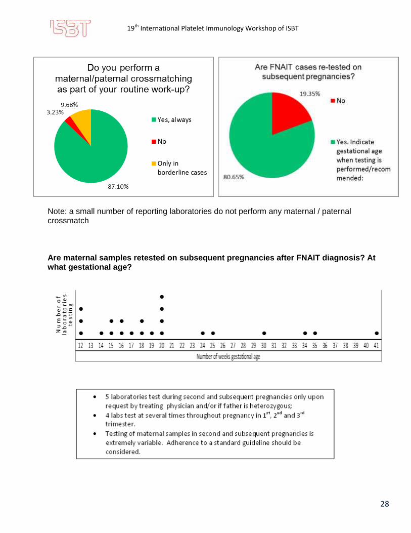
Note: a small number of reporting laboratories do not perform any maternal / paternal crossmatch Are maternal samples retested on subsequent pregnancies after FNAIT diagnosis? At what gestational age?
19th International Platelet Immunology Workshop of ISBT
29
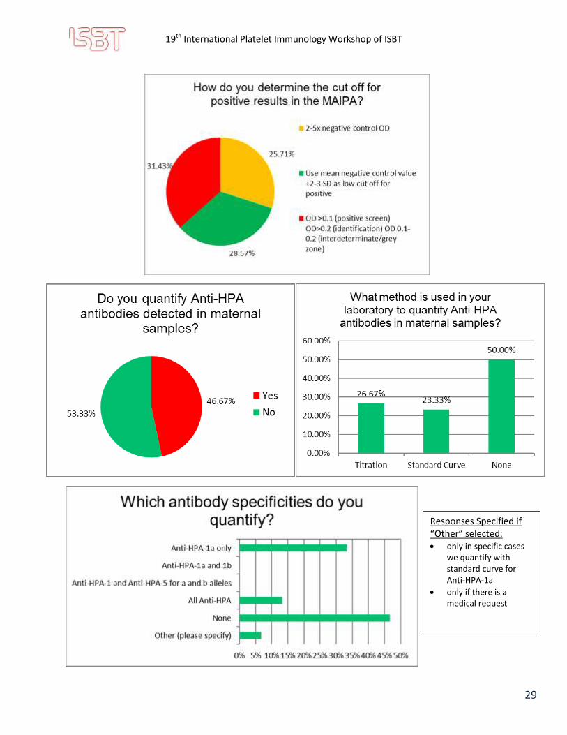
Responses Specified if “Other” selected: only in specific cases
we quantify with standard curve for Anti-HPA-1a
only if there is a medical request
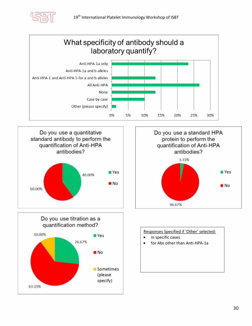
19th International Platelet Immunology Workshop of ISBT
30
Responses Specified if ‘Other’ selected:
in specific cases
for Abs other than Anti-HPA-1a
19th International Platelet Immunology Workshop of ISBT
31
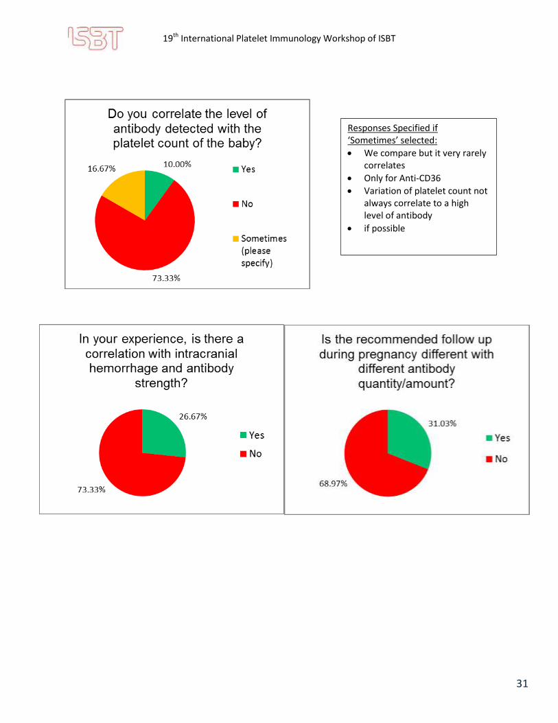
Responses Specified if ‘Sometimes’ selected:
We compare but it very rarely correlates
Only for Anti-CD36
Variation of platelet count not always correlate to a high level of antibody
if possible

19th International Platelet Immunology Workshop of ISBT
32
Participating laboratories indicated extremely variable recommendations regarding follow up testing which was inconsistent regardless of high or low levels of antibody. The antibody strength, even when measured, did not always result in increased laboratory monitoring.

19th International Platelet Immunology Workshop of ISBT
33
Responses Specified if ‘Sometimes’ selected:
for anti HPA-1a quantification only
If available
determined on a Case by case
if current assay is the same
We never quantify antibodies
Responses Specified if ‘Other’ selected:
When an antibody is identified, the genotyping is performed on the mother and the father to predict if the foetus will be affected, in some case the amniotic liquid is also tested
19th International Platelet Immunology Workshop of ISBT
34
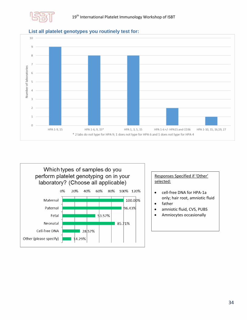
List all platelet genotypes you routinely test for:
Responses Specified if ‘Other’ selected:
cell-free DNA for HPA-1a only; hair root, amniotic fluid
father
amniotic fluid, CVS, PUBS
Amniocytes occasionally
19th International Platelet Immunology Workshop of ISBT
35
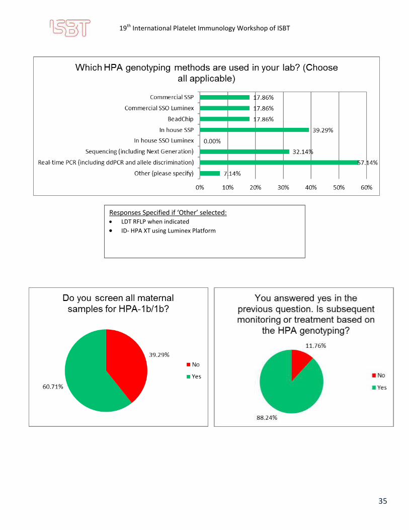
Responses Specified if ‘Other’ selected: LDT RFLP when indicated
ID- HPA XT using Luminex Platform
19th International Platelet Immunology Workshop of ISBT
36
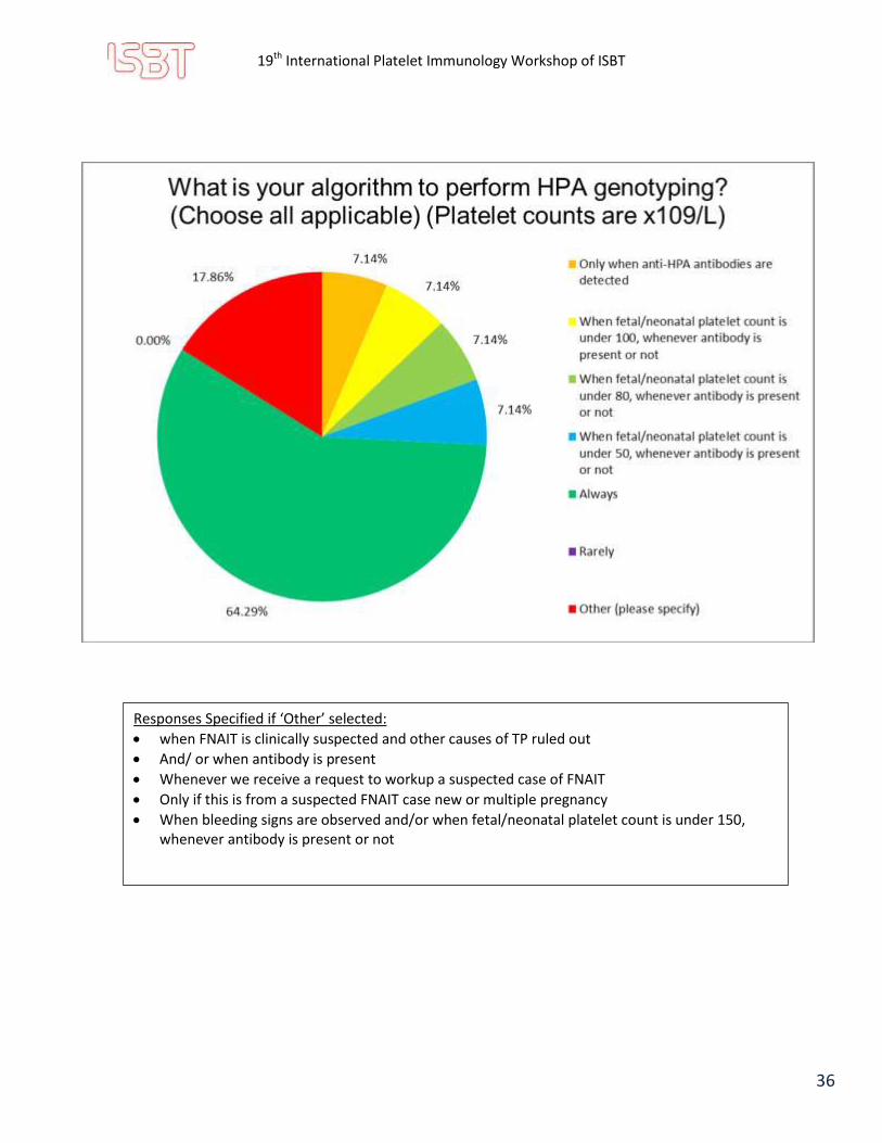
Responses Specified if ‘Other’ selected:
when FNAIT is clinically suspected and other causes of TP ruled out
And/ or when antibody is present
Whenever we receive a request to workup a suspected case of FNAIT
Only if this is from a suspected FNAIT case new or multiple pregnancy
When bleeding signs are observed and/or when fetal/neonatal platelet count is under 150, whenever antibody is present or not
19th International Platelet Immunology Workshop of ISBT
37
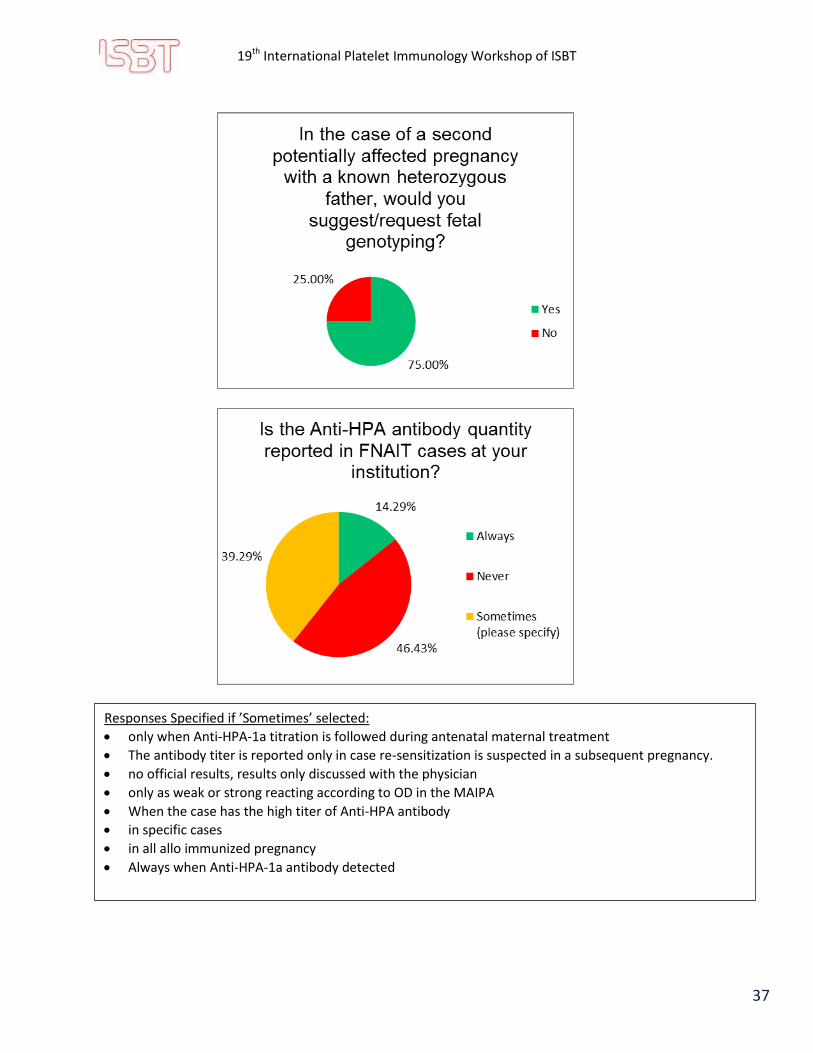
Responses Specified if ’Sometimes’ selected:
only when Anti-HPA-1a titration is followed during antenatal maternal treatment
The antibody titer is reported only in case re-sensitization is suspected in a subsequent pregnancy.
no official results, results only discussed with the physician
only as weak or strong reacting according to OD in the MAIPA
When the case has the high titer of Anti-HPA antibody
in specific cases
in all allo immunized pregnancy
Always when Anti-HPA-1a antibody detected
19th International Platelet Immunology Workshop of ISBT
38
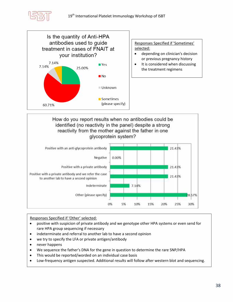
Responses Specified if ‘Other’ selected:
positive with suspicion of private antibody and we genotype other HPA systems or even send for rare HPA group sequencing if necessary
indeterminate and referral to another lab to have a second opinion
we try to specify the LFA or private antigen/antibody
never happens
We sequence the father's DNA for the gene in question to determine the rare SNP/HPA
This would be reported/worded on an individual case basis
Low-frequency antigen suspected. Additional results will follow after western blot and sequencing.
Responses Specified if ’Sometimes’ selected:
depending on clinician's decision or previous pregnancy history
It is considered when discussing the treatment regimens
19th International Platelet Immunology Workshop of ISBT
39
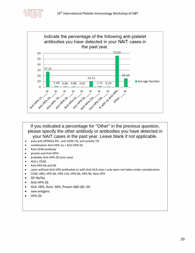
If you indicated a percentage for "Other" in the previous question, please specify the other antibody or antibodies you have detected in
your NAIT cases in the past year. Leave blank if not applicable. auto anti GPIIbIIIa 4% ; anti CD36 1%; anti private 1%
combination Anti-HPA-1a + Anti-HPA-5b
Anti-CD36 antibody
private and Anti-GPIV
probably Anti-HPA-2b (one case)
HLA-I, CD36
Anti-HPA 6b and 9b
cases without Anti-HPA antibodies or with Anti-HLA class I only were not taken under consideration
CD36, ABO, HPA-6b, HPA-11b, HPA-4b, HPA-9b, New HPA
GP IIb/IIIa
Anti-HPA-2b
HLA: 28%, Auto: 36%, Proven ABO (B): 4%
new antigens
HPA-2b
19th International Platelet Immunology Workshop of ISBT
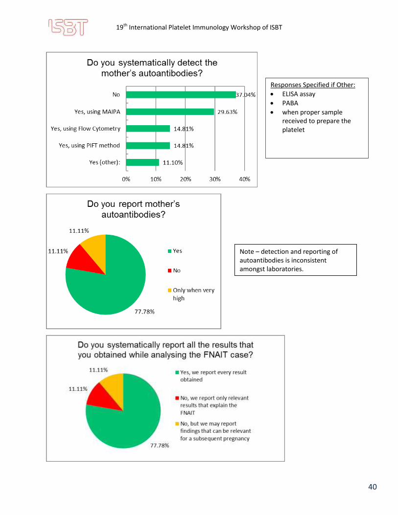
40
Responses Specified if Other:
ELISA assay
PABA
when proper sample received to prepare the platelet
Note – detection and reporting of autoantibodies is inconsistent amongst laboratories.
19th International Platelet Immunology Workshop of ISBT
41
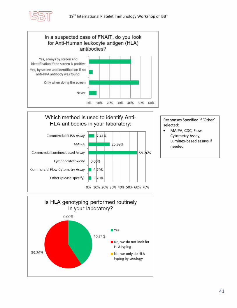
Responses Specified if ‘Other’ selected:
MAIPA, CDC, Flow Cytometry Assay, Luminex-based assays if needed
19th International Platelet Immunology Workshop of ISBT
42
Responses Specified if ‘Other’ selected:
HLA type only when platelet donor is needed with 100% maternal HLA antibody
Only for HPA-1bb women without abs to determine the risk, e.g. sister of an affected woman
Only in cases with strong maternal HLA antibodies
When Anti-HPA is negative and Anti-HLA is suspected to be the cause
Responses Specified if ‘Other’ selected:
We have the ability to HLA type (SSO and SSP) but don't perform testing for NAIT
19th International Platelet Immunology Workshop of ISBT
43
19th International Platelet Immunology Workshop of ISBT
44
Conclusion The survey results indicate consistency in the methodology used for identification of Anti-HPA antibodies and fairly consistent assessment of HPA types. Marked variability was noted in reporting cut off values. In addition practice is variable with respect to assessment of antibody quantity as well as the method used, for those labs that quantify the antibody. For those labs which performed assessment of antibody quantity, result reporting was inconsistent. Additional areas of variable practice that may benefit from guidelines for testing would include a recommended approach to the timing and frequency of follow up test samples in pregnancies subsequent to the index pregnancy. Achieving better agreement on the significance and on the reporting and re- testing of autoantibodies is of extreme relevance for laboratory quality improvement and should be a major goal of future workshops, as recently addressed by the working party.
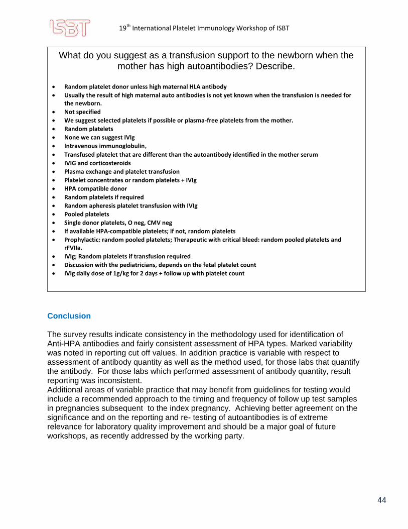
What do you suggest as a transfusion support to the newborn when the mother has high autoantibodies? Describe.
Random platelet donor unless high maternal HLA antibody
Usually the result of high maternal auto antibodies is not yet known when the transfusion is needed for the newborn.
Not specified
We suggest selected platelets if possible or plasma-free platelets from the mother.
Random platelets
None we can suggest IVIg
Intravenous immunoglobulin,
Transfused platelet that are different than the autoantibody identified in the mother serum
IVIG and corticosteroids
Plasma exchange and platelet transfusion
Platelet concentrates or random platelets + IVIg
HPA compatible donor
Random platelets if required
Random apheresis platelet transfusion with IVIg
Pooled platelets
Single donor platelets, O neg, CMV neg
If available HPA-compatible platelets; if not, random platelets
Prophylactic: random pooled platelets; Therapeutic with critical bleed: random pooled platelets and rFVIIa.
IVIg; Random platelets if transfusion required
Discussion with the pediatricians, depends on the fetal platelet count
IVIg daily dose of 1g/kg for 2 days + follow up with platelet count

19th International Platelet Immunology Workshop of ISBT
45
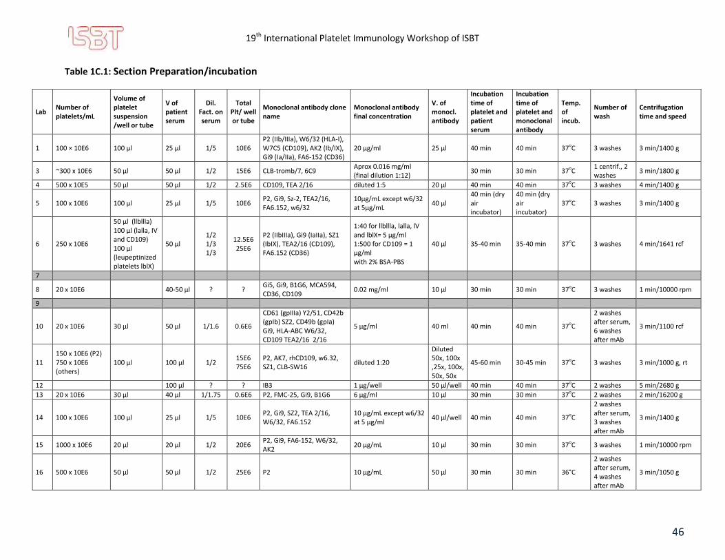
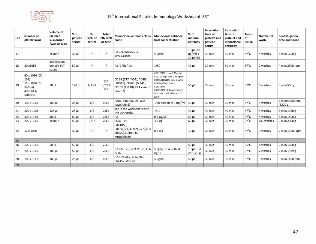
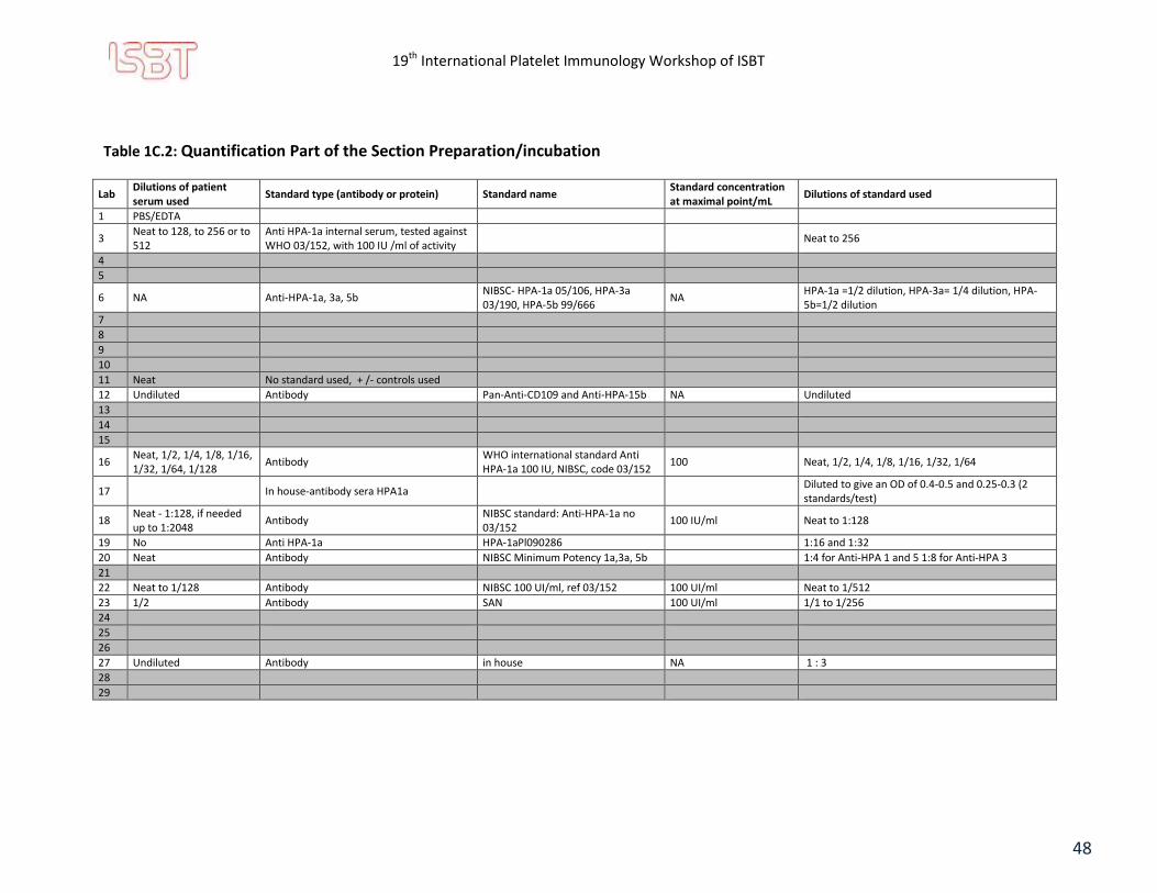
Quantitative MAIPA Protocol (Section C of the questionnaire)
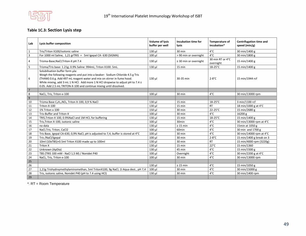
A total of 24/28 (85.71%) labs answered the section C of the worksheet that was dedicated to quantification protocol. However, only around 46% of the participating labs mentioned in the survey doing quantitative MAIPA. In concordance with the latter, 12/28 (42.86%) labs reported using a standard protein or antibody for quantification; other labs left this section blank. There was probably a misunderstanding surrounding this section of Exercise 1 and the intent was probably not clear enough. We realize that not only those labs doing quantitative MAIPA did fill this section but also labs not performing quantitative MAIPA. Nevertheless, we looked at all the protocols generously shared by the participants. We divided the MAIPA into different steps which are 1) the preparation/incubation, 2) the lysis, 3) the attachment to solid phase, 4) the conjugate antibody and 5) the colorimetry. On the following pages are grouped the different tables to be described in the text. A lot of variation was found between labs concerning some of the different steps of the MAIPA protocol.
19th International Platelet Immunology Workshop of ISBT
46
Table 1C.1: Section Preparation/incubation
Lab Number of platelets/mL
Volume of platelet suspension /well or tube
V of patient serum
Dil. Fact. on serum
Total Plt/ well or tube
Monoclonal antibody clone name
Monoclonal antibody final concentration
V. of monocl. antibody
Incubation time of platelet and patient serum
Incubation time of platelet and monoclonal antibody
Temp. of incub.
Number of wash
Centrifugation time and speed
1 100 × 10E6 100 µl 25 µl 1/5 10E6 P2 (IIb/IIIa), W6/32 (HLA-I), W7C5 (CD109), AK2 (Ib/IX), Gi9 (Ia/IIa), FA6-152 (CD36)
20 μg/ml 25 µl 40 min 40 min 37oC 3 washes 3 min/1400 g
3 ~300 x 10E6 50 µl 50 µl 1/2 15E6 CLB-tromb/7, 6C9 Aprox 0.016 mg/ml (final dilution 1:12)
30 min 30 min 37oC 1 centrif., 2 washes
3 min/1800 g
4 500 x 10E5 50 µl 50 µl 1/2 2.5E6 CD109, TEA 2/16 diluted 1:5 20 µl 40 min 40 min 37oC 3 washes 4 min/1400 g
5 100 x 10E6 100 µl 25 µl 1/5 10E6 P2, Gi9, Sz-2, TEA2/16, FA6.152, w6/32
10µg/mL except w6/32 at 5µg/mL
40 µl 40 min (dry air incubator)
40 min (dry air incubator)
37oC 3 washes 3 min/1400 g
6 250 x 10E6
50 µl (llbllla) 100 µl (lalla, IV and CD109) 100 µl (leupeptinized platelets lblX)
50 µl 1/2 1/3 1/3
12.5E6 25E6
P2 (IIbIIIa), Gi9 (IaIIa), SZ1 (IbIX), TEA2/16 (CD109), FA6.152 (CD36)
1:40 for llbllla, lalla, lV and lblX= 5 µg/ml 1:500 for CD109 = 1 µg/ml with 2% BSA-PBS
40 µl 35-40 min 35-40 min 37oC 3 washes 4 min/1641 rcf
7
8 20 x 10E6
40-50 µl ? ? Gi5, Gi9, B1G6, MCA594, CD36, CD109
0.02 mg/ml 10 µl 30 min 30 min 37oC 3 washes 1 min/10000 rpm
9
10 20 x 10E6 30 µl 50 µl 1/1.6 0.6E6
CD61 (gpIIIa) Y2/51, CD42b (gpIb) SZ2, CD49b (gpIa) Gi9, HLA-ABC W6/32, CD109 TEA2/16 2/16
5 µg/ml 40 ml 40 min 40 min 37oC
2 washes after serum, 6 washes after mAb
3 min/1100 rcf
11 150 x 10E6 (P2) 750 x 10E6 (others)
100 µl 100 µl 1/2 15E6 75E6
P2, AK7, rhCD109, w6.32, SZ1, CLB-SW16
diluted 1:20
Diluted 50x, 100x ,25x, 100x, 50x, 50x
45-60 min 30-45 min 37oC 3 washes 3 min/1000 g, rt
12
100 µl ? ? IB3 1 μg/well 50 μl/well 40 min 40 min 37oC 2 washes 5 min/2680 g
13 20 x 10E6 30 µl 40 µl 1/1.75 0.6E6 P2, FMC-25, Gi9, B1G6 6 µg/ml 10 µl 30 min 30 min 37oC 2 washes 2 min/16200 g
14 100 x 10E6 100 µl 25 µl 1/5 10E6 P2, Gi9, SZ2, TEA 2/16, W6/32, FA6.152
10 µg/mL except w6/32 at 5 µg/ml
40 µl/well 40 min 40 min 37oC
2 washes after serum, 3 washes after mAb
3 min/1400 g
15 1000 x 10E6 20 µl 20 µl 1/2 20E6 P2, Gi9, FA6-152, W6/32, AK2
20 µg/mL 10 µl 30 min 30 min 37oC 3 washes 1 min/10000 rpm
16 500 x 10E6 50 µl 50 µl 1/2 25E6 P2 10 µg/mL 50 µl 30 min 30 min 36°C
2 washes after serum, 4 washes after mAb
3 min/1050 g
19th International Platelet Immunology Workshop of ISBT
47
Lab Number of platelets/mL
Volume of platelet suspension /well or tube
V of patient serum
Dil. Fact. on serum
Total Plt/ well or tube
Monoclonal antibody clone name
Monoclonal antibody final concentration
V. of monocl. antibody
Incubation time of platelet and patient serum
Incubation time of platelet and monoclonal antibody
Temp. of incub.
Number of wash
Centrifugation time and speed
17
2x10E7 30 µl ? ? P2,Gi9,FMC25,CLB-SW16,B1G9
5 µg/ml 10 µl( 20 µg/ml) + 30 µl PBS
30 min 30 min 37oC 4 washes 3 min/1200 g
18 20 x10E6 depends on donor's PLT count
50 µl ? ? P2 (GPIIb/IIIa) 1/50 40 µl 30 min 30 min 37oC 2 washes 4 min/2500 rpm
19
60 x 10E6 (CD 109) 15 x 10E6 (Gp IIb/IIIa) 40 x 10E6 (others)
50 µl 120 µl 1/1.42 3E6
0,75E6 2E6
CD 61 (C17, Y/51), CD49b (10G11), CD42b (MB45), CD109 (15E10), HLA class I (W6.32)
CD61 (C17) f conc 1.55µg/ml
CD61 (Y/51) f conc 0.25 µg/ml
CD49b (10G11) f conc 5 µg/ml
CD42b (MB45) f conc
0.255µg/ml
CD109 (15E10) f conc 2µg/ml
HLA class I (W6.32) f conc 10
µg/ml
50 µl 30 min 30 min 37oC 5 washes 5 min/550 g
20 100 x 10E6 100 µl 25 µl 1/5 10E6 PAB1, P16, CD109 ( also have PAB 6)
1:50 diution of 1 mg/ml 40 µl 30 min 30 min 37oC 3 washes 3 mim/4000 rpm (3220 g)
21 200 x 10E6 125 µl 25 µl 1/6 25E6 see in the worksheets with the OD results
1/10 40 µl 40 min 40 min 37oC 3 washes 3 min/1360 g
22 500 x 10E6 50 µl 50 µl 1/2 25E6 P2 0.5 µg/µl 50 µl 30 min 30 min 37oC 4 washes 3 min/1500 g
23 200 x 10E6 2x10E7 50 µl 1/3? 20E6 CD41 - P2 3.3 µg 60 µl 30 min 30 min 37oC 2x3 washes 3 min/2000 g
24 12 x 10E6
40 µl ? ?
Cd41(P2), Cd42a(SZ1),CD42b(SZ2),CD49b(Gi9),CD109, b2 micrglobulin
0.2 mg 10 µl 30 min 30 min 37oC 3 washes 2 min/13000 rpm
25
26 500 x 10E6 50 µl 50 µl 1/2 25E6
50 µl 30 min 30 min 35°C 8 washes 3 min/1050 g
27 200 x 10E6 100 µl 50 µl 1/3 20E6 P2, FMC 25, Gi 9, B1G6, TEA 2/16
5 ng/µl, TEA 2/16: 8 ng/µl
10 µl, TEA 2/16 20 µl
30 min 30 min 37°C 2 washes 2 min/1539 g
28 100 x 10E6 100 µl 25 µl 1/5 10E6 P2, Gi9, AK2, TEA2/16, FA6152, W6/32
5 µg/ml 40 µl 30 min 30 min 37°C 3 washes 3 min/1400 rpm
29
19th International Platelet Immunology Workshop of ISBT
48
Table 1C.2: Quantification Part of the Section Preparation/incubation
Lab Dilutions of patient serum used
Standard type (antibody or protein) Standard name Standard concentration at maximal point/mL
Dilutions of standard used
1 PBS/EDTA
3 Neat to 128, to 256 or to 512
Anti HPA-1a internal serum, tested against WHO 03/152, with 100 IU /ml of activity
Neat to 256
4
5
6 NA Anti-HPA-1a, 3a, 5b NIBSC- HPA-1a 05/106, HPA-3a 03/190, HPA-5b 99/666
NA HPA-1a =1/2 dilution, HPA-3a= 1/4 dilution, HPA-5b=1/2 dilution
7
8
9
10
11 Neat No standard used, + /- controls used
12 Undiluted Antibody Pan-Anti-CD109 and Anti-HPA-15b NA Undiluted
13
14
15
16 Neat, 1/2, 1/4, 1/8, 1/16, 1/32, 1/64, 1/128
Antibody WHO international standard Anti HPA-1a 100 IU, NIBSC, code 03/152
100 Neat, 1/2, 1/4, 1/8, 1/16, 1/32, 1/64
17 In house-antibody sera HPA1a Diluted to give an OD of 0.4-0.5 and 0.25-0.3 (2 standards/test)
18 Neat - 1:128, if needed up to 1:2048
Antibody NIBSC standard: Anti-HPA-1a no 03/152
100 IU/ml Neat to 1:128
19 No Anti HPA-1a HPA-1aPl090286 1:16 and 1:32
20 Neat Antibody NIBSC Minimum Potency 1a,3a, 5b 1:4 for Anti-HPA 1 and 5 1:8 for Anti-HPA 3
21
22 Neat to 1/128 Antibody NIBSC 100 UI/ml, ref 03/152 100 UI/ml Neat to 1/512
23 1/2 Antibody SAN 100 UI/ml 1/1 to 1/256
24
25
26
27 Undiluted Antibody in house NA 1 : 3
28
29
19th International Platelet Immunology Workshop of ISBT
49
Table 1C.3: Section Lysis step
Lab Lysis buffer composition Volume of lysis buffer per well
Incubation time for lysis
Temperature of incubation*
Centrifugation time and speed (min/g)
1 Tris/Triton X100/isotonic saline 130 μl 30 min 4°C 30 min/1400 g
3 For 1000 ml Saline, 1,21 grTRIS + 5ml Igepal CA- 630 (SIGMA) 100 μl > 90 min or overnight 4°C 30 min/1800 g
4 Trizma-Base;NaCl;Triton-X pH 7.4 130 μl ≥ 30 min or overnight 30 min RT or 4°C overnight
15 min/1400 g
5 Trizma/Tris base: 1.21g; 0.9% Saline: 994mL; Triton X100: 5mL. 130 μl 15 min 18-25°C 15 min/1400 g
6
Solubilisation buffer form μla: Weigh the following reagents and put into a beaker: Sodium Chloride 4.5 g Tris (THAM) 0.6 g. Add 497 mL reagent water and mix on stirrer in fume hood. While mixing, add 3 mL 1 N HCl. Add more 1 N HCl dropwise to adjust pH to 7.4 ± 0.05. Add 2.5 mL TRITON-X-100 and continue mixing until dissolved.
130 μl 30-35 min 2-8°C 15 min/1944 rcf
7
8 NaCL, Tris, Triton-x-100 100 μl 30 min 4°C 30 min/13000 rpm
9
10 Trizma Base C4H11NO3, Triton X-100, 0,9 % NaCl 130 μl 15 min 18-25°C 3 min/1100 rcf
11 Triton-X-100 130 μl 15 min RT 18 min/1000 g at 4°C
12 1% Triton x-100 150 μl 30 min 22-25°C 15 min/2680 g
13 Tris-Buffer and Triton-X 100 μl 30 min 4°C 16200 g
14 TRIS,Trtion-X 100, 0.9%NaCl and 1M HCL for buffering 130 μl 15 min 18-25°C 15 min/1400 g
15 Tris,Triton X-100, isotonic saline 100 μl 30min 4°C 30 min/13000 rpm at 4°C
16 no data 130 µl ≥ 15 min 4°C 15min at 1050 g
17 NaCl,Tris, Triton, CaCl2 100 µl 60min 4°C 30 min and 1700 g
18 Tris Base, Igepal CA-630, 0,9% NaCl, pH is adjusted to 7,4, buffer is stored at 4°C 100 μl 30 min 4°C 30 min/14000 rpm at 4°C
19 Tris /NaCl/Igepal 100 µl 30 min 2-8°C 15 min/1400 g break on 3
20 10ml (10xTBS)+0.5ml Triton X100 made up to 100ml 130 μl 30 min RT 15 min/4000 rpm (3220g)
21 Triton X 130 μl 15 min 22°C 15 min/1360
22 Unknown (ApDia) 130 μl 45 min 4°C 15 min/1500 g
23 TBS (TRIS 100 mM - NaCl 1,5 M) / Nonidet P40 100 µl Overnight 4°C 30 min/2200 g at 4°C
24 NaCL, Tris, Triton-x-100 100 µl 30 min 4°C 30 min/13000 rpm
25
26
130 μl ≥ 15 min 4°C 15 min/1050 g
27 1,21g Trishydrxymethylaminomethan, 5ml TritonX100, 9g NaCl, 1l Aqua dest., pH 7,4 100 µl 30 min 4°C 30 min/15000 g
28 Tris, isotonic saline, Nonidet P40 (pH to 7.4 using HCl) 130 μl 30 min 4°C 30 min/1400 rpm
29
*: RT = Room Temperature
19th International Platelet Immunology Workshop of ISBT
50
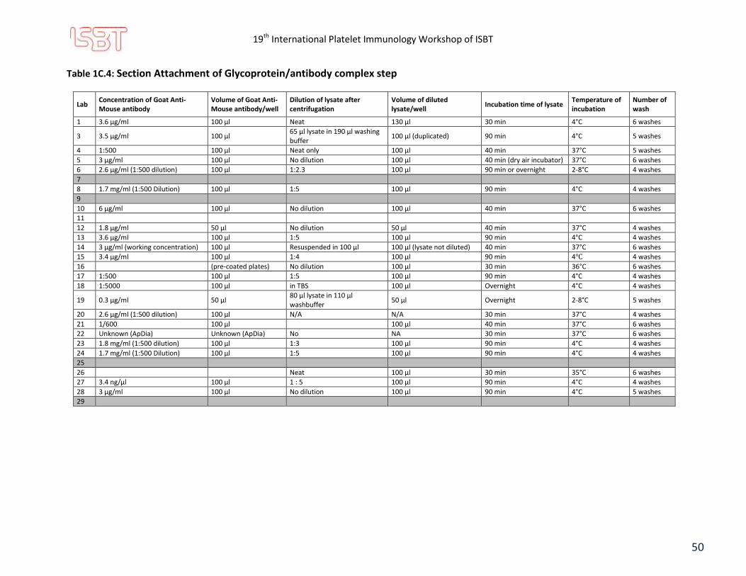
Table 1C.4: Section Attachment of Glycoprotein/antibody complex step
Lab Concentration of Goat Anti-Mouse antibody
Volume of Goat Anti-Mouse antibody/well
Dilution of lysate after centrifugation
Volume of diluted lysate/well
Incubation time of lysate Temperature of incubation
Number of wash
1 3.6 μg/ml 100 μl Neat 130 μl 30 min 4°C 6 washes
3 3.5 μg/ml 100 μl 65 μl lysate in 190 μl washing buffer
100 μl (duplicated) 90 min 4°C 5 washes
4 1:500 100 μl Neat only 100 μl 40 min 37°C 5 washes
5 3 μg/ml 100 μl No dilution 100 μl 40 min (dry air incubator) 37°C 6 washes
6 2.6 µg/ml (1:500 dilution) 100 μl 1:2.3 100 μl 90 min or overnight 2-8°C 4 washes
7
8 1.7 mg/ml (1:500 Dilution) 100 μl 1:5 100 μl 90 min 4°C 4 washes
9
10 6 µg/ml 100 μl No dilution 100 μl 40 min 37°C 6 washes
11
12 1.8 µg/ml 50 μl No dilution 50 μl 40 min 37°C 4 washes
13 3.6 µg/ml 100 μl 1:5 100 μl 90 min 4°C 4 washes
14 3 μg/ml (working concentration) 100 μl Resuspended in 100 µl 100 μl (lysate not diluted) 40 min 37°C 6 washes
15 3.4 μg/ml 100 μl 1:4 100 μl 90 min 4℃ 4 washes
16
(pre-coated plates) No dilution 100 μl 30 min 36°C 6 washes
17 1:500 100 μl 1:5 100 μl 90 min 4°C 4 washes
18 1:5000 100 μl in TBS 100 μl Overnight 4°C 4 washes
19 0.3 µg/ml 50 µl 80 µl lysate in 110 µl washbuffer
50 μl Overnight 2-8°C 5 washes
20 2.6 μg/ml (1:500 dilution) 100 μl N/A N/A 30 min 37°C 4 washes
21 1/600 100 μl
100 μl 40 min 37°C 6 washes
22 Unknown (ApDia) Unknown (ApDia) No NA 30 min 37°C 6 washes
23 1.8 mg/ml (1:500 dilution) 100 μl 1:3 100 μl 90 min 4°C 4 washes
24 1.7 mg/ml (1:500 Dilution) 100 μl 1:5 100 μl 90 min 4°C 4 washes
25
26
Neat 100 μl 30 min 35°C 6 washes
27 3.4 ng/µl 100 μl 1 : 5 100 μl 90 min 4°C 4 washes
28 3 μg/ml 100 μl No dilution 100 μl 90 min 4°C 5 washes
29

19th International Platelet Immunology Workshop of ISBT
51
Table 1C.5: Section Anti-Human conjugate and Colorimetry
Lab Concentration of conjugate Anti-Human antibody
Volume of conjugate antibody/well
Incubation time of conjugate
Temperature of incubation
Number of wash
Name of substrate
Incubation time of substrate
Temperature of incubation*
Stop solution used Wave length for reading
1 0.2 μg/ml 100 μl 120 min 4°C 6 washes OPD 15-20 min RT H2SO4 492/620 nm
3 80 ng/ml 100 μl 90 min 4°C 6 washes OPD 15-20 min RT H2SO4 490 nm
4 1:6000 100 μl 60 min RT 5 washes OPD 2HCL 10-20 min RT 2N H2SO4 490 nm
5 0.8 mg/ml, N:12 000 100 μl 60 min 18-25°C 6 washes OPD 20 min 18-25°C 0.5M H2SO4 490 nm
6 1:4000 = 0.2 μg/ml 100 μl 90 min 2-8°C 6 washes TMB, OPD 15-18 min RT TMB= 100uL of 0.2M 0.4N H2SO44, Sigma=50uL of 2.5N H2SO4
TMB=450 nm, OPD=490 nm
7
8 1:4000 100 μl 2 hours 4°C 4 washes OPD 15 min RT 2.5M H2SO4 492/620 nm
9
10 0.07 ug/ml 100 μl 40 min 37°C 6 washes 3.3′, 5.5′- TMB 10 min 18-25°C 1M H2SO4 450 nm
11
50 μl 30 min RT 3 washes
12 0.1 μg/ml 100 μl/well 40 min 22-25°C 4 washes OPD, H2O2 12 min 22-25°C 2.5N H2SO4 492 nm
13 32 ng/ml 100 μl 90 min 4°C 6 washes OPD 12 min 20°C 2.5N H2SO4 492 nm
14 0.26 μg/ml 100 μl 40 min 18-20°C 6 washes OPD 20 min 18-25°C 0.5M H2SO4 490 nm
15 0.2 μg/ml 100 μl 90 min 4°C 4 washes OPD 15 min RT 2.5N H2SO4 492 nm
16 no data 100 μl 30min 36°C 6 washes TMB 15 min RT H2SO4 450 nm (background 590nm)
17 1 :4000 100 μl 90min 4°C 5 washes TMB 10 min RT 100µl H2SO4 450/620 nm
18 1 :5000 100 μl overnight 4°C 6 washes 3.3’, 5.5’- TMB 2-5 min RT 1N H2SO4 450 nm
19 50 µl overnight 2-8°C 5 washes OPD max 30 min RT H2SO4 492 nm
20 1:2000 100 μl 1 hour 2-8°C 6 washes OPD 10 min RT 0.5M H2SO4 492/620 nm
21 100 μl 60 min 18-20°C 6 washes OPD 9 min 22°C H2SO4 492 nm
22 Unknown (ApDia) 100 μl 30 min 37°C 6 washes TMB 15 min 37°C TMB stop solution 450 nm
23 0.8mg/ml, 1 :10000 100 μl 90 min 4°C 6 washes TMB 10-20 min RT 1N H2SO4 450 nm
24 1:4000 100 μl 2 hours 4°C 4 washes OPD 15 min RT 2.5M H2SO4 492/620 nm
25
26
100 μl 30 min 35°C 6 washes 3.3', 5.5'-TMB 15 min 35°C H2SO4 450/650 nm
27 0.266 ng/µl 100 μl 120 min 4°C 6 washes OPD 15 min RT H2SO4 492 nm
28 0.13 ug/ml 100 μl 90 min 4°C 5 washes TMB 5 min RT H2SO4 450 nm
29
*: RT = Room Temperature
19th International Platelet Immunology Workshop of ISBT
52
Preparation/Incubation section The starting platelet concentration varied a lot; from 50x105 to 10x108 platelet/mL, in a volume of 20 to 125µL. This gives a range of available platelets varying from 0.6 to 75x106 platelet/tube or well. The dilution factor applied on the patient serum during contact with platelets varies from 1/1.42 to 1/6. Monoclonal concentration and volume also vary considerably. However, some labs gave information for all the monoclonals they use and some labs gave information for a specific one, giving no information about the others. It is though difficult to compare and conclude on this point. One thing observed overall is that there is a wide range of final concentration and volume used for all the monoclonals. Sixteen/24 labs (66.67%) provided a concentration value with µg/mL or µg/µL units, 5/24 (20.83%) provided a dilution value, 2/24 (8.33%) gave a quantity in either mg and µg and 1/24 (4.17%) did not answered. Incubation times for platelet and patient’s serum and for monoclonal incubation are relatively consistent between labs and go from 30 to 40 min (except one lab with longer incubation times) with a temperature of 37°C. These conditions are stable among participants as well as the number of washes (3±1 washes for serum and monoclonal steps, except 3 labs with 5, 6 and 8 washes). Quantification part of the Preparation/Incubation section A total of 12/24 (50%) participants answered using a standard or making dilutions of either the sample or the standard. Of these 12 labs, 6 (50%) mentioned using a WHO (NIBSC) antibody standard and 5 (41.67%) mentioned using another source or an “in house” standard. Five/12 (41.67%) performed serial dilutions of the sample to be tested while 4 of these 5 (80.00%) also mentioned doing serial dilutions of the sample to be tested. Six/12 (50.00%) are using fixed or single dilution or undiluted standard. Lysis section Parameters which differ most in the lysis step in the incubation time and the temperature at which the lysis occurs. Sixteen/24 (66.67%) labs perform lysis at 4°C and incubation time from 15 min to overnight. Seven labs (29.17%) are lysing at room temperature for 15 to 30 min. One lab (4.17%) reports using both possibilities. TritonX is used by the majority (16/24, 66.66%) as a detergent in the buffer composition. Three (12.50%) are using Igepal, 2 (8.33%) are using Nonidet and 3 (12.50%) did not mention. Attachment to solid phase section Some variation is seen in the concentration of the Goat Anti-mouse antibody used by the participants but this could be specific to the brand and the lot number. Twenty/24 (83.33%) gave information on the antibody concentration and 2/24 (8.33%) mentioned using pre-coated plates or ApDia. On the 20 who did answer, 18 (90.00%) are using a volume of 100 µL and 2/20 (10.00%) are using 50 µL. A good consensus is also reached when looking at the lysate volume where 18/21 (85.71%) are using 100 µL, 2/21 (9.52%)
19th International Platelet Immunology Workshop of ISBT
53
are using 50 µL, only 1/21 (4.76%) is using 130 µL and 3 of the 24 participants did not answer. Where there is much more variation is on the lysate dilution. Two labs did not answer and 2 labs gave no precision on the 24 participants. On the 20 labs who provided details, 10 (50.00%) are using the lysate without dilution, 2 (10.00%) are using a 1/2.3 dilution, 1 (5.00%) is using a 1/3 dilution, 2 (10.00%) are using a 1/ 4 dilution and 5 (25.00%) are using a 1/5 dilution. Incubation time also varies, the majority 10/23 (43.48%) incubate for 90 min, 6/23 (26.09%) incubate 40 min, 5/23 (21.74%) incubate 30 min and 2/23 (8.70%) incubate overnight. One/24 (4.17%) did not answer. The temperature of incubation is more evenly distributed between those incubating at 4°C (13/23, 56.52 %) and those incubating at 35-37°C (10/23, 43.48%). Conjugate antibody section The concentration of the conjugate Anti-human antibody varied a lot although the volume is quite the same among labs. Some labs reported a dilution factor instead of a concentration, making the comparison difficult. Colorimetry section The most commonly used colorimetric procedure is the one with OPD (14/24, 58.33%) despite the fact that OPD is very toxic and that many companies stopped producing it few years ago. Eight/24 (33.33%) labs mentioned using TMB, 1/24 (4.17%) is using both OPD and TMB and 1/24 (4.17%) did not answer this part of the questionnaire. Conclusion We are far from harmonization of the MAIPA protocol. The variation seen in the protocols may contribute to variations in results. It would be beneficial for all participants to try the protocols that are suggested by the NIBSC at this link: http://www.nibsc.org/science_and_research/biotherapeutics/platelets.aspx The ISBT Platelet Immunology Working Party may want to consider strategies for working on standardization of the MAIPA protocol and also on establishing uniformity of the laboratory analytical approach to be developped when investigating a patient case.
19th International Platelet Immunology Workshop of ISBT
54
Exercise 2 FNAIT caused by HLA Specific Alloantibodies
Aim:
1) To detect the presence and identify HLA-specific alloantibodies in a case of FNAIT caused by Anti-HLA antibodies.
2) To determine the HLA of the mother, the father and the child.
Case History:
Both parents are Caucasians and blood group O Rh(D) positive.The mother gave birth of a first child with a severe thrombocytopenia (5x10⁹ platelet count) and intracranial bleed. Anti-HPA antibody could not be found but Anti-HLA were present. Two years later, the mother was refferred again at 20 weeks of pregnancy for a monthly follow-up. No Anti-HPA antibody could be found but strong Anti-HLA antibody specificities could be identified and corresponded to the HLA of the father. While Anti-HLA antibodies were identified in cord blood plasma, an eluate could not be performed due to insufficient numbers of neonatal platelets.
The second child also had a severe thrombocytopenia at birth (7x10⁹ platelet count) but had no bleeding. Nevertheless, he was transfused with HLA selected platelets.
Materials Supplied:
Participating laboratories were provided with:
1 maternal serum sample (S6) (1.5 mL)
3 DNA samples (H1 - maternal, 70 µL of 27 ng/µL; H2 - paternal, 70 µL of 39 ng/µL; H3 - child, 20 µL of 25 ng/µL)
Methods:
The serum sample was to be tested for the presence of platelet-specific alloantibodies and HLA antibodies. The 3 DNA samples were to be genotyped for HLA class I, loci A and B. Participating laboratories were to:
1) Test the serum sample according to routine techniques used in the investigation of FNAIT cases.
2) Test serum to determine specificity of HLA antibodies using routine techniques.
3) Test all 3 DNA samples with their current HLA genotyping technique for loci A and B.
Results:
Assay data and the identified specificity of platelet and/or HLA-specific alloantibodies were reported in the Excel answer grid provided.

19th International Platelet Immunology Workshop of ISBT
55
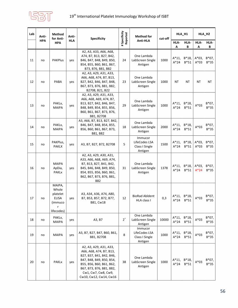
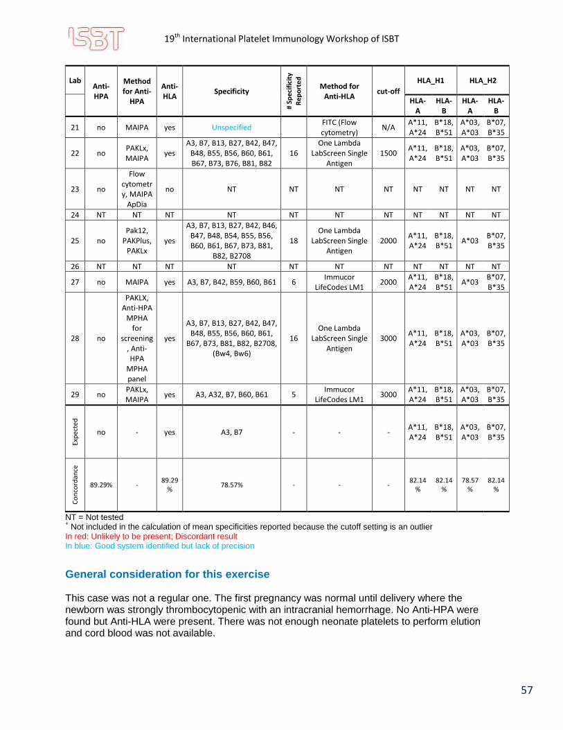
Results for Exercise 2 FNAIT caused by HLA Specific Alloantibodies
Antibody Detection
This case was different from the ordinary FNAIT cases because no Anti-HPA antibody could be demonstrated. Instead, Anti-HLA were found to be the cause of the thrombocytopenia of the neonate. Table 2.1 Results for Anti-HLA Identification and HLA Genotyping for Exercise 2
Lab Anti- HPA
Method for Anti-
HPA
Anti- HLA
Specificity
# Sp
eci
fici
ty
Re
po
rte
d
Method for Anti-HLA
cut-off
HLA_H1 HLA_H2
HLA-
A HLA-
B HLA-
A HLA-
B
1 no MAIPA yes
A2, A3, A29, A31, A33, A66, A68, A74, B7, B13, B27, B42, B46, B47, B48, B49, B54, B55, B56, B60, B61, B67, B73, B76, B81,
B82, B2708
27 One Lambda
LabScreen Single Antigen
500 A*11, A*24
B*18, B*51
A*03, A*03
B*07, B*35
3 no PAKPlus, PAKLx, MAIPA
yes A3, B7, B27, B50, B2708,
(B27:05, B27:03) 5
Immucor LifeCodes LSA Class I Single
Antigen
1500 A*11, A*24
B*18, B*51
A*03, A*03
B*07:02,
B*35
4 no PAKLx yes
A3, A66, B7, B27, B42, B46, B47, B48, B54, B55, B56, B60, B61, B67, B73, B81,
B82
17 One Lambda
LabScreen Single Antigen
2500 A*11, A*24
B*18, B*51
A*03, A*03
B*07, B*35
5 no MAIPA yes A3, B7, B13, B27, B42, B48,
B55, B56, B60, B61, B67, B73, B81, B82, B2708
16 One Lambda
LabScreen Single Antigen
2000 A*11, A*24
B*18, B*51
A*03, A*03
B*07, B*35
6 no PAKLx, MAIPA
yes
A2, A3, A29, A31, A33, A66, A68, A74, B7, B13, B27, B42, B46, B47, B48, B49, B50, B54, B55, B56, B60, B61, B67, B73, B76,
B81, B82, B2708
28 One Lambda
LabScreen Single Antigen
1000 A*11, A*24
B*18, B*51
A*03 B*07, B*35
7 no MAIPA yes
A3, A66, B7, B13, B27, B42, B47, B55, B56, B60, B61,
B64, B67, B73, B81, B2708, (Bw4, Bw6)
16
Immucor LifeCodes LSA Class I Single
Antigen
750 A*11, A*24
B*18, B*51
A*03, A*03
B*07, B*35
8 HPA 5b MAIPA yes
A2, A3, A29, A31, A33, A66, A69, A74, B7, B13, B27, B42, B46, B47, B48, B49, B50, B54, B55, B56, B60, B61, B67, B73, B76,
B81, B82
27 One Lambda
LabScreen Single Antigen
1000 A*11, A*24
B*18, B*51
A*03, A*03
B*07, B*35
9 no PAKLx, MAIPA
yes Unspecified NT NT (screening) NT A*11, A*24
B*18, B*51
A*03 B*07, B*35
10 no PAKLx yes Unspecified NT NT (Screening) NT NT NT NT NT
19th International Platelet Immunology Workshop of ISBT
56
Lab Anti- HPA
Method for Anti-
HPA
Anti- HLA
Specificity
# Sp
eci
fici
ty
Re
po
rte
d
Method for Anti-HLA
cut-off
HLA_H1 HLA_H2
HLA-
A HLA-
B HLA-
A HLA-
B
11 no PAKPlus yes
A2, A3, A33, A66, A68, A74, B7, B13, B27, B42,
B46, B47, B48, B49, B50, B54, B55, B60, B61, B67,
B73, B76, B81, B82
24 One Lambda
LabScreen Single Antigen
1000 A*11, A*24
B*18, B*51
A*03, A*03
B*07, B*35
12 no PABA yes
A2, A3, A29, A31, A33, A66, A68, A74, B7, B13, B27, B42, B46, B47, B48, B67, B73, B76, B81, B82,
B2708, B21, B22
23 One Lambda
LabScreen Single Antigen
1000 NT NT NT NT
13 no PAKLx, MAIPA
yes
A2, A3, A29, A31, A33, A66, A68, A69, A74, B7, B13, B27, B42, B46, B47, B48, B49, B54, B55, B56, B60, B61, B67, B73, B76,
B81, B2708
29 One Lambda
LabScreen Single Antigen
1000 A*11, A*24
B*18, B*51
A*03 B*07, B*35
14 no PAKLx, MAIPA
yes
A3, A66, B7, B13, B27, B42, B46, B47, B48, B54, B55, B56, B60, B61, B67, B73,
B81, B82
18 One Lambda
LabScreen Single Antigen
2000 A*11, A*24
B*18, B*51
A*03 B*07, B*35
15 no PAKPlus,
PAKLX yes A3, B7, B27, B72, B2708 5
Immucor LifeCodes LSA Class I Single
Antigen
1500 A*11, A*24
B*18, B*51
A*03, A*03
B*07, B*35
16 no MAIPA ApDia, PAKLx
yes
A2, A3, A29, A30, A31, A33, A66, A68, A69, A74, B7, B13, B27, B41, B42,
B45, B46, B48, B49, B50, B54, B55, B56, B60, B61, B62, B67, B73, B76, B81,
B82
31 One Lambda
LabScreen Single Antigen
1378 A*11, A*24
B*18, B*51
A*03, A*24
B*07, B*35
17 no
MAIPA, Whole
platelet ELISA
(immucor
lifecodes)
yes A3, A34, A36, A74, A80, B7, B53, B57, B72, B77,
B81, Cw18 12
BioRad Abldent HLA class I
0,3 A*11, A*24
B*18, B*51
A*03 B*07, B*35
18 no PAKLx, MAIPA
yes A3, B7 2+
One Lambda LabScreen Single
Antigen 10000
A*11, A*24
B*18, B*51
A*03 B*07, B*35
19 no MAIPA yes A3, B7, B27, B47, B60, B61,
B81, B2708 8
Immucor LifeCodes LSA Class I Single
Antigen
1000 A*11, A*24
B*18, B*51
A*03 B*07, B*35
20 no PAKLx yes
A2, A3, A29, A31, A33, A66, A68, A74, B7, B13, B27, B37, B41, B42, B46, B47, B48, B49, B50, B54, B55, B56, B60, B61, B62, B67, B73, B76, B81, B82,
Cw1, Cw7, Cw8, Cw9, Cw10, Cw12, Cw14, Cw16
38 One Lambda
LabScreen Single Antigen
1000 A*11, A*24
B*18, B*51
A*03 B*07, B*35
19th International Platelet Immunology Workshop of ISBT
57
Lab Anti- HPA
Method for Anti-
HPA
Anti- HLA
Specificity
# Sp
eci
fici
ty
Re
po
rte
d
Method for Anti-HLA
cut-off
HLA_H1 HLA_H2
HLA-
A HLA-
B HLA-
A HLA-
B
21 no MAIPA yes Unspecified FITC (Flow cytometry)
N/A A*11, A*24
B*18, B*51
A*03, A*03
B*07, B*35
22 no PAKLx, MAIPA
yes A3, B7, B13, B27, B42, B47,
B48, B55, B56, B60, B61, B67, B73, B76, B81, B82
16 One Lambda
LabScreen Single Antigen
1500 A*11, A*24
B*18, B*51
A*03, A*03
B*07, B*35
23 no
Flow cytometry, MAIPA
ApDia
no NT NT NT NT NT NT NT NT
24 NT NT NT NT NT NT NT NT NT NT NT
25 no Pak12,
PAKPlus, PAKLx
yes
A3, B7, B13, B27, B42, B46, B47, B48, B54, B55, B56, B60, B61, B67, B73, B81,
B82, B2708
18 One Lambda
LabScreen Single Antigen
2000 A*11, A*24
B*18, B*51
A*03 B*07, B*35
26 NT NT NT NT NT NT NT NT NT NT NT
27 no MAIPA yes A3, B7, B42, B59, B60, B61 6 Immucor
LifeCodes LM1 2000
A*11, A*24
B*18, B*51
A*03 B*07, B*35
28 no
PAKLX, Anti-HPA
MPHA for
screening, Anti-HPA
MPHA panel
yes
A3, B7, B13, B27, B42, B47, B48, B55, B56, B60, B61,
B67, B73, B81, B82, B2708, (Bw4, Bw6)
16 One Lambda
LabScreen Single Antigen
3000 A*11, A*24
B*18, B*51
A*03, A*03
B*07, B*35
29 no PAKLx, MAIPA
yes A3, A32, B7, B60, B61 5 Immucor
LifeCodes LM1 3000
A*11, A*24
B*18, B*51
A*03, A*03
B*07, B*35
Exp
ecte
d
no - yes A3, B7 - - - A*11, A*24
B*18, B*51
A*03, A*03
B*07, B*35
Co
nco
rdan
ce
89.29% - 89.29
% 78.57% - - -
82.14%
82.14%
78.57%
82.14%
NT = Not tested + Not included in the calculation of mean specificities reported because the cutoff setting is an outlier
In red: Unlikely to be present; Discordant result In blue: Good system identified but lack of precision
General consideration for this exercise This case was not a regular one. The first pregnancy was normal until delivery where the newborn was strongly thrombocytopenic with an intracranial hemorrhage. No Anti-HPA were found but Anti-HLA were present. There was not enough neonate platelets to perform elution and cord blood was not available.
19th International Platelet Immunology Workshop of ISBT
58
At the second pregnancy, the case was refferred during midterm. Again no Anti-HPA could be found but the Anti-HLA were rising as compared to their level from the first pregnancy. Anti-HLA antibodies were identified and corresponded to the HLA of the father. Because the father was homozygous for HLA-A locus and was likely to give the allele to the second child, because the Anti-HLA were rising up during pregnancy, and also because the first child had been dramatically impacted, the physician decided to prepare for C-section with a transfusion of HLA-compatible platelet if needed. We provided a platelet unit that was negative for the HLA antigens of the father. At birth, the newborn had a platelet count of only 7 but did not suffer from intracranial hemorrhage. Nevertheles, he received the platelet transfusion. We could demonstrate the presence of Anti-HLA antibody in the cord blood plasma and an eluate could not be performed due to insufficient numbers of neonatal platelets. It is always a challenge to confirm HLA as a causative reason for FNAIT since not all the labs are able to perform Anti-HLA antibody identification and HLA genotyping in a routine basis. However, the vast majority of participants 26/28 (92.86%) did verify the absence of Anti-HPA antibody and 25/28 (89.29%) verified the presence of Anti-HLA antibody. The specificity of Anti-HLA antibody was determined by 22/28 (78.57%) of the participants. Table 2.2 Results for Anti-HPA Antibody Detection
Frequency Percent
Anti-HPA-5b (?) 1 3.57
Negative 25 89.29
Not tested 2 7.14
There is a strong consensus (89.29%) on the absence of Anti-HPA antibodies. One center (Lab #8) reported a suspicion of Anti-HPA-5b which is unlikely to be present. A total of 2 centers (Lab #24 and #26) did not test for Anti-HPA. Some centers (12) reported an Anti-HLA in the section of Anti-HPA reporting. We therefore considered that the 12 centers did not detect any Anti-HPA specificity and found similar result than the expected result. All centers that tested for Anti-HLA reported Anti-HLA (100%). Because 3 centers did not test for Anti-HLA, consensus rate was only 89.29%. Table 2.3 Anti-HLA specificity for concordance results
Frequency Percent
A3 and B7 22 78.57
not reported 3 10.71
not tested 3 10.71
Of the 25 centers that tested for Anti-HLA and found an antibody, 3 did not report the specificity of the antibody found. Nevertheless, all the centers that tested for Anti-HLA did found and reported at least an Anti-HLA-A3 and an Anti-HLA-B7, resulting in a concordance of 78.57% (this concordance rises to 100% when using only respondent center)
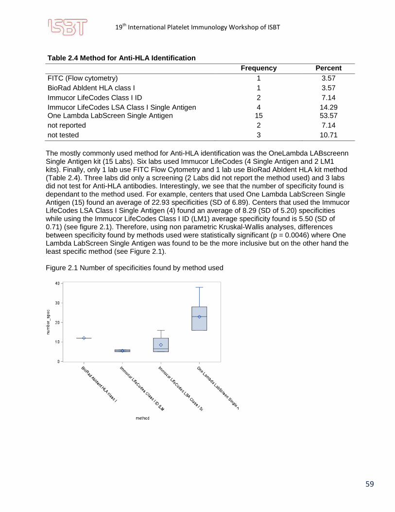
19th International Platelet Immunology Workshop of ISBT
59
Table 2.4 Method for Anti-HLA Identification
Frequency Percent
FITC (Flow cytometry) 1 3.57
BioRad Abldent HLA class I 1 3.57
Immucor LifeCodes Class I ID 2 7.14
Immucor LifeCodes LSA Class I Single Antigen 4 14.29 One Lambda LabScreen Single Antigen 15 53.57
not reported 2 7.14
not tested 3 10.71
The mostly commonly used method for Anti-HLA identification was the OneLambda LABscreenn Single Antigen kit (15 Labs). Six labs used Immucor LifeCodes (4 Single Antigen and 2 LM1 kits). Finally, only 1 lab use FITC Flow Cytometry and 1 lab use BioRad AbIdent HLA kit method (Table 2.4). Three labs did only a screening (2 Labs did not report the method used) and 3 labs did not test for Anti-HLA antibodies. Interestingly, we see that the number of specificity found is dependant to the method used. For example, centers that used One Lambda LabScreen Single Antigen (15) found an average of 22.93 specificities (SD of 6.89). Centers that used the Immucor LifeCodes LSA Class I Single Antigen (4) found an average of 8.29 (SD of 5.20) specificities while using the Immucor LifeCodes Class I ID (LM1) average specificity found is 5.50 (SD of 0.71) (see figure 2.1). Therefore, using non parametric Kruskal-Wallis analyses, differences between specificity found by methods used were statistically significant (p = 0.0046) where One Lambda LabScreen Single Antigen was found to be the more inclusive but on the other hand the least specific method (see Figure 2.1). Figure 2.1 Number of specificities found by method used
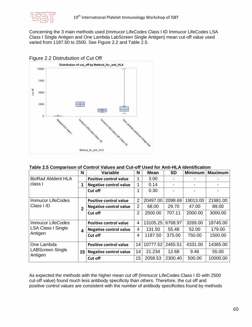
19th International Platelet Immunology Workshop of ISBT
60
Concerning the 3 main methods used (Immucor LifeCodes Class I ID Immucor LifeCodes LSA Class I Single Antigen and One Lambda LabScreen Single Antigen) mean cut-off value used varied from 1187.50 to 2500. See Figure 2.2 and Table 2.5.
Figure 2.2 Distrubution of Cut Off
Table 2.5 Comparison of Control Values and Cut-off Used for Anti-HLA Identification
N Variable N Mean SD Minimum Maximum
BioRad Abldent HLA class I
Positive control value 1 3.00 - - -
1 Negative control value 1 0.14 - - -
Cut off 1 0.30 - - -
Immucor LifeCodes Class I ID
2
Positive control value 2 20497.00 2098.69 19013.00 21981.00
Negative control value 2 68.00 29.70 47.00 89.00
Cut off 2 2500.00 707.11 2000.00 3000.00
Immucor LifeCodes LSA Class I Single Antigen
4
Positive control value 4 13105.25 6768.97 3269.00 18745.00
Negative control value 4 131.50 55.48 52.00 179.00
Cut off 4 1187.50 375.00 750.00 1500.00
One Lambda LABScreen Single Antigen
15
Positive control value 14 10777.52 2465.51 4331.00 14365.00
Negative control value 14 21.234 12.68 9.46 55.00
Cut off 15 2058.53 2300.40 500.00 10000.00
As expected the methods with the higher mean cut off (Immucor LifeCodes Class I ID with 2500 cut-off value) found much less antibody specificity than others. Therefore, the cut off and positive control values are consistent with the number of antibody specificities found by methods

19th International Platelet Immunology Workshop of ISBT
61
(i.e. the more the cut off and positive control increased, the less the number of specificities was found), see Table 2.6. Table 2.6 Median cutoff by the number of specificity found and the method used
N Median Q1 Q3
0 to 8 specificity found 2000 1500 3000
Immucor LifeCodes LM1 Class I ID 2 2500.00 2000.00 3000.00
Immucor LifeCodes LSA Class I Single Antigen 2 1500.00 1500.00 1500.00
One Lambda LabScreen Single Antigen 1 10000.00 - -
9 to 18 specificity found 1500 1000 2000
BioRad Abldent HLA class I 1 0.30 - - Immucor LifeCodes LSA Class I Single Antigen 1 1000.00 - -
One Lambda LabScreen Single Antigen 3 2000.00 1500.00 2500.00
19 – 27 specificity found 1500 1000 2000
Immucor LifeCodes LSA Class I Single Antigen 1 750.00 - - One Lambda LabScreen Single Antigen 5 2000.00 1000.00 2000.00
More than 27 specificity found 1000 1000 1000
One Lambda LabScreen Single Antigen 6 1000.00 1000.00 1000.00
Cut-off values were stratified by the number of specificities found. All centers that tested for Anti-HLA specificities were included in the analyses (including Lab 18 with an outlier cut-off of 10000) and, in order to estimate the central trend of data, medians and interquartile ranges were shown. Independently with the methods used we can see that the more the cut-off decrease the less the number of specificities was found (median cut-off were found to be 2000, 1500, 1500 and 1000 for group 0 to 8; 9 to 18; 19 to 27 and more than 27 specificities found, respectively). The determination of the cut off has a direct impact on the number of specificities that will be reported and we could see a big variability among labs.
Specificity of the results reported for Anti-HLA Generally speaking, we could see variations in specificity and sensitivity among the different methods but also among different labs, according to the cut off setting. The major point to report here would be that all the antibody identification method did report at least the Anti-HLA-A3 and Anti-HLA-B7 that were expected from the father genotype.
HLA Genotyping Mother
Table 2.7 HLA Genotyping for H1 (mother)
Allele A Allele B Frequency Percent
A*11, A*24 B*18, B*51 23 82.14
Not tested Not tested 5 17.86
Concerning the DNA of the mother, there was a participating rate of 82.14% (23/28) in the HLA genotyping exercise. Among the 23 participating labs, there was a 100% consensus and
19th International Platelet Immunology Workshop of ISBT
62
concordance with the original lab with the identification of A*11, A*24 on the A locus and B*18, B*51 on the B locus. Expected results: H1: Mother DNA HLA-A*11, A*24, B*18, B*51 Father
Table 2.8 HLA Genotyping for H2 (father)
Allele A Allele B Frequency Percent
A*03, A*03 B*07, B*35 22 78.57
A*03, A*24 B*07, B*35 1 3.57
Not tested Not tested 5 17.86
These results also reached a very good consensus and concordance with the original lab. Like for the Mother, 5 centers did HLA genotype the Father (17.86%) and only one center (Lab #16) reported a wrong specificity (A*3, A*24). Finally, all the other centers (n = 22) found the expected specificities in the Father DNA for the A locus (A*03, A*03) and the B locus (B*07, B*35) given a concordance rate of 95.65% (22/23). Expected results: H2: Father DNA HLA-A*03, A*03, B*07, B*35 Baby Because the DNA of the baby was found to be contaminated by many labs, calculation of consensus results concerning this HLA genotyping was not performed. Expected results: H3: Baby DNA HLA-A*03, A*11, B*07, B*18
Conclusion This case was unusual because of the direct implication of the Anti-HLA antibodies in the FNAIT. The gravity of the clinical status made it also of special importance. The first pregnancy of this couple ended out with an infant dramatically injured by an intracranial hemorrhage. The second pregnancy was also affected, however, the neonate could be delivered without injury. Both first and second neonates were drastically thrombopenic with a platelet count of 5 and 7 respectively. Anti-HLA are not always investigated in FNAIT cases. However, this case has proven their direct implication in severe FNAIT.
19th International Platelet Immunology Workshop of ISBT
63
Exercise 3 HPA Genotyping
Aim:
1) To determine the ability of the participating labs to genotype HPA-1 through 15.
2) To provide the labs with DNA having interesting HPA combination.
Materials Supplied:
Participating laboratories were provided with:
5 DNA samples (D1, D2, D3, D4, D5) containing between 45 and 100 µL of DNA at a concentration of 30 to 70 ng/µL each.
Methods:
The five DNA samples were to be analyzed using routine HPA genotyping method.
Results:
Assay data and the identified platelet genotype were to be reported in the Excel answer grid provided.
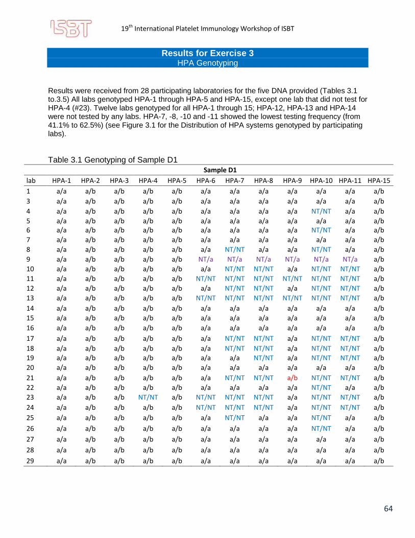
19th International Platelet Immunology Workshop of ISBT
64
Results for Exercise 3 HPA Genotyping
Results were received from 28 participating laboratories for the five DNA provided (Tables 3.1 to.3.5) All labs genotyped HPA-1 through HPA-5 and HPA-15, except one lab that did not test for HPA-4 (#23). Twelve labs genotyped for all HPA-1 through 15; HPA-12, HPA-13 and HPA-14 were not tested by any labs. HPA-7, -8, -10 and -11 showed the lowest testing frequency (from 41.1% to 62.5%) (see Figure 3.1 for the Distribution of HPA systems genotyped by participating labs).
Table 3.1 Genotyping of Sample D1 Sample D1
lab HPA-1 HPA-2 HPA-3 HPA-4 HPA-5 HPA-6 HPA-7 HPA-8 HPA-9 HPA-10 HPA-11 HPA-15
1 a/a a/b a/b a/b a/b a/a a/a a/a a/a a/a a/a a/b
3 a/a a/b a/b a/b a/b a/a a/a a/a a/a a/a a/a a/b
4 a/a a/b a/b a/b a/b a/a a/a a/a a/a NT/NT a/a a/b
5 a/a a/b a/b a/b a/b a/a a/a a/a a/a a/a a/a a/b 6 a/a a/b a/b a/b a/b a/a a/a a/a a/a NT/NT a/a a/b
7 a/a a/b a/b a/b a/b a/a a/a a/a a/a a/a a/a a/b
8 a/a a/b a/b a/b a/b a/a NT/NT a/a a/a NT/NT a/a a/b
9 a/a a/b a/b a/b a/b NT/a NT/a NT/a NT/a NT/a NT/a a/b
10 a/a a/b a/b a/b a/b a/a NT/NT NT/NT a/a NT/NT NT/NT a/b
11 a/a a/b a/b a/b a/b NT/NT NT/NT NT/NT NT/NT NT/NT NT/NT a/b
12 a/a a/b a/b a/b a/b a/a NT/NT NT/NT a/a NT/NT NT/NT a/b
13 a/a a/b a/b a/b a/b NT/NT NT/NT NT/NT NT/NT NT/NT NT/NT a/b
14 a/a a/b a/b a/b a/b a/a a/a a/a a/a a/a a/a a/b
15 a/a a/b a/b a/b a/b a/a a/a a/a a/a a/a a/a a/b
16 a/a a/b a/b a/b a/b a/a a/a a/a a/a a/a a/a a/b
17 a/a a/b a/b a/b a/b a/a NT/NT NT/NT a/a NT/NT NT/NT a/b
18 a/a a/b a/b a/b a/b a/a NT/NT NT/NT a/a NT/NT NT/NT a/b
19 a/a a/b a/b a/b a/b a/a a/a NT/NT a/a NT/NT NT/NT a/b
20 a/a a/b a/b a/b a/b a/a a/a a/a a/a a/a a/a a/b
21 a/a a/b a/b a/b a/b a/a NT/NT NT/NT a/b NT/NT NT/NT a/b
22 a/a a/b a/b a/b a/b a/a a/a a/a a/a NT/NT a/a a/b
23 a/a a/b a/b NT/NT a/b NT/NT NT/NT NT/NT a/a NT/NT NT/NT a/b
24 a/a a/b a/b a/b a/b NT/NT NT/NT NT/NT a/a NT/NT NT/NT a/b
25 a/a a/b a/b a/b a/b a/a NT/NT a/a a/a NT/NT a/a a/b
26 a/a a/b a/b a/b a/b a/a a/a a/a a/a NT/NT a/a a/b
27 a/a a/b a/b a/b a/b a/a a/a a/a a/a a/a a/a a/b
28 a/a a/b a/b a/b a/b a/a a/a a/a a/a a/a a/a a/b
29 a/a a/b a/b a/b a/b a/a a/a a/a a/a a/a a/a a/b

19th International Platelet Immunology Workshop of ISBT
65
Table 3.2 Genotyping of Sample D2
Sample
D2
lab HPA-1 HPA-2 HPA-3 HPA-4 HPA-5 HPA-6 HPA-7 HPA-8 HPA-9 HPA-10 HPA-11 HPA-15
1 a/a b/b b/b a/a a/b a/a a/a a/a a/a a/a a/a b/b
3 a/a b/b b/b a/a a/b a/a a/a a/a a/a a/a a/a b/b
4 a/a b/b b/b a/a a/b a/a a/a a/a a/a NT/NT a/a b/b
5 a/a b/b b/b a/a a/b a/a a/a a/a a/a a/a a/a b/b
6 a/a b/b b/b a/a a/b a/a a/a a/a a/a NT/NT a/a b/b
7 a/a b/b b/b a/a a/b a/a a/a a/a a/a a/a a/a b/b
8 a/a b/b b/b a/a a/b a/a NT/NT a/a a/a NT/NT a/a b/b
9 a/a b/b b/b a/a a/b NT/a NT/a NT/a NT/a NT/a NT/a b/b
10 a/a b/b b/b a/a a/b a/a NT/NT NT/NT a/a NT/NT NT/NT b/b
11 a/a b/b b/b a/a a/b NT/NT NT/NT NT/NT NT/NT NT/NT NT/NT b/b
12 a/a b/b b/b a/a a/b a/a NT/NT NT/NT a/a NT/NT NT/NT b/b
13 a/a b/b b/b a/a a/b NT/NT NT/NT NT/NT NT/NT NT/NT NT/NT b/b
14 a/a b/b b/b a/a a/b a/a a/a a/a a/a a/a a/a b/b
15 a/a b/b b/b a/a a/b a/a a/a a/a a/a a/a a/a b/b
16 a/a b/b b/b a/a a/b a/a a/a a/a a/a a/a a/a b/b
17 a/a b/b b/b a/a a/b a/a NT/NT NT/NT a/a NT/NT NT/NT b/b
18 a/a b/b b/b a/a a/b a/a NT/NT NT/NT a/a NT/NT NT/NT b/b
19 a/a b/b b/b a/a a/b a/a a/a NT/NT a/a NT/NT NT/NT b/b
20 a/a b/b b/b a/a a/b a/a a/a a/a a/a a/a a/a b/b
21 a/a b/b b/b a/a a/b a/a NT/NT NT/NT a/b NT/NT NT/NT b/b
22 a/a b/b b/b a/a a/b a/a a/a a/a a/a NT/NT a/a b/b
23 a/a b/b b/b NT/NT a/b NT/NT NT/NT NT/NT a/a NT/NT NT/NT b/b
24 a/a b/b b/b a/a a/b NT/NT NT/NT NT/NT a/a NT/NT NT/NT b/b
25 a/a b/b b/b a/a a/b a/a NT/NT a/a a/a NT/NT a/a b/b
26 a/a b/b b/b a/a a/b a/a a/a a/a a/a NT/NT a/a b/b
27 a/a b/b b/b a/a a/b a/a a/a a/a a/a a/a a/a b/b
28 a/a b/b b/b a/a a/b a/a a/a a/a a/a a/a a/a b/b
29 a/a b/b b/b a/a a/b a/a a/a a/a a/a a/a a/a b/b
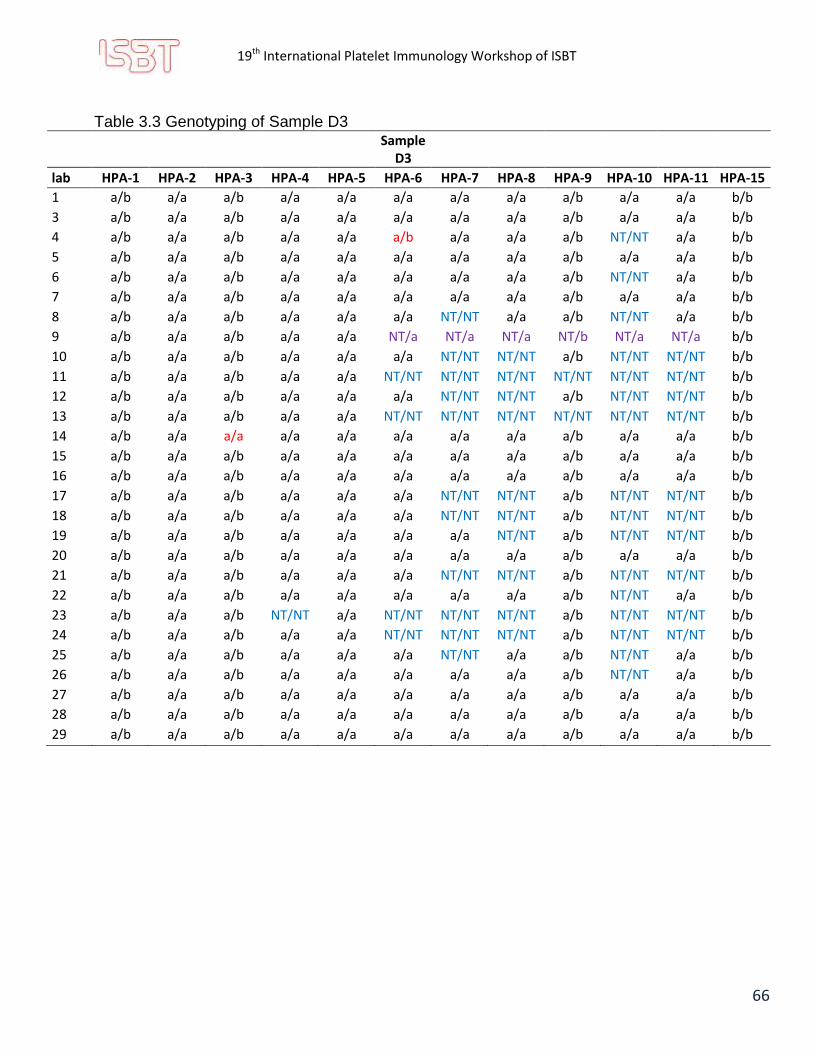
19th International Platelet Immunology Workshop of ISBT
66
Table 3.3 Genotyping of Sample D3
Sample
D3
lab HPA-1 HPA-2 HPA-3 HPA-4 HPA-5 HPA-6 HPA-7 HPA-8 HPA-9 HPA-10 HPA-11 HPA-15
1 a/b a/a a/b a/a a/a a/a a/a a/a a/b a/a a/a b/b
3 a/b a/a a/b a/a a/a a/a a/a a/a a/b a/a a/a b/b
4 a/b a/a a/b a/a a/a a/b a/a a/a a/b NT/NT a/a b/b
5 a/b a/a a/b a/a a/a a/a a/a a/a a/b a/a a/a b/b
6 a/b a/a a/b a/a a/a a/a a/a a/a a/b NT/NT a/a b/b
7 a/b a/a a/b a/a a/a a/a a/a a/a a/b a/a a/a b/b
8 a/b a/a a/b a/a a/a a/a NT/NT a/a a/b NT/NT a/a b/b
9 a/b a/a a/b a/a a/a NT/a NT/a NT/a NT/b NT/a NT/a b/b
10 a/b a/a a/b a/a a/a a/a NT/NT NT/NT a/b NT/NT NT/NT b/b
11 a/b a/a a/b a/a a/a NT/NT NT/NT NT/NT NT/NT NT/NT NT/NT b/b
12 a/b a/a a/b a/a a/a a/a NT/NT NT/NT a/b NT/NT NT/NT b/b
13 a/b a/a a/b a/a a/a NT/NT NT/NT NT/NT NT/NT NT/NT NT/NT b/b
14 a/b a/a a/a a/a a/a a/a a/a a/a a/b a/a a/a b/b
15 a/b a/a a/b a/a a/a a/a a/a a/a a/b a/a a/a b/b
16 a/b a/a a/b a/a a/a a/a a/a a/a a/b a/a a/a b/b
17 a/b a/a a/b a/a a/a a/a NT/NT NT/NT a/b NT/NT NT/NT b/b
18 a/b a/a a/b a/a a/a a/a NT/NT NT/NT a/b NT/NT NT/NT b/b
19 a/b a/a a/b a/a a/a a/a a/a NT/NT a/b NT/NT NT/NT b/b
20 a/b a/a a/b a/a a/a a/a a/a a/a a/b a/a a/a b/b
21 a/b a/a a/b a/a a/a a/a NT/NT NT/NT a/b NT/NT NT/NT b/b
22 a/b a/a a/b a/a a/a a/a a/a a/a a/b NT/NT a/a b/b
23 a/b a/a a/b NT/NT a/a NT/NT NT/NT NT/NT a/b NT/NT NT/NT b/b
24 a/b a/a a/b a/a a/a NT/NT NT/NT NT/NT a/b NT/NT NT/NT b/b
25 a/b a/a a/b a/a a/a a/a NT/NT a/a a/b NT/NT a/a b/b
26 a/b a/a a/b a/a a/a a/a a/a a/a a/b NT/NT a/a b/b
27 a/b a/a a/b a/a a/a a/a a/a a/a a/b a/a a/a b/b
28 a/b a/a a/b a/a a/a a/a a/a a/a a/b a/a a/a b/b
29 a/b a/a a/b a/a a/a a/a a/a a/a a/b a/a a/a b/b
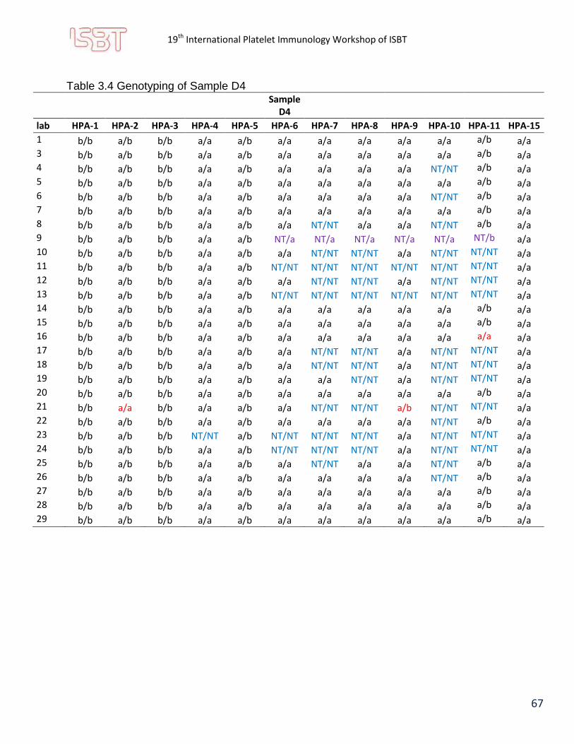
19th International Platelet Immunology Workshop of ISBT
67
Table 3.4 Genotyping of Sample D4
Sample
D4
lab HPA-1 HPA-2 HPA-3 HPA-4 HPA-5 HPA-6 HPA-7 HPA-8 HPA-9 HPA-10 HPA-11 HPA-15
1 b/b a/b b/b a/a a/b a/a a/a a/a a/a a/a a/b a/a 3 b/b a/b b/b a/a a/b a/a a/a a/a a/a a/a a/b a/a 4 b/b a/b b/b a/a a/b a/a a/a a/a a/a NT/NT a/b a/a 5 b/b a/b b/b a/a a/b a/a a/a a/a a/a a/a a/b a/a 6 b/b a/b b/b a/a a/b a/a a/a a/a a/a NT/NT a/b a/a 7 b/b a/b b/b a/a a/b a/a a/a a/a a/a a/a a/b a/a 8 b/b a/b b/b a/a a/b a/a NT/NT a/a a/a NT/NT a/b a/a 9 b/b a/b b/b a/a a/b NT/a NT/a NT/a NT/a NT/a NT/b a/a 10 b/b a/b b/b a/a a/b a/a NT/NT NT/NT a/a NT/NT NT/NT a/a 11 b/b a/b b/b a/a a/b NT/NT NT/NT NT/NT NT/NT NT/NT NT/NT a/a 12 b/b a/b b/b a/a a/b a/a NT/NT NT/NT a/a NT/NT NT/NT a/a 13 b/b a/b b/b a/a a/b NT/NT NT/NT NT/NT NT/NT NT/NT NT/NT a/a 14 b/b a/b b/b a/a a/b a/a a/a a/a a/a a/a a/b a/a 15 b/b a/b b/b a/a a/b a/a a/a a/a a/a a/a a/b a/a 16 b/b a/b b/b a/a a/b a/a a/a a/a a/a a/a a/a a/a 17 b/b a/b b/b a/a a/b a/a NT/NT NT/NT a/a NT/NT NT/NT a/a 18 b/b a/b b/b a/a a/b a/a NT/NT NT/NT a/a NT/NT NT/NT a/a 19 b/b a/b b/b a/a a/b a/a a/a NT/NT a/a NT/NT NT/NT a/a 20 b/b a/b b/b a/a a/b a/a a/a a/a a/a a/a a/b a/a 21 b/b a/a b/b a/a a/b a/a NT/NT NT/NT a/b NT/NT NT/NT a/a 22 b/b a/b b/b a/a a/b a/a a/a a/a a/a NT/NT a/b a/a 23 b/b a/b b/b NT/NT a/b NT/NT NT/NT NT/NT a/a NT/NT NT/NT a/a 24 b/b a/b b/b a/a a/b NT/NT NT/NT NT/NT a/a NT/NT NT/NT a/a 25 b/b a/b b/b a/a a/b a/a NT/NT a/a a/a NT/NT a/b a/a 26 b/b a/b b/b a/a a/b a/a a/a a/a a/a NT/NT a/b a/a 27 b/b a/b b/b a/a a/b a/a a/a a/a a/a a/a a/b a/a 28 b/b a/b b/b a/a a/b a/a a/a a/a a/a a/a a/b a/a 29 b/b a/b b/b a/a a/b a/a a/a a/a a/a a/a a/b a/a
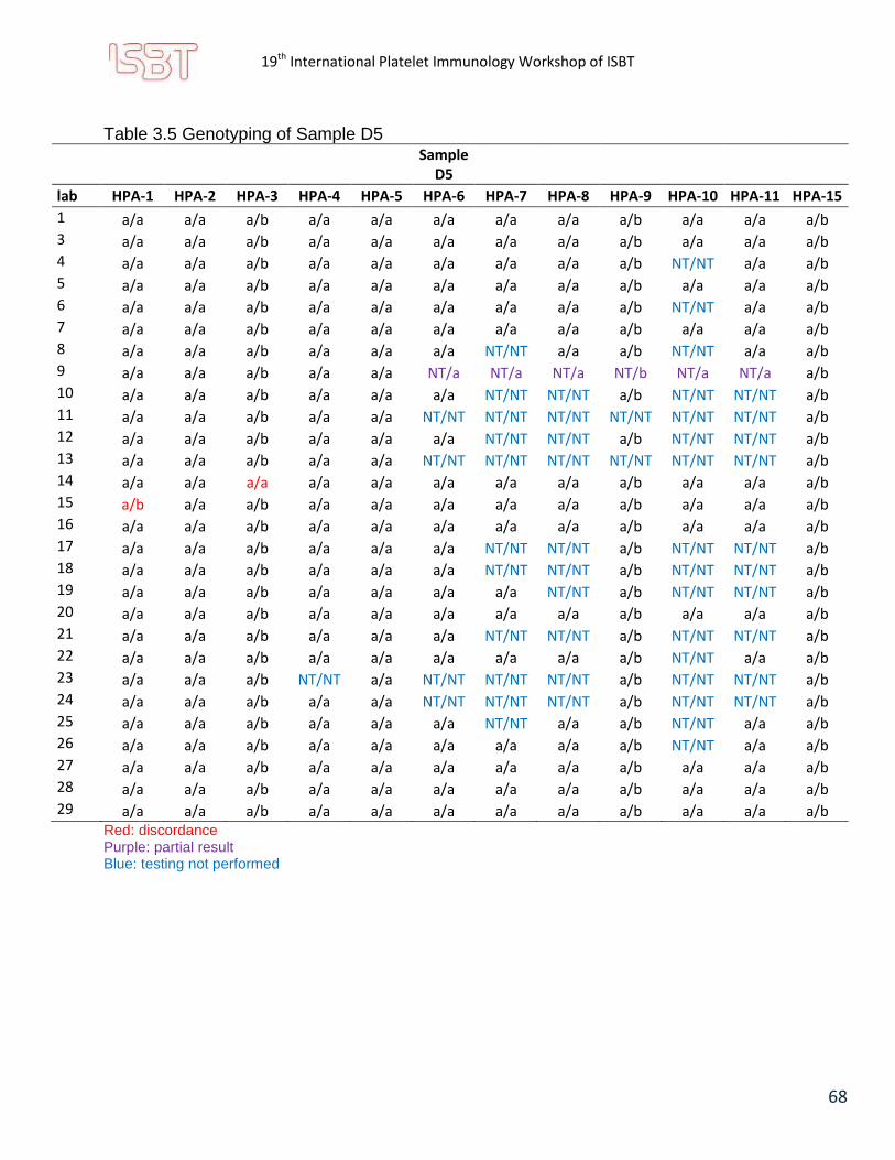
19th International Platelet Immunology Workshop of ISBT
68
Table 3.5 Genotyping of Sample D5
Sample
D5
lab HPA-1 HPA-2 HPA-3 HPA-4 HPA-5 HPA-6 HPA-7 HPA-8 HPA-9 HPA-10 HPA-11 HPA-15
1 a/a a/a a/b a/a a/a a/a a/a a/a a/b a/a a/a a/b 3 a/a a/a a/b a/a a/a a/a a/a a/a a/b a/a a/a a/b 4 a/a a/a a/b a/a a/a a/a a/a a/a a/b NT/NT a/a a/b 5 a/a a/a a/b a/a a/a a/a a/a a/a a/b a/a a/a a/b 6 a/a a/a a/b a/a a/a a/a a/a a/a a/b NT/NT a/a a/b 7 a/a a/a a/b a/a a/a a/a a/a a/a a/b a/a a/a a/b 8 a/a a/a a/b a/a a/a a/a NT/NT a/a a/b NT/NT a/a a/b 9 a/a a/a a/b a/a a/a NT/a NT/a NT/a NT/b NT/a NT/a a/b 10 a/a a/a a/b a/a a/a a/a NT/NT NT/NT a/b NT/NT NT/NT a/b 11 a/a a/a a/b a/a a/a NT/NT NT/NT NT/NT NT/NT NT/NT NT/NT a/b 12 a/a a/a a/b a/a a/a a/a NT/NT NT/NT a/b NT/NT NT/NT a/b 13 a/a a/a a/b a/a a/a NT/NT NT/NT NT/NT NT/NT NT/NT NT/NT a/b 14 a/a a/a a/a a/a a/a a/a a/a a/a a/b a/a a/a a/b 15 a/b a/a a/b a/a a/a a/a a/a a/a a/b a/a a/a a/b 16 a/a a/a a/b a/a a/a a/a a/a a/a a/b a/a a/a a/b 17 a/a a/a a/b a/a a/a a/a NT/NT NT/NT a/b NT/NT NT/NT a/b 18 a/a a/a a/b a/a a/a a/a NT/NT NT/NT a/b NT/NT NT/NT a/b 19 a/a a/a a/b a/a a/a a/a a/a NT/NT a/b NT/NT NT/NT a/b 20 a/a a/a a/b a/a a/a a/a a/a a/a a/b a/a a/a a/b 21 a/a a/a a/b a/a a/a a/a NT/NT NT/NT a/b NT/NT NT/NT a/b 22 a/a a/a a/b a/a a/a a/a a/a a/a a/b NT/NT a/a a/b 23 a/a a/a a/b NT/NT a/a NT/NT NT/NT NT/NT a/b NT/NT NT/NT a/b 24 a/a a/a a/b a/a a/a NT/NT NT/NT NT/NT a/b NT/NT NT/NT a/b 25 a/a a/a a/b a/a a/a a/a NT/NT a/a a/b NT/NT a/a a/b 26 a/a a/a a/b a/a a/a a/a a/a a/a a/b NT/NT a/a a/b 27 a/a a/a a/b a/a a/a a/a a/a a/a a/b a/a a/a a/b 28 a/a a/a a/b a/a a/a a/a a/a a/a a/b a/a a/a a/b 29 a/a a/a a/b a/a a/a a/a a/a a/a a/b a/a a/a a/b
Red: discordance Purple: partial result Blue: testing not performed
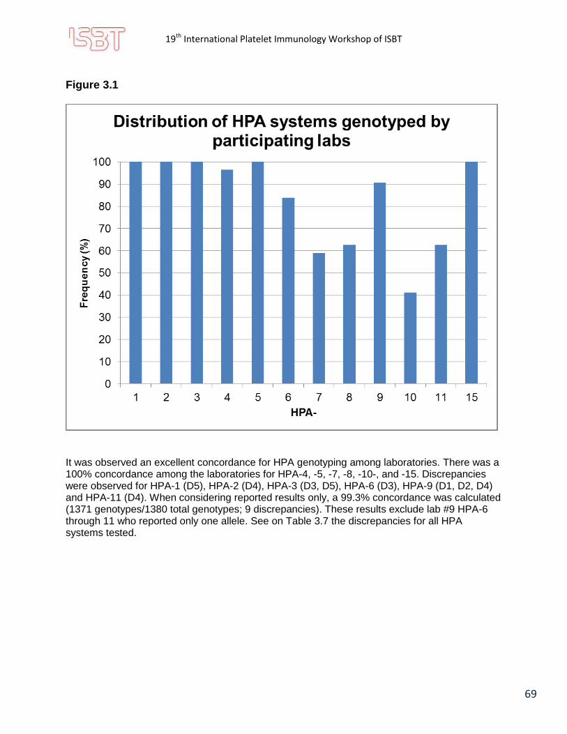
19th International Platelet Immunology Workshop of ISBT
69
Figure 3.1
It was observed an excellent concordance for HPA genotyping among laboratories. There was a 100% concordance among the laboratories for HPA-4, -5, -7, -8, -10-, and -15. Discrepancies were observed for HPA-1 (D5), HPA-2 (D4), HPA-3 (D3, D5), HPA-6 (D3), HPA-9 (D1, D2, D4) and HPA-11 (D4). When considering reported results only, a 99.3% concordance was calculated (1371 genotypes/1380 total genotypes; 9 discrepancies). These results exclude lab #9 HPA-6 through 11 who reported only one allele. See on Table 3.7 the discrepancies for all HPA systems tested.
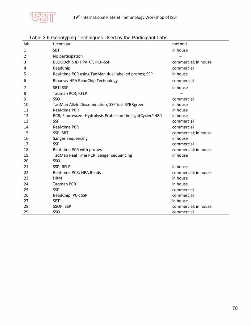
19th International Platelet Immunology Workshop of ISBT
70
Table 3.6 Genotyping Techniques Used by the Participant Labs
lab technique method
1 SBT in house
2 No participation –
3 BLOODchip ID HPA XT; PCR-SSP commercial; in house
4 BeadChip commercial
5 Real-time PCR using TaqMan dual labelled probes; SSP in house
6 Bioarray HPA BeadChip Technology commercial
7 SBT; SSP in house
8 Taqman PCR; RFLP – 9 SSO commercial 10 TaqMan Allele Discrimination; SSP test SYBRgreen in house 11 Real-time PCR in house 12 PCR; Fluorescent Hydrolysis Probes on the LightCycler® 480 in house 13 SSP commercial
14 Real-time PCR commercial
15 SSP; SBT commercial; in house 16 Sanger Sequencing in house 17 SSP commercial
18 Real-time PCR with probes commercial; in house 19 TaqMan Real Time PCR; Sanger sequencing in house 20 SSO –
21 SSP; RFLP in house
22 Real-time PCR; HPA Beads commercial; in house 23 HRM in house
24 Taqman PCR in house
25 SSP commercial 26 BeadChip; PCR SSP commercial 27 SBT in house 28 SSOP; SSP commercial; in house 29 SSO commercial
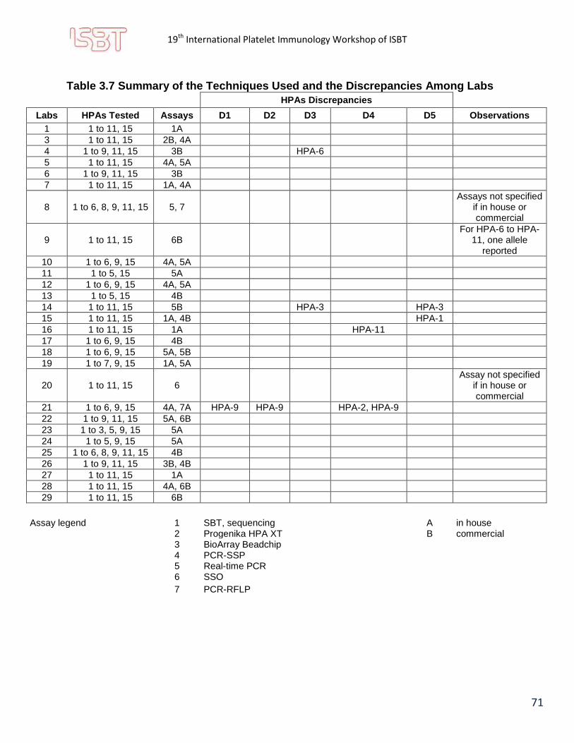
19th International Platelet Immunology Workshop of ISBT
71
Table 3.7 Summary of the Techniques Used and the Discrepancies Among Labs
HPAs Discrepancies
Labs HPAs Tested Assays D1 D2 D3 D4 D5 Observations
1 1 to 11, 15 1A
3 1 to 11, 15 2B, 4A
4 1 to 9, 11, 15 3B HPA-6
5 1 to 11, 15 4A, 5A
6 1 to 9, 11, 15 3B
7 1 to 11, 15 1A, 4A
8 1 to 6, 8, 9, 11, 15 5, 7 Assays not specified
if in house or commercial
9 1 to 11, 15 6B For HPA-6 to HPA-
11, one allele reported
10 1 to 6, 9, 15 4A, 5A
11 1 to 5, 15 5A
12 1 to 6, 9, 15 4A, 5A
13 1 to 5, 15 4B
14 1 to 11, 15 5B HPA-3 HPA-3
15 1 to 11, 15 1A, 4B HPA-1
16 1 to 11, 15 1A HPA-11
17 1 to 6, 9, 15 4B
18 1 to 6, 9, 15 5A, 5B
19 1 to 7, 9, 15 1A, 5A
20 1 to 11, 15 6 Assay not specified
if in house or commercial
21 1 to 6, 9, 15 4A, 7A HPA-9 HPA-9 HPA-2, HPA-9
22 1 to 9, 11, 15 5A, 6B
23 1 to 3, 5, 9, 15 5A
24 1 to 5, 9, 15 5A
25 1 to 6, 8, 9, 11, 15 4B
26 1 to 9, 11, 15 3B, 4B
27 1 to 11, 15 1A
28 1 to 11, 15 4A, 6B
29 1 to 11, 15 6B
Assay legend 1 SBT, sequencing
A in house
2 Progenika HPA XT
B commercial
3 BioArray Beadchip
4 PCR-SSP
5 Real-time PCR
6 SSO
7 PCR-RFLP
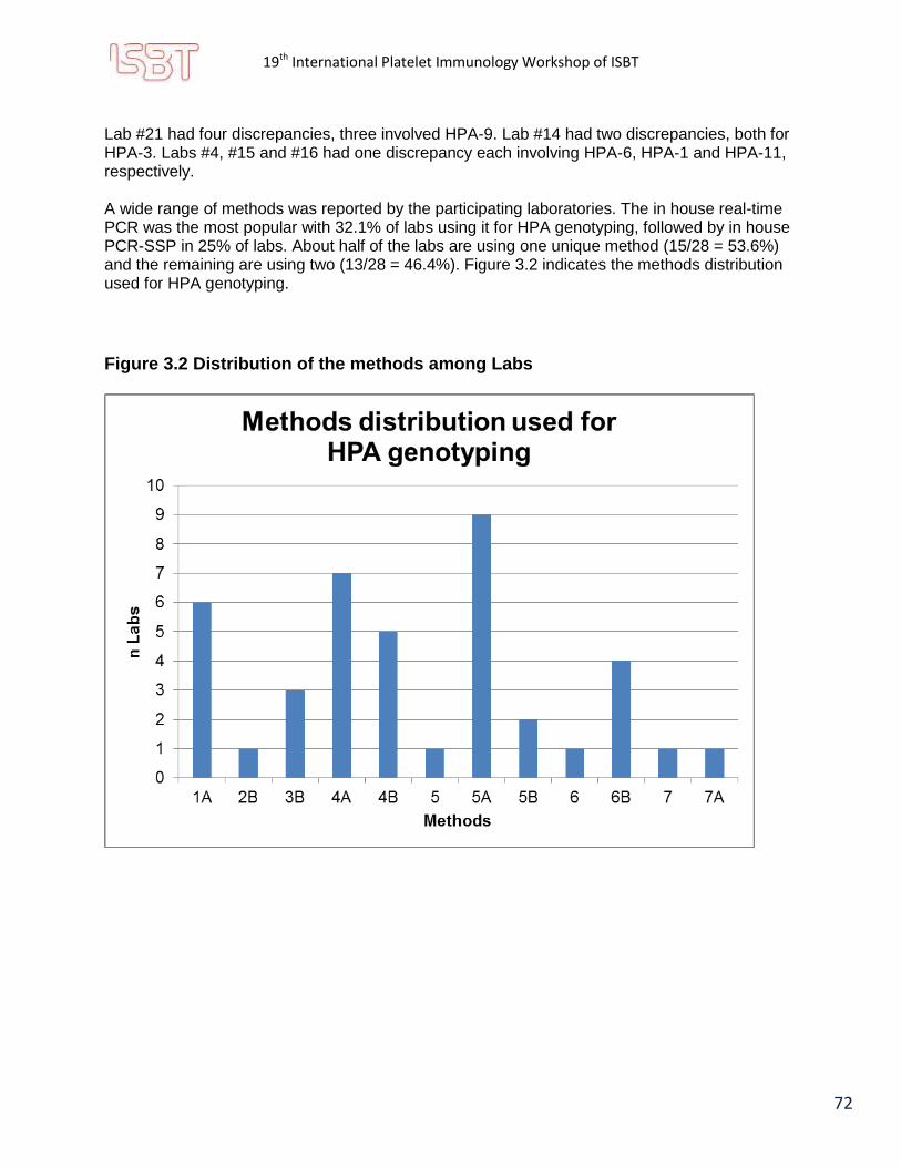
19th International Platelet Immunology Workshop of ISBT
72
Lab #21 had four discrepancies, three involved HPA-9. Lab #14 had two discrepancies, both for HPA-3. Labs #4, #15 and #16 had one discrepancy each involving HPA-6, HPA-1 and HPA-11, respectively. A wide range of methods was reported by the participating laboratories. The in house real-time PCR was the most popular with 32.1% of labs using it for HPA genotyping, followed by in house PCR-SSP in 25% of labs. About half of the labs are using one unique method (15/28 = 53.6%) and the remaining are using two (13/28 = 46.4%). Figure 3.2 indicates the methods distribution used for HPA genotyping.
Figure 3.2 Distribution of the methods among Labs
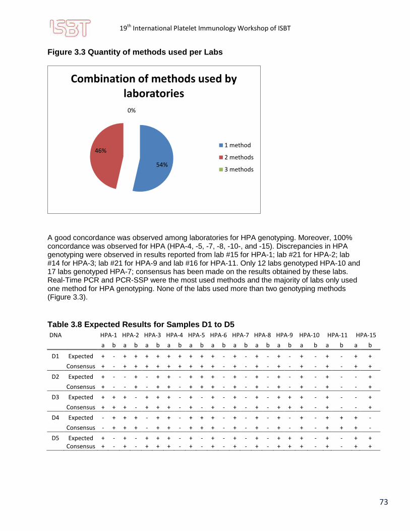
19th International Platelet Immunology Workshop of ISBT
73
Figure 3.3 Quantity of methods used per Labs
A good concordance was observed among laboratories for HPA genotyping. Moreover, 100% concordance was observed for HPA (HPA-4, -5, -7, -8, -10-, and -15). Discrepancies in HPA genotyping were observed in results reported from lab #15 for HPA-1; lab #21 for HPA-2; lab #14 for HPA-3; lab #21 for HPA-9 and lab #16 for HPA-11. Only 12 labs genotyped HPA-10 and 17 labs genotyped HPA-7; consensus has been made on the results obtained by these labs. Real-Time PCR and PCR-SSP were the most used methods and the majority of labs only used one method for HPA genotyping. None of the labs used more than two genotyping methods (Figure 3.3).
Table 3.8 Expected Results for Samples D1 to D5
DNA
HPA-1 HPA-2 HPA-3 HPA-4 HPA-5 HPA-6 HPA-7 HPA-8 HPA-9 HPA-10 HPA-11 HPA-15
a b a b a b a b a b a b a b a b a b a b a b a b
D1 Expected + - + + + + + + + + + - + - + - + - + - + - + +
Consensus + - + + + + + + + + + - + - + - + - + - + - + +
D2 Expected + - - + - + + - + + + - + - + - + - + - + - - +
Consensus + - - + - + + - + + + - + - + - + - + - + - - +
D3 Expected + + + - + + + - + - + - + - + - + + + - + - - +
Consensus + + + - + + + - + - + - + - + - + + + - + - - +
D4 Expected - + + + - + + - + + + - + - + - + - + - + + + -
Consensus - + + + - + + - + + + - + - + - + - + - + + + -
D5 Expected + - + - + + + - + - + - + - + - + + + - + - + +
Consensus + - + - + + + - + - + - + - + - + + + - + - + +
54%
46%
0%
Combination of methods used by laboratories
1 method
2 methods
3 methods
19th International Platelet Immunology Workshop of ISBT
74
Conclusion All laboratories are genotyping for the most important HPA systems (HPA-1, -2, -3, -5 and -15) and between 42 to 68% are genotyping for extended genotypes that are less frequently involved in antibody development and platelet disorders (HPA-4, -6 to -11). An excellent reproducibility for overall HPA genotyping was observed among the participating labs (99.3%). Real-time PCR is now the most popular method used for HPA genotyping. Few labs are genotyping for HPA-12 to -14 and HPA-16 to -28. They reported their results in the comment section for the five DNA samples being of the aa genotype for all these HPA. One lab reported variants for HPA-6a in samples D1, D3 and D4.
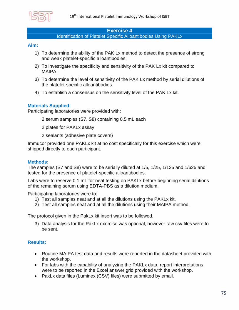
19th International Platelet Immunology Workshop of ISBT
75
Exercise 4 Identification of Platelet Specific Alloantibodies Using PAKLx
Aim:
1) To determine the ability of the PAK Lx method to detect the presence of strong and weak platelet-specific alloantibodies.
2) To investigate the specificity and sensitivity of the PAK Lx kit compared to MAIPA.
3) To determine the level of sensitivity of the PAK Lx method by serial dilutions of the platelet-specific alloantibodies.
4) To establish a consensus on the sensitivity level of the PAK Lx kit.
Materials Supplied: Participating laboratories were provided with:
2 serum samples (S7, S8) containing 0,5 mL each
2 plates for PAKLx assay
2 sealants (adhesive plate covers)
Immucor provided one PAKLx kit at no cost specifically for this exercise which were shipped directly to each participant.
Methods: The samples (S7 and S8) were to be serially diluted at 1/5, 1/25, 1/125 and 1/625 and tested for the presence of platelet-specific alloantibodies.
Labs were to reserve 0.1 mL for neat testing on PAKLx before beginning serial dilutions of the remaining serum using EDTA-PBS as a dilution medium.
Participating laboratories were to: 1) Test all samples neat and at all the dilutions using the PAKLx kit. 2) Test all samples neat and at all the dilutions using their MAIPA method.
The protocol given in the PakLx kit insert was to be followed.
3) Data analysis for the PakLx exercise was optional, however raw csv files were to be sent.
Results:
Routine MAIPA test data and results were reported in the datasheet provided with the workshop.
For labs with the capability of analyzing the PAKLx data; report interpretations were to be reported in the Excel answer grid provided with the workshop.
PakLx data files (Luminex (CSV) files) were submitted by email.
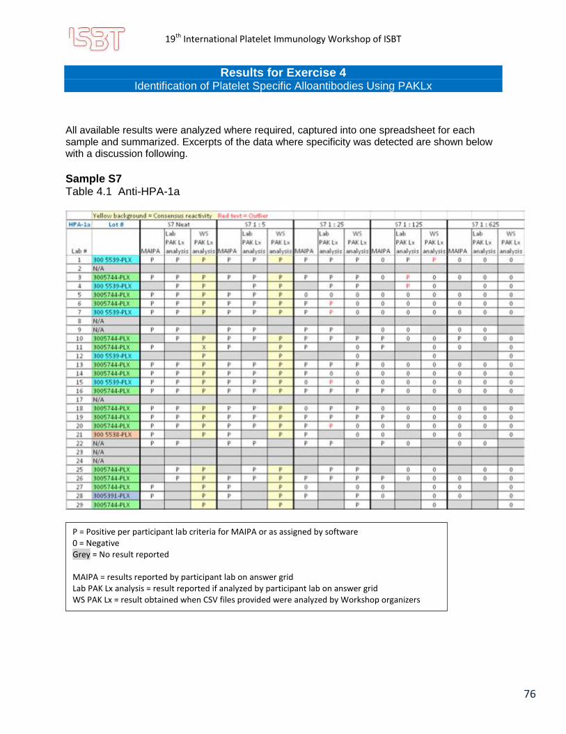
19th International Platelet Immunology Workshop of ISBT
76
Results for Exercise 4 Identification of Platelet Specific Alloantibodies Using PAKLx
All available results were analyzed where required, captured into one spreadsheet for each sample and summarized. Excerpts of the data where specificity was detected are shown below with a discussion following.
Sample S7 Table 4.1 Anti-HPA-1a
P = Positive per participant lab criteria for MAIPA or as assigned by software 0 = Negative Grey = No result reported MAIPA = results reported by participant lab on answer grid Lab PAK Lx analysis = result reported if analyzed by participant lab on answer grid WS PAK Lx = result obtained when CSV files provided were analyzed by Workshop organizers

19th International Platelet Immunology Workshop of ISBT
77
Table 4.2 Anti-HPA-5a
P = Positive per participant lab criteria for MAIPA or as assigned by software 0 = Negative Grey = No result reported MAIPA = results reported by participant lab on answer grid Lab PAK Lx analysis = result reported if analyzed by participant lab on answer grid WS PAK Lx = result obtained when CSV files provided were analyzed by Workshop organizers

19th International Platelet Immunology Workshop of ISBT
78
Table 4.3 Anti-HLA
P = Positive per participant lab criteria for MAIPA or as assigned by software 0 = Negative Grey = No result reported MAIPA = results reported by participant lab on answer grid Lab PAK Lx analysis = result reported if analyzed by participant lab on answer grid WS PAK Lx = result obtained when CSV files provided were analyzed by Workshop organizers
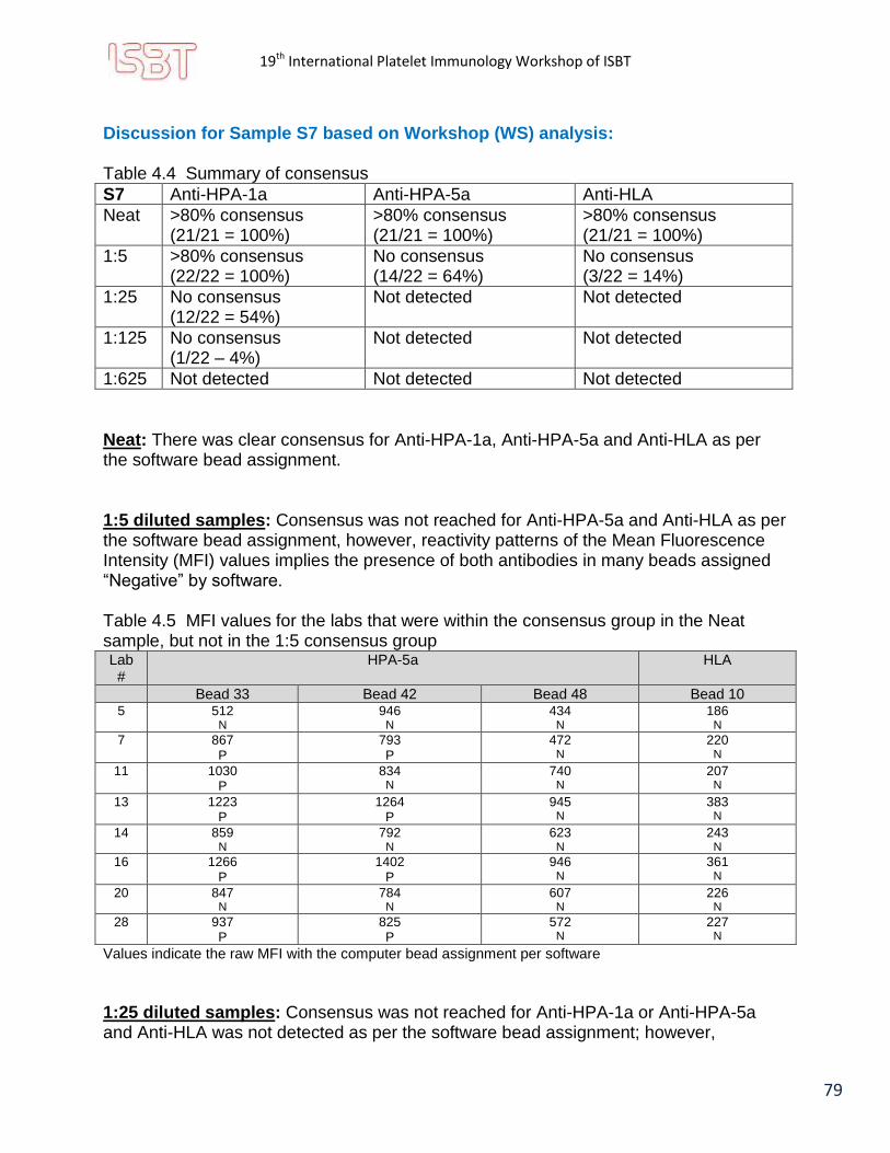
19th International Platelet Immunology Workshop of ISBT
79
Discussion for Sample S7 based on Workshop (WS) analysis: Table 4.4 Summary of consensus
S7 Anti-HPA-1a Anti-HPA-5a Anti-HLA
Neat >80% consensus (21/21 = 100%)
>80% consensus (21/21 = 100%)
>80% consensus (21/21 = 100%)
1:5 >80% consensus (22/22 = 100%)
No consensus (14/22 = 64%)
No consensus (3/22 = 14%)
1:25 No consensus (12/22 = 54%)
Not detected Not detected
1:125 No consensus (1/22 – 4%)
Not detected Not detected
1:625 Not detected Not detected Not detected
Neat: There was clear consensus for Anti-HPA-1a, Anti-HPA-5a and Anti-HLA as per the software bead assignment. 1:5 diluted samples: Consensus was not reached for Anti-HPA-5a and Anti-HLA as per the software bead assignment, however, reactivity patterns of the Mean Fluorescence Intensity (MFI) values implies the presence of both antibodies in many beads assigned “Negative” by software. Table 4.5 MFI values for the labs that were within the consensus group in the Neat sample, but not in the 1:5 consensus group Lab #
HPA-5a HLA
Bead 33 Bead 42 Bead 48 Bead 10 5 512
N 946
N 434
N 186
N
7 867 P
793 P
472 N
220 N
11 1030 P
834 N
740 N
207 N
13 1223 P
1264 P
945 N
383 N
14 859 N
792 N
623 N
243 N
16 1266 P
1402 P
946 N
361 N
20 847 N
784 N
607 N
226 N
28 937 P
825 P
572 N
227 N
Values indicate the raw MFI with the computer bead assignment per software
1:25 diluted samples: Consensus was not reached for Anti-HPA-1a or Anti-HPA-5a and Anti-HLA was not detected as per the software bead assignment; however,
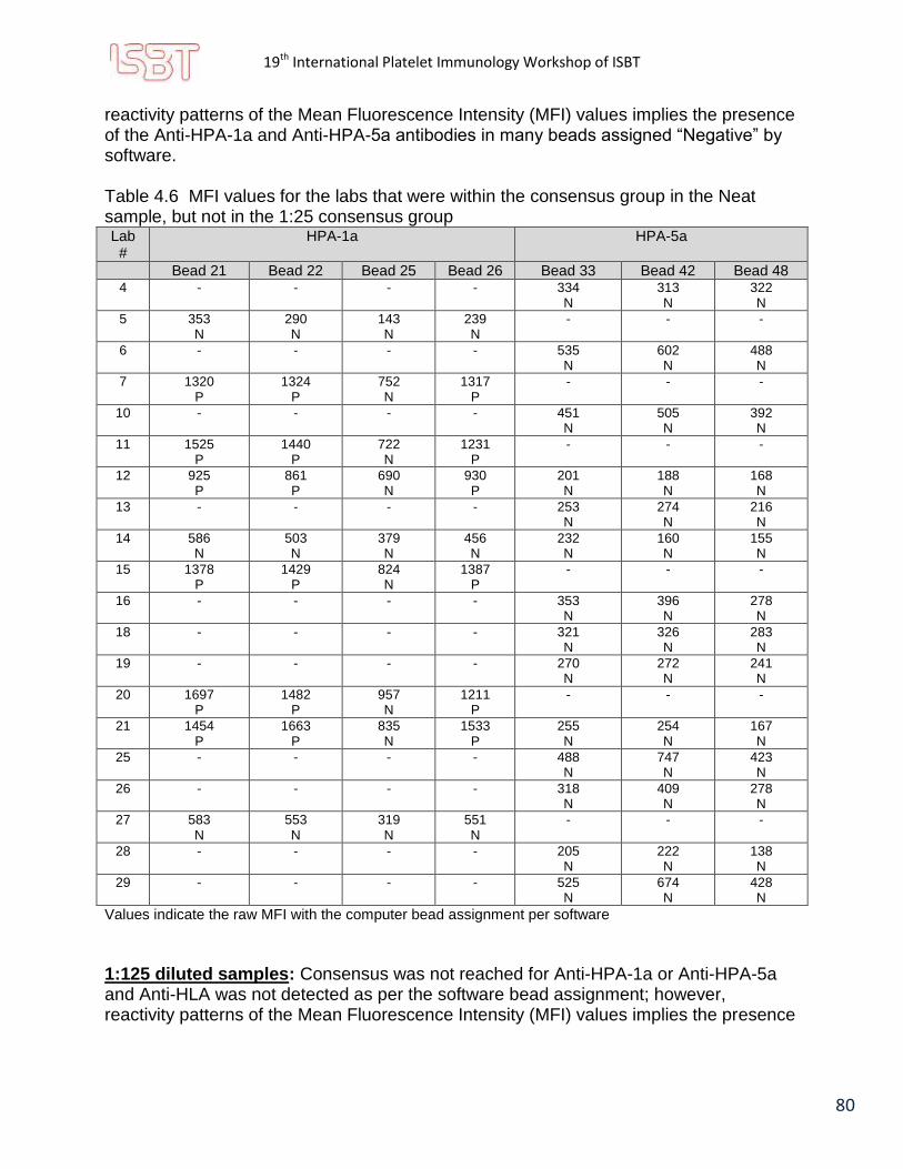
19th International Platelet Immunology Workshop of ISBT
80
reactivity patterns of the Mean Fluorescence Intensity (MFI) values implies the presence of the Anti-HPA-1a and Anti-HPA-5a antibodies in many beads assigned “Negative” by software. Table 4.6 MFI values for the labs that were within the consensus group in the Neat sample, but not in the 1:25 consensus group Lab #
HPA-1a HPA-5a
Bead 21 Bead 22 Bead 25 Bead 26 Bead 33 Bead 42 Bead 48 4 - - - - 334
N 313 N
322 N
5 353 N
290 N
143 N
239 N
- - -
6 - - - - 535 N
602 N
488 N
7 1320 P
1324 P
752 N
1317 P
- - -
10 - - - - 451 N
505 N
392 N
11 1525 P
1440 P
722 N
1231 P
- - -
12 925 P
861 P
690 N
930 P
201 N
188 N
168 N
13 - - - - 253 N
274 N
216 N
14 586 N
503 N
379 N
456 N
232 N
160 N
155 N
15 1378 P
1429 P
824 N
1387 P
- - -
16 - - - - 353 N
396 N
278 N
18 - - - - 321 N
326 N
283 N
19 - - - - 270 N
272 N
241 N
20 1697 P
1482 P
957 N
1211 P
- - -
21 1454 P
1663 P
835 N
1533 P
255 N
254 N
167 N
25 - - - - 488 N
747 N
423 N
26 - - - - 318 N
409 N
278 N
27 583 N
553 N
319 N
551 N
- - -
28 - - - - 205 N
222 N
138 N
29 - - - - 525 N
674 N
428 N
Values indicate the raw MFI with the computer bead assignment per software
1:125 diluted samples: Consensus was not reached for Anti-HPA-1a or Anti-HPA-5a and Anti-HLA was not detected as per the software bead assignment; however, reactivity patterns of the Mean Fluorescence Intensity (MFI) values implies the presence
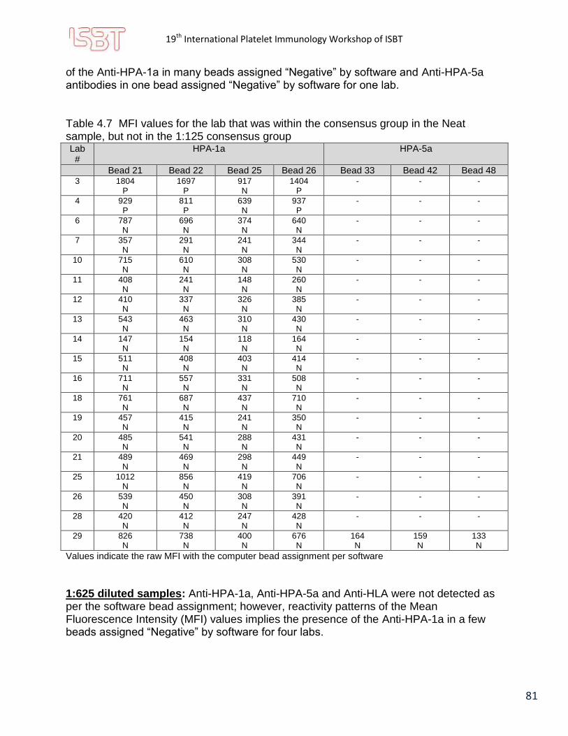
19th International Platelet Immunology Workshop of ISBT
81
of the Anti-HPA-1a in many beads assigned “Negative” by software and Anti-HPA-5a antibodies in one bead assigned “Negative” by software for one lab. Table 4.7 MFI values for the lab that was within the consensus group in the Neat sample, but not in the 1:125 consensus group Lab #
HPA-1a HPA-5a
Bead 21 Bead 22 Bead 25 Bead 26 Bead 33 Bead 42 Bead 48 3 1804
P 1697
P 917 N
1404 P
- - -
4 929 P
811 P
639 N
937 P
- - -
6 787 N
696 N
374 N
640 N
- - -
7 357 N
291 N
241 N
344 N
- - -
10 715 N
610 N
308 N
530 N
- - -
11 408 N
241 N
148 N
260 N
- - -
12 410 N
337 N
326 N
385 N
- - -
13 543 N
463 N
310 N
430 N
- - -
14 147 N
154 N
118 N
164 N
- - -
15 511 N
408 N
403 N
414 N
- - -
16 711 N
557 N
331 N
508 N
- - -
18 761 N
687 N
437 N
710 N
- - -
19 457 N
415 N
241 N
350 N
- - -
20 485 N
541 N
288 N
431 N
- - -
21 489 N
469 N
298 N
449 N
- - -
25 1012 N
856 N
419 N
706 N
- - -
26 539 N
450 N
308 N
391 N
- - -
28 420 N
412 N
247 N
428 N
- - -
29 826 N
738 N
400 N
676 N
164 N
159 N
133 N
Values indicate the raw MFI with the computer bead assignment per software
1:625 diluted samples: Anti-HPA-1a, Anti-HPA-5a and Anti-HLA were not detected as per the software bead assignment; however, reactivity patterns of the Mean Fluorescence Intensity (MFI) values implies the presence of the Anti-HPA-1a in a few beads assigned “Negative” by software for four labs.
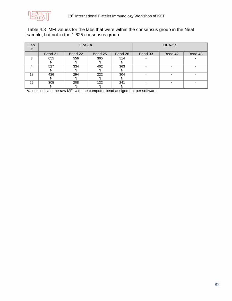
19th International Platelet Immunology Workshop of ISBT
82
Table 4.8 MFI values for the labs that were within the consensus group in the Neat sample, but not in the 1:625 consensus group Lab #
HPA-1a HPA-5a
Bead 21 Bead 22 Bead 25 Bead 26 Bead 33 Bead 42 Bead 48 3 655
N 556 N
305 N
514 N
- - -
4 527 N
334 N
402 N
363 N
- - -
18 426 N
294 N
222 N
304 N
- - -
29 305 N
208 N
122 N
241 N
- - -
Values indicate the raw MFI with the computer bead assignment per software
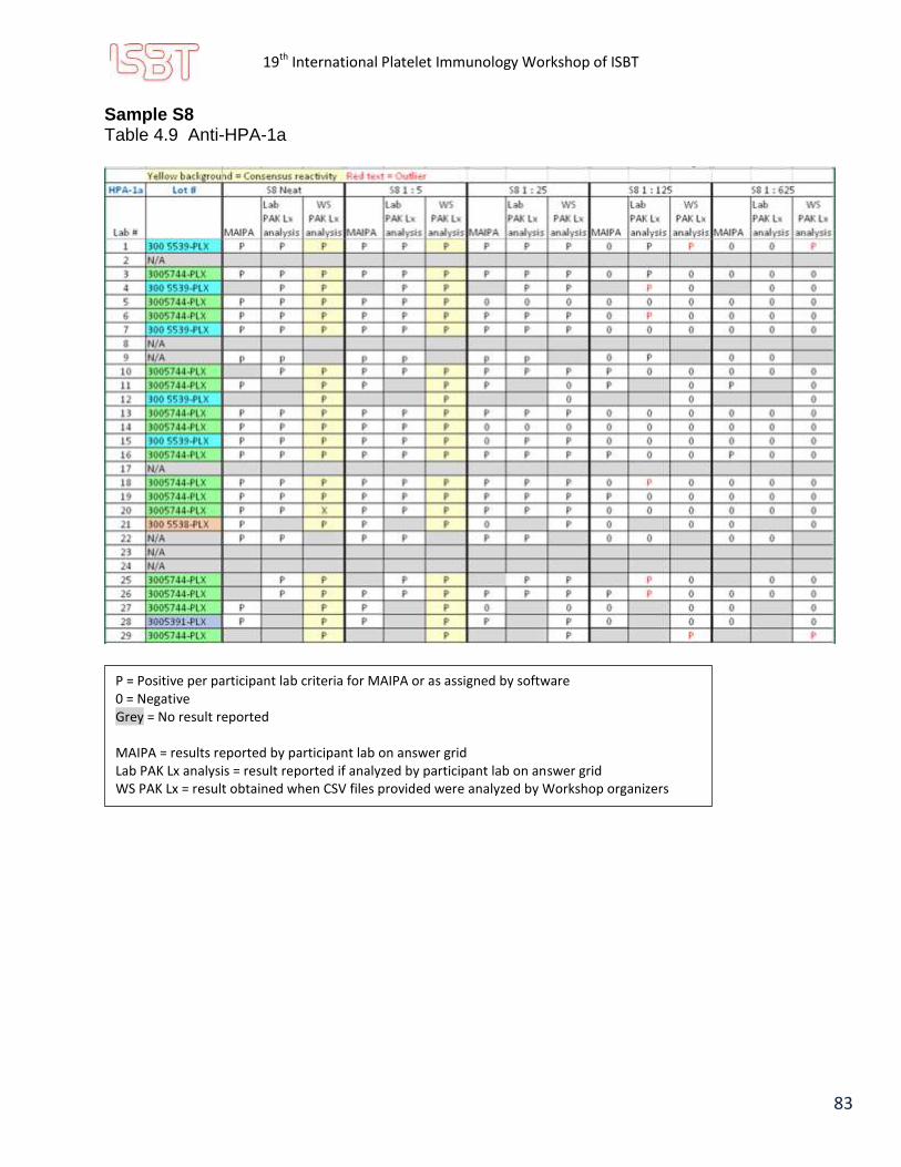
19th International Platelet Immunology Workshop of ISBT
83
Sample S8 Table 4.9 Anti-HPA-1a
P = Positive per participant lab criteria for MAIPA or as assigned by software 0 = Negative Grey = No result reported MAIPA = results reported by participant lab on answer grid Lab PAK Lx analysis = result reported if analyzed by participant lab on answer grid WS PAK Lx = result obtained when CSV files provided were analyzed by Workshop organizers
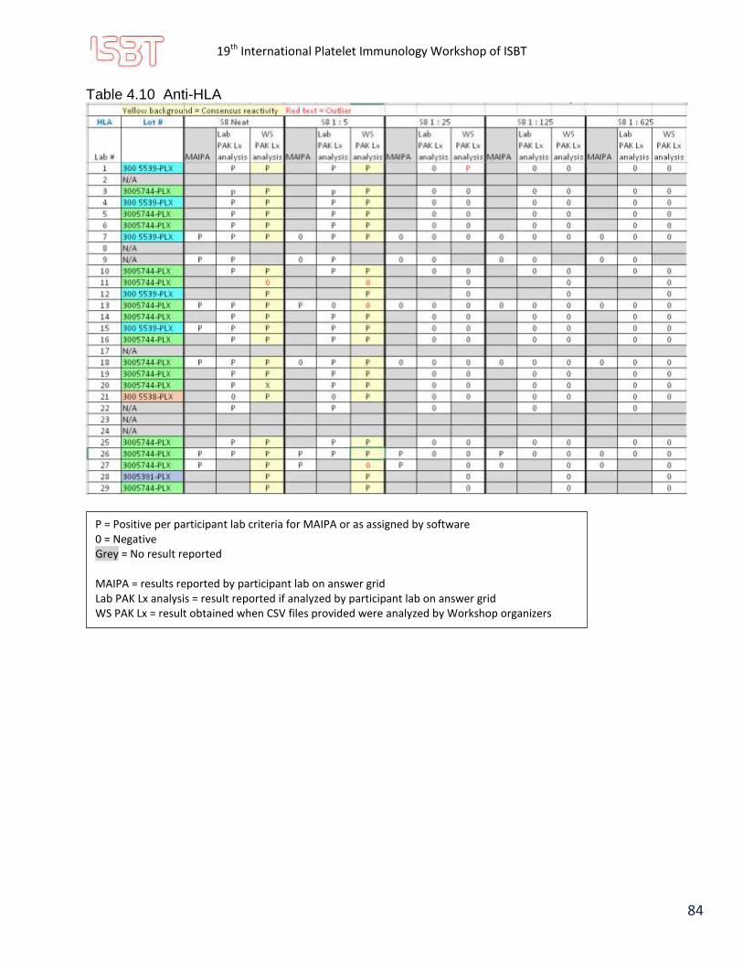
19th International Platelet Immunology Workshop of ISBT
84
Table 4.10 Anti-HLA
P = Positive per participant lab criteria for MAIPA or as assigned by software 0 = Negative Grey = No result reported MAIPA = results reported by participant lab on answer grid Lab PAK Lx analysis = result reported if analyzed by participant lab on answer grid WS PAK Lx = result obtained when CSV files provided were analyzed by Workshop organizers

19th International Platelet Immunology Workshop of ISBT
85
Discussion for Sample S8 based on Workshop (WS) analysis: Table 4.11 Summary of consensus
S8 Anti-HPA-1a Anti-HLA Anti-GP IV
Neat >80% consensus (21/21 = 100%)
>80% consensus (20/21 – 95%)
Not detected
1:5 >80% consensus (21/21 = 100%)
>80% consensus (19/22 – 86%)
Not detected
1:25 No consensus (17/22 = 77%)
No consensus (1/22 = 4%)
Not detected
1:125 No consensus (2/22 – 9%)
Not detected Not detected
1:625 No consensus (2/22 – 9%)
Not detected No consensus Note: (1/22 = 4% Considered False Positive)
Neat: There was clear consensus for Anti-HPA-1a and Anti-HLA as per the software bead assignment. Table 4.12 MFI value for the one lab that did not detect Anti-HLA in the Neat sample group Lab #
HLA
Bead 10 11 1382
N
Values indicate the raw MFI with the computer bead assignment per software
1:5 diluted samples: There was clear consensus for Anti-HPA-1a and Anti-HLA as per the software bead assignment. Table 4.13 MFI value for the three labs that did not detect Anti-HLA in the 1:5 consensus group Lab #
HLA
Bead 10 11 242
N
13 618 N
27 500 N
Values indicate the raw MFI with the computer bead assignment per software
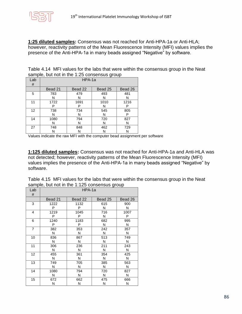
19th International Platelet Immunology Workshop of ISBT
86
1:25 diluted samples: Consensus was not reached for Anti-HPA-1a or Anti-HLA; however, reactivity patterns of the Mean Fluorescence Intensity (MFI) values implies the presence of the Anti-HPA-1a in many beads assigned “Negative” by software. Table 4.14 MFI values for the labs that were within the consensus group in the Neat sample, but not in the 1:25 consensus group Lab #
HPA-1a
Bead 21 Bead 22 Bead 25 Bead 26 5 783
N 479 N
493 N
481 N
11 1722 P
1691 P
1010 N
1216 P
12 738 N
734 N
545 N
805 P
14 1080 N
794 N
720 N
827 N
27 748 N
848 N
462 N
729 N
Values indicate the raw MFI with the computer bead assignment per software
1:125 diluted samples: Consensus was not reached for Anti-HPA-1a and Anti-HLA was not detected; however, reactivity patterns of the Mean Fluorescence Intensity (MFI) values implies the presence of the Anti-HPA-1a in many beads assigned “Negative” by software. Table 4.15 MFI values for the labs that were within the consensus group in the Neat sample, but not in the 1:125 consensus group Lab #
HPA-1a
Bead 21 Bead 22 Bead 25 Bead 26 3 1222
P 1132
P 615 N
900 N
4 1219 P
1045 P
716 N
1007 P
6 1240 P
1183 P
682 N
995 N
7 382 N
353 N
242 N
357 N
10 836 N
867 N
513 N
749 N
11 306 N
236 N
211 N
243 N
12 455 N
361 N
354 N
425 N
13 749 N
705 N
385 N
563 N
14 1080 N
794 N
720 N
827 N
15 672 N
662 N
475 N
666 N
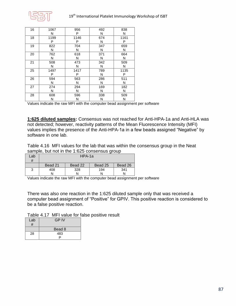
19th International Platelet Immunology Workshop of ISBT
87
16 1067 N
956 P
492 N
838 N
18 1199 P
1146 P
674 N
1161 P
19 822 N
704 N
347 N
659 N
20 762 N
618 N
371 N
664 N
21 508 N
473 N
342 N
509 N
25 1497 P
1417 P
789 N
1135 P
26 594 N
563 N
286 N
511 N
27 274 N
294 N
169 N
182 N
28 608 N
596 N
338 N
509 N
Values indicate the raw MFI with the computer bead assignment per software
1:625 diluted samples: Consensus was not reached for Anti-HPA-1a and Anti-HLA was not detected; however, reactivity patterns of the Mean Fluorescence Intensity (MFI) values implies the presence of the Anti-HPA-1a in a few beads assigned “Negative” by software in one lab. Table 4.16 MFI values for the lab that was within the consensus group in the Neat sample, but not in the 1:625 consensus group Lab #
HPA-1a
Bead 21 Bead 22 Bead 25 Bead 26 3 408
N 328 N
194 N
341 N
Values indicate the raw MFI with the computer bead assignment per software
There was also one reaction in the 1:625 diluted sample only that was received a computer bead assignment of “Positive” for GPIV. This positive reaction is considered to be a false positive reaction. Table 4.17 MFI value for false positive result Lab #
GP IV
Bead 8 28 483
P
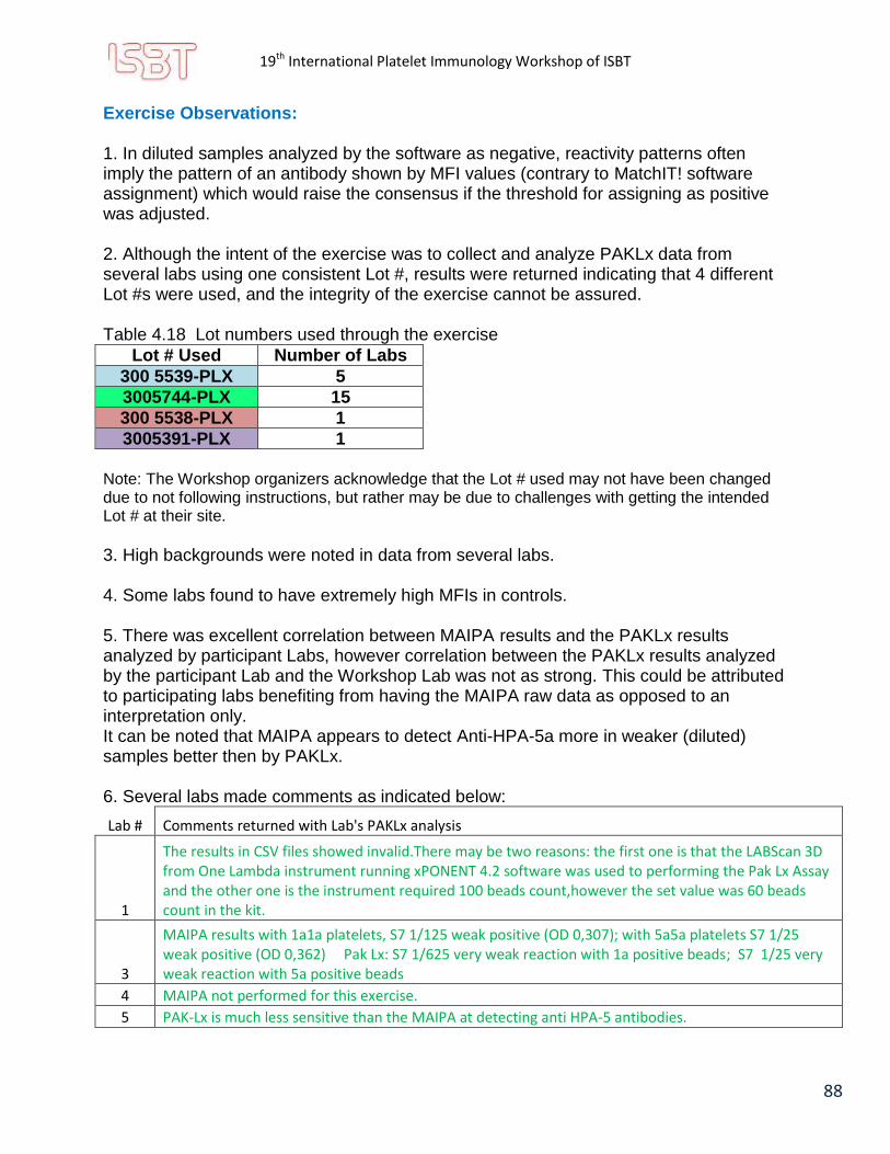
19th International Platelet Immunology Workshop of ISBT
88
Exercise Observations: 1. In diluted samples analyzed by the software as negative, reactivity patterns often imply the pattern of an antibody shown by MFI values (contrary to MatchIT! software assignment) which would raise the consensus if the threshold for assigning as positive was adjusted. 2. Although the intent of the exercise was to collect and analyze PAKLx data from several labs using one consistent Lot #, results were returned indicating that 4 different Lot #s were used, and the integrity of the exercise cannot be assured. Table 4.18 Lot numbers used through the exercise
Lot # Used Number of Labs
300 5539-PLX 5
3005744-PLX 15
300 5538-PLX 1
3005391-PLX 1
Note: The Workshop organizers acknowledge that the Lot # used may not have been changed due to not following instructions, but rather may be due to challenges with getting the intended Lot # at their site.
3. High backgrounds were noted in data from several labs. 4. Some labs found to have extremely high MFIs in controls. 5. There was excellent correlation between MAIPA results and the PAKLx results analyzed by participant Labs, however correlation between the PAKLx results analyzed by the participant Lab and the Workshop Lab was not as strong. This could be attributed to participating labs benefiting from having the MAIPA raw data as opposed to an interpretation only. It can be noted that MAIPA appears to detect Anti-HPA-5a more in weaker (diluted) samples better then by PAKLx. 6. Several labs made comments as indicated below:
Lab # Comments returned with Lab's PAKLx analysis
1
The results in CSV files showed invalid.There may be two reasons: the first one is that the LABScan 3D from One Lambda instrument running xPONENT 4.2 software was used to performing the Pak Lx Assay and the other one is the instrument required 100 beads count,however the set value was 60 beads count in the kit.
3
MAIPA results with 1a1a platelets, S7 1/125 weak positive (OD 0,307); with 5a5a platelets S7 1/25 weak positive (OD 0,362) Pak Lx: S7 1/625 very weak reaction with 1a positive beads; S7 1/25 very weak reaction with 5a positive beads
4 MAIPA not performed for this exercise.
5 PAK-Lx is much less sensitive than the MAIPA at detecting anti HPA-5 antibodies.
19th International Platelet Immunology Workshop of ISBT
89
6
S7= Anti-HPA-1a and Anti-HPA- 5a detected. S8= Anti-HPA-1a detected. As well 1/125 for S8 only 2/4 beads are assigned as positive. MFI although low still shows a pattern of reactivity regardless of PAKLx interpretation.
7
The samples have been delayed to arrive at our lab for two months as the problems of customs. The PAKLx kit we used in this project was bought ourselves from the immucor agent in <country where lab located>. The lot number of PAKLx kit is 3005539.
10
Routine MAIPA is normally performed with 2 parallells à 50 ul per well. Due to limited sample material in ws sample, it was tested with one well only. Neither was there enough material to include homozygous screening cells to discriminate in all systems.
12
Due to insufficient sample volume, the lab was unable to complete the MAIPA portion of this exercise. The lab does not have the software capability of analyzing the PakLx data. The PakLx raw data output file will be submitted.
14 PAK-Lx : HPA-4b antibodies have not been excluded as their presence is masked when antibodies to HPA-1a are detected. MAIPA panel cells are not typed for HPA-4 but are unlikely to be HPA-4b positive.
15 Our results show that PAKLx method can be used to detect platelet-specific alloantibodies, and the specificity and sensitivity seem better than MAIPA.
19
Altough PakLx scored S7 anti HPA-1a negative for 1:125 dilution, the MFI pattern still indicates the presence of anti HPA-1a The same accounts for S7 anti HPA-5a 1:25, S8 anti HPA-1a 1:125 and 1:625 (see results sheet Pakx)
20 Please note PAKLx run at half bead assay could detect HPA 1a at 1/25 for S7 , S8 and HPA 5a at 1/5 for S7 only
21 We did not receive the PAK Lx so far due to <country where lab located> customs problems.
25 sample S8 neat - suspected to Anti-GPIb/IX, sample S8 (1/125) - HPA-1a weak positive
26 MAIPA more sensible than PakLx
7. During the execution of the exercise, some labs found that the data could not be analyzed due to a discrepancy between the kit template for data collection on the Luminex (60 events), and the bead count requirement of the MatchIT! software (100 events). This may have limited the ability of participant labs to submit their analysis. Note: All Workshop analysis was performed using the software fix provided by Immucor. 8. There was one false positive result detected with a (false) specificity of Anti-GPIV. This single result could have implications for the testing lab and for patient management decisions. Exercise Conclusions:
1) PAKLx demonstrated its ability to pick up strong platelet-specific alloantibodies with ease. MatchIT! Software is an excellent screening tool, however as samples were diluted down, a deficit was observed with the MatchIT! software algorithm itself. Software struggled to assign weak/diluted samples positive. MFI values obtained suggested the presence of an antibody however software’s algorithm assigned beads as negative. The MatchIT! software does not appear to take into consideration the MFI spread between beads. Patterns or reactivity based on MFI
19th International Platelet Immunology Workshop of ISBT
90
results could be observed, even when the software assigned the beads as negative. The indication is that there is a missed opportunity to detect antibodies that may be weaker or still developing; and that this may in certain clinical situations be relevant and/or impact patient management decisions. Vendor should consider revisiting their software and implement a grey zone of “Indeterminate”.
2) Overall there is a good correlation between PAKLx and MAIPA when comparing samples containing strong antibody titres. As samples are diluted down PAKLx becomes less sensitive. It also appears that antibody specificity continues to be a challenge, especially at lower titres.ie. HPA-3 (exercise 1) and HPA-5.
3) The method itself appears to pick up weaker titre antibodies when looking at the MFI values alone and not by software bead result assignments. However software’s algorithm needs to be reviewed. Currently MAIPA continues to be more sensitive and open for overall interpretation by testing personnel. As always testing personnel must be cognizant of the need to review all information provided by the software and not go by the software bead assignment alone when assigning a negative result, or determining whether subsequent testing is warranted.
19th International Platelet Immunology Workshop of ISBT
91
Exercise 5 Delay for Platelet Preparation
Aim:
1) To determine the maximum delay for platelet preparation from blood before losing HPA-3 antigens on the surface of the platelets.
2) To investigate the impact of the delay in platelet preparation on the detection of Anti-HPA-3 specific alloantibodies by MAIPA.
3) To evaluate the possibility of missing Anti-HPA-3 specific alloantibodies.
4) To establish a consensus on the delay for platelet preparation from blood.
Materials Supplied:
Participating laboratories were provided with:
2 serum samples (S9-500 µL, S10-150 µL)
Methods:
The participating laboratory collected three samples on two platelet donors. One platelet donor with a typing of HPA-1a/1a, 3a/3a and the second platelet donor with a typing of HPA-1a/1a, 3b/3b. Platelets from each sample collected were to be isolated at predefined time intervals. Once entire panel was prepared, testing was to be performed by MAIPA against GPIIbIIIa (HPA-3). Both sera were to be tested by MAIPA at 5-7 days after the last platelet isolation (72 hours). Only S9 was to be tested by MAIPA at 23-28 days after the last platelet isolation (72 hours).
Participating laboratories were to:
1) Collect three samples from two platelet donors (one platelet donor with HPA-1a/1a, 3a/3a typing and the second platelet donor with HPA-1a/1a, 3b/3b typing)
2) Prepare the platelet suspensions in isotonic buffer to create the panel. One sample from each donor was to be isolated at 24 hours, the second at 48 hours and the third at 72 hours.
3) Test S9 and S10 serum samples against the platelet panel at 5-7 days after the last platelet isolation, using 20 µL/well.
4) Test only S9 serum sample against the platelet panel at 23-28 days after the last platelet isolation, using 20 µL/well.
Results:
Assay data was to be reported in the Excel answer grid provided.
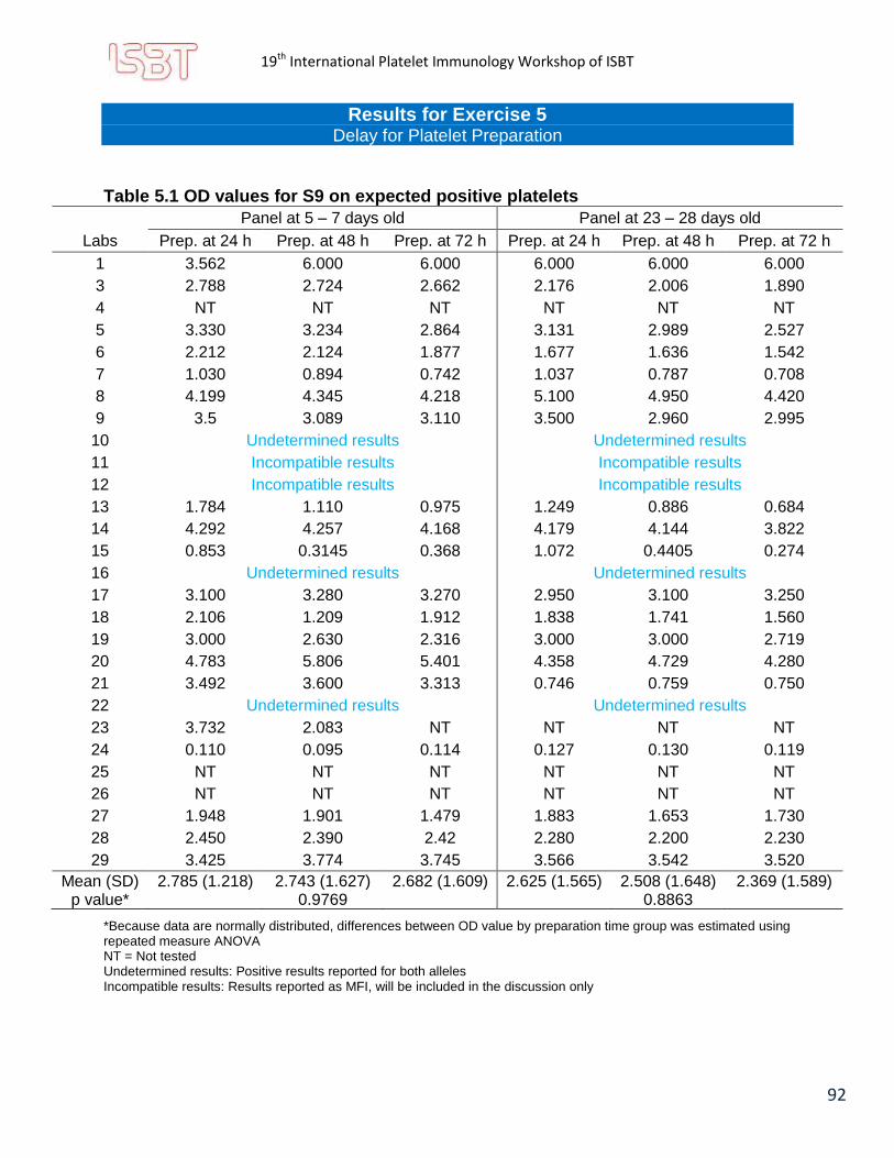
19th International Platelet Immunology Workshop of ISBT
92
Results for Exercise 5 Delay for Platelet Preparation
Table 5.1 OD values for S9 on expected positive platelets
Panel at 5 – 7 days old Panel at 23 – 28 days old
Labs Prep. at 24 h Prep. at 48 h Prep. at 72 h Prep. at 24 h Prep. at 48 h Prep. at 72 h
1 3.562 6.000 6.000 6.000 6.000 6.000
3 2.788 2.724 2.662 2.176 2.006 1.890
4 NT NT NT NT NT NT
5 3.330 3.234 2.864 3.131 2.989 2.527
6 2.212 2.124 1.877 1.677 1.636 1.542
7 1.030 0.894 0.742 1.037 0.787 0.708
8 4.199 4.345 4.218 5.100 4.950 4.420
9 3.5 3.089 3.110 3.500 2.960 2.995
10 Undetermined results Undetermined results
11 Incompatible results Incompatible results
12 Incompatible results Incompatible results
13 1.784 1.110 0.975 1.249 0.886 0.684
14 4.292 4.257 4.168 4.179 4.144 3.822
15 0.853 0.3145 0.368 1.072 0.4405 0.274
16 Undetermined results Undetermined results
17 3.100 3.280 3.270 2.950 3.100 3.250
18 2.106 1.209 1.912 1.838 1.741 1.560
19 3.000 2.630 2.316 3.000 3.000 2.719
20 4.783 5.806 5.401 4.358 4.729 4.280
21 3.492 3.600 3.313 0.746 0.759 0.750
22 Undetermined results Undetermined results
23 3.732 2.083 NT NT NT NT
24 0.110 0.095 0.114 0.127 0.130 0.119
25 NT NT NT NT NT NT
26 NT NT NT NT NT NT
27 1.948 1.901 1.479 1.883 1.653 1.730
28 2.450 2.390 2.42 2.280 2.200 2.230
29 3.425 3.774 3.745 3.566 3.542 3.520
Mean (SD) 2.785 (1.218) 2.743 (1.627) 2.682 (1.609) 2.625 (1.565) 2.508 (1.648) 2.369 (1.589) p value* 0.9769 0.8863
*Because data are normally distributed, differences between OD value by preparation time group was estimated using repeated measure ANOVA NT = Not tested Undetermined results: Positive results reported for both alleles Incompatible results: Results reported as MFI, will be included in the discussion only
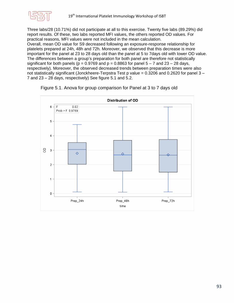
19th International Platelet Immunology Workshop of ISBT
93
Three labs/28 (10.71%) did not participate at all to this exercise. Twenty five labs (89.29%) did report results. Of these, two labs reported MFI values, the others reported OD values. For practical reasons, MFI values were not included in the mean calculation. Overall, mean OD value for S9 decreased following an exposure-response relationship for platelets prepared at 24h, 48h and 72h. Moreover, we observed that this decrease is more important for the panel at 23 to 28 days old than the panel at 5 to 7days old with lower OD value. The differences between a group’s preparation for both panel are therefore not statistically significant for both panels (p = 0.9769 and p = 0.8863 for panel 5 – 7 and 23 – 28 days, respectively). Moreover, the observed decreased trends between preparation times were also not statistically significant (Jonckheere-Terpstra Test p value = 0.3206 and 0.2620 for panel 3 – 7 and 23 – 28 days, respectively) See figure 5.1 and 5.2.
Figure 5.1. Anova for group comparison for Panel at 3 to 7 days old
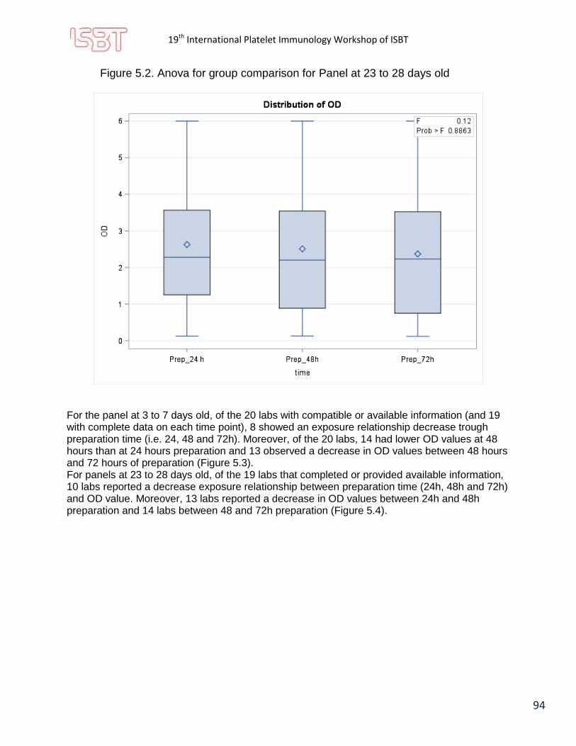
19th International Platelet Immunology Workshop of ISBT
94
Figure 5.2. Anova for group comparison for Panel at 23 to 28 days old
For the panel at 3 to 7 days old, of the 20 labs with compatible or available information (and 19 with complete data on each time point), 8 showed an exposure relationship decrease trough preparation time (i.e. 24, 48 and 72h). Moreover, of the 20 labs, 14 had lower OD values at 48 hours than at 24 hours preparation and 13 observed a decrease in OD values between 48 hours and 72 hours of preparation (Figure 5.3). For panels at 23 to 28 days old, of the 19 labs that completed or provided available information, 10 labs reported a decrease exposure relationship between preparation time (24h, 48h and 72h) and OD value. Moreover, 13 labs reported a decrease in OD values between 24h and 48h preparation and 14 labs between 48 and 72h preparation (Figure 5.4).

19th International Platelet Immunology Workshop of ISBT
95
Figure 5.3 Reactivity of Panel at 5 – 7 days old
Figure 5.4 Reactivity of Panel at 23 – 28 days old
When comparing reactivity between freshly prepared and older panels there is a trend for lower reactivity in the older panels for the 3 the preparation time conditions (Table 5.2). The loss of
0
1
2
3
4
5
6
7
Prep. at 24 h Prep. at 48 h Prep. at 72 h
Lab 1
Lab 3
Lab 5
Lab 6
Lab 7
Lab 8
Lab 9
Lab 13
Lab 14
Lab 15
Lab 17
0
1
2
3
4
5
6
7
Prep. at 24 h Prep. at 48 h Prep. at 72 h
Lab 1
Lab 3
Lab 5
Lab 6
Lab 7
Lab 8
Lab 9
Lab 13
Lab 14
Lab 15
Lab 17
Lab 18
Lab 19
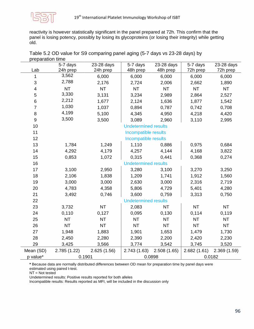
19th International Platelet Immunology Workshop of ISBT
96
reactivity is however statistically significant in the panel prepared at 72h. This confirm that the panel is losing potency, possibly by losing its glycoproteins (or losing their integrity) while getting old.
Table 5.2 OD value for S9 comparing panel aging (5-7 days vs 23-28 days) by preparation time
Lab 5-7 days 24h prep
23-28 days 24h prep
5-7 days 48h prep
23-28 days 48h prep
5-7 days 72h prep
23-28 days 72h prep
1 3,562 6,000 6,000 6,000 6,000 6,000
3 2,788 2,176 2,724 2,006 2,662 1,890
4 NT NT NT NT NT NT
5 3,330 3,131 3,234 2,989 2,864 2,527
6 2,212 1,677 2,124 1,636 1,877 1,542
7 1,030 1,037 0,894 0,787 0,742 0,708
8 4,199 5,100 4,345 4,950 4,218 4,420
9 3,500 3,500 3,089 2,960 3,110 2,995
10 Undetermined results
11 Incompatible results
12 Incompatible results
13 1,784 1,249 1,110 0,886 0,975 0,684
14 4,292 4,179 4,257 4,144 4,168 3,822
15 0,853 1,072 0,315 0,441 0,368 0,274
16 Undetermined results
17 3,100 2,950 3,280 3,100 3,270 3,250
18 2,106 1,838 1,209 1,741 1,912 1,560
19 3,000 3,000 2,630 3,000 2,316 2,719
20 4,783 4,358 5,806 4,729 5,401 4,280
21 3,492 0,746 3,600 0,759 3,313 0,750
22 Undetermined results
23 3,732 NT 2,083 NT NT NT
24 0,110 0,127 0,095 0,130 0,114 0,119
25 NT NT NT NT NT NT
26 NT NT NT NT NT NT
27 1,948 1,883 1,901 1,653 1,479 1,730
28 2,450 2,280 2,390 2,200 2,420 2,230
29 3,425 3,566 3,774 3,542 3,745 3,520
Mean (SD) 2.785 (1.22) 2.625 (1.56) 2.743 (1.63) 2.508 (1.65) 2.682 (1.61) 2.369 (1.59)
p value* 0.1901 0.0898 0.0182
* Because data are normally distributed differences between OD mean for preparation time by panel days were
estimated using paired t-test. NT = Not tested Undetermined results: Positive results reported for both alleles Incompatible results: Results reported as MFI, will be included in the discussion only
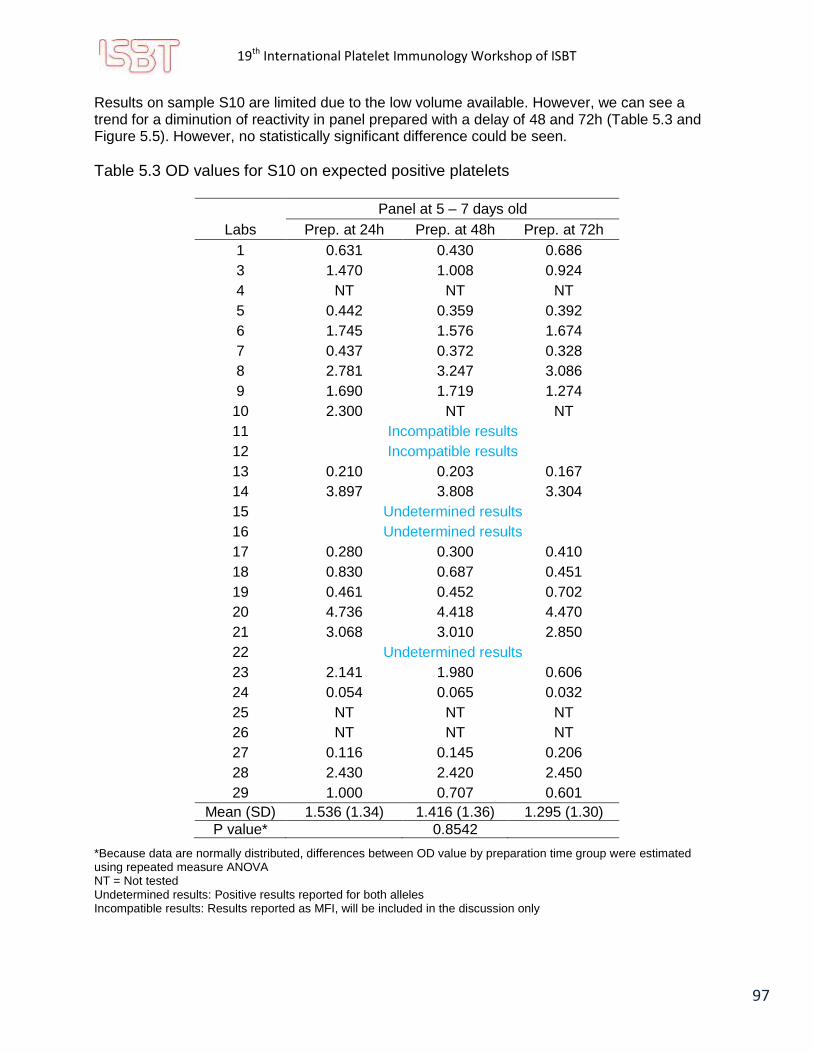
19th International Platelet Immunology Workshop of ISBT
97
Results on sample S10 are limited due to the low volume available. However, we can see a trend for a diminution of reactivity in panel prepared with a delay of 48 and 72h (Table 5.3 and Figure 5.5). However, no statistically significant difference could be seen.
Table 5.3 OD values for S10 on expected positive platelets
Panel at 5 – 7 days old
Labs Prep. at 24h Prep. at 48h Prep. at 72h
1 0.631 0.430 0.686
3 1.470 1.008 0.924
4 NT NT NT
5 0.442 0.359 0.392
6 1.745 1.576 1.674
7 0.437 0.372 0.328
8 2.781 3.247 3.086
9 1.690 1.719 1.274
10 2.300 NT NT
11 Incompatible results
12 Incompatible results
13 0.210 0.203 0.167
14 3.897 3.808 3.304
15 Undetermined results
16 Undetermined results
17 0.280 0.300 0.410
18 0.830 0.687 0.451
19 0.461 0.452 0.702
20 4.736 4.418 4.470
21 3.068 3.010 2.850
22 Undetermined results
23 2.141 1.980 0.606
24 0.054 0.065 0.032
25 NT NT NT
26 NT NT NT
27 0.116 0.145 0.206
28 2.430 2.420 2.450
29 1.000 0.707 0.601
Mean (SD) 1.536 (1.34) 1.416 (1.36) 1.295 (1.30)
P value* 0.8542
*Because data are normally distributed, differences between OD value by preparation time group were estimated using repeated measure ANOVA NT = Not tested Undetermined results: Positive results reported for both alleles Incompatible results: Results reported as MFI, will be included in the discussion only
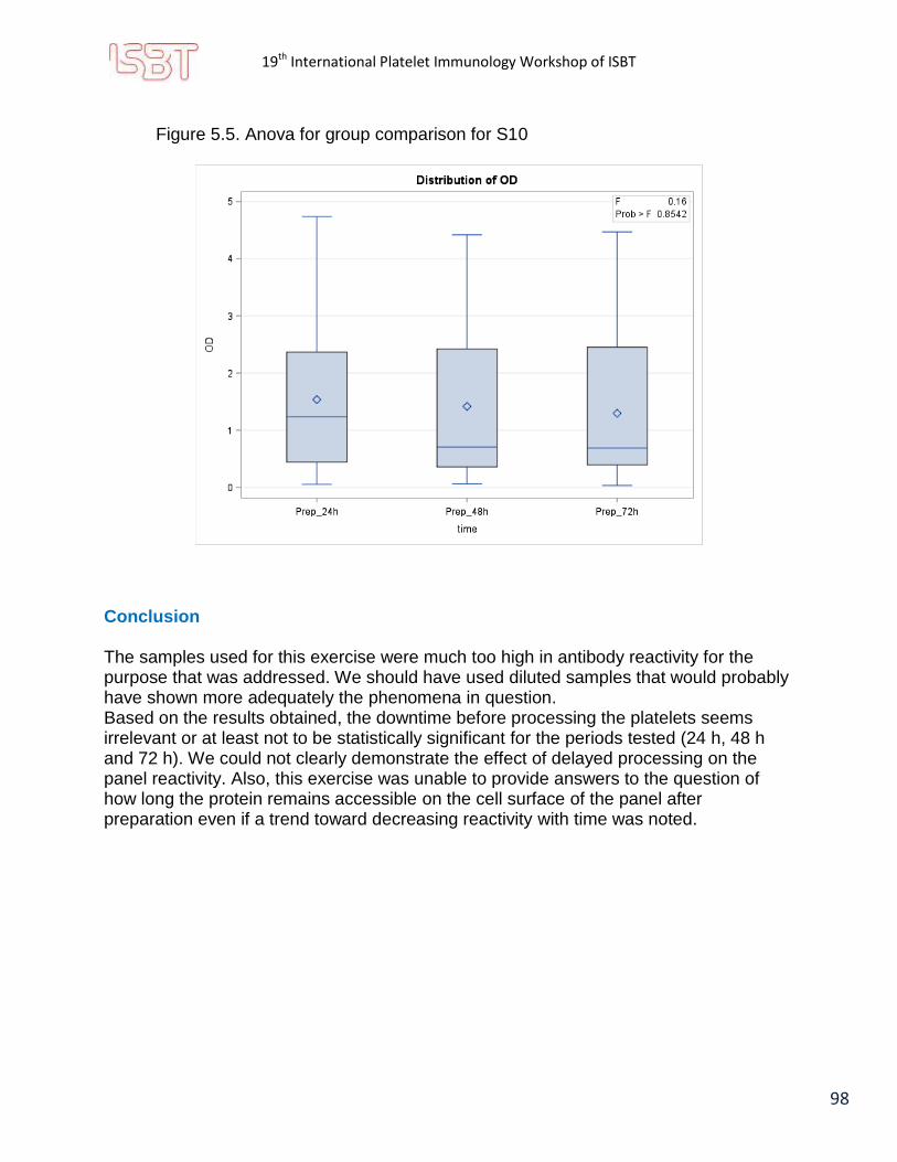
19th International Platelet Immunology Workshop of ISBT
98
Figure 5.5. Anova for group comparison for S10
Conclusion The samples used for this exercise were much too high in antibody reactivity for the purpose that was addressed. We should have used diluted samples that would probably have shown more adequately the phenomena in question. Based on the results obtained, the downtime before processing the platelets seems irrelevant or at least not to be statistically significant for the periods tested (24 h, 48 h and 72 h). We could not clearly demonstrate the effect of delayed processing on the panel reactivity. Also, this exercise was unable to provide answers to the question of how long the protein remains accessible on the cell surface of the panel after preparation even if a trend toward decreasing reactivity with time was noted.
19th International Platelet Immunology Workshop of ISBT
99
Exercise 6 Comparison of Anti-CD109 for HPA-15 Specific Alloantibodies
Aim:
1) To verify the potency of Anti-CD109 antibodies available on the market.
2) To compare the results obtained with the current data from each lab.
3) To improve the sensitivity of the MAIPA technique for Anti-HPA-15 alloantibody detection.
Materials Supplied:
Participating laboratories were provided with:
2 serum samples (S11, S12) containing 1.2 mL
4 Anti-CD109 clones from different sources (clones 1 to 4) containing 200 µL of a 10X solution
Methods:
The MAIPA for Anti-HPA-15 alloantibody detection was to be performed by the labs. Participating laboratories were to:
1) Prepare a platelet panel of 3 cells for the HPA-15 MAIPA. Be sure to have one platelet HPA-15a/a, one platelet HPA-15a/b, and one platelet HPA-15b/b in your panel.
2) Test the provided samples containing Anti-HPA-15 alloantibodies using their routine MAIPA method for HPA-15.
3) Use the 4 different Anti-CD109 monoclonals provided (each diluted 1 in 10 in PBS-EDTA) and their routine Anti-CD109 monoclonal for the isolation of the HPA-15 (CD109) protein.
Results:
Assay data and the identified specificity of platelet-specific alloantibodies was to be reported in the Excel answer grid provided.
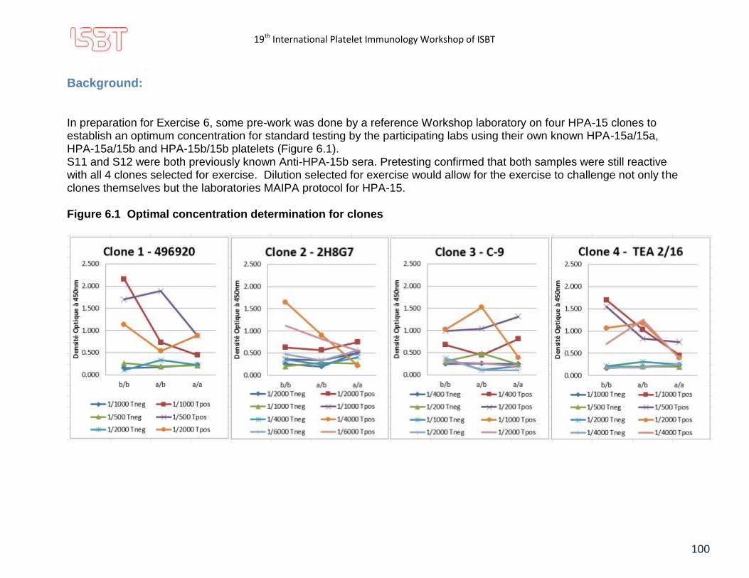
19th International Platelet Immunology Workshop of ISBT
100
Background: In preparation for Exercise 6, some pre-work was done by a reference Workshop laboratory on four HPA-15 clones to establish an optimum concentration for standard testing by the participating labs using their own known HPA-15a/15a, HPA-15a/15b and HPA-15b/15b platelets (Figure 6.1). S11 and S12 were both previously known Anti-HPA-15b sera. Pretesting confirmed that both samples were still reactive with all 4 clones selected for exercise. Dilution selected for exercise would allow for the exercise to challenge not only the clones themselves but the laboratories MAIPA protocol for HPA-15. Figure 6.1 Optimal concentration determination for clones
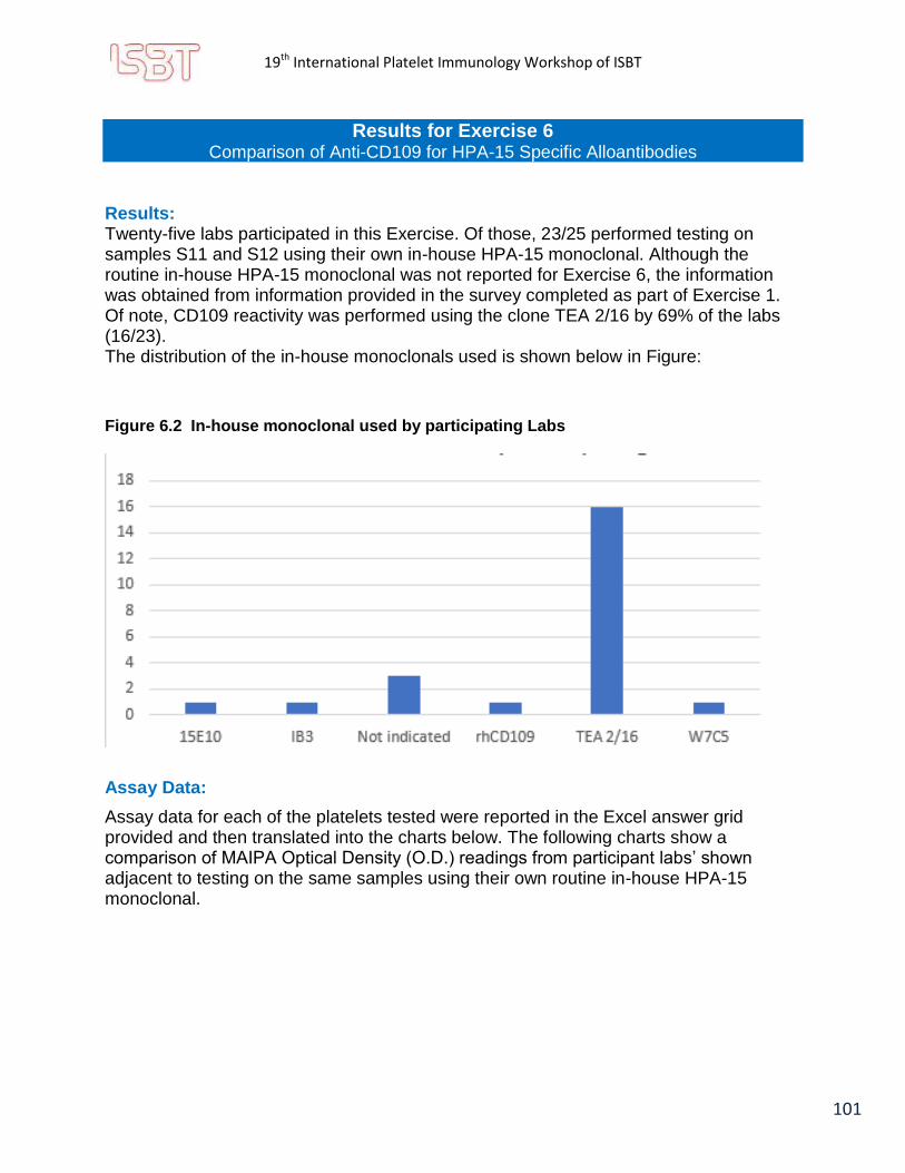
19th International Platelet Immunology Workshop of ISBT
101
Results for Exercise 6 Comparison of Anti-CD109 for HPA-15 Specific Alloantibodies
Results: Twenty-five labs participated in this Exercise. Of those, 23/25 performed testing on samples S11 and S12 using their own in-house HPA-15 monoclonal. Although the routine in-house HPA-15 monoclonal was not reported for Exercise 6, the information was obtained from information provided in the survey completed as part of Exercise 1. Of note, CD109 reactivity was performed using the clone TEA 2/16 by 69% of the labs (16/23). The distribution of the in-house monoclonals used is shown below in Figure:
Figure 6.2 In-house monoclonal used by participating Labs
Assay Data:
Assay data for each of the platelets tested were reported in the Excel answer grid provided and then translated into the charts below. The following charts show a comparison of MAIPA Optical Density (O.D.) readings from participant labs’ shown adjacent to testing on the same samples using their own routine in-house HPA-15 monoclonal.
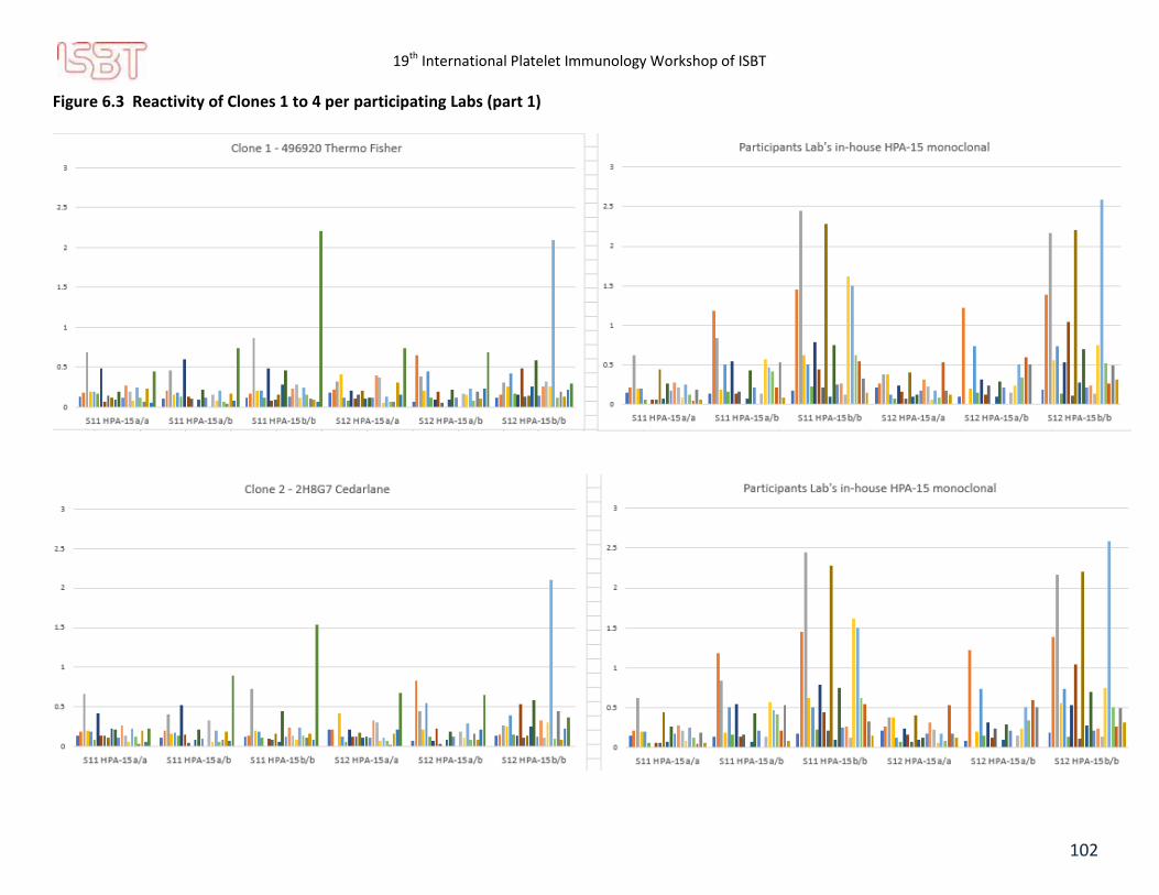
19th International Platelet Immunology Workshop of ISBT
102
Figure 6.3 Reactivity of Clones 1 to 4 per participating Labs (part 1)
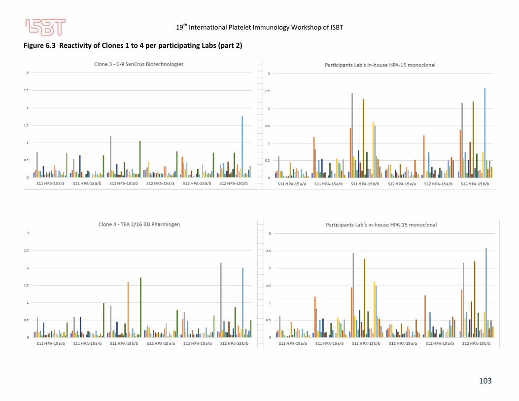
19th International Platelet Immunology Workshop of ISBT
103
Figure 6.3 Reactivity of Clones 1 to 4 per participating Labs (part 2)
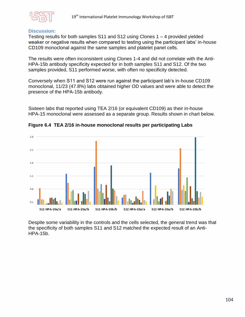
19th International Platelet Immunology Workshop of ISBT
104
Discussion: Testing results for both samples S11 and S12 using Clones 1 – 4 provided yielded weaker or negative results when compared to testing using the participant labs’ in-house CD109 monoclonal against the same samples and platelet panel cells. The results were often inconsistent using Clones 1-4 and did not correlate with the Anti-HPA-15b antibody specificity expected for in both samples S11 and S12. Of the two samples provided, S11 performed worse, with often no specificity detected. Conversely when S11 and S12 were run against the participant lab’s in-house CD109 monoclonal, 11/23 (47.8%) labs obtained higher OD values and were able to detect the presence of the HPA-15b antibody. Sixteen labs that reported using TEA 2/16 (or equivalent CD109) as their in-house HPA-15 monoclonal were assessed as a separate group. Results shown in chart below. Figure 6.4 TEA 2/16 in-house monoclonal results per participating Labs
Despite some variability in the controls and the cells selected, the general trend was that the specificity of both samples S11 and S12 matched the expected result of an Anti-HPA-15b.
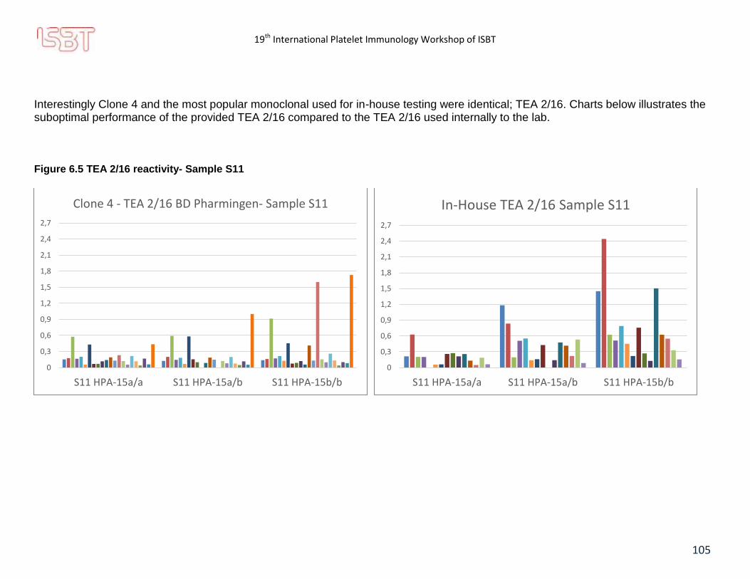
19th International Platelet Immunology Workshop of ISBT
105
Interestingly Clone 4 and the most popular monoclonal used for in-house testing were identical; TEA 2/16. Charts below illustrates the suboptimal performance of the provided TEA 2/16 compared to the TEA 2/16 used internally to the lab. Figure 6.5 TEA 2/16 reactivity- Sample S11
0
0,3
0,6
0,9
1,2
1,5
1,8
2,1
2,4
2,7
S11 HPA-15a/a S11 HPA-15a/b S11 HPA-15b/b
Clone 4 - TEA 2/16 BD Pharmingen- Sample S11
0
0,3
0,6
0,9
1,2
1,5
1,8
2,1
2,4
2,7
S11 HPA-15a/a S11 HPA-15a/b S11 HPA-15b/b
In-House TEA 2/16 Sample S11
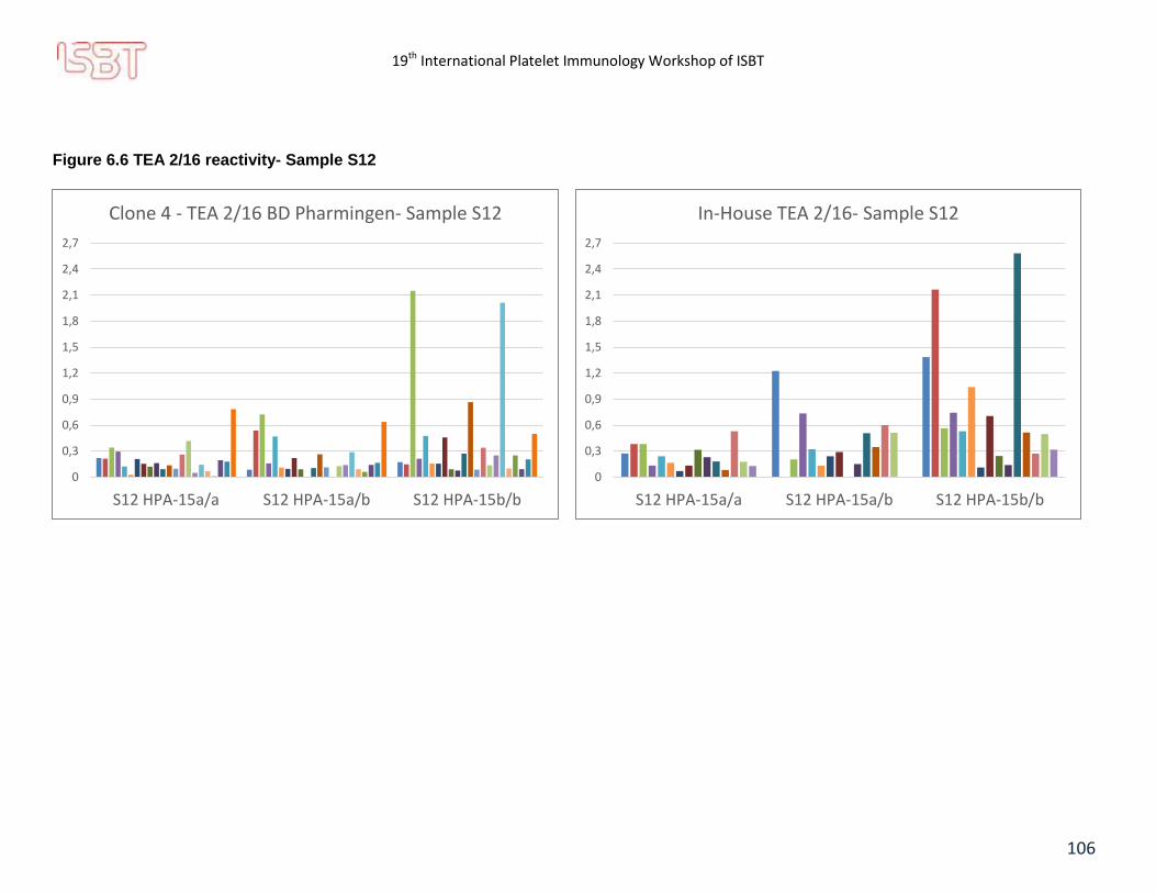
19th International Platelet Immunology Workshop of ISBT
106
Figure 6.6 TEA 2/16 reactivity- Sample S12
0
0,3
0,6
0,9
1,2
1,5
1,8
2,1
2,4
2,7
S12 HPA-15a/a S12 HPA-15a/b S12 HPA-15b/b
Clone 4 - TEA 2/16 BD Pharmingen- Sample S12
0
0,3
0,6
0,9
1,2
1,5
1,8
2,1
2,4
2,7
S12 HPA-15a/a S12 HPA-15a/b S12 HPA-15b/b
In-House TEA 2/16- Sample S12
19th International Platelet Immunology Workshop of ISBT
107
As with any type of research, experiment failures can occur. Such was the case with Exercise 6 as there is a strong indication that the monoclonals in the Workshop packages sent to participants were compromised. At this time it is unclear as to the causative reason as to why the provided monoclonals failed when they had worked successfully in pre-workshop runs. Possible causes include shipping, as some participants had expected reactions. Other causes may also have a role in the failure of reactivity. Regardless of the pitfalls of the exercise some information reported by the participants can be gleaned and is still considered to be valuable. Conclusion First, poor results obtained using all four CD109 clones are likely attributed to the clones themselves; however it is unclear what the root cause is. Second, lack of standardization in the MAIPA method for CD109 is clearly evident in results obtains from lab to lab using the same monoclonal TEA 2/16. Finally, of the labs participating in this exercise, it was obvious that even with the challenges of the monoclonals provided; select labs were still able to detect the HPA-15b antibody. OD values, using the provided clones, had been reduced however their overall MAIPA protocol appears to be more robust. It would be beneficial for future workshops to look at these labs, to determine what may be best practices when performing MAIPA using CD109 monoclonals.
19th International Platelet Immunology Workshop of ISBT
108
Final Conclusions
Although the ISBT platelet workshop is a very important activity for development and improvement of platelet immunohematology, the 19th International Platelet Workshop has been a tremendous challenge for us to prepare, to analyze, and then to conclude. The workshop participation was a successful worldwide representation of our working group, comprising 28 laboratories from 17 countries. Because of the large number of participating centers disseminated all over the world, the biggest challenge for the organizer laboratories were to provide sufficient material and to fulfill the international shipment restrictions for biological sample transportation (i.e. international biological shipping and governmental authorization, transportation condition and shipment integrity). Nevertheless, interesting findings could be drawn from each one of the various exercises. Interest for technical improvement and standardization has always guided us through the different exercises that we designed. We therefore strongly recommend that the ISBT Platelet Working Group subcommittee needs to provide a good guidance for the next International Workshop. Finally, we would like to thank all participants for their dedication to platelet immunology.