19/2: treatment of gingival smile: a case report
TRANSCRIPT
© International Academy of Periodontology
Journal of the International Academy of Periodontology 2017 19/2: 51–56
Correspondence to: Carmen L. Mueller Storrer, Professor of Graduate Program in Clinical Dentistry, Universidade Positivo, Rua. Prof. Pedro Viriato Parigot de Souza, 5300. Campo Com-prido – Curitiba – PR – Brazil CEP: 81280-330. Phone: +55 41 3317-3454; Fax: +55 41 3317-3000; [email protected]
Introduction
Many patients seek dental treatment to improve not only their oral health but also their smile. Thus, it is necessary for health professionals to stay abreast of new forms of treatment for gingival smile correction (Gaddale et al., 2014; Storrer et al., 2014).
Many dental professionals consider that a harmonious smile requires the upper lip be positioned at the level of the gingival margin of the maxillary central incisors. The fi eld of dentistry concerned with both diseases of the teeth and facial aesthetics is periodontology (Seixas et al., 2011; Gaddale et al., 2014).
Excessive exposure of the gums (more than 3 mm) while smiling is known as a gingival smile. Its aetiology is varied, including gingival enlargement, lip hyperactivity, vertical maxillary excess (VME), altered passive eruption (APE), or combinations thereof (Allen, 1988; Garber and Salama, 1996; Robbins, 1999; Monaco et al., 2004; Ribeiro et al., 2012).
The aesthetics of a smile involve relationships between the teeth, the shape of the lips, and the gingival anatomy
Treatment of Gingival Smile: A Case ReportCarmen Lucia Mueller Storrer, Naylin Danyele de Oliveira, Tatiana Miranda Deliberador, Luana Toledo Ori, Sara Moncada Guerrero, Felipe Rychuv Santos and Fernando Henrique Puppel Osternack
Abstract
There are various alternatives for correcting a gingival smile, ranging from techniques such as gingivectomy to more complex and invasive procedures such as orthognathic surgery. Gingival smile is one of the most common complaints of patients seeking an aesthetic smile. This article describes the alternative treatments available for lip repositioning and improving smile aesthetics, and presents the clinical course of a gingival smile patient after undergoing the surgical technique of gingival recontouring (GR) associated with lip repositioning with restorative aesthetic treatment. The surgery performed corrected altered passive eruption of the maxillary anterior teeth and lengthened the lateral beam of the elevator muscle of the upper lip and wing of the nose, and achieved containment with sutures, reducing the gingival smile. After surgery, it was apparent that the high smile line had been successfully corrected without compromising labial harmony. The patient expressed high satisfaction with the treatment. The surgical technique proposed in this study may be a therapeutic option for lip repositioning to achieve smile harmony.
Key words: aesthetic surgery, dental aesthetics, gingival recontouring, gummy smile, gums
Universidade Positivo, Curitiba, Parana, Brazil
(Garber and Salama, 1996). Depending on the patient’s teeth and the shape of their lips, the clinician should determine the optimal type of smile – high, medium or low (Lee, 2004). Where there is a gingival smile, it must be diagnosed and classifi ed prior to the formulation of a treatment plan (Sahoo et al., 2012).
According to Garber and Salama (1996), a smile can be high, medium or low. A high smile or ‘gummy’ smile is exhibited when the patient presents a substantial amount of soft tissue above the upper teeth. This feature can be the re-sult of the combination of several factors, including gingival enlargement, lip hyperactivity, VME or APE. We propose a modifi cation of Garber and Salama’s (1996) classifi cation system for the diagnosis and treatment of various forms of gingival smile (Table 1).
If the tooth crown anatomy is larger in width (mesio-distal) than in height (cervical-incisal), the problem results from APE. Lee (2004) proposed the utilization of cosmetic prostheses in such cases, after fi rst evaluating the biological width of each tooth, and identifying whether or not clinical crown augmentation procedures are required. If tooth shape is not changed but there is a considerable amount of gingival tissue (attached gingiva) from the bottom vestibule to the line of the gingival zenith, the result is VME. In most cases, however, a gingival smile is a consequence of both APE and VME. Efforts to improve smile aesthetics and dental-facial imbalances have led to the development of new surgical techniques to correct gingival smiles via correct diagnoses.
52 Journal of the International Academy of Periodontology (2017) 19/2
While some previous studies have approached gingival smile as a muscular problem caused by a hyper-active upper lip (Sahoo et al., 2012; Storrer et al., 2014) the etiological causes of gingival smile evidently include skeletal (VME), gingival (APE) and muscular factors. The use of botulinum toxin is considered a minimally invasive technique involving the administration of toxin injections into the hyperactive muscle of the upper lip to treat the defect (Sahoo et al., 2012). In some cases it is not possible to treat the patient solely via gingival recontouring (GR), however, because the amount of gingival display remains signifi cant (Storrer et al., 2014).
This report describes the alternative treatments available for lip repositioning and improving smile aesthetics, and presents the clinical course of a gingival smile patient after undergoing the surgical technique of GR associated with lip repositioning with restorative aesthetic treatment.
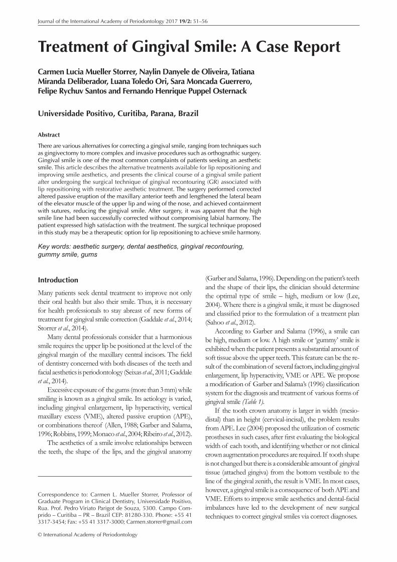
Case description and results A 23-year-old female Brazilian patient attended the dental clinic at Positivo University reporting excessive exposure of her gums when she smiled. On clinical examination, it was noted that the periodontium was healthy, and periodontal clinical parameters were within normal limits. However, the patient was dissatisfi ed with the cosmetic appearance of her teeth, stating that they seemed ‘very small’ and her gums ‘very large.’ During a happy natural smile, the patient’s teeth were visible from the maxillary right second pre-molar to the maxillary left second pre-molar, with 8 mm of excessive gingival tissue display among the incisor teeth from the gingival margin to the bottom edge of the upper lip. During extravagant smiling, we could observe an overexuberant lip raising, which led to a diagnosis of a hyperactive upper lip (Figure 1).
It was suggested to the patient that surgery would be required for the resolution of her case. After evaluation of the medical and family history of the patient, it was determined that she was medically fi t for surgery. With the aid of a millimeter periodontal probe, her anterior teeth were then probed to the upper right and left canine, identifying a probing depth of 4 mm and an absence of bleeding and periodontal pockets. The patient had tooth crown anatomy larger in width than in height, a considerable amount of gingival tissue (attached gingiva) display and lip hyperactivity. Thus, she was diagnosed with APE Type 1A or 1B and AVM Grade 2, and the treatment planning was GR with or without osteotomy plus lip repositioning and restorative aesthetic treatment to improve the height and width of the dental crown, and minimize the gummy smile, as orthognathic surgery was not the patient’s option. The lip repositioning tech-nique selected was the one fi rst described by Storrer et al. (2014) where the elevator muscle of upper lip and wing of nose (EMULWN) is tensioned and contained.
Condition Treatment
APE Type 1A: Normal biological space GR and/or periodontal surgery lip repositioning
APE Type 1B: Without biological space (bone crest coincides with CEJ)
GR with retail survey and osteotomia and/or periodontal surgery lip repositioning
APE Type 2: Short teeth without excess gum tissue Realization of aesthetics prosthesis with or without osteotomy
VME Grade 1: Mucosa and gingival range 2 – 4 mm Orthodontic intrusion, periodontics or orthodontics and peri-odontics and aesthetic restorative treatment
VME Grade 2: Mucosa and gingival range 4 – 8 mm GR + lip repositioning and restorative aesthetic treatment or orthognathic surgery
VME Grade 3: Mucosa and gingival range ³ 8 mm GR + lip repositioning and aesthetic restorative treatment or orthognathic surgery
APE + VME Grade 2 or 3 GR with or without osteotomy + lip repositioning and restora-tive aesthetic treatment or orthognathic surgery
Table 1. Diagnosis and treatment of gingival smile
APE, altered passive eruption; VME, vertical maxillary excess; CEJ, cementoenamel junction; GR, gingival recontouring
Fig 1
Fig 2
Figure 1. A) Vestibular aspect of gingival smile correction before surgery. Modifi ed Garber and Salama (1996) classifi cation; APE + VME grade 2. B) Intraoral view with gingival exposure. The distance from the lip edge to the gingival margin was 8 mm. Increasing the clinical crown of the incisors and lip repositioning were proposed to improve smile aesthetics in this case. APE, altered passive eruption; VME, vertical maxillary excess.
Mueller Storrer et al.: Treatment of Gingival Smile 53
This muscle is described as being long and slender, ex-tending from the frontal process of the maxilla at the corner of the eye to the upper lip. EMULWN is divided into medial and lateral cords: the lateral cord raises and everts the upper lip and raises, deepens and increases the curvature of the upper part of the nasolabial groove (Storrer et al., 2014).
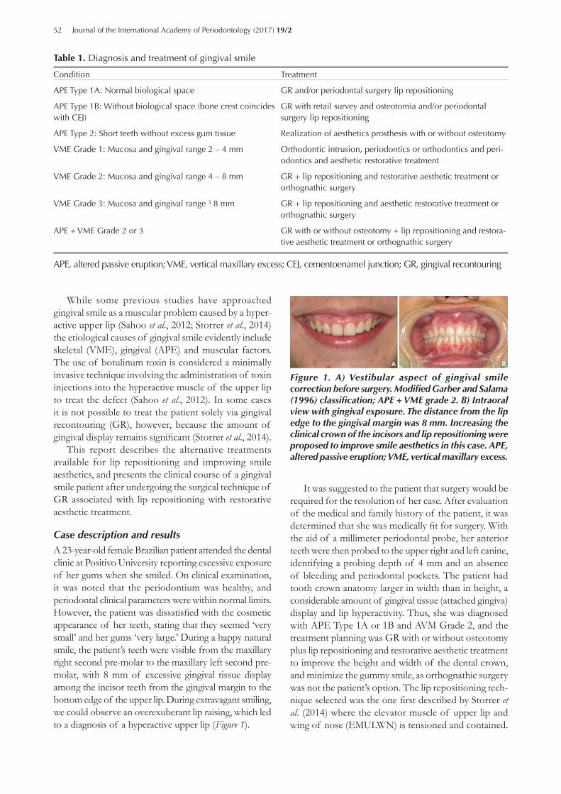
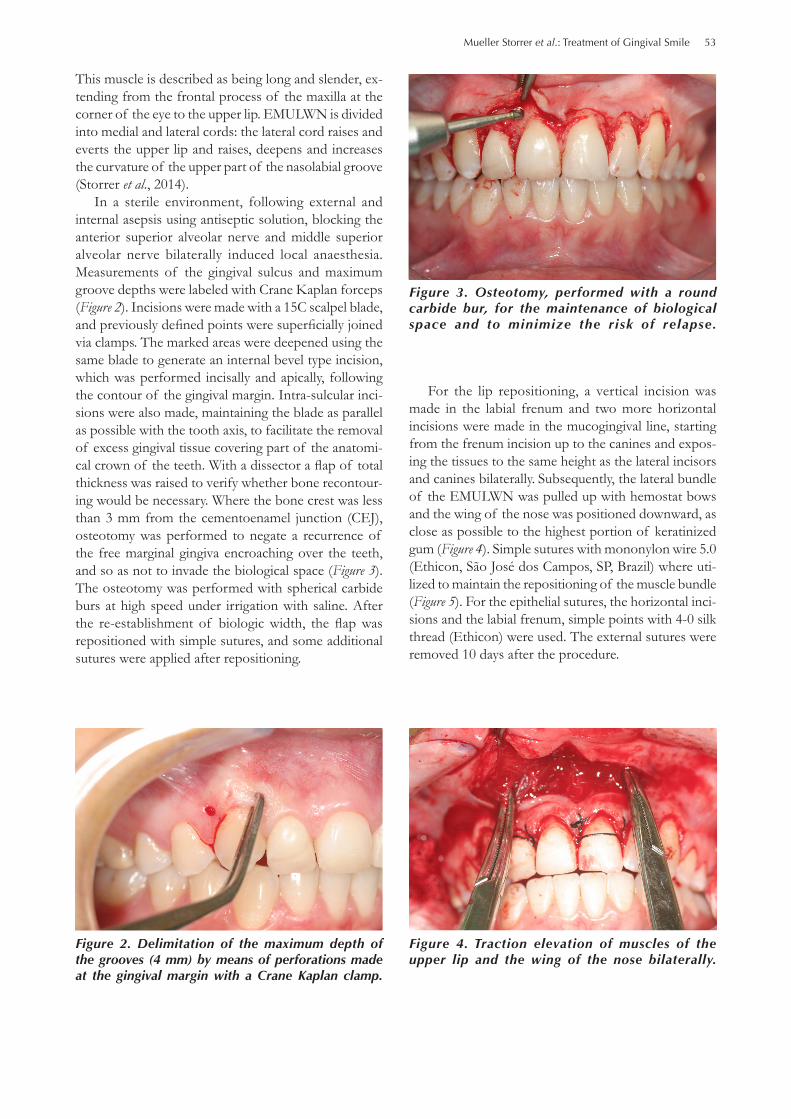
In a sterile environment, following external and internal asepsis using antiseptic solution, blocking the anterior superior alveolar nerve and middle superior alveolar nerve bilaterally induced local anaesthesia. Measurements of the gingival sulcus and maximum groove depths were labeled with Crane Kaplan forceps (Figure 2). Incisions were made with a 15C scalpel blade, and previously defi ned points were superfi cially joined via clamps. The marked areas were deepened using the same blade to generate an internal bevel type incision, which was performed incisally and apically, following the contour of the gingival margin. Intra-sulcular inci-sions were also made, maintaining the blade as parallel as possible with the tooth axis, to facilitate the removal of excess gingival tissue covering part of the anatomi-cal crown of the teeth. With a dissector a fl ap of total thickness was raised to verify whether bone recontour-ing would be necessary. Where the bone crest was less than 3 mm from the cementoenamel junction (CEJ), osteotomy was performed to negate a recurrence of the free marginal gingiva encroaching over the teeth, and so as not to invade the biological space (Figure 3). The osteotomy was performed with spherical carbide burs at high speed under irrigation with saline. After the re-establishment of biologic width, the fl ap was repositioned with simple sutures, and some additional sutures were applied after repositioning.
For the lip repositioning, a vertical incision was made in the labial frenum and two more horizontal incisions were made in the mucogingival line, starting from the frenum incision up to the canines and expos-ing the tissues to the same height as the lateral incisors and canines bilaterally. Subsequently, the lateral bundle of the EMULWN was pulled up with hemostat bows and the wing of the nose was positioned downward, as close as possible to the highest portion of keratinized gum (Figure 4). Simple sutures with mononylon wire 5.0 (Ethicon, São José dos Campos, SP, Brazil) where uti-lized to maintain the repositioning of the muscle bundle (Figure 5). For the epithelial sutures, the horizontal inci-sions and the labial frenum, simple points with 4-0 silk thread (Ethicon) were used. The external sutures were removed 10 days after the procedure.
Fig 1
Fig 2Figure 2. Delimitation of the maximum depth of the grooves (4 mm) by means of perforations made at the gingival margin with a Crane Kaplan clamp.
Fig 3
Fig 4
Figure 3. Osteotomy, performed with a round carbide bur, for the maintenance of biological space and to minimize the risk of relapse.
Fig 3
Fig 4Figure 4. Traction elevation of muscles of the upper lip and the wing of the nose bilaterally.
54 Journal of the International Academy of Periodontology (2017) 19/2
Postoperative recommendations included avoiding physical exertion for at least 48 hours, ice packs on the upper lip, limiting facial movements for a period of 7 days and using chlorhexidine gluconate 0.12% for 7 days. Amoxicillin 500 mg every 8 hours for 7 days, nimesulide 100 mg every 12 hours for 3 days and tramadol 50 mg every 6 hours for 3 days were utilized for the control of pain.
Follow-up was performed after 7, 30, 60 and 90 days. During the postoperative period, bruising, swelling or hematoma were not observed, but some tension while smiling was seen during the fi rst three months. The pa-tient also reported no pain when smiling or talking. After 90 days, no notable scar was seen on patient smiling. There was improved harmony between her lips, teeth and gums with noticeable indications of periodontal health including pale pink coloration and embrasures completely fi lled by the papillae. Change in lip symmetry after surgical treatment was not observed (Figure 6).
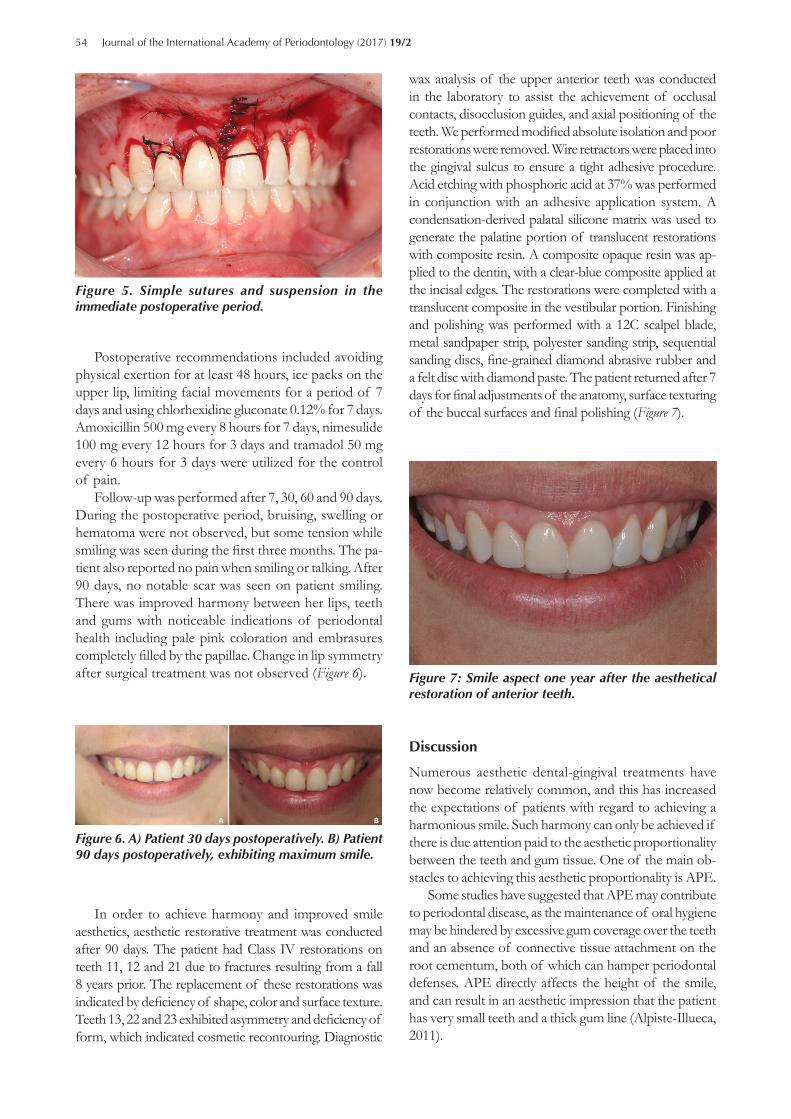
wax analysis of the upper anterior teeth was conducted in the laboratory to assist the achievement of occlusal contacts, disocclusion guides, and axial positioning of the teeth. We performed modifi ed absolute isolation and poor restorations were removed. Wire retractors were placed into the gingival sulcus to ensure a tight adhesive procedure. Acid etching with phosphoric acid at 37% was performed in conjunction with an adhesive application system. A condensation-derived palatal silicone matrix was used to generate the palatine portion of translucent restorations with composite resin. A composite opaque resin was ap-plied to the dentin, with a clear-blue composite applied at the incisal edges. The restorations were completed with a translucent composite in the vestibular portion. Finishing and polishing was performed with a 12C scalpel blade, metal sandpaper strip, polyester sanding strip, sequential sanding discs, fi ne-grained diamond abrasive rubber and a felt disc with diamond paste. The patient returned after 7 days for fi nal adjustments of the anatomy, surface texturing of the buccal surfaces and fi nal polishing (Figure 7).
Fig 5
Fig 6
Figure 5. Simple sutures and suspension in the immediate postoperative period.
Fig 5
Fig 6Figure 6. A) Patient 30 days postoperatively. B) Patient 90 days postoperatively, exhibiting maximum smile.
In order to achieve harmony and improved smile aesthetics, aesthetic restorative treatment was conducted after 90 days. The patient had Class IV restorations on teeth 11, 12 and 21 due to fractures resulting from a fall 8 years prior. The replacement of these restorations was indicated by defi ciency of shape, color and surface texture. Teeth 13, 22 and 23 exhibited asymmetry and defi ciency of form, which indicated cosmetic recontouring. Diagnostic
Figure 7Figure 7: Smile aspect one year after the aesthetical restoration of anterior teeth.
Discussion
Numerous aesthetic dental-gingival treatments have now become relatively common, and this has increased the expectations of patients with regard to achieving a harmonious smile. Such harmony can only be achieved if there is due attention paid to the aesthetic proportionality between the teeth and gum tissue. One of the main ob-stacles to achieving this aesthetic proportionality is APE.
Some studies have suggested that APE may contribute to periodontal disease, as the maintenance of oral hygiene may be hindered by excessive gum coverage over the teeth and an absence of connective tissue attachment on the root cementum, both of which can hamper periodontal defenses. APE directly affects the height of the smile, and can result in an aesthetic impression that the patient has very small teeth and a thick gum line (Alpiste-Illueca, 2011).
Mueller Storrer et al.: Treatment of Gingival Smile 55
Several factors have been implicated in the occur-rence of APE, including occlusal interference during the eruptive phase, the presence of thick and fi brotic gums, and hereditary conditions. During the active eruption phase, the gum moves with the crown until the erupting tooth reaches the occlusal plane of its antagonist. In the passive eruption stage, the gum migrates apically until the gingival margin arrives at a stable cervical level. Notably, however, some authors have reported that when the bony crest coincides with the CEJ, gum migration in an apical direction does not occur (Alpiste-Illueca, 2011). This phenomenon potentially explains what may have occurred during the passive eruption process in the case reported herein.
Once APE has been diagnosed, the indication is surgical GR with or without repositioning of the elevator muscle of the upper lip and the wing of the nose (Alpiste-Illueca, 2011; Gaddale et al., 2014). The nasal septum muscle containment surgical technique has been suggested by some authors as an alternative for the treatment of gingival smile, as this muscle has a strong infl uence on the smile and has been associated with excessive lifting of the upper lip. In the current study, a technique incorporating bilateral muscle re-straint resulted in favorable lowering of the upper lip. To correct upper lip asymmetry, where the patient raises one side more than the other when smiling, restraint can be applied on one side only (Storrer et al., 2014).
In 2014, Storrer et al. proposed a muscle reposi-tioning technique to lift the upper lip and nose wing via resorbable muscle sutures positioned close to the alveolar process. In the case reported herein, an amend-ment to that suture technique was utilized to minimize the recurrence of adverse repositioning. To increase the visible dental crown, recontouring gingival surgery was also performed. The necessity for osteotomy in some regions was due to the fact that after the folding of the fl ap, it was apparent that the patient had APE in addition to the previously diagnosed VME. Thus, the CEJ of some teeth coincided with the bone crest. Therefore, osteotomy was utilized to avoid the recur-rence of gum encroachment.
The surgical lip repositioning technique in conjunc-tion with GR performed in the case reported herein resulted in an adequate smile height, with a lower line and improved aesthetics. The gingival display at baseline was 8 mm, among the incisive teeth, and decreased to 2 mm in the medial line and 4 mm at the lateral incisors within 24 months of follow-up. It is also important to note that the proportion of the size of the clinical crown was restored after GR surgery and restorative procedures. At the beginning (Figure 1), the patient had crowns with a square format. After the restorative and surgical procedures, the mesio-distal proportion of the clinical crown has been re-established (Figure 7).
The GR techniques resulting in lip repositioning were achieved in a single operation, which was performed un-der local anesthesia. In addition, the procedure may be associated with relatively less discomfort after surgery than other methods in patients with no recurrence, as the sutures do not need to be changed to mononylon. With regard to synthetic yarn, nylon yarn is reportedly associated with better biocompatibility with tissue, gen-erating only a small and brief infl ammatory response that evidently does not damage the tissues used for internal sutures (Castelli et al., 1978).
Notably, both GR and surgical lip repositioning are considered safe with regard to the risk of relapse, as long as a regimented surgical protocol is followed and the patient has adequate postoperative follow-up. Various cases treated with corrective cosmetic surgery have been reported in the literature, including patients with facial paralysis. In such cases, silicone implantation in the foyer of the fund at the base of the nasal spine, administration of botulinum toxin, and resective proce-dures in the muscles responsible for upper lip mobility are techniques commonly used for corrections. Such techniques have reportedly proved successful when utilized solely for the cosmetic correction of smile height (Seixas et al., 2011).
Botulinum toxin generates muscle paralysis-induced depression of the nasal septum, and thus reduces movement of the upper lip, resulting in the appearance of less gingival tissue during smiling. However, ‘limited action time lock’ and the potential for undesired muscle anesthesia are negative aspects of this technique. In the case reported herein, the surgical technique used to contain the muscle was more intrusive than the use of botulinum toxin, but the result was more aestheti-cally favorable, and longer lasting. In the current study, as well as upper lip lowering there was also apparent avoidance of eversion, resulting in excellent aesthetic results (Storrer et al., 2014). Only in cases of silicone implantation in the foyer fund would the methods reported herein be contraindicated for the aesthetic resolution of gingival smile. The current case has now been followed up for more than 1 year post-surgery, and it can be concluded that the gingival recontour-ing and surgical lip repositioning procedures utilized represent an excellent option for the correction of gingival smile.
Conclusion
Within the limitations of this case report, it can be concluded that the surgical technique proposed in this study may be a therapeutic option for lip repositioning to achieve smile harmony. However, a careful anamnesis and management of correct diagnosis should be con-sidered to achieve success.
56 Journal of the International Academy of Periodontology (2017) 19/2
References
Allen EP. Use of mucogingival surgical procedures to enhance esthetics. Dental Clinics North America 1988; 32:307-330.
Alpiste-Illueca F. Altered passive eruption (APE): A little-known clinical situation. Medicina Oral, Patología Oral y Cirugía Buccal 2011; 16:e100-104.
Castelli WA, Nasjleti CE, Caffesse RE and Diaz-Perez R. Gingival response to silk, cotton, and nylon suture materials. Oral Surgery, Oral Medicine, and Oral Pathology 1978; 45:179-185.
Gaddale R, Desai SR, Mudda JA and Karthikeyan I. Lip repositioning. Journal of the Indian Society of Periodontology 2014; 18:254-258.
Garber DA and Salama MA. The aesthetic smile: diagnosis and treatment. Periodontology 2000 1996; 11:18-28.
Lee EA. Aesthetic crown lengthening: classifi cation, biologic rationale, and treatment planning considerations. Practical Procedures and Aesthetic Dentistry 2004; 16:769-778.
Monaco A, Streni O, Marci MC, et al. Gummy smile: clinical parameters useful for diagnosis and therapeutical ap-proach. Journal of Clinical Pediatric Dentistry 2004; 29:19-25.
Ribeiro FS, Garção FCC, Martins AT, Sakakura CE, de Toledo BEC and Pontes AEF. A modifi ed technique that decreases the height of the upper lip in the treat-ment of gummy smile patients: A case series study. Journal of Dentistry and Oral Hygiene 2012; 4:21-28.
Robbins JW. Differential diagnosis and treatment of ex-cess gingival display. Practical Periodontics and Aesthetic Dentistry 1999; 11:265-272.
Sahoo KC, Raghunath N and Shivalinga BM. Botox in gummy smile - A review. Indian Journal of Dental Sciences 2012; 4:51-53.
Seixas MR, Costa-Pinto RA and De Araújo TM. Check-list of esthetic features to consider in diagnosing and treating excessive gingival display (gummy smile). Dental Press Journal of Orthodontics 2011; 16:131-157.
Storrer CL, Valverde FK, Santos FR and Deliberador TM. Treatment of gummy smile: Gingival recontour-ing with the containment of the elevator muscle of the upper lip and wing of nose. A surgery innovation technique. Journal of the Indian Society of Periodontology 2014; 18:656-660.