1471-2377-11-109
TRANSCRIPT

STUDY PROTOCOL Open Access
Risk factors and prognosis of young stroke. TheFUTURE study: A prospective cohort study. Studyrationale and protocolLoes CA Rutten-Jacobs1, Noortje AM Maaijwee1, Renate M Arntz1, Mayte E Van Alebeek1,Pauline Schaapsmeerders2, Henny C Schoonderwaldt1, Lucille DA Dorresteijn3, Sebastiaan Overeem1, Gea Drost1,Mirian C Janssen4, Waander L van Heerde5, Roy PC Kessels2,6,7, Marcel P Zwiers8,9, David G Norris9,Maureen J van der Vlugt10, Ewoud J van Dijk1 and Frank-Erik de Leeuw1*
Abstract
Background: Young stroke can have devastating consequences with respect to quality of life, the ability to work,plan or run a family, and participate in social life. Better insight into risk factors and the long-term prognosis isextremely important, especially in young stroke patients with a life expectancy of decades. To date, detailedinformation on risk factors and the long-term prognosis in young stroke patients, and more specific risk ofmortality or recurrent vascular events, remains scarce.
Methods/Design: The FUTURE study is a prospective cohort study on risk factors and prognosis of young ischemicand hemorrhagic stroke among 1006 patients, aged 18-50 years, included in our study database between 1-1-1980and 1-11-2010. Follow-up visits at our research centre take place from the end of 2009 until the end of 2011.Control subjects will be recruited among the patients’ spouses, relatives or social environment. Information onmortality and incident vascular events will be retrieved via structured questionnaires. In addition, participants areinvited to the research centre to undergo an extensive sub study including MRI.
Discussion: The FUTURE study has the potential to make an important contribution to increase the knowledge onrisk factors and long-term prognosis in young stroke patients. Our study differs from previous studies by having amaximal follow-up of more than 30 years, including not only TIA and ischemic stroke but also hemorrhagic stroke,the addition of healthy controls and prospectively collect data during an extensive follow-up visit. Completion ofthe FUTURE study may provide better information for treating physicians and patients with respect to theprognosis of young stroke.
BackgroundUp to 12% of all stroke occur in patients between 18-50years ("young” stroke) [1], affecting about 5000 patientseach year in the Netherlands and about 2 million youngpeople each year worldwide. In a substantial proportionof roughly one third the etiology remains unelucidated.In terms of prognosis a “young” stroke has a dramaticinfluence on independency and quality of life as itoccurs in the period of life that people start to form
families, make decisive career moves, and have an activesocial life. Uncertainty about long term prognosis affectschoices and planning affiliated with these life events.Whereas risk factors and prognosis in patients who
develop a stroke at higher ages (usually over 70 years)are among the best studied topics in clinical medicine,this does not hold true for young stroke. At higher ages,almost all risk factors have atherosclerosis in their finalcommon pathway. However, this cannot simply beextrapolated to young stroke as the underlying cause ofstroke is usually different from that in elderly and maytherefore also have a different prognosis both withrespect to functional stroke outcome as to risks of
* Correspondence: [email protected] Institute for Brain, Cognition and Behaviour, Centre forNeuroscience, Department of Neurology, Radboud University NijmegenMedical Centre, PO Box 9101, 6500 HB Nijmegen, The NetherlandsFull list of author information is available at the end of the article
Rutten-Jacobs et al. BMC Neurology 2011, 11:109http://www.biomedcentral.com/1471-2377/11/109
© 2011 Rutten-Jacobs et al; licensee BioMed Central Ltd. This is an Open Access article distributed under the terms of the CreativeCommons Attribution License (http://creativecommons.org/licenses/by/2.0), which permits unrestricted use, distribution, andreproduction in any medium, provided the original work is properly cited.
recurrent stroke or other major vascular events. Evenmore, the identification of risk factors for young strokeso far has often been based on the occurrence of pre-sumed risk factors in consecutive series of young strokepatients, without methodological sound comparisonwith controls.The “long-term” perspective in an on average over 70
years “old” stroke patient differs from that of a 30 years“young” stroke patient, and particularly studies with a long-term follow-up of more than 10 years are lacking in theyoung stroke field. Studies thus far, usually with a meanfollow-up duration of less than 7 years, report highly vari-able post-stroke mortality and risk of incident vascular dis-ease [2-7]. These large differences across studies are wellexplained because young stroke is a heterogeneous diseaseand most studies were small, had different selection cri-teria, did not investigate patients in person but relied ontelephone interviews and outcome assessments and follow-up planning was not uniform and often suboptimal.Although stroke includes both ischemic and hemorrhagicstroke, almost all studies have excluded the investigation ofetiology and prognosis of young hemorrhagic stroke.Except for recurrent vascular disease and persistent
motor and language impairments, post-"young” strokequality of life will most likely also be determined by cog-nitive dysfunction, depressive symptoms, fatigue, andspecific post-stroke complications such as epilepsy,because those determine the ability to (return to) workand to have a normal family and social life. Data onthose aspects in the very long-term follow-up of youngstroke patients are even more scarce.Although the absolute number of young stroke is
lower than stroke among the elderly, the total numberof years that young stroke patients as a whole will livewith the consequences of the stroke exceeds that ofolder stroke survivors due to far longer survival.This justifies a properly designed and executed study
on risk factors and prognosis of young stroke, comparedwith controls. We therefore set up the FUTURE study(Follow-Up of Transient ischemic attack and strokepatients and Unelucidated Risk factor Evaluation study),the largest single-centre prospective cohort study on riskfactors and prognosis of young TIA, ischemic stroke andhemorrhagic stroke patients (n = 1006) and controls.
Methods/DesignThe FUTURE study is a prospective cohort study thataims to investigate the causes and consequences of ayoung stroke. The Medical Review Ethics Committeeregion Arnhem-Nijmegen approved the study.
PatientsThe department of neurology has a long-standing inter-est in the etiology and prognosis of young stroke and
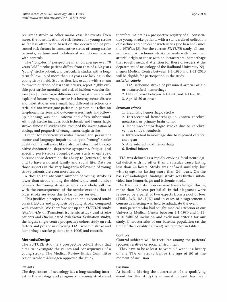
therefore maintains a prospective registry of all consecu-tive young stroke patients with a standardized collectionof baseline and clinical characteristics (see baseline) sincethe 1970’ies [8]. For the current FUTURE-study, all con-secutive TIA, ischemic stroke patients with presumedarterial origin or those with an intracerebral hemorrhagethat sought medical attention for these disorders at thedepartment of neurology of the Radboud University Nij-megen Medical Centre between 1-1-1980 and 1-11-2010will be eligible for participation in the study.Inclusion criteria
1. TIA, ischemic stroke of presumed arterial originor intracerebral hemorrhage2. Date of onset between 1-1-1980 and 1-11-20103. Age 18-50 at onset
Exclusion criteria1. Traumatic hemorrhagic stroke2. Intracerebral hemorrhage in known cerebralmetastasis or primary brain tumor3. Ischemic/hemorrhagic stroke due to cerebralvenous sinus thrombosis4. Intracerebral hemorrhage due to ruptured cerebralaneurysm5. Any subarachnoid hemorrhage6. Retinal infarct
TIA was defined as a rapidly evolving focal neurologi-cal deficit with no other than a vascular cause lastingless than 24 hours. Stroke was defined similarly, butwith symptoms lasting more than 24 hours. On thebasis of radiological findings, stroke was further subdi-vided into hemorrhagic and ischemic stroke.As the diagnostic process may have changed during
more than 30-year period all initial diagnoses werereviewed by a panel of two experts from a pool of four(FEdL, EvD, RA, LJD) and in cases of disagreement aconsensus meeting was held to adjudicate the event.1006 patients who had sought medical attention at our
University Medical Center between 1-1-1980 and 1-11-2010 fulfilled inclusion and exclusion criteria for ourstudy. Characteristics of our baseline population (at thetime of their qualifying event) are reported in table 1.
ControlsControl subjects will be recruited among the patients’spouses, relatives or social environment.They have to be at least 18 years old without a history
of any TIA or stroke before the age of 50 at themoment of inclusion.
BaselineAt baseline (during the occurrence of the qualifyingevent for the study) a minimal dataset has been
Rutten-Jacobs et al. BMC Neurology 2011, 11:109http://www.biomedcentral.com/1471-2377/11/109
Page 2 of 8

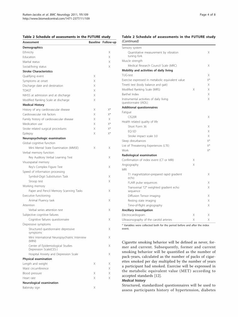
collected that consists of demographics, stroke subtype,risk factors and additional investigations (table 2). Thecompleteness of the baseline dataset varies amongpatients due to changes in standard diagnostic proce-dures over the last thirty years.Current common rating scales for the severity and
cause of stroke did not exist at the time when a sub-stantial proportion of our patients experienced theirqualifying event. Therefore a rating of both the severity(NIHSS) and cause (TOAST) was done for all cases ret-rospectively by a validated approach [9].
Follow-upInformation on the vital status will be available eitherfrom hospital data or through coupling of patientrecords with data from the municipality registry. Allpatients alive will be approached for the follow-upassessment according to a two-step approach.First, all patients will be contacted by letter to inform
them about the study; subsequently they will be con-tacted by phone. In case the patient has moved, themunicipality register of the last known residence will becontacted to trace the patient. In cases of an invalidphone number, a second letter will be sent asking thepatient to contact our centre to provide a correct phonenumber. Subsequently, when a patient does not respondto the second letter, the last known general practitionerwill be contacted to provide us with updated contactdetails. The patient will be considered lost to follow-upwhen known alive, but when untraceable via the proce-dure described above.Subsequently, patients will be given the opportunity to
participate in an extensive sub study. If they agree to doso, they will be invited to visit our research centre foradditional investigations including a structured inter-view, cognitive assessment, physical and neurologicalexamination, an extensive MRI protocol, an electrocar-diogram and an ultrasonography of the carotid arteries(Table 2). In addition, blood samples (serum/plasma/DNA) will be taken for future analysis. When patientsare not able to visit our research centre the same inves-tigations will be performed at their homes, except for
the ultrasonography of the carotid arteries, electrocar-diogram and MRI scan. Controls will undergo the sameprotocol as patients.The follow-up has started at the end of 2009 and is
planned to finish at the end of 2011.All these participants signed an informed consent.
Outcome eventsThe primary outcome of the study will be all-causemortality and the composite endpoint of death from allvascular causes; non-fatal stroke, non-fatal (silent) myo-cardial infarction, cardiovascular procedures (coronaryartery bypass grafting, percutaneous transluminal coron-ary angioplasty, carotid endarterectomy and other arter-ial revascularization procedures), whichever occurredfirst. We will perform separate analysis for the occur-rence of fatal or non-fatal stroke. Causes of death willbe categorized into ischemic stroke, intracerebralhemorrhage, cardiac causes, other vascular causes ornon-vascular causes. If we cannot obtain informationabout the cause of death, the event will be classified asunspecified.Secondary outcomes are seizures (classified according
to the ILAE [10]) and dementia (according to DSM-IV).Whenever an outcome event is suspected with the aid
of a standardized, structured questionnaire, informationretrieved will be verified and adjudicated by physiciansfrom the appropriate specialty. In case a patient hasdied, this information will be retrieved from their gen-eral practitioner or a relative. If there is no informationavailable, the event will be classified as a possible event.
Assessment of variables during follow-upDemographics and life styleStandardized questionnaires on demographics, education(classified using seven categories; one being less thanprimary school and seven reflecting an academic degree)[11], marital status, living conditions, and life stylehabits (alcohol consumption, smoking, exercise) will beadministered. Alcohol consumption will be defined asunits per day and the age at which alcohol consumptionhad started (and ended if stopped) will be noted.
Table 1 Baseline population characteristics
Total population Time of index event
1980-1989 1990-1999 2000-2010
n 1006 223 249 534
Male, n (%) 470 (46.7) 110 (49.3) 128 (51.4) 232 (43.4)
Age at index event, mean (sd) 40.2 (7.9) 39.3 (8.3) 39.7 (8.6) 40.8 (7.4)
Index event
TIA, n (%) 277 (27.5) 52 (23.3) 40 (16.1) 185 (34.6)
Infarction, n (%) 630 (62.6) 146 (65.6) 189 (75.9) 295 (55.2)
Hemorrhage, n (%) 99 (9.8) 25 (11.2) 20 (8.0) 54 (10.1)
Rutten-Jacobs et al. BMC Neurology 2011, 11:109http://www.biomedcentral.com/1471-2377/11/109
Page 3 of 8
Cigarette smoking behavior will be defined as never, for-mer and current. Subsequently, former and currentsmoking behavior will be quantified as the number ofpack-years, calculated as the number of packs of cigar-ettes smoked per day multiplied by the number of yearsa participant had smoked. Exercise will be expressed inthe metabolic equivalent value (MET) according toaccepted standards [12].Medical historyStructured, standardized questionnaires will be used toassess participants history of hypertension, diabetes
Table 2 Schedule of assessments in the FUTURE study
Assessment Baseline Follow-up
Demographics
Ethnicity X
Education X
Marital status X
Social/living status X
Stroke Characteristics
Qualifying event X
Symptoms at onset X
Discharge date and destination X
TOAST X
NIHSS at admission and at discharge X
Modified Ranking Scale at discharge X
Medical History
History of any cardiovascular disease X X*
Cardiovascular risk factors X X*
Family history of cardiovascular disease X X
Medication use X X*
Stroke related surgical procedures X X*
Epilepsy X X*
Neuropsychologic examination
Global cognitive function
Mini Mental State Examination (MMSE) X X
Verbal memory function
Rey Auditory Verbal Learning Test X
Visuospatial memory
Rey’s Complex Figure Test X
Speed of information processing
Symbol-Digit Substitution Task X
Stroop test X
Working memory
Paper and Pencil Memory Scanning Tasks X
Executive functioning
Animal Fluency task X
Attention
Verbal series attention test X
Subjective cognitive failures
Cognitive failures questionnaire X
Depressive symptoms
Structured questionnaire depressivesymptoms
X
Mini International Neuropsychiatric Interview(MINI)
X
Center of Epidemiological StudiesDepression Scale(CES-)
X
Hospital Anxiety and Depression Scale X
Physical examination
Length and weight X X
Waist circumference X
Blood pressure X X
Heart rate X X
Neurological examination
Babinsky sign X
Table 2 Schedule of assessments in the FUTURE study(Continued)
Sensory system
Quantitative measurement by vibrationtuning fork
X
Muscle strength
Medical Research Council Scale (MRC) X
Mobility and activities of daily living
TUG-test X
Exercise expressed in metabolic equivalent value X*
Tinetti test (body balance and gait) X
Modified Ranking Scale (MRS) X X
Barthel Index X
Instrumental activities of daily livingquestionnaire (IADL)
X
Additional questionnaires
Fatigue
CIS20R X
Health related quality of life
Short Form 36 X
EQ-5D X
Stroke impact scale 3.0 X
Sleep disturbances X*
List of Threatening Experiences (LTE) X*
Work X*
Radiological examination
Confirmation of index event (CT or MRI) X
Angiography X
MRI
T1 magnetization-prepared rapid gradientecho
X
FLAIR pulse sequences X
Transversal T2* weighted gradient echosequence
X
Diffusion Tensor imaging X
Resting state imaging X
Time-of-flight angiography X
Ancillary investigation
Electrocardiogram X X
Ultrasonography of the carotid arteries X X
* Variables were collected both for the period before and after the indexevent.
Rutten-Jacobs et al. BMC Neurology 2011, 11:109http://www.biomedcentral.com/1471-2377/11/109
Page 4 of 8
mellitus, atrial fibrillation, TIA, stroke, myocardialinfarction, coronary artery bypass grafting, percutaneoustransluminal coronary angioplasty, carotid endarterect-omy and other arterial revascularization procedures[13-16], migraine with or without aura [17], pregnancyand malignancy. Whenever a primary or secondary out-come event is suspected with the aid of this standar-dized, structured questionnaire, information retrievedwill be verified and adjudicated by physicians from theappropriate specialty (see outcome events). The pre-sence of a family history of myocardial infarction, cere-brovascular disease and diabetes mellitus in next of kinwill be recorded.EpilepsyEach patient will be evaluated for a history of epilepsyby means of a standardized, structured questionnaire.Whenever epilepsy is suspected, information will beretrieved from the treating physician and verified andadjudicated by a neurologist (FEdL). Epilepsy will beclassified according to the ILAE criteria [10]. Post-strokeepilepsy will be subdivided into early (≤ 7 days post-stroke) and late (> 7 days) post-stroke epilepsy.Current medicationCurrent medication use and the age at which medica-tion use started will be noted and classified according tothe Anatomical Therapeutic Chemical (ATC) classifica-tion system. (World Health Organization, WHO Colla-borating Centre for drug statistics and methodology,http://www.whocc.no/atcddd/)Neuropsychological assessmentWe will administer an extensive neuropsychological testbattery that encompasses items from other large-scaleepidemiological studies covering the main cognitivedomains [18,19]. Global cognitive function will beassessed using the Mini Mental State Examination(MMSE) [20]. Verbal episodic memory function will beassessed by the three-trial version of the Rey AuditoryVerbal Learning Test (RAVLT) that also includes adelayed free-recall and recognition trial, a test used toevaluate the ability to acquire and retain new verbalinformation [7]. Visuospatial episodic memory will beadministered by the Rey Complex Figure Test (RCFT),that consists of three trials: a copy trial, an immediaterecall trial after 3 minutes and a delayed-recall trial after30 minutes [21]. To evaluate speed of information pro-cessing and executive function, two tests will be used;the abbreviated Stroop Color Word Test (three subtasks,the interference trial measuring response inhibition) [22]and the Symbol-Digit Substitution Task, which is amodified version of the Symbol Digit Modalities Test[23]. A verbal fluency task in which as many animals aspossible have to be named within 60 seconds will beused to test semantic memory and executive functioning(response generation). To assess working memory, the
Paper and Pencil Memory Scanning Task (four subtasks)[24] will be used. To evaluate attention, the verbal seriesattention test (VSAT) will be used [25]. To register sub-jective cognitive failures we will administer the modifiedCognitive Failures Questionnaire (CFQ) [26]. Theassessments will be carried out under standard circum-stances in quiet rooms.A standardized structured questionnaire used in pre-
vious large-scale epidemiological studies will be used toassess the history of depressive symptoms; normal reac-tions to stressful events or normal grief will carefully beexcluded [27]. In case of a depressive episode, age ofonset, the medical advice and medication use will beregistered. We defined ‘depression’ as those depressiveepisodes that have required attention of a general practi-tioner, psychologist, or psychiatrist. This definitionincludes minor depression, as well as more severedepression syndromes such as major depression andbipolar depression [27].In addition participants will be screened for current
depressive symptoms by means of the Mini InternationalNeuropsychiatric Interview (MINI), part A, which is ashort diagnostic structured interview based on the DSMIV [28]. Additionally, presence of actual depressivesymptoms will be assessed by two self report question-naires, the Centre of Epidemiologic Studies DepressionScale (CES-D) [29] and the Hospital Anxiety andDepression Scale (HADS) [30].Physical and Neurological ExaminationHeight and weight will be measured without shoes inlight clothing. The body mass index (BMI) will be calcu-lated as weight divided by height (in meters) squared.The maximal waist circumference will be measuredwithout shirt, in standing position, between the lowestrib and the iliac crest, at the end of normal expiration[31]. Blood pressure and pulse rate will be measured intriplicate in supine position after 5 minutes rest. Subse-quently one measurement is performed after 1 minutein upright position [15].The strength of the biceps, hand grip, iliopsoas, quad-
riceps and foot extensor muscles on both sides will bescored according to the medical research council scale(MRC).The sensory system will be assessed by a quantitative
measurement by vibration tuning fork (Rydel-Seiffer®)on both first toes and both medial malleolus, also regis-tering ankle edema and the ankle jerk reflex.Gait and balanceWe will use a widely used modified version of the origi-nal Tinetti test with 17 items: 9 for body balance (score0-16) and 8 for gait (score 0-12), with a maximum scoreof 28 [32]. It grades balance while sitting, standing witheyes open and closed, nudging and turning, gait initia-tion, stride length and width and symmetry. Functional
Rutten-Jacobs et al. BMC Neurology 2011, 11:109http://www.biomedcentral.com/1471-2377/11/109
Page 5 of 8
mobility will be classified by using the widely-usedTUG-test which is a timed test during which the partici-pant is asked to rise from a standard armchair, walk 3m, turn, walk back and sit down again [33]. Each parti-cipant will perform the test three times.Functional performanceAs a measure of disability the Barthel Index and modi-fied Ranking Scale will be used [34]. The activities ofdaily living will be assessed by the instrumental activitiesof daily living questionnaire [35].Additional Self-report questionnairesSeveral primary sleep disorders are addressed using anumber of screening questions. The presence of possiblesleep disordered breathing is based on a history of snor-ing, witnessed sleep-related apneas and non-restorativesleep. Non-REM and REM parasomnias are addressedbased on a history of sleepwalking, dream-enactingbehavior. Excessive daytime sleepiness will be assessedbased on the presence of continuous feelings of sleepi-ness, sleep attacks or a combination of both. Finally, thepresence of sleep-onset and/or sleep-maintenanceinsomnia is recorded.For the assessment of fatigue we will use the Checklist
on Individual Strength (CIS20R) [36]. The overall healthstatus (quality of life) will be assessed with the ShortForm 36 (SF-36) [37,38], the EQ-5D [39] and the StrokeImpact Scale 3 [40].Adverse life events will be assessed with the 12-item
List of Threatening Events (LTE), 6 months before theindex event and subsequently the period after the indexevent [41].Patients will be asked for their employment status in
the month before their index event, within the first yearafter their index event and at time of the follow-up visit.Each period includes a description of occupation, work-ing hours a week, adjustments in tasks, use of support-ing devices and reasons for not working.Ancillary InvestigationsMRI protocol MRI scanning will be performed on a 1.5-Tesla Magnetom scanner (Siemens, Erlangen, Germany).The scanning protocol includes whole brain 3D T1magnetization-prepared rapid gradient-echo (MPRAGE)sequence (TR/TE/TI 2730/2.95/1000 ms; flip angle 7°;voxel size 1.0 × 1.0 × 1.0 mm); FLAIR pulse sequences(TR/TE/TI 12220/85/2200 ms; voxel size 1.0 × 1.2 × 3.0mm; slice gap 0.6 mm); transversal T2-weighted turbospin echo sequence (TR/TE 7440/96 ms; voxel size 0.9× 0.9 × 3.0 mm; slice gap 0.6 mm); Multi-slab 3D timeof flight angiography sequence (TR/TE 24/7 ms; voxelsize 0.8 × 0.5 × 1.0 mm) will be made of the carotidarteries and the circle of Willis. Gradient echo suscept-ibility weighted imaging sequence (TR/TE 49/40 ms;voxel size 0.8 × 0.7 × 1.0 mm); DTI (TR/TE 9100/98ms; voxel size 2.2 × 2.2 × 2.2 mm; 7 unweighted scans,
61 diffusion weighted scans, with non co-linear orienta-tion of the diffusion-weighting gradient, and b value1000 s/mm2) and resting state imaging using a gradientecho EPI (TR/TE 1870/35 ms; voxel size 3.5 × 3.5 × 3.0mm; slice gap 0.5 mm). During resting state, participantswill be told not to concentrate on any particular subject,but just to relax with their eyes closed. The completescanning protocol takes approximately 60 minutes.ECG An electrocardiogram (ECG) will be performedand evaluated by a standardized assessment by anexperienced cardiologist, registering frequency, cardiacrhythm, cardiac ectopias, cardiac axis, conduction timeover the PQ, QRS and QTC intervals, conduction dis-turbances, left ventricle hypertrophy, pathologic Qs,infarction, repolarisation disturbances and acute ische-mia. A final diagnosis is defined as normal, abnormalwithout clinical significance, abnormal with clinical con-sequences or pathologic ECG with immediate consulta-tion of a cardiologist when necessary.Carotid ultrasound A carotid ultrasound assessmentwill be performed at which the intima media thickness(IMT) will be measured in the distal (near the bulbus)left and right common carotid artery. All measurementswill be performed using a phased array real-time scan-ner (Philips i-u22, The Netherlands) with a 17-5 MHzbroadband linear transducer. The IMT will be automati-cally measured by QLab® qualification software (V.4.2.1.) according to previously described procedures[42]. All ultrasound measurements will be performed bythree experienced and specific trained clinical neurophy-siology technicians.Vena puncture Fasting blood samples will be taken.Immediate analysis will include glucose, creatinine, lipidprofile and complete blood count. Additional serum,plasma and DNA will be stored (-80°C) for future bio-chemical and genetic analyses.
Statistical analysisCumulative risk of primary and secondary outcomes willbe estimated with Kaplan-Meier analysis. In the analysisof vascular events, patients who had died from otherthan the defined fatal endpoints will be censored at thetime of death. Cox proportional hazard models will beused to calculate the risk of suffering from any of theprimary or secondary outcomes in the follow-up period,with adjustments for the necessary covariates. The rela-tive risk (hazards ratios) will be calculated with theircorresponding 95% confidence intervals.Cross-sectional analysis (for example in the compari-
son between patients and controls of data acquired dur-ing the follow-up) of continuous variables will be donewith Student’s t test or analysis of variance or in case ofskewed distributions which cannot be normalized corre-sponding nonparametric tests will be used. Chi-squared
Rutten-Jacobs et al. BMC Neurology 2011, 11:109http://www.biomedcentral.com/1471-2377/11/109
Page 6 of 8

test will be used for cross-sectional analysis of categori-cal variables.
DiscussionDetailed information on risk factors and the long-termprognosis in young stroke patients, and more specificthe risk of mortality and recurrent vascular events,remains scarce. These data are often derived fromselected patients (often with the exclusion of TIA andhemorrhagic stroke patients) in small sized studies withshort follow-up without in person assessment of riskfactors and outcomes. We therefore performed theFUTURE study, designed to investigate risk factors andto prospectively assess prognosis in a large cohort ofyoung stroke patients.Strong elements of our study are the inclusion of both
TIA and hemorrhagic and ischemic stroke patients, thevery long follow-up (up to 30 years), its sample size ofover 1000 potential participants and the availability ofbaseline data of all consecutive patients in a single uni-versity medical centre. In addition, the extensive investi-gation during a follow-up visit, including advancedneuroimaging has the potential of major contributionsto the field. Our study differs from many other youngstroke studies due to the inclusion of controls thatenable us to compare the frequency of some presumed,but also unknown, risk factors between patients andcontrols. Detailed risk factor analysis can be done, notonly for commonly documented risk factors but also forthose that are rarely documented in medical records,like physical inactivity and sleep disturbances. Moreover,the inclusion of healthy controls provides the opportu-nity to distinguish consequences of a young stroke fromother factors like aging effects.We feel that completion of our study may contribute
to a better understanding of the etiology of young strokeand may provide better information for treating physi-cians and patients with respect to the prognosis ofyoung stroke.
AcknowledgementsThis study was supported by the ‘Dutch Epilepsy Fund” (grant 10-18).
Author details1Donders Institute for Brain, Cognition and Behaviour, Centre forNeuroscience, Department of Neurology, Radboud University NijmegenMedical Centre, PO Box 9101, 6500 HB Nijmegen, The Netherlands.2Radboud University Nijmegen Medical Centre, Department of MedicalPsychology, Radboud, 6500 HB Nijmegen, The Netherlands. 3MedischSpectrum Twente, Department of Neurology, P.O. Box 50 000, 7500 KA,Enschede, The Netherlands. 4Radboud University Nijmegen Medical Centre,Department of Internal Medicine, PO Box 9101, 6500 HB Nijmegen, TheNetherlands. 5Radboud University Nijmegen Medical Centre, CentralLaboratory for Haematology, PO Box 9101, 6500 HB Nijmegen, TheNetherlands. 6Radboud University Nijmegen Medical Centre, Department ofGeriatrics, Radboud, 6500 HB Nijmegen, The Netherlands. 7Donders Institutefor Brain, Cognition and Behaviour, Centre for Cognition, Radboud University
Nijmegen, 6500 HE, The Netherlands. 8Donders Institute for Brain, Cognitionand Behaviour, Centre for Neuroscience, Department of Psychiatry, RadboudUniversity Nijmegen Medical Centre, The Netherlands. 9Donders Institute forBrain, Cognition and Behaviour, Centre for Cognitive Neuroimaging,Radboud University Nijmegen, 6500 HE Nijmegen, The Netherlands.10Radboud University Nijmegen Medical Centre, Department of Cardiology,PO Box 9101, 6500 HB Nijmegen, The Netherlands.
Authors’ contributionsLRJ, NM, RA, LD, FEDL, EVD: contribution to conception and design;acquisition of data; involvement in drafting the manuscript; final approval ofthe version to be publishedMVA, PS: contribution to conception and design; acquisition of data; revisingthe manuscript critically; final approval of the version to be publishedHS, SO, GD, MVDV, MJ, WVH, RK, MZ, DN: contribution to conception anddesign; revising the manuscript critically; final approval of the version to bepublished
Competing interestsThe authors declare that they have no competing interests.
Received: 29 July 2011 Accepted: 20 September 2011Published: 20 September 2011
References1. Varona JF, Bermejo F, Guerra JM, Molina JA: Long-term prognosis of
ischemic stroke in young adults. Study of 272 cases. Journal of neurology2004, 251:1507-1514.
2. Camerlingo M, Casto L, Censori B, Ferraro B, Caverni L, Manara O, Finazzi G,Radice E, Drago G, De Tommasi SM, Gotti E, Barbui T, Mamoli A:Recurrence after first cerebral infarction in young adults. Acta neurologicaScandinavica 2000, 102:87-93.
3. Hindfelt B, Nilsson O: Long-term prognosis of ischemic stroke in youngadults. Acta neurologica Scandinavica 1992, 86:440-445.
4. Naess H, Nyland HI, Thomassen L, Aarseth J, Myhr KM: Long-term outcomeof cerebral infarction in young adults. Acta neurologica Scandinavica 2004,110:107-112.
5. Nedeltchev K, der Maur TA, Georgiadis D, Arnold M, Caso V, Mattle HP,Schroth G, Remonda L, Sturzenegger M, Fischer U, Baumgartner RW:Ischaemic stroke in young adults: predictors of outcome and recurrence.Journal of neurology, neurosurgery, and psychiatry 2005, 76:191-195.
6. Putaala J, Haapaniemi E, Metso AJ, Metso TM, Artto V, Kaste M,Tatlisumak T: Recurrent ischemic events in young adults after first-everischemic stroke. Annals of neurology 2010, 68:661-671.
7. Van der Elst W, van Boxtel MP, van Breukelen GJ, Jolles J: Rey’s verballearning test: normative data for 1855 healthy participants aged 24-81years and the influence of age, sex, education, and mode ofpresentation. J Int Neuropsychol Soc 2005, 11:290-302.
8. Boers GH, Smals AG, Trijbels FJ, Fowler B, Bakkeren JA, Schoonderwaldt HC,Kleijer WJ, Kloppenborg PW: Heterozygosity for homocystinuria inpremature peripheral and cerebral occlusive arterial disease. The NewEngland journal of medicine 1985, 313:709-715.
9. Kasner SE, Chalela JA, Luciano JM, Cucchiara BL, Raps EC, McGarvey ML,Conroy MB, Localio AR: Reliability and validity of estimating the NIHstroke scale score from medical records. Stroke; a journal of cerebralcirculation 1999, 30:1534-1537.
10. Proposal for revised clinical and electroencephalographic classificationof epileptic seizures. From the Commission on Classification andTerminology of the International League Against Epilepsy. Epilepsia1981, 22:489-501.
11. Hochstenbach J, Mulder T, van Limbeek J, Donders R, Schoonderwaldt H:Cognitive decline following stroke: a comprehensive study of cognitivedecline following stroke. Journal of clinical and experimentalneuropsychology 1998, 20:503-517.
12. Weuve J, Kang JH, Manson JE, Breteler MM, Ware JH, Grodstein F: Physicalactivity, including walking, and cognitive function in older women. Jama2004, 292:1454-1461.
13. Bots ML, van der Wilk EC, Koudstaal PJ, Hofman A, Grobbee DE: Transientneurological attacks in the general population. Prevalence, risk factors,and clinical relevance. Stroke; a journal of cerebral circulation 1997,28:768-773.
Rutten-Jacobs et al. BMC Neurology 2011, 11:109http://www.biomedcentral.com/1471-2377/11/109
Page 7 of 8

14. de Leeuw FE, de Groot JC, Oudkerk M, Kors JA, Hofman A, van Gijn J,Breteler MM: Atrial fibrillation and the risk of cerebral white matterlesions. Neurology 2000, 54:1795-1801.
15. de Leeuw FE, de Groot JC, Oudkerk M, Witteman JC, Hofman A, van Gijn J,Breteler MM: Hypertension and cerebral white matter lesions in aprospective cohort study. Brain 2002, 125:765-772.
16. Ott A, Stolk RP, van Harskamp F, Pols HA, Hofman A, Breteler MM: Diabetesmellitus and the risk of dementia: The Rotterdam Study. Neurology 1999,53:1937-1942.
17. Classification and diagnostic criteria for headache disorders, cranialneuralgias and facial pain. Headache Classification Committee of theInternational Headache Society. Cephalalgia 1988, 8(Suppl 7):1-96.
18. de Groot JC, de Leeuw FE, Oudkerk M, van Gijn J, Hofman A, Jolles J,Breteler MM: Cerebral white matter lesions and cognitive function: theRotterdam Scan Study. Annals of neurology 2000, 47:145-151.
19. Moller JT, Cluitmans P, Rasmussen LS, Houx P, Rasmussen H, Canet J,Rabbitt P, Jolles J, Larsen K, Hanning CD, Langeron O, Johnson T,Lauven PM, Kristensen PA, Biedler A, van Beem H, Fraidakis O, Silverstein JH,Beneken JE, Gravenstein JS: Long-term postoperative cognitivedysfunction in the elderly ISPOCD1 study. ISPOCD investigators.International Study of Post-Operative Cognitive Dysfunction. Lancet 1998,351:857-861.
20. Folstein MF, Folstein SE, McHugh PR: “Mini-mental state”. A practicalmethod for grading the cognitive state of patients for the clinician.Journal of psychiatric research 1975, 12:189-198.
21. Osterrieth P: Le test de copie d’une figure complexe: Contribution a l’étude de la perception et de la mémoire. Arch de Psychologie 1944,30:206-353.
22. Houx PJ, Jolles J, Vreeling FW: Stroop interference: aging effects assessedwith the Stroop Color-Word Test. Experimental aging research 1993,19:209-224.
23. Lezak M, Ed.: Neuropsychologic assesment. New York: Oxford UniversityPress; 1976.
24. Sternberg S: Memory-scanning: mental processes revealed by reactiontime experiments. Am Sci 1969, 57:421-457.
25. Mahurin RKCN: Verbal Series Attention Test: Clinical utility in theassessment of dementia. Clinical Neuropsychologist 1996, 10:43-53.
26. Broadbent DE, Cooper PF, FitzGerald P, Parkes KR: The Cognitive FailuresQuestionnaire (CFQ) and its correlates. The British journal of clinicalpsychology/the British Psychological Society 1982, 21(Pt 1):1-16.
27. de Groot JC, de Leeuw FE, Oudkerk M, Hofman A, Jolles J, Breteler MM:Cerebral white matter lesions and depressive symptoms in elderlyadults. Archives of general psychiatry 2000, 57:1071-1076.
28. Sheehan DV, Lecrubier Y, Sheehan KH, Amorim P, Janavs J, Weiller E,Hergueta T, Baker R, Dunbar GC: The Mini-International NeuropsychiatricInterview (M.I.N.I.): the development and validation of a structureddiagnostic psychiatric interview for DSM-IV and ICD-10. The Journal ofclinical psychiatry 1998, 59(Suppl 20):22-33, quiz 34-57.
29. Radloff S: The CES-D Scale: A self-report depression-scale for research inthe general population. Appl Psychol Measurem 1977, 385-401.
30. Zigmond AS, Snaith RP: The hospital anxiety and depression scale. Actapsychiatrica Scandinavica 1983, 67:361-370.
31. Thompson CJ, Ryu JE, Craven TE, Kahl FR, Crouse JR: Central adiposedistribution is related to coronary atherosclerosis. Arteriosclerosis andThrombosis 1991, 11:327-333.
32. Tinetti ME: Performance-oriented assessment of mobility problems inelderly patients. Journal of the American Geriatrics Society 1986, 34:119-126.
33. Podsiadlo D, Richardson S: The timed “Up & Go": a test of basicfunctional mobility for frail elderly persons. Journal of the AmericanGeriatrics Society 1991, 39:142-148.
34. Mahoney FI, Barthel DW: Functional Evaluation: the Barthel Index.Maryland state medical journal 1965, 14:61-65.
35. Lawton MP, Brody EM: Assessment of older people: self-maintaining andinstrumental activities of daily living. The Gerontologist 1969, 9:179-186.
36. Vercoulen JH, Swanink CM, Fennis JF, Galama JM, van der Meer JW,Bleijenberg G: Dimensional assessment of chronic fatigue syndrome.Journal of psychosomatic research 1994, 38:383-392.
37. Razavi D, Gandek B: Testing Dutch and French translations of the SF-36Health Survey among Belgian angina patients. Journal of clinicalepidemiology 1998, 51:975-981.
38. Ware JE Jr, Sherbourne CD: The MOS 36-item short-form health survey(SF-36). I. Conceptual framework and item selection. Medical care 1992,30:473-483.
39. EuroQol–a new facility for the measurement of health-related quality oflife. The EuroQol Group. Health policy (Amsterdam, Netherlands) 1990,16:199-208.
40. Duncan PW, Bode RK, Min Lai S, Perera S: Rasch analysis of a new stroke-specific outcome scale: the Stroke Impact Scale. Archives of physicalmedicine and rehabilitation 2003, 84:950-963.
41. Brugha TS, Cragg D: The List of Threatening Experiences: the reliabilityand validity of a brief life events questionnaire. Acta psychiatricaScandinavica 1990, 82:77-81.
42. Wendelhag I, Liang Q, Gustavsson T, Wikstrand J: A new automatedcomputerized analyzing system simplifies readings and reduces thevariability in ultrasound measurement of intima-media thickness. Stroke;a journal of cerebral circulation 1997, 28:2195-2200.
Pre-publication historyThe pre-publication history for this paper can be accessed here:http://www.biomedcentral.com/1471-2377/11/109/prepub
doi:10.1186/1471-2377-11-109Cite this article as: Rutten-Jacobs et al.: Risk factors and prognosis ofyoung stroke. The FUTURE study: A prospective cohort study. Studyrationale and protocol. BMC Neurology 2011 11:109.
Submit your next manuscript to BioMed Centraland take full advantage of:
• Convenient online submission
• Thorough peer review
• No space constraints or color figure charges
• Immediate publication on acceptance
• Inclusion in PubMed, CAS, Scopus and Google Scholar
• Research which is freely available for redistribution
Submit your manuscript at www.biomedcentral.com/submit
Rutten-Jacobs et al. BMC Neurology 2011, 11:109http://www.biomedcentral.com/1471-2377/11/109
Page 8 of 8