14 modular approach - home - the pelvic floor society
TRANSCRIPT

193
14
Laparoscopic ventral mesh rectopexy: A standardised modular approachPETER ALEXANDER NEWMAN AND TONY DIXON Q1
Introduction 194Background 194Patient selection and optimisation 195Indications and contraindications 197Preparation and consent 198Exposure 198Mobilisation 200Mesh 203Closure 207
Post-operative considerations 207Complications 209Take-home message 210Appendix 14.1: Pelvic Floor Society published procedure-based assessment for laparoscopic ventral mesh rectopexy (1) 210References 212
LEARNING OBJECTIVES
▪ Gain a greater understanding of a standardised approach to performing laparoscopic ventral mesh rectopexy.
▪ Understand and apply a standard modular and safe approach to performing laparoscopic ventral mesh rectopexy in females with a uterus, post-hysterectomy females, and male patients.
▪ Understand the principles of functional and anatomical pelvic floor disorders and the role of surgical intervention (C1-3, PL1).
▪ Consider appropriate surgical approaches taking into account history, examination, and investigation findings (PL2).
▪ Describe the equipment, devices, and materials needed (PL3). ▪ Demonstrate appropriate preparation and positioning of patient and equipment (PL3-5). ▪ Describe port placement for efficient and ergonomic operating (E1, IT12). ▪ Understand how to gain maximal exposure of the pelvis; dissect the lateral and
anterior pelvic peritoneum; and demonstrate awareness of ureters, pelvic nerves, and relevant organs, for females with a uterus, hysterectomised females, and males (E2-3, IT1-2,5-7,9,13-14).
▪ Understand how to select and prepare the appropriate mesh, and placement and securing of mesh with an appropriate suture (IT16-17).
▪ Understand how to close the peritoneum and abdominal wall (IT18,19). ▪ Consider common intra-operative problems and pitfalls (IT 3,7-8). ▪ Describe expected post-operative recovery (C5).
K25113_C014.indd 193 19-08-2016 17:45:40

194
Laparoscopic ventral mesh rectopexy
This chapter should be used in conjunction with the Pelvic Floor Society published procedure–based assessment for laparoscopic ventral mesh rectopexy (see Appendix 14.1) (1).
INTRODUCTION
Laparoscopic ventral mesh rectopexy (LVMR) has gained in popularity by colorectal sur-geons as a procedure for internal and external rectal prolapse. The fundamental principle of this operation is to correct morphological abnormalities of the posterior and middle compart-ments, while preserving neuromuscular function of the pelvic floor, the outcome of which is to improve the functional symptoms associated with prolapse. This operation is technically demanding and requires expert levels of skill and judgement in patient selection and laparo-scopic pelvic surgical technique in order to achieve a high level of clinical and quality-of-life outcome improvements.
BACKGROUND
ANATOMY OF THE PELVIC FLOOR
The pelvic floor is complex consisting of deep and superficial myofascial structures supported by strong osteoligamentous attachments. Separating the lesser pelvis from the perineum is the pelvic floor that is pierced by urogenital and anorectal hiatuses. The puborectalis, pubococ-cygeus, and iliococcygeus form the levator ani which together with the coccygeus acts as a diaphragm; tonically contracted to support abdominopelvic viscera during changes in intra-abdominal pressure (2). Puborectalis tonicity maintains the anorectal angle allowing stool to fill within the rectum, during defecation reflex inhibition relaxes the puborectalis, straighten-ing the angle, and aids expulsion. The superficial muscles include the internal and external anal sphincters, perineal body, and transverse perineal muscles, together these are largely respon-sible for controlling evacuation (3). Pelvic fascia is continuous with endoabdominal fascia deep to the peritoneum and sweeps over the pelvic floor and envelops the visceral organs. This fascia consists of loose areolar tissue (e.g. presacral space) and regions of thickened fibrous tissue sur-rounding neurovascular structures and providing mechanical support (e.g. hypogastric sheath) (4). Somatic innervation is provided by the anterior rami of the sacral nerve roots and auto-nomic innervation by S2-S4 via the inferior hypogastric plexus.
MECHANISM OF PELVIC SUPPORT
It is not just the muscles, tendons, and ligaments that provide support, the quality of the con-nective tissue fascia that envelops and invests the visceral organs as they penetrate the uro-genital and anal hiatus plays a significant role. This extracellular matrix is rich in collagen type I providing tensile strength and III providing more elastic support (5), glycoprotein col-lagen forming cross-linkage between fibrils (6) and elastin (7). The vaginal suspensory axis is a collagen-rich thickening of this connective tissue running from the sacrum to the perineum, acting as a leash (8). Obstetric damage during delivery can cause overwhelming shearing forces that result in tears within the endopelvic fascia. The peri-cervical ring can be torn from the uterosacral ligaments (superiorly) or from the rectovaginal septum (inferiorly). Disruption to the connective tissue support results in areas of lower resting pressure into which organs will
Q2
K25113_C014.indd 194 19-08-2016 17:45:40

195
Patient selection and optimisation
preferentially herniate during periods of increased intra-abdominal pressure. Histologically prolapsed tissue has a lower concentration of collagen (9), a lower collagen I:III ratio (10), and up to four times greater concentration of lytic proteases (11).
AETIOLOGY OF RECTAL PROLAPSE
External rectal prolapse (ERP) is a full thickness protrusion of the rectum through the anus involving all layers. Internal rectal prolapse (IRP) is rectal intussusception (RI) which can be graded according to the Oxford prolapse grade (OPG); from 1 (high rectorectal intussuscep-tion) through to 5 (ERP) (12). Rectal prolapse can occur independently or in association with other pelvic floor disorders (PFDs) such as solitary rectal ulcer, rectocele, enterocele, uterine or vaginal vault prolapse. Aetiology is unclear but associated with conditions that increase the intra-abdominal pressure and decrease the strength of support mechanisms (13).
The key events are aging, pregnancy, and vaginal delivery. Aging will disrupt the extracel-lular tissue matrix and decrease the effectiveness of muscular hypertrophy in response to stress (14). Pregnancy-related hormonal changes result in decreased collagen levels with subsequent softening of connective tissue (15). Vaginal delivery can tear the fascia, avulse the pelvic dia-phragm from its origin on the levator tendon, and cause a stretch neuropathy of the pudendal nerve (8). Nulliparous women and men form a significant subset of those with rectal prolapse, up to 20%. Here the underlying mechanism is likely to be decreased quality of the connective tissue due to genetic factors (e.g. Ehlers-Danlos syndrome or benign joint hypermobility syn-drome) aging, or behavioral factors such as smoking, eating disorders, and chronic straining (8).
During defecation increased intra-abdominal force is transmitted to the luminal contents of the rectum, which with a straightened ano-rectal junction allows expulsion. This force needs to be resisted in the pelvis by the rectum, uterus, and pelvis and hence channelled intra-luminally. The force vector will be inefficient as energy will be transmitted to herniating organs in prefer-ence to the luminal contents.
Anatomical features include diastasis of the levator ani, redundant sigmoid, loss of the verti-cal position of the rectum and sacral attachments, and a deep rectouterine or rectovesical recess (3). Performing a hysterectomy will reduce the pelvic support and the recurrence risk fails to diminish with each re-operative attempt (16).
PATIENT SELECTION AND OPTIMISATION
PATIENT ASSESSMENT
A thorough proctological history should be sought including assessment of symptoms such as a mass or bulge; when this occurs, spontaneous reduction or not, also inquire about functional components including incontinence of bowel and bladder, constipation, obstructive symptoms, bleeding, pain, and sexual function. Urological and gynaecological symptoms need assessment along with effect on quality of life. Validated symptom questionnaires such as the Cleveland Clinic Incontinence Score (CCIS), Wexner constipation score, and quality of life such as the Birmingham bowel and urinary symptoms questionnaire-22 (BBSQ-22) should be used at the time of initial assessment and during follow-up thereafter (17). Appropriate past medical, sur-gical, obstetric, and gynaecological history need taking into account. Often there is a signifi-cant psychological overlay, and understanding patient views and expectations is of paramount importance when considering further investigation and intervention.
K25113_C014.indd 195 19-08-2016 17:45:40

196
Laparoscopic ventral mesh rectopexy
All patients should have a pelvic floor examination of posterior and middle compartments supine, in the left lateral position, and while squatting. Endoluminal studies with or without biopsies to exclude colonic pathology should be performed as appropriate. Dynamic defecogra-phy and anorectal physiology studies in patients with concomitant symptoms of incontinence and constipation can help in consideration of appropriate surgical approach. Examination under anaesthesia with a circular anoscope that allows prolapsed tissue to ‘drop’ into view is useful in patients with clinical suspicion of prolapse but in whom the outpatient clinical assess-ment was equivocal (18). Laparoscopy to gauge and understand the pelvic floor anatomy may also be required prior to undertaking rectopexy.
As PFDs are complex and often involve different surgical specialities with limited high-quality evidence base for therapy, patients ideally need discussing in a pelvic floor multidisci-plinary team meeting bringing together specialist surgeons, physicians, radiologists, specialist nurses, and physiologists. The aim of which should provide an individualised treatment care plan, an opportunity to be involved in clinical trials, continuity of care, and clear communica-tion between primary, secondary, and tertiary care and with the patient (19).
SURGICAL APPROACH
The treatment of rectal prolapse is exclusively surgical, and multiple operations have been described. Approaches include perineal or abdominal, open or laparoscopic or robotic, varying degrees of mobilisation, with mesh or sutured rectopexy with or without colonic resection (13). A Cochrane Review of 15 randomised studies concluded that laparoscopic surgery was associ-ated with lower morbidity (0% versus 21%, p < .05) and a shorter hospital length of stay (mean difference 2.35 days, p < .01), division of lateral ligaments was associated with a lower recur-rence of prolapse (OR 15.35, CI 0.73–321.58) but a higher incidence of constipation (OR O.32, CI 0.08–1.23) and bowel resection resulting in less constipation (OR 0.14, CI 0.04–0.44) (20). The Prolapse Surgery: Perineal or Rectopexy (PROSPER) trial found no significant difference in out-comes between abdominal or perineal procedures or suture or resection rectopexy. This trial was limited with difficulty in recruitment and randomisation (21). Laparoscopic rectopexy was first described by Berman in 1992 (22) and LVMR was described in 2004 (23). LVMR has been shown to be safe and effective in the treatment of ERP, IRP, solitary rectal ulcer syndrome (SRUS), asso-ciated genital prolapse, mechanical obstruction, in elderly and male patients (24–29).
The principles of any surgical intervention should aim to correct the underlying abnormal-ity rather than just the pathological effect. This is especially true for rectal prolapse surgery where understanding the anatomy and pathophysiology is key prior to undertaking corrective surgery. LVMR restores anatomy to the posterior and middle compartment by elevating the rectum, perineal body, cervix, and vaginal vault. The rectovaginal septum and rectovaginal (rectovesicular) recess are reconstructed and peritonealised correcting any rectocele or entero-cele. LVMR is effective for controlling ERP and IRP causing faecal incontinence and obstruc-tive defecation syndrome; indeed the worse the symptoms, the more effective the surgery and benefits are maintained in the medium to long term (23–29). Posterior rectopexy does not treat middle compartment prolapse and denervation surgery leads to unacceptably high rates of con-stipation up to 50% (23), whereas LVMR improves evacuatory function in 80%–84% (27) with infrequent new onset constipation (23). Full continence returns to 90% of those undergoing LVMR (27).
Surgeons have to adapt to the patient, pathology, and problem in front of them and LVMR is no different. This is a technically demanding operation working within the confines of the pelvic cavity often at the limit of laparoscopic instruments. The complexity of the operation is
K25113_C014.indd 196 19-08-2016 17:45:40
197
Indications and contraindications
reflected in the learning curve of over 100 operations to ensure predictable functional outcomes (30). As with all operations good outcomes relate to surgery done correctly without cutting cor-ners. We would recommend starting the learning curve under the guidance of a mentor, using the procedure-based assessment (Appendix 14.1), commencing with external rectal prolapse. Predictably more challenging patients are those with raised body mass index (BMI), the frail, those who had previous surgery with likely or known adhesions, and those with SRUS. This surgical approach, however, allows for correction of PFDs that may not have been planned for (e.g. posterior rectal prolapse or cystocele).
ADVANTAGES AND DISADVANTAGES OF LAPAROSCOPIC VENTRAL MESH RECTOPEXY
Advantages:
• Laparoscopic hence lower morbidity and reduced length of stay even in the elderly (28)• Ventral mobilisation• Prevention of long-term constipation seen in almost half of patients with a posterior
rectopexy by retaining the autonomic nerve supply to the rectum (23)• Reduced morbidity (e.g. avoid bleeding from pre-sacral venous plexus)• Treatment of middle compartment• Reinforcement of the rectovaginal septum and treatment of genital prolapse, rectocele,
and enterocele, as posterior colporrhaphy and vaginal sacrocolpopexy are performed at the same time (particularly important in the post-hysterectomy female who has lost cardinal-uterosacral ligaments) (27).
• Treatment of anterior compartment• Ability to perform anterior colporrhaphy and vaginal sacrocolpopexy with a second
mesh, secured on top of the first (27).
Disadvantages:
• Mesh-related complications• Risk of infection, fistulation, stricturing, and technical failure due to placement of foreign
material within the body, not unique to LVMR (Polyester mesh has been shown to significantly increase mesh-related complications and should not be used.) (30)
• Protracted learning curve• Demonstrated learning curve for clinical and quality-of-life outcomes of 80–100 cases;
shown to be up to 150 operations for similar complex colorectal laparoscopic operations (31); mentoring, fellowships, and workshops important (30)
• Evidence base• Lack of high-quality evidence regarding surgical interventions for rectal prolapse (17)
INDICATIONS AND CONTRAINDICATIONS
The consensus statement on ventral rectopexy classifies the indications into definitive and rela-tive, and the contraindications into absolute and relative (17):
Definitive indication:
• External rectal prolapse where age should not preclude selection
K25113_C014.indd 197 19-08-2016 17:45:40

198
Laparoscopic ventral mesh rectopexy
Relative indication:
• High-grade internal rectal prolapse (OPG 3–4) with• Significant symptoms of obstruction defecation (ODS) or faecal incontinence (FI) that
have failed to respond to conservative measures• Or complex rectocele or enterocele• Or SRUS
Absolute contraindications:
• Pregnancy• No demonstrable anatomical abnormality• Severe intra-abdominal adhesions• Active proctitis• Psychological instability• Anismus resistant to conventional treatment
Relative contraindications:
• Male (as more difficult and greater potential risks performing laparoscopic surgery in male pelvis, however good outcomes have been reported) (29)
• Body mass index >40 kg/m2• Endometriosis• Pelvic radiotherapy• Surgery for sigmoid diverticulitis• Minimal bowel dysfunction
PREPARATION AND CONSENT
Preoperatively a full discussion with the patient explaining the potential benefits and risks of the operation needs to take place, including, if appropriate, inclusion in clinical research. An explanation of alternative therapies including no intervention must be explained. The patient will require time to consider options, be guided to accessing correct and up-to-date informa-tion, and have questions and concerns answered in order to make an informed decision. This may require a further outpatient appointment. If the patient has medical comorbidities, an anaesthetic review prior to operation should be sought.
All patients are given a phosphate enema, are risk assessed for thromboembolic events and prescribed thromboembolic deterrent (TED) stockings and low molecular weight heparin as appropriate; the administration of which is determined after the surgical procedure. Prophylactic antimicrobials are administered at induction; in our unit amoxicillin 1 g, metronidazole 500 mg, and gentamicin 5 mg/kg (ideal body weight) IV are given to non-penicillin allergic patients.
EXPOSURE
CORRECT THEATRE SETUP AND EQUIPMENT
The following equipment should be available:
• High-definition camera and monitor with the screen on the patient’s left• 30° laparoscope
K25113_C014.indd 198 19-08-2016 17:45:40

199
Exposure
• Ports – 10 mm umbilical, two 5 mm right iliac fossa• Atraumatic bowel graspers• Left and right laparoscopic needle holders• Laparoscopic diathermy hook• Laparoscopic scissors• Vaginal Spackmann Cannula• ProTack 5-mm Fixation Device• Titanium-coated lightweight polypropylene mesh (20 × 4 cm; Tilene, PFM, Nuremburg,
Germany)• 3/0 Polydioxanone suture (PDS) on round-bodied one half circle needle (to secure mesh)• 3/0 PDS on ‘J’ needle (to close 10-mm and 12-mm ports)• 2/0 Absorbable V-Loc 180 on curved needle (to close peritoneum over mesh)• 2/0 Polyethylene terephthalate (Ethibond) for posterior rectal prolapse repair• 3/0 Polyglecaprone (Monocryl) on curved cutting needle (to close skin)
APPROPRIATE PATIENT POSITIONING
Patients are placed into the modified Lloyd-Davies on the operating table lying on a non-slip gel mat to allow significant Trendelenburg and lateral tilt positioning. The arms are wrapped with gel and incontinence pads. The legs are placed into Allen stirrups. All patients are catheterised. Intermittent pneumatic compression devices are placed on the lower limb. Hair removal is per-formed, and an electrosurgery split pad is placed on the thigh. A digital rectal examination or examination under anaesthesia with an anoscope is performed prior to the procedure. A World Health Organization surgical checklist is performed to ensure patient safety. The patient’s abdomen from xiphisternum to pubis is prepared with alcoholic 2% chlorhexidine gluconate solution and allowed to evaporate prior to drape placement.
SAFE ACCESS TECHNIQUES
Pneumoperitoneum is established by placement of the 10-mm umbilical port using a modified Hasson technique. Once confirmed within the peritoneal cavity CO2 insufflation is initiated with a maximum pressure of 12 cmH2O and maximum flow rate of 20 L/min. Two 5-mm work-ing ports are inserted in the right lower quadrant under direct visualisation with appropriate triangulation to allow for ergonomic positioning. One is placed low down and as lateral as possible, the other a handbreath superior to this at the level of the umbilicus just lateral to the rectus abdominus (Photo 14.1).
EXPOSURE OF THE OPERATING FIELD
An assistant competent in the use of a 30° laparoscope holds the camera for the duration of the procedure. A laparoscopic assessment of the peritoneal cavity is made to assess for any poten-tial difficulties during the procedure, such as adhesions, diverticular disease, and bulky fibroid uterus. The stomach is also assessed and if distended the anaesthetist can place a nasogastric tube to aspirate the contents, which can be removed at the end of the procedure. The patient is placed in steep Trendelenburg position and the omentum and small bowel are delivered from the pelvis, a swab can be placed to prevent the small bowel reentering the operative field. The sigmoid colon is removed from the pelvis by grasping the appendix epiploic. Retraction of the sigmoid will aid dissection, and this can be performed by fixing two or more appendices
K25113_C014.indd 199 19-08-2016 17:45:40

200
Laparoscopic ventral mesh rectopexy
epiploicae onto the anterolateral left abdominal wall just superior to the pelvic brim with a few fixation devices using the ProTack (Photo 14.2). Optimal exposure is essential for safe and effective laparoscopic surgery, especially within a confined space like the pelvis. To aid vision and dissection the scrub nurse can retract the uterus anteriorly using the vaginal Spackman cannula and if necessary the uterus can be sutured to the anterior abdominal wall.
MOBILISATION
DISSECTION
Consistent with surgical principles the retracting hand does the majority of the work – displaying the correct tissue planes under the right amount of tension for the diathermy to
Photo 14.2 Retraction of the sigmoid by fixation of two appendices epiploicae using ProTack.
Photo 14.1 Port placement.
K25113_C014.indd 200 19-08-2016 17:45:41

201
Mobilisation
cauterise and divide the tissues in a safe and avascular fashion. With the upper rectum retracted cranially, anteriorly, and to the patient’s left, the mesorectum is displayed. The peritoneum is opened superficially just to the right of the sacral promontory (Photo 14.3). This incision is con-tinued caudally along the plane of the right outer border of mesorectum, down to the right side of the pouch of Douglas. Care is taken to avoid the right hypogastric nerve, gonadal vessels, ureter, and the deeper structures of the pelvic side wall.
FEMALE DISSECTION
At the level of the pouch of Douglas the peritoneal incision is continued from right to left over the ventral aspect of the rectum (Photo 14.4). The rectovaginal septum is incised, and dissection continues caudally in this plane facilitated by traction on the rectum cranially, the pneumoperitoneum, and vaginal Spackmann cannula. Dissection continues as inferiorly as possible, to the level of the levator plate and laterally to cardinal ligaments and pelvic side walls (Photo 14.5). The 30° laparoscope is invaluable in providing a clear view of the dissection plane by swinging the light lead to the 6 o’clock position. A second incision is made at the most cra-nial level of the pouch of Douglas in the midline (Photo 14.6). The peritoneum and underlying
Photo 14.3 Incision to the right of the sacral promontory.
Photo 14.4 Incision continued from right to left.
K25113_C014.indd 201 19-08-2016 17:45:41

202
Laparoscopic ventral mesh rectopexy
fibroadipose tissue of the pouch of Douglas is dissected off the ventral rectum as these two incisions unite; a pouchectomy is thus performed and this tissue is removed through the 12-mm port (Photo 14.7). Any remaining anterior peritoneum or fibro-adipose tissue can be cleared. In symptomatic anterior compartment prolapse (e.g. complex cystocele or anterior enterocele), the vesicovaginal plane is incised and the bladder is carefully mobilised from the anterior vaginal wall. This dissection is the same for post-hysterectomised females.
MALE DISSECTION
Common anatomical features in males with rectal prolapse include tall slim men, wide gyne-coid pelvis with widely spaced ischial spines, deep rectovesicle pouch, a mobile mesorectum, and paucity of Denonvilliers fascia (19). The dissection is the same as described above except that it is the plane between Denonvilliers fascia and seminal vesicles which is incised to allow dissection to the pelvic floor (Photo 14.8).
Photo 14.6 Incision at the cranial aspect of the pouch of Douglas.
Photo 14.5 To the level of the pelvic floor.
K25113_C014.indd 202 19-08-2016 17:45:42

203
Mesh
MESH
MESH SELECTION
Mesh can be biological or synthetic. The consensus statement on ventral rectopexy recom-mends the use of titanium-coated lightweight polypropylene mesh (17). Standard polypropyl-ene or poliglecaprone may stretch, heavy polypropylene can shrink, and both can result in recurrence of symptoms. Polyester mesh is associated with a significant increase in complica-tions and hence should not be used (30). Biological mesh can be considered in younger patients, women of reproductive age, patients with a higher risk for mesh infection and fistulation, revi-sional surgery, contamination during surgery, diabetics, smokers, pelvic radiation, and inflam-matory bowel disease.
Photo 14.8 Dissection to the pelvic floor in a male.
Photo 14.7 Pouchectomy.
K25113_C014.indd 203 19-08-2016 17:45:42
204
Laparoscopic ventral mesh rectopexy
MESH PLACEMENT IN FEMALE PATIENTS
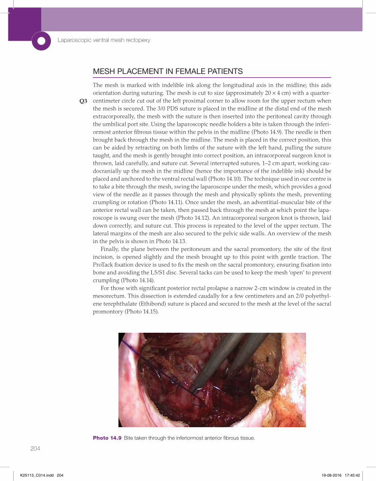
The mesh is marked with indelible ink along the longitudinal axis in the midline; this aids orientation during suturing. The mesh is cut to size (approximately 20 × 4 cm) with a quarter-centimeter circle cut out of the left proximal corner to allow room for the upper rectum when the mesh is secured. The 3/0 PDS suture is placed in the midline at the distal end of the mesh extracorporeally, the mesh with the suture is then inserted into the peritoneal cavity through the umbilical port site. Using the laparoscopic needle holders a bite is taken through the inferi-ormost anterior fibrous tissue within the pelvis in the midline (Photo 14.9). The needle is then brought back through the mesh in the midline. The mesh is placed in the correct position, this can be aided by retracting on both limbs of the suture with the left hand, pulling the suture taught, and the mesh is gently brought into correct position, an intracorporeal surgeon knot is thrown, laid carefully, and suture cut. Several interrupted sutures, 1–2 cm apart, working cau-docranially up the mesh in the midline (hence the importance of the indelible ink) should be placed and anchored to the ventral rectal wall (Photo 14.10). The technique used in our centre is to take a bite through the mesh, swing the laparoscope under the mesh, which provides a good view of the needle as it passes through the mesh and physically splints the mesh, preventing crumpling or rotation (Photo 14.11). Once under the mesh, an adventitial-muscular bite of the anterior rectal wall can be taken, then passed back through the mesh at which point the lapa-roscope is swung over the mesh (Photo 14.12). An intracorporeal surgeon knot is thrown, laid down correctly, and suture cut. This process is repeated to the level of the upper rectum. The lateral margins of the mesh are also secured to the pelvic side walls. An overview of the mesh in the pelvis is shown in Photo 14.13.
Finally, the plane between the peritoneum and the sacral promontory, the site of the first incision, is opened slightly and the mesh brought up to this point with gentle traction. The ProTack fixation device is used to fix the mesh on the sacral promontory, ensuring fixation into bone and avoiding the L5/S1 disc. Several tacks can be used to keep the mesh ‘open’ to prevent crumpling (Photo 14.14).
For those with significant posterior rectal prolapse a narrow 2-cm window is created in the mesorectum. This dissection is extended caudally for a few centimeters and an 2/0 polyethyl-ene terephthalate (Ethibond) suture is placed and secured to the mesh at the level of the sacral promontory (Photo 14.15).
Q3
Photo 14.9 Bite taken through the inferiormost anterior fibrous tissue.
K25113_C014.indd 204 19-08-2016 17:45:42

205
Mesh
Photo 14.12 Laparoscope over the mesh once a bite is taken through the anterior rectal wall and back through the mesh.
Photo 14.11 Laparoscope under the mesh splinting it and providing a good view of the needle.
Photo 14.10 Mesh secured to anterior rectum in pelvis.
K25113_C014.indd 205 19-08-2016 17:45:43

206
Laparoscopic ventral mesh rectopexy
Photo 14.15 Placement of suture.
Photo 14.13 Mesh secured in pelvis (light lead at 6 o’clock looking towards pelvic floor).
Photo 14.14 Mesh secured on sacral promontory with ProTack device.
K25113_C014.indd 206 19-08-2016 17:45:44

207
Post-operative considerations
MESH PLACEMENT IN MALE PATIENTS
Mesh placement is the same as described for females (Photo 14.16).
CLOSURE
A 3/0 V-Loc with a welded loop is inserted through the umbilical port into the pelvis. Bites are taken of the peritoneum and the mesh to cover the mesh with peritoneum and obliterate potential space between the two. In females bites can be taken through the posterior vaginal wall, peritoneum, mesh, and anterior rectal wall (Photo 14.17). This reconstitutes the rectovagi-nal septum, as the vaginal wall is secured to the mesh a posterior colporrhaphy is performed, a neo-pouch of Douglas is formed, which is much shallower and prevents enterocele. This continues caudocranially until, at the site of the first incision, the peritoneum is closed over the fixation tacks.
Photos 14.18 to 14.20 show the pelvis at the completion of the procedure.Haemostasis is ensured, the 5-mm ports are withdrawn under direct vision, then the umbil-
ical port and the pneumoperitoneum is deflated. The 10-mm port is closed in standard fashion using a 3/0 PDS on a ‘J’-shaped needle. Local anaesthetic (e.g. 0.5% bupivacaine) is applied to the wounds. Skin is closed with 3/0 polyglecaprone, and dressings or skin glue is applied. An examination under anaesthesia (EUA) should be performed to assess the rectopexy and if required a sutured mucopexy can be performed.
POST-OPERATIVE CONSIDERATIONS
The catheter is removed prior to the patient waking. A gauze tampon is placed in the vagina to provide support for the first 24 hours. Patients are managed with enhanced recovery prin-ciples with the aim of going home the day after surgery, although same-day discharge has been shown to be safe and feasible (32). The use of opiates should be discouraged due to their
Q4
Photo 14.16 Placement of mesh in a male.
K25113_C014.indd 207 19-08-2016 17:45:44

208
Laparoscopic ventral mesh rectopexy
Photo 14.17 V-Loc suture taken through peritoneum, posterior vaginal wall, mesh, and anterior rectal wall.
Photo 14.18 Female (with uterus) pelvis at end of procedure.
Photo 14.19 Female (post-hysterectomy) pelvis at end of procedure.
K25113_C014.indd 208 19-08-2016 17:45:45

209
Complications
significant gastrointestinal side effect profile, alternative analgesics such as Gabapentin should be considered. If the patient has significant chronic pelvic pain a 24-hour ketamine infusion should be considered. Patients should be followed up long term with the completion of vali-dated symptom and quality-of-life questionnaires.
COMPLICATIONS
Complications arise during all forms of surgical intervention. The most common complication is urinary retention post procedure (17). Specific complications to be mindful of include haem-orrhage from the posterior vaginal wall, perforation of the rectum or vagina, urinary reten-tion, worsening of symptoms or non-resolution of symptoms, and sexual dysfunction. Incorrect placement of the ProTack into the L5/S1 disc can result in spondylodiscitis (33). Mesh-related complications are a reality and include erosion 2%–3% through vagina, rectum, or bladder, infection, stricture of the rectum, and technical failure (i.e. incorrect placement or securing) (34). However, these patients do respond to revisional surgery carried out by an experienced operator with sustained functional outcomes (35).
Intra-operative perforation may occur. If this is in the vagina this can be repaired and the operation continued, if the rectum is perforated this will require immediate repair and defunc-tioning, enterotomies will require repair. In our experience it is rare for patients to develop adhesional obstruction, although there is a risk with fixation tacks that bowel may become caught resulting in obstructive symptoms. Many patients will develop a systemic response to the surgery and mesh, resulting in pyrexia; they will of course secrete serous fluid in reaction to the mesh and hence any imaging of these patients will invariably show fluid collection at the level of the mesh, which may be misinterpreted as an intra-abdominal source of sepsis. If mesh is infected this can be treated with antibiotics and the mesh allowed to fistulate through the vagina or rectum where it can be retrieved in a safer fashion via an EUA rather than a transab-dominal approach.
Photo 14.20 Male pelvis at end of procedure.
K25113_C014.indd 209 19-08-2016 17:45:45

210
Laparoscopic ventral mesh rectopexy
TAKE-HOME MESSAGE
Laparoscopic ventral mesh rectopexy requires modular training and a multidisciplinary approach.
APPENDIX 14.1: PELVIC FLOOR SOCIETY PUBLISHED PROCEDURE-BASED ASSESSMENT FOR LAPAROSCOPIC VENTRAL MESH RECTOPEXY (1)
KEY POINTS
▪ Thorough clinical assessment is required with sound knowledge and understanding. ▪ PFDs are complex, and patients should be referred to and discussed at pelvic floor MDTs. ▪ LVMR addresses the underlying pathophysiological and anatomical abnormalities. ▪ LVMR has a proven safety and efficacy profile. ▪ LVMR is effective for ERP and IRP causing FI or ODS. ▪ Surgery is technically challenging with a prolonged learning curve. ▪ Using a modular approach with a mentor is advised. ▪ Take time to ensure the operation is done correctly. ▪ Mesh-related complications happen, but they do respond well to revisional surgery by an
experienced operator.
SUMMARY
Laparoscopic ventral mesh rectopexy is a safe and effective operation for external rectal prolapse or internal rectal prolapse with ODS, FI, complex cystocele or enterocele, and SRUS. This is a techni-cally demanding operation, and surgeons need to develop skills through mentorship, fellowships, and lab-based sessions outside the operating theatre. Using a standardised modular approach can help surgeons through the learning curve (36).
Procedure-based assessment: Laparoscopic ventral mesh rectopexy
I. Consent Rating Comment
C1 Demonstrates sound knowledge of indications and
contraindications including alternatives to surgery.
C2 Demonstrates awareness of different surgical approaches and
advantages and disadvantages.
C3 Demonstrates sound knowledge of complications of surgery
and rates of recurrence.
C4 Explains the procedure to the patient/relatives/carers and
checks understanding.
C5 Explains likely outcome and time to recovery and checks
understanding.
K25113_C014.indd 210 19-08-2016 17:45:45

211
Appendix 14.1
II. Preoperative planning Rating Comment
PL1 Demonstrates recognition of anatomical and pathological
abnormalities, and their likely clinical relevance, and
selects appropriate operative strategies/techniques to deal
with these.
PL2 Demonstrates ability to make reasoned choices of appropriate
equipment, materials, or devices (e.g. mesh) taking into
account appropriate investigations (e.g. proctograms).
PL3 Checks materials, equipment, and device requirements with
operating staff.
PL4 Checks operative records; personally reviews investigations.
III. Preoperative planning Rating Comment
PR1 Checks in theatre that consent has been obtained.
PR2 Gives effective briefing to theatre team.
PR3 Ensures proper and safe positioning of the patient on the
operating table.
PR4 Demonstrates careful skin preparation.
PR5 Demonstrates careful draping of the patient’s operative field.
PR6 Ensures general equipment and materials are deployed safely
(e.g. diathermy).
PR7 Ensures appropriate drugs are administered.
IV. Exposure and closure Rating Comment
E1 Demonstrates knowledge of optimum skin incision/portal/
access.
E2 Achieves an adequate exposure through purposeful dissection
in correct tissue planes and identifies all structures correctly.
E3 Uses graspers, diathermy, and other energy devices so as to
minimise the risk of iatrogenic injury.
E4 Completes a sound wound repair.
E5 Protects the wound with dressings or glue.
V. Intra-operative technique: global (G) and task-specific items (T) Rating Comment
IT1 (G) Follows an agreed-on, logical sequence or protocol for the
procedure.
IT2 (G) Consistently handles tissue well with minimal damage.
IT3 (G) Controls bleeding promptly by an appropriate method.
IT4 (G) Demonstrates a sound technique of knots and sutures.
IT5 (G) Uses instruments appropriately and safely.
IT6 (G) Proceeds at appropriate pace with economy of movement.
IT7 (G) Anticipates and responds appropriately to variation, for
example, anatomy.
IT8 (G) Deals calmly and effectively with unexpected events/
complications.
IT9 (G) Uses assistant(s) to the best advantage at all times.
IT10 (G) Communicates clearly and consistently with scrub team.
IT11 (G) Communicates clearly and consistently with the anaesthetist.
Q5
K25113_C014.indd 211 19-08-2016 17:45:45

212
Laparoscopic ventral mesh rectopexy
REFERENCES
1. The Pelvic Floor Society. 2014. Procedure Based Assessment—Laparoscopic Ventral Mesh Rectopexy. [Online]. [Accessed 10 January 2016]. Available at: http://thepelvicfloorsoci-ety.co.uk/pages.php?t=Training-&-Education&s=Training-and-Education&id=102
2. Moore KL, Dalley AF. 2006. Clinically Oriented Anatomy. 5th ed. Baltimore: Lippincott Williams and Wilkins.
3. Bordeianou L, Hicks CW, Kaiser AM, Alavi K, Sudan R, Wise PE. Rectal prolapse: An overview of clinical features, diagnosis, and patient-specific management strategies. J Gastrointest Surg 18(5):1059–69.
4. Ellis H, Mahadevan V. Clinical Anatomy: Applied Anatomy for Students and Junior Doctors. 12th ed. Chichester: Wiley-Blackwell, 2010.
5. Lammers K, Lince SL, Spath MA, van Kempen LC, Hendriks JC, Vierhout ME, Kluivers KB, 2012. Pelvic organ prolapse and collagen-associated disorders. Int Urogynecol J 2014;23(3):313–9.
6. Lin S, Tee Y, Ng S, Chang H, Lin P, Chen G. 2007. Changes in the extracellular matrix in the anterior vagina of women with or without prolapse. Int Urogynecol J 18(1):43–8.
7. Bai SW, Choe BH, Kim JY, Park KH. 2002. Pelvic organ prolapse and connective tissue abnormalities in Korean women. J Reprod Med 47(3):231–4.
8. Dixon AR. 2010. Pathophysiological approach to obstruction defecation. In I. Lindsey, K. Nugent, A.R. Dixon (Eds.) Pelvic Floor Disorders for the Colorectal Surgeon. Oxford: Oxford University Press, pp. 57–68.
V. Intra-operative technique: global (G) and task-specific items (T) Rating Comment
IT12 (T) Safely obtains pneumoperitoneum and places ports in suitable
positions.
IT13 (T) Ensures small bowel is safely delivered out of the pelvis, and
the rectum is clearly exposed.
IT14 (T) Performs dissection of the lateral pelvic peritoneum and
continues dissection anteriorly. Shows awareness of ureters,
pelvic nerves and vagina in females, seminal vesicles in males.
IT15 (T) Appropriately uses traction and maintains field of view to
facilitate dissection.
IT16 (T) Identifies the pelvic floor and performs insertion and suturing of
mesh.
IT17 (T) Safely secures mesh to sacral promontory.
IT18 (T) Safely performs peritoneal closure.
IT19 (T) Performs abdominal wall closure.
VI. Post-operative management Rating Comment
PM1 Ensures the patient is transferred safely from the operating
table to the bed.
PM2 Constructs a clear operation note.
PM3 Records clear and appropriate post-operative instruction.
PM4 Deals with specimens (if appropriate). Labels and orientates
specimens appropriately.
K25113_C014.indd 212 19-08-2016 17:45:45

213
References
9. Soderberg MW, Falconer C, Bystrom B, Malmstrom A, Ekman G. 2004. Young women with genital prolapse have a low collagen concentration. Acta Obstet Gynecol Scand 83(12):1193–8.
10. Jackson S, Avery NC, Tarlton JF, Eckford SD, Abrams P, Bailey AJ. 1996. Changes in metabolism of collagen in genitourinary prolapse. Lancet 347(9016):1658–61.
11. Moalli PA, Shand SH, Zyczynski HM, Gordy SC, Men LA. 2005. Remodelling of vaginal connective tissue in patients with prolapse. Obstet Gynecol 106(5 Pt 1):593–63.
12. Collinson R, Cunningham C, D’Costa H, Lindsey I. 2009. Rectal intussusception and unex-plained faecal incontinence: Findings of a proctographic study. Colorectal Dis 11(1):77–83.
13. Rickert A, Kienle P. Laparoscopic surgery for rectal prolapse and pelvic floor disorders. World J Gastrointest Endosc 2015;7(12):1045–54.
14. Marzetti E, Leeuwenburgh C. Skeletal muscle apoptosis, sarcopenia and frailty in old age. Exp Gerontol 2006;41(12):1234–8.
15. Sangsawang B. Risk factors for the development of stress urinary incontinence during pregnancy in primigravidae: A review of the literature. Eur J Obstet Gynecol Reprod Biol 2014;178:27–34.
16. Clark AL, Gregory T, Smith VJ, Edwards R. Epidemiological evaluation of reopera-tion for surgically treated pelvic organ prolapse and urinary incontinence. Am J Obstet Gynaecol 2003;189(5):1261–7.
17. Mercer-Jones MA, D’Hoore A, Dixon AR, Lehur P, Lindsey I, Mellgren A, Stevenson AR. Consensus on ventral rectopexy: Report of a panel of experts. Colorectal Dis 2014;16(2):82–8.
18. Hompes R, Jones OM, Cunningham C, Lindsey I. What causes chronic idiopathic peri-neal pain? Colorectal Dis 2011;13(9):1035–39.
19. The Pelvic Floor Society. 2014. QA and Standards. [Online]. [Accessed 10th January 2016]. Available at: http://thepelvicfloorsociety.co.uk/pages.php?t=QA-&-standards&s=QA-&-Governance&id=101
20. Tou S, Brown SR, Nelson RL. Surgery for complete (full-thickness) rectal pro-lapse in adults. The Cochrane Database of Systematic Reviews 2015, Issue 11. Art. No.: CD001758.
21. Senapati A, Gray RG, Middleton LJ, Harding J, Hills RK, Armitage NC, Buckley L, Northover JM. PROSPER Collaborative Group. PROSPER: A randomised comparison of surgical treatments for rectal prolapse. Colorectal Dis 2013;15(7):858–8.
22. Berman IR. Sutureless laparoscopic rectopexy for procidentia: Technique and implica-tions. Dis Colon Rectum 1992;35(7):689–93.
23. D’Hoore A, Cadoni R, Penninckx F. Long-term outcome of laparoscopic ventral recto-pexy for total rectal prolapse. Br J Surg 2004;91(11):1500–5.
24. Randall J, Smyth E, McCarthy K, Dixon AR. Outcome of laparoscopic ventral mesh rectopexy for external rectal prolapse. Colorectal Dis 2014;16(11):914–9.
25. Collinson R, Wijffels N, Cunningham C, Lindsey I. Laparoscopic ventral rectopexy for internal rectal prolapse: Short-term functional results. Colorectal Dis 2010;12(2):97–104.
26. Badrek-Amoudi AH, Roe T, Mabey K, Carter H, Mills A, Dixon AR. Laparoscopic ven-tral mesh rectopexy in the management of solitary rectal ulcer syndrome: A cause for optimism? Colorectal Dis 2013;15(5):575–81.
27. Slawik S, Soulsby R, Carter H, Payne H, Dixon AR. Laparoscopic ventral rectopexy, posterior colporrhaphy and vaginal sacrocolpopexy for the treatment of recto-genital prolapse and mechanical outlet obstruction. Colorectal Dis 2008;10(2):138–43.
K25113_C014.indd 213 19-08-2016 17:45:45

214
Laparoscopic ventral mesh rectopexy
28. Wijffels N, Cunningham C, Dixon A, Greenslade G, Lindsey I. Laparoscopic ventral rectopexy for external rectal prolapse is safe and effective in the elderly. Does this make perineal procedures obsolete? Colorectal Dis 2011;13(5):561–6.
29. Owais AE, Sumrien H, Mabey K, McCarthy K, Greenslade GL, Dixon AR. Laparoscopic ventral mesh rectopexy in male patients with internal or external rectal prolapse. Colorectal Dis 2014;16(12):995–1000.
30. Mackenzie H, Dixon AR. Proficiency gain curve and predictors of outcome for laparo-scopic ventral mesh rectopexy. Surgery 2014;156(1):158–67.
31. Miskovic D, Ni M, Wyles SM, Tekkis P, Hanna GB. Learning curve and case selection in laparoscopic colorectal surgery: Systematic review and international multicenter analy-sis of 4852 cases. Dis Colon Rectum 2012;55(12):1300–10.
32. Powar MP, Ogilvie JW Jr, Stevenson AR 2013. Day-case laparoscopic ventral rectopexy: An achievable reality. Colorectal Dis 15(6):700–6.
33. Propst K, Tunitsky-Bitton E, Schimpf MO, Ridgeway B. 2014. Pyogenic spondylodiscitis associated with sacral colpopexy and rectopexy: Report of two cases and evaluation of the literature. Int Urogynecol J 2014;25(1):21–31.
34. Evans C, Stevenson AR, Sileri P, Mercer-Jones MA, Dixon AR, Cunningham C, Jones OM, Lindsey I, 2015. A multicenter collaboration to assess the safety of laparoscopic ventral rectopexy. Dis Colon Rectum 58(8):799–807.
35. Badrek-Al Amoudi AH, Greenslade GL, Dixon AR. 2013. How to deal with complica-tions after laparoscopic ventral mesh rectopexy: Lessons learnt from a tertiary referral centre. Colorectal Dis 2013;15(6):707–12.
36. Hemandas A, Flashman KG, Farrow J, O’Leary DP, Parvaiz A. 2011. Modular training in laparoscopic colorectal surgery maximizes training opportunities without clinical compromise. World J Surg 2011;35(2):409–14.
K25113_C014.indd 214 19-08-2016 17:45:45

Cat#: K25113 Chapter: 014
TO: CORRESPONDING AUTHOR
AUTHOR QUERIES – TO BE ANSWERED BY THE AUTHOR
The following queries have arisen during the typesetting of your manuscript. Please answer these queries by marking the required corrections at the appropriate point in the
text.
Query No.
Query Response
Q1 Please confirm first author’s name as “Peter Alexander Newman” (in provided TOC it is “Peter Newman”)
Q2 We have renumbered the references in sequenctial order both in text and reference list. Please check.
Q3 Circle size measurement OK? Q4 Please confirm EUA is spelled out correctly Q5 Please check shorted running head is okay here.