14 cerebellopontine angle masses on computed tomography
TRANSCRIPT

14 Cerebellopontine Angle Masses on Computed
Tomography

CLINICAL IMAGAGINGAN ATLAS OF DIFFERENTIAL DAIGNOSIS
EISENBERG
DR. Muhammad Bin Zulfiqar PGR-FCPS III SIMS/SHL
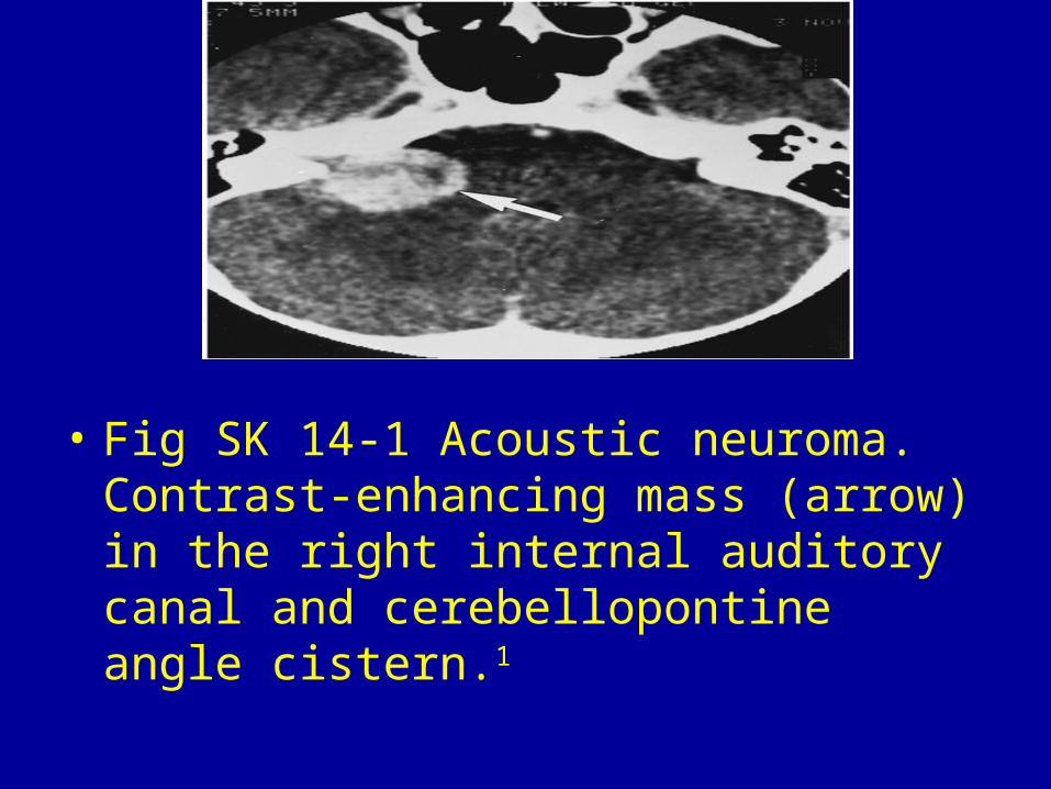
• Fig SK 14-1 Acoustic neuroma. Contrast-enhancing mass (arrow) in the right internal auditory canal and cerebellopontine angle cistern.1

• Fig SK 14-2 Neurofibromatosis. Bilateral acoustic neuromas (A) in a young girl with progressive bilateral sensorineural hearing loss.

• Fig SK 14-3 Intracanalicular acoustic neuroma. Air injected into the subarachnoid space shows the cerebellopontine angle cistern (open arrows) and outlines the small tumor (arrowheads).

• Fig SK 14-4 Meningioma. Dense enhancing lesion (arrows) that is more broadly based along the petrous bone than a typical acoustic neuroma.
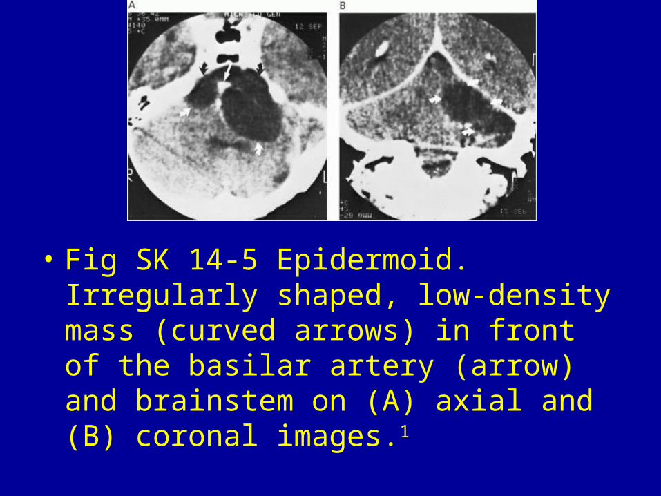
• Fig SK 14-5 Epidermoid. Irregularly shaped, low-density mass (curved arrows) in front of the basilar artery (arrow) and brainstem on (A) axial and (B) coronal images.1
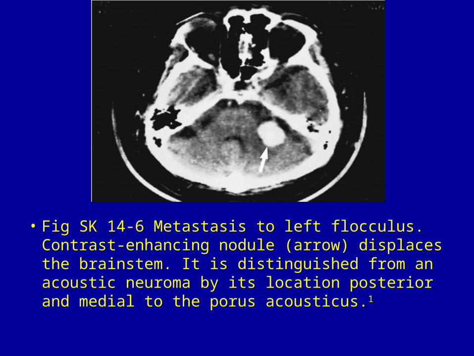
• Fig SK 14-6 Metastasis to left flocculus. Contrast-enhancing nodule (arrow) displaces the brainstem. It is distinguished from an acoustic neuroma by its location posterior and medial to the porus acousticus.1

• Fig SK 14-7 Arachnoid cyst. Slightly irregular cystic mass (arrow) of cerebrospinal fluid density that displaces the brainstem and basilar artery to the right.1
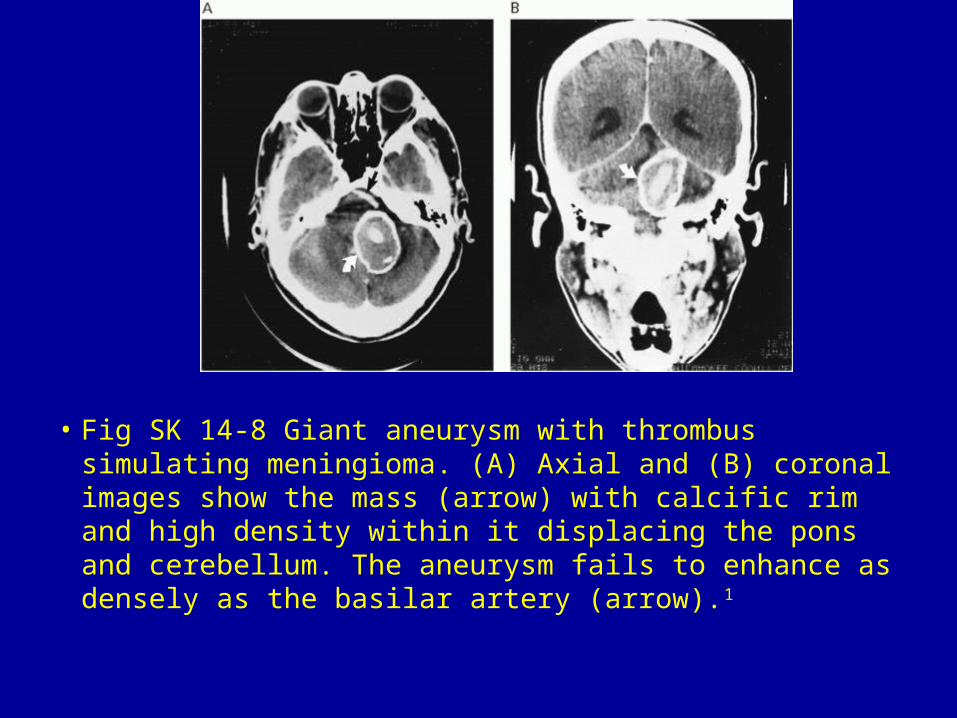
• Fig SK 14-8 Giant aneurysm with thrombus simulating meningioma. (A) Axial and (B) coronal images show the mass (arrow) with calcific rim and high density within it displacing the pons and cerebellum. The aneurysm fails to enhance as densely as the basilar artery (arrow).1

• Fig SK 14-9 Glomus jugulare tumor. Densely enhancing mass (arrow) that has eroded the osseous margins adjacent to the right jugular foramen.1

