10e12z cla alters adipocyte differentiation and adipocyte cytokine expression and induces macrophage...
TRANSCRIPT
Available online at www.sciencedirect.com
Journal of Nutritional Biochemistry 23 (2012) 510–518
10E12Z CLA alters adipocyte differentiation and adipocyte cytokine expressionand induces macrophage proliferation☆
Benjamin J. Belda1, Jerry T. Thompson, Pinar O. Eser, John P. Vanden Heuvel⁎
The Department of Veterinary and Biomedical Sciences, The Pennsylvania State University, University Park, PA 16802, USA
Received 12 November 2010; received in revised form 16 February 2011; accepted 17 February 2011
Abstract
The trans-10, cis-12 (10e12z) conjugated linoleic acid (CLA) isomer of CLA is responsible for loss of lipid storage or adipose tissue in vitro or in vivo. Thisisomer also induces inflammatory signaling in both mouse and human adipocytes in vitro. However, when these events occur and whether they are significantenough to affect other cell types are unclear. In these experiments, the 3T3-L1 cell line has been used to examine the interaction between inflammatory signalingand decreased differentiation or lipid storage induced by 10e12z CLA. In assays measuring both lipid accumulation and gene expression, differentiating 3T3-L1cells exhibit concurrent induction of inflammatory signaling, as measured by cyclooxygenase-2 expression, and a decrease in adipocyte marker gene expression.Furthermore, in fully differentiated adipocytes, as identified in microarray assays and confirmed with real-time polymerase chain reaction, 10e12z CLA alsosignificantly affected expression of both matrix metalloprotein-3 (MMP-3), collagen VI α 3 ColVI alpha 3 (VIα3) and the cytokine epiregulin, demonstrating thatthe effects of 10e12z broadly impact adipocyte function. In agreement with other experimental systems, 10e12z CLA inhibited RAW 264.7 cell proliferation;however, in response to adipocyte-conditioned media, 10e12z-CLA-treated adipocytes induced proliferation of this cell line, suggesting that the effect of 10e12zCLA is context dependent. These results are largely consistent with the known activation of the inflammatory mediator nuclear factor-κB in adipocytes in vitroand in vivo by 10e12z CLA treatment and demonstrate that adipose is an important target tissue of this isomer that impacts other cell types.© 2012 Elsevier Inc. All rights reserved.
Keywords: Inflammation; 3T3-L1 adipocytes; Paracrine effects; Extracellular matrix; Cyclooxygenase
1. Introduction
The adipocyte plays an integral role in energy homeostasis of thewhole organism. Adipocytes are important in regulating energymetabolism through the release of a broad array of endocrine-actingfactors [1]. A prominent example of this ability is the secretion ofadiponectin, which modulates energy utilization in distant tissuesincluding liver and muscle [2–4]. Adipocytes also contribute to thedetrimental symptoms of metabolic syndrome and diabetes as theyare competent to initiate inflammatory signaling leading to macro-phage infiltration and obesity-associated inflammation and insulinresistance [1,5]. Similar to leukocyte-induced inflammatory signaling,adipose-inflammatory signaling through cytokines such as tumornecrosis factor (TNF)-α, interleukin (IL)-6 and monocyte chemoat-tractant protein (MCP)-1 results in increased macrophage infiltrationthat further enhances cytokine and chemokine production [6,7] and
☆ This work was funded by the Department of Defense Breast CancerResearch Program Predoctoral Award to B.J.B. (Number W81XWH-06-1-0773).
⁎ Corresponding author.E-mail address: [email protected] (J.P. Vanden Heuvel).1 Current address: Western Human Nutrition Reserch Center, Obesity
andMetabolism Unit, USDA. 430West Health Sciences Drive, Davis CA 95616
0955-2863/$ - see front matter © 2012 Elsevier Inc. All rights reserved.doi:10.1016/j.jnutbio.2011.02.009
.
plays a specific role in increased insulin resistance [8]. Maintainingproper adipocyte function is an important health goal, and dietary andenvironmental chemicals or diseases that reduce adipocyte functionare of great concern [9,10].
Conjugated linoleic acid (CLA) isomers have gained notoriety fortheir anti-inflammatory actions in multiple model systems andantiproliferative effects in cancer cell lines [11–13]. Conjugatedlinoleic acid isomers exhibit a variety of anti-inflammatory mecha-nisms in vitro including inhibition of nuclear factor (NF)-κBtranscriptional activity in several cell lines [14–16]. The trans-10,cis-12 (10e12z) CLA isomer exhibits anti-inflammatory effects inmacrophage cell lines, while in fully differentiated adipocytes, thisisomer decreases lipid storage and increases insulin resistance [14].These effects are also seen in mice and humans, especially inoverweight or obese subjects [17]. Mice fed 10e12z CLA develophepatic steatosis and increase adipose expression of inflammatorymarkers such as IL-6 and IL-8 as well as decrease expression ofadiponectin [18–20], suggesting that the effects of 10e12z CLA onadipose are directly opposed to their effects in other tissues [14].In vitro studies illustrate that 10e12z CLA reduces insulin sensitivity ofadipocytes and reduces glucose uptake while increasing rates oflipolysis [21]. These events lead to an increase in circulating glucoseand lipotoxicity in mice and humans [18,20,22] and have beenassociated with increased NF-κB activity and cyclooxygenase (COX)-2

511B.J. Belda et al. / Journal of Nutritional Biochemistry 23 (2012) 510–518
expression in fully differentiated human adipocytes [23–25]. Thesecell-specific effects are especially significant given that the effects onadipose are the most consistent effect of 10e12z CLA feeding in mice.Although decreased adipocyte differentiation occurs in adipocytestreated with 10e12z CLA [21,26,27] and has been proposed to bepartially responsible for the decrease in body fat caused by 10e12zCLA [28], no specific inflammatory involvement has been demon-strated in differentiating adipocytes.
In these experiments, the effect of 10e12z CLA on differentiatingand fully differentiated 3T3-L1 cells was studied. 10e12z CLAdecreased the extent of 3T3-L1 adipocyte differentiation whentreated throughout the differentiation period. These changes becameevident only midway through the differentiation protocol andoccurred concurrent with increased COX-2 expression, a marker ofNF-κB activity. Analysis of gene expression of fully differentiated cellsdemonstrated multiple changes in adipocyte function and substan-tiated the involvement of NF-κB in altering adipocyte function inresponse to 10e12z CLA. Finally, the response of 10e12z-CLA-treatedadipocytes was sufficient to impact other cell types as conditionedmedia from 10e12z-CLA-treated adipocytes affected proliferation ofthe macrophage cell line RAW264.7, suggesting that increasedmacrophage numbers described in vivo [20] are due to theadipocyte-specific effect of this CLA isomer.
2. Materials and methods
2.1. Cells and cell culture 3T3-L1
Cells were purchased from ATCC (Manassas, VA, USA) and grown in high-glucoseDulbecco's modified Eagle's medium (HGDMEM; Sigma, Saint Louis, MO, USA)supplemented with 10% calf serum (Hyclone, Logan, UT, USA) and 100 U of penicillinand 100 μg/ml of streptomycin (Invitrogen). 3T3-L1 fibroblasts were differentiated byallowing the cells to grow to confluency (Fig. 1). Two days after confluency, the cellswere initiated to differentiate by changing the serum source to fetal bovine serum(FBS) and supplementing with 1 μg/ml insulin, 1 μM dexamethasone and 100 μM 3-isobutyl-1-methylxanthine (IBMX; MDI) for 2 days. Cells were either treated 2 dayslater with test compounds in HGDMEM containing 10% FBS plus 1 μg/ml insulinduring differentiation experiments or were refed every 2 days with HGDMEM with 1μg/ml of insulin for 10 days for experiments with fully differentiated adipocytes. After10 days, BSA-treated controls all contained large lipid droplets and numerous smalllipid droplets. Therefore, 10 days was considered full differentiation. In someinstances, the BSA controls did not differentiate fully within the 10-day period; inthese cases, the cells were not analyzed. Other chemicals used included rosiglitazone(rosi; Cayman Chemical), cis-9, trans-11 (9z11e) CLA and 10e12z CLA (Matreya,Pleasant Gap, PA, USA). Linoleic acid (LA) and all other chemicals were from Sigma(Saint Louis, MO, USA).
2.2. Fatty acid conjugates
10e12z CLA is nearly insoluble in aqueous solutions; therefore, all fatty acidswere conjugated to fatty-acid-free BSA. This was accomplished following establishedprotocols [29], maintaining a molar ratio of 4:1 (fatty acid:BSA). Fatty acid stocksand conjugates were stored at −20°C under argon. The resulting conjugates weretested for endotoxin contamination using the Limulus Amebocyte Lysate assay (LAL,E-Toxate, Sigma). In all cases, the conjugates contained b0.5 EU/ml endotoxin.
Fig. 1. Differentiation protocol for 3T3-L1 adipocytes used in this study. Timeline of differentiasection. CS, calf serum; MDI, IBMX, dexamethasone and insulin; Exp day; experimental day;
2.3. Quantitative oil red O staining
3T3-L1 cells were differentiated in 24-well plates as described above. After theappropriate treatment period, the cells were washed twice in phosphate-bufferedsaline. The cells were fixed, stained and extracted, and neutral lipid was quantifiedspectrophotometrically at 510 nm as previously described [30].
2.4. RNA analysis
Cells were lysed in Tri-reagent (Sigma), and total RNA was extracted followingthe manufacturer's protocol. One microgram of total RNA was reverse transcribedusing the High Capacity cDNA Archive Kit (ABI, Foster City, CA, USA). ComplementaryDNA was diluted 1:200, and real-time polymerase chain reaction (PCR) wasconducted with an ABI 7000 thermal cycler using Sybr Green PCR Master Mix(ABI). Gene specific primers were designed using Primer Express software (ABI) tospan introns where possible. Primer sequence information can be found in thesupplementary online materials. All messenger RNA (mRNA) expression data werecorrected for 18s ribosomal RNA (rRNA) expression prior to analysis. Data arepresented as the fold change relative to the single most appropriate treatment ortime point in the study.
2.5. Microarray hybridizations and analysis
The Mouse Genome Oligo Set, Version 2, was purchased from OperonTechnologies and printed onto amino-silane-treated slides using GeneMachinesOmnigrid (San Carlos, CA, USA) at the Penn State University DNA Microarray Facility.Total RNA from fully differentiated 3T3-L1 adipocytes treated with either 50 μM10e12z CLA or 50 μM LA was isolated by Tri-reagent (Sigma-Aldrich) according themanufacturer's instructions. Total RNA (1 μg) was amplified and labeled for twocolored arrays using the Amino Allyl MessageAmp II aRNA Amplification Kit (ABI) andthe Cye Dye Post Reactive Labeling Kit (GE Healthcare, Piscataway, NJ, USA) followingthe manufacturer's instructions. The arrays were then blocked, hybridized withamplified RNA and washed following previously reported protocols [31]. Arrays werescanned with a GenePix 4000A scanner (Axon Instruments Inc., Foster City, CA, USA),and image intensity information was collected with Genepix 3.0 software (AxonInstruments). Data from Genepix files were filtered to remove bad spots and spotsthat were not significantly different (P≤.001) from background. After filtering the dataset, ratio data from approximately 12,000 genes was collected and normalized usinglowess normalization and standard deviation regularization within a slide by Midas2.16 from the TM4 suite [32]. Significantly regulated genes were determined usingStatistical Analysis of Microarrays [33] with a median false discovery rate equal to13.5% Delta of 0.323 as implemented in TM4. Using this approach, 329 genes, or 3%,were found to be increased by 10e12z CLA, while less than 1%, or 58 genes, weredecreased by 10e12z CLA. These data sets were uploaded either together orindependently into The Database for Annotation, Visualization and IntegratedDiscovery (DAVID) [34] to interpret the resulting gene lists by functional annotation,with a P value ≤.05 considered significant.
2.6. Proliferation assays
Treatments were prepared in HGDMEM containing insulin and 0.1% heat-inactivated FBS. Media were conditioned in empty wells (unconditioned media;UCM) or in wells containing fully differentiated 3T3-L1 adipocytes (adipocyteconditioned media; ACM) for 26 h and stored at −80°C until the macrophages wereready to be treated. RNA was extracted from the differentiated adipocytes to validategene induction of inflammatory marker COX-2.
RAW264.7 macrophages were plated in clear 96-well plates at 10,000 cells perwell in HGDMEM containing 10% heat-inactivated FBS. Cells were allowed to recoverovernight; then serum was starved in media containing 0.1% heat-inactivated FBS for18 h prior to treatment. Adipocyte and unconditioned BSA, CLA and epiregulintreatments were randomized across the plate. A duplicate standard curve, treatedwith UCM containing BSA, was generated for quantification of the results.
tion used in these studies. The details are contained within the Materials and MethodsMCE, mitotic clonal expansion.
512 B.J. Belda et al. / Journal of Nutritional Biochemistry 23 (2012) 510–518
Proliferation was determined after 72 h. Media were removed from the wells byaspiration. CellTiter-Glo Cell Viability Assay reagent (Promega, Madison, WI, USA)was diluted 1:3 in Opti-MEM, and 45 μl of diluted reagent was added to each well.Contents of the wells were transferred to a white plate, and luminescencemeasurements were taken using a Tecan GENios Pro (Männedorf, Switzerland)plate reader.
2.7. Statistical analysis
Experiments were typically set up in a completely randomized or a randomizedcomplete block design. Experiments that used multiple treatments or time andtreatment conditions were analyzed as factorial design. All data were analyzed fornormalcy and homogeneity of variance prior to analysis of variance (ANOVA) using theGLM procedure in Minitab V. 14 (State College, PA, USA). Data that did not meet theassumptions of the ANOVA were transformed using the Box–Cox procedure asimplemented in Minitab. Treatment groups that differ are identified in graphs withdifferent letters or with an asterisk. Significant differences between multiple treatmentgroups were distinguished by Tukey's test with Pb.05 considered significant. All dataare represented in graphs as the mean (±S.E.M.).
3. Results
3.1. 10e12z CLA treatment during differentiation inhibits adipocytespecific gene expression and induces COX-2
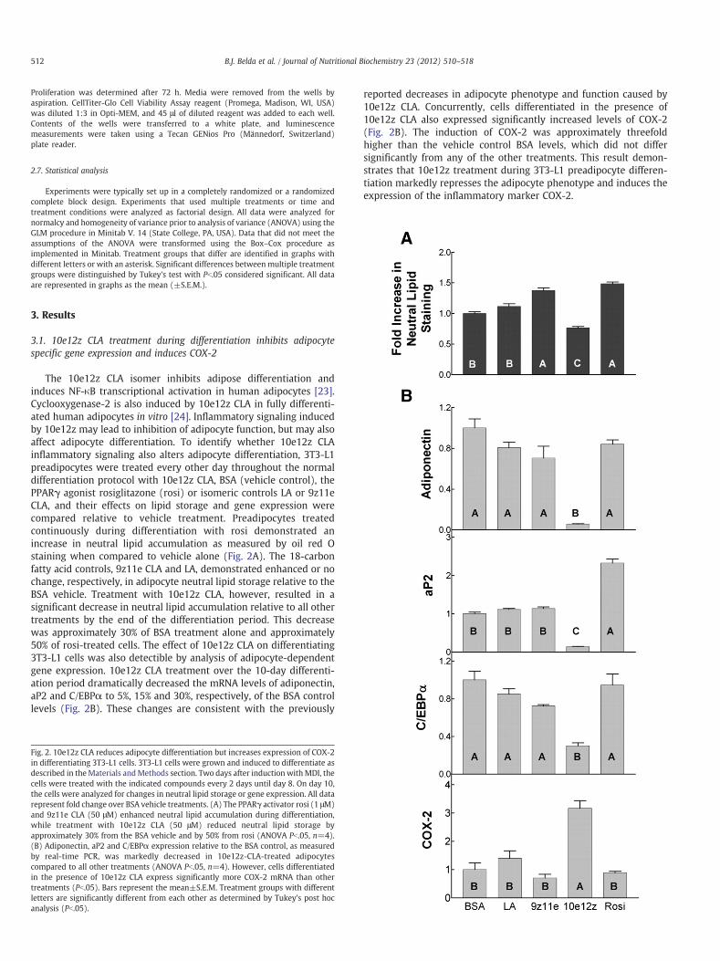
The 10e12z CLA isomer inhibits adipose differentiation andinduces NF-κB transcriptional activation in human adipocytes [23].Cyclooxygenase-2 is also induced by 10e12z CLA in fully differenti-ated human adipocytes in vitro [24]. Inflammatory signaling inducedby 10e12z may lead to inhibition of adipocyte function, but may alsoaffect adipocyte differentiation. To identify whether 10e12z CLAinflammatory signaling also alters adipocyte differentiation, 3T3-L1preadipocytes were treated every other day throughout the normaldifferentiation protocol with 10e12z CLA, BSA (vehicle control), thePPARγ agonist rosiglitazone (rosi) or isomeric controls LA or 9z11eCLA, and their effects on lipid storage and gene expression werecompared relative to vehicle treatment. Preadipocytes treatedcontinuously during differentiation with rosi demonstrated anincrease in neutral lipid accumulation as measured by oil red Ostaining when compared to vehicle alone (Fig. 2A). The 18-carbonfatty acid controls, 9z11e CLA and LA, demonstrated enhanced or nochange, respectively, in adipocyte neutral lipid storage relative to theBSA vehicle. Treatment with 10e12z CLA, however, resulted in asignificant decrease in neutral lipid accumulation relative to all othertreatments by the end of the differentiation period. This decreasewas approximately 30% of BSA treatment alone and approximately50% of rosi-treated cells. The effect of 10e12z CLA on differentiating3T3-L1 cells was also detectible by analysis of adipocyte-dependentgene expression. 10e12z CLA treatment over the 10-day differenti-ation period dramatically decreased the mRNA levels of adiponectin,aP2 and C/EBPα to 5%, 15% and 30%, respectively, of the BSA controllevels (Fig. 2B). These changes are consistent with the previously
Fig. 2. 10e12z CLA reduces adipocyte differentiation but increases expression of COX-2in differentiating 3T3-L1 cells. 3T3-L1 cells were grown and induced to differentiate asdescribed in theMaterials andMethods section. Two days after induction withMDI, thecells were treated with the indicated compounds every 2 days until day 8. On day 10,the cells were analyzed for changes in neutral lipid storage or gene expression. All datarepresent fold change over BSA vehicle treatments. (A) The PPARγ activator rosi (1 μM)and 9z11e CLA (50 μM) enhanced neutral lipid accumulation during differentiation,while treatment with 10e12z CLA (50 μM) reduced neutral lipid storage byapproximately 30% from the BSA vehicle and by 50% from rosi (ANOVA Pb.05, n=4).(B) Adiponectin, aP2 and C/EBPα expression relative to the BSA control, as measuredby real-time PCR, was markedly decreased in 10e12z-CLA-treated adipocytescompared to all other treatments (ANOVA Pb.05, n=4). However, cells differentiatedin the presence of 10e12z CLA express significantly more COX-2 mRNA than othertreatments (Pb.05). Bars represent the mean±S.E.M. Treatment groups with differentletters are significantly different from each other as determined by Tukey's post hocanalysis (Pb.05).
reported decreases in adipocyte phenotype and function caused by10e12z CLA. Concurrently, cells differentiated in the presence of10e12z CLA also expressed significantly increased levels of COX-2(Fig. 2B). The induction of COX-2 was approximately threefoldhigher than the vehicle control BSA levels, which did not differsignificantly from any of the other treatments. This result demon-strates that 10e12z treatment during 3T3-L1 preadipocyte differen-tiation markedly represses the adipocyte phenotype and induces theexpression of the inflammatory marker COX-2.

Fig. 3. Expression of COX-2 in differentiating adipocytes is concurrent with the onset ofdecreased adipocyte differentiation by 10e12z CLA. (A) 3T3-L1 fibroblasts wereinduced to differentiate and were treated on day 0 with (open symbols) or without 1μM rosiglitazone (closed symbols) plus 75 μM 10e12z CLA (circles), 75 μM 9z11e CLA(diamonds) or 18.75 μM BSA (squares) every 2 days. Rosi-treated cells storedsignificantly more lipid than did cells treated in the absence of rosi. On day 4, cellstreated without rosi did not differ from one another, nor did cells treated with rosi.However, when comparing between days 4 and 6, the cells treated with 10e12z CLA didnot increase lipid storage, whereas both BSA and 9z11e CLA treatments did,irrespective of the presence of rosi (ANOVA N=4, Pb.05). (B) Differentiating 3T3-L1cells were treated 50 μM10e12z CLA (circles) or 12.5 μMBSA (squares) every 2 days for10 days. On the days indicated, cells were harvested and mRNA was extracted.Expression of aP2mRNA (2B, upper panel) is significantly and rapidly increased by rosi,illustrating that PPAR γ activity is inducible as early as day 2. However, 10e12z CLAsuppresses the increase that occurs in the presence of BSA alone (N=4, Pb.05). In10e12z-CLA-treated cells, expression of COX-2 mRNA (2B, lower panel) is inducedapproximately sixfold from days 5–10 over BSA- or rosi-treated cells (N=4, Pb.05),concurrent with the onset of 10e12z CLA sensitivity as measured by lipid staining indifferentiating 3T3-L1 cells.
513B.J. Belda et al. / Journal of Nutritional Biochemistry 23 (2012) 510–518
3.2. CLA induction of COX-2 occurs concurrent with altereddifferentiation program
In human adipocytes, 10e12z CLA elicits an inflammatoryresponse concurrent with a decrease in adipocyte character, andinhibition of NF-κB activity can restore that function [23]. To addresswhether 10e12z CLA elicits the same response in differentiating 3T3-L1 cells, the timing of sensitivity to 10e12z CLA during differentiationwas examined by neutral lipid staining and gene expression analysis.Cells were treated with BSA, 9z11e or 10e12z CLA in the presence orabsence of rosi. After 4 days of treatment in the presence of the PPARγactivator, cells stored significantlymore lipid than those treated in theabsence of rosi, although no differences in lipid accumulation werefound between the fatty acid or vehicle treatments (Fig. 3A). By day 6,cells that were treated in the absence or presence of rosi with either9z11e CLA or BSA accumulated more neutral lipid as determined byoil red O staining. Cells treated with 10e12z CLA, however, did notincrease the amount of lipid accumulation, suggesting that thedifferentiating 3T3-L1 cells become sensitive to the presence of10e12z CLA around days 4–6 independent of the presence of thepotent PPARγ activator rosi.
The timing of the effect of 10e12z CLA on adipogenesis was alsoexamined via analysis of aP2 mRNA expression in differentiatingadipocytes. In the presence of rosi, expression of aP2 was induced 40-fold over the vehicle control as early as day 2 and continued toincrease to about 80-fold by day 10 (Fig. 3B, top panel). In cells treatedwith the BSA vehicle, aP2 expression remained low until betweendays 5 and 10, when it increased by 30-fold over BSA at day 2,reaching approximately 50% of the rosi-stimulated cells on day 10.aP2 expression in cells treated with 10e12z CLA was not induced atany time point. In contrast to the expression of aP2, COX-2 wassignificantly induced by 10e12z CLA by day 5 and was elevatedthrough day 10, an effect that was not seen in completelydifferentiated vehicle-treated controls (Fig. 3B) or in undifferentiated3T3-L1 cells (data not shown). Together, these results demonstratethat 3T3-L1 cells measurably convert to an adipocyte phenotypebetween days 4 and 6 as measured by neutral lipid accumulation andafter day 5 as measured by the expression of the PPARγ target geneaP2. In 3T3-L1 cells induced to differentiate but treated with 10e12z,these events are preceded by induction of inflammatory response asmeasured by COX-2 expression and as seen in completely differen-tiated adipocytes treated with 10e12z CLA, suggesting that a relatedmechanism may alter both the differentiation of adipocytes and thefunction of fully differentiated adipocytes [35].
3.3. Examination of pathways altered by CLA treatment of adipocytes:decreased lipid metabolism and increased cytokine signaling
Differentiating and fully differentiated adipocytes treated with10e12z CLA both exhibit an inflammatory profile that may beresponsible for the altered ability of these cells to differentiate orfunction normally. Therefore, global changes in gene expression of10e12z-CLA-treated fully differentiated adipocytes were examined bymicroarray to examine the extent of inflammatory phenotype in thesecells. Fully differentiated, day-10 adipocytes were treated for 12 hwith 50 μM 10e12z CLA or LA as a fatty acid control. Treatment with10e12z CLA significantly increased the expression of 329 genes andrepressed expression of 58 genes relative to LA treatment asdetermined by SAM as implemented in TMEV (SupplementaryTable 1). Consistent with a decrease in adipocyte function, the10e12z-CLA-repressed gene set was significantly overrepresented ingenes associated with lipid metabolism (Tables 1 and 2) includingPPARγ co-regulator-1α, in agreement with a decrease of adipocytefunction. Significantly repressed genes include C/EBPα and Spot 14,both of which are induced in the differentiating andmature adipocyte
[36,37]. Significant changes in expression of a subset of these geneswere validated by real-time PCR (Fig. 4). The 10e12z-CLA-inducedgene set was significantly overrepresented in genes associated withmitogen-activated protein kinase cascade, cytokine signaling and thetoll-like receptor (TLR) pathways, consistent with an inflammatorystate induced by 10e12z CLA (Table 1). This set of genes includesknown 10e12z CLA targets such as ATF3, MCP-1, NF-κB and IL-6(Table 2) [20,23,38,39].
Annotation analysis by DAVID indicated that a significant numberof genes involved in the TLR signaling pathway were induced. TLR4 is
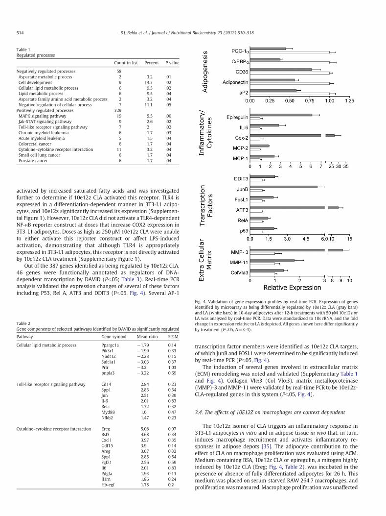
Table 1Regulated processes
Count in list Percent P value
Negatively regulated processes 58Aspartate metabolic process 2 3.2 .01Cell development 9 14.3 .02Cellular lipid metabolic process 6 9.5 .02Lipid metabolic process 6 9.5 .04Aspartate family amino acid metabolic process 2 3.2 .04Negative regulation of cellular process 7 11.1 .05Positively regulated processes 329MAPK signaling pathway 19 5.5 .00Jak-STAT signaling pathway 9 2.6 .02Toll-like receptor signaling pathway 7 2 .02Chronic myeloid leukemia 6 1.7 .03Acute myeloid leukemia 5 1.5 .04Colorectal cancer 6 1.7 .04Cytokine–cytokine receptor interaction 11 3.2 .04Small cell lung cancer 6 1.7 .04Prostate cancer 6 1.7 .04
514 B.J. Belda et al. / Journal of Nutritional Biochemistry 23 (2012) 510–518
activated by increased saturated fatty acids and was investigatedfurther to determine if 10e12z CLA activated this receptor. TLR4 isexpressed in a differentiation-dependent manner in 3T3-L1 adipo-cytes, and 10e12z significantly increased its expression (Supplemen-tal Figure 1). However, 10e12z CLA did not activate a TLR4-dependentNF-κB reporter construct at doses that increase COX2 expression in3T3-L1 adipocytes. Doses as high as 250 µM 10e12z CLA were unableto either activate this reporter construct or affect LPS-inducedactivation, demonstrating that although TLR4 is appropriatelyexpressed in 3T3-L1 adipocytes, this receptor is not directly activatedby 10e12z CLA treatment (Supplementary Figure 1).
Out of the 387 genes identified as being regulated by 10e12z CLA,46 genes were functionally annotated as regulators of DNA-dependent transcription by DAVID (Pb.05; Table 3). Real-time PCRanalysis validated the expression changes of several of these factorsincluding P53, Rel A, ATF3 and DDIT3 (Pb.05, Fig. 4). Several AP-1
Table 2Gene components of selected pathways identified by DAVID as significantly regulated
Pathway Gene symbol Mean ratio S.E.M.
Cellular lipid metabolic process Ppargc1a −1.79 0.14Pik3r1 −1.99 0.33Nudt12 −2.28 0.15Sult1a1 −3.03 0.37Prlr −3.2 1.03pnpla3 −3.22 0.69
Toll-like receptor signaling pathway Cd14 2.84 0.23Spp1 2.85 0.54Jun 2.51 0.39Il-6 2.01 0.83Rela 1.72 0.32Myd88 1.6 0.47Nfkb2 1.47 0.23
Cytokine–cytokine receptor interaction Ereg 5.08 0.97Bsf3 4.68 0.34Cxcl1 3.97 0.35Gdf15 3.9 0.14Areg 3.07 0.32Spp1 2.85 0.54Fgf21 2.56 0.59Il6 2.01 0.83Pdgfa 1.93 0.13Il1rn 1.86 0.24Hb-egf 1.78 0.2
Fig. 4. Validation of gene expression profiles by real-time PCR. Expression of genesidentified by microarray as being differentially regulated by 10e12z CLA (gray bars)and LA (white bars) in 10-day adipocytes after 12-h treatments with 50 μM 10e12z orLA was analyzed by real-time PCR. Data were standardized to 18s rRNA, and the foldchange in expression relative to LA is depicted. All genes shown here differ significantlyby treatment (Pb.05, N=3-4).
transcription factor members were identified as 10e12z CLA targets,of which JunB and FOSL1 were determined to be significantly inducedby real-time PCR (Pb.05, Fig. 4).
The induction of several genes involved in extracellular matrix(ECM) remodeling was noted and validated (Supplementary Table 1and Fig. 4). Collagen VIα3 (Col VIα3), matrix metalloproteinase(MMP)-3 andMMP-11were validated by real-time PCR to be 10e12z-CLA-regulated genes in this system (Pb.05, Fig. 4).
3.4. The effects of 10E12Z on macrophages are context dependent
The 10e12z isomer of CLA triggers an inflammatory response in3T3-L1 adipocytes in vitro and in adipose tissue in vivo that, in turn,induces macrophage recruitment and activates inflammatory re-sponses in adipose depots [35]. The adipocyte contribution to theeffect of CLA on macrophage proliferation was evaluated using ACM.Medium containing BSA, 10e12z CLA or epiregulin, a mitogen highlyinduced by 10e12z CLA (Ereg; Fig. 4, Table 2), was incubated in thepresence or absence of fully differentiated adipocytes for 26 h. Thismedium was placed on serum-starved RAW 264.7 macrophages, andproliferationwasmeasured. Macrophage proliferationwas unaffected
Table 3Transcription factors regulated by 10e12z CLA as identified by DAVID
Name Gene symbol GenBank accession Normalized ratio S.E.M.
Activating transcription factor 3 Atf3 U19118 4.16 0.24Aryl-hydrocarbon receptor Ahr – 3.24 0.13Basic helix–loop–helix domain containing, class B2 Bhlhb2 AF010305 1.59 0.28Bromodomain containing 2 Brd2 AF045462 1.55 0.25CCAAT/enhancer binding protein (C/EBP), alpha Cebpa BC011118 −2.45 0.61CCAAT/enhancer binding protein (C/EBP), delta Cebpd X61800 2.32 0.11E26 avian leukemia oncogene 2, 3′ domain Ets2 BC005486 2.07 0.39Early growth response 1 Egr1 M22326 3.41 0.08Early growth response 2 Egr2 X06746 2.41 0.18Ets variant gene 5 Etv5 AK003461 1.83 0.2Forkhead box C2 Foxc2 – 1.71 0.17GATA binding protein 2 Gata2 AB000096 1.63 0.19Heat shock factor 1 Hsf1 BC013716 1.42 0.26High mobility group AT-hook 1 Hmga1 J04179 3.25 0.4Inhibitor of DNA binding 3 Id3 M60523 2.15 0.03Interferon activated gene 203 Ifi203 AF022371 2.72 0.74Interferon activated gene 204 Ifi204 M31419 3.42 0.54Jun oncogene Jun J04115 2.51 0.39Jun proto-oncogene related gene d1 Jund1 J05205 2.08 0.07Kruppel-like factor 16 Klf16 AF283891 1.98 0.23Kruppel-like factor 2 (lung) Klf2 U25096 2.79 0.12Kruppel-like factor 4 (gut) Klf4 U70662 2.14 0.29Kruppel-like factor 5 Klf5 BC006646 2.27 0.6Kruppel-like factor 6 Klf6 AK053584 2.69 0.46Leucine-zipper-like transcriptional regulator, 1 Lztr1 AK083411 1.49 0.08LIM homeobox protein 6 Lhx6 AB031040 1.62 0.27Lin-9 homolog (C. elegans) Lin9 AK012271 −2.49 0.24MAD homolog 3 (Drosophila) Smad3 AK048626 1.62 0.4Myelocytomatosis oncogene Myc X01023 2.19 0.37MyoD family inhibitor domain containing Mdfic AK048821 1.46 0.15Nuclear factor of kappa light chain gene enhancer in B-cells inhibitor, beta Nfkbib U19799 1.64 0.43Nuclear factor of kappa light polypeptide gene enhancer in B-cells 2, p49/p100 Nfkb2 AF155373 1.47 0.23Nuclear transcription factor, X-box binding-like 1 Nfxl1 AK005913 1.77 0.64Peroxisome proliferative activated receptor, gamma, coactivator 1 beta Ppargc1b AF453324 −2.68 0.5Peroxisome proliferative activated receptor, gamma, coactivator-related 1 Pprc1 BC013720 1.82 0.21PWP1 homolog (S. cerevisiae) Pwp1 AK009972 1.93 0.68SERTA domain containing 2 Sertad2 AB041541 1.68 0.06Single stranded DNA binding protein 4 Ssbp4 AK004835 2.34 0.86SRY-box containing gene 11 Sox11 AK012306 2.19 0.53SRY-box containing gene 11 Sox11 AF009414 1.94 0.36SWI/SNF related, matrix associated, actin dependent regulator of chromatin,
subfamily a, member 5Smarca5 AF325921 1.54 0.41
TG interacting factor 1 Tgif1 X89749 4.69 0.52Thyroid hormone responsive SPOT14 homolog (Rattus) Thrsp BG244447 −3.41 0.43Transformation related protein 53 Trp53 BC005448 1.64 0.14V-maf musculoaponeurotic fibrosarcoma oncogene family, protein K (avian) Mafk BC014295 2.92 0.26V-rel reticuloendotheliosis viral oncogene homolog A (avian) Rela M61909 1.72 0.32Zinc finger protein 113 Zfp113 BC052453 2.87 0.39
515B.J. Belda et al. / Journal of Nutritional Biochemistry 23 (2012) 510–518
by BSA in either ACM or UCM, demonstrating that the ACM did notalter macrophage proliferation by itself (Fig. 5). 10e12z CLA in UCMreduced macrophage numbers relative to BSA by as much as 60% inUCM, while ACM generated from 10e12z-CLA-treated adipocytesstrongly stimulated proliferation by nearly 200% relative to BSA ACM,demonstrating that the effect of 10e12z CLA on macrophageproliferation is context dependent. The role of 10e12z-CLA-respon-sive epiregulin in relation to macrophage proliferation was alsoexamined. As with 10e12z CLA, epiregulin in UCM repressedmacrophage proliferation by approximately 30%, while ACM gener-ated from epiregulin-treated adipocytes stimulated macrophageproliferation by nearly 50%.
4. Discussion
In an isomer-specific manner, 10e12z CLA decreases adipocytefunction and adipocyte differentiation [21,26,27,39]. Inflammatorysignaling induced by 10e12z CLA activation of NF-κB is responsible forthis effect in fully differentiated adipocytes [23,25], although a similarmechanism has not been identified in differentiating adipocytes. In
differentiating adipocytes of either human or mouse origin, 10e12zCLA represses PPARγ-dependent gene transcription and TG accumu-lation. In isolated human stromal–vascular cells, inflammatorysignaling is mediated by adipocytes and potentiated by preadipocytes[25]. Consistent with these findings, we demonstrate here that 3T3-L1cells treated throughout differentiation exhibit decreased lipidstorage and adipocyte-specific gene expression consistent withdecreased PPARγ activity. Concurrently, these cells express increasedCOX-2 expression, consistent with the activation of NF-κB transcrip-tional activity.
As with human preadipocytes [25], 3T3-L1 fibroblasts do notrespond to 10e12z CLA with an increase in inflammatory markers asdetermined by microarray [40] or examination of COX-2 expression(data not shown). An examination of the window of sensitivity ofdifferentiating 3T3-L1 adipocytes demonstrated that treatment with10e12z CLA for at least 4 days after induction to an adipogenicprogram was required prior to the observation of any change in thedifferentiation program. The first identifiable alteration in thedifferentiation program was the measurement of COX-2 at day 5that occurred prior to measurable alterations in aP2 expression, a

Fig. 5. Adipocyte-dependent effects on macrophage proliferation in vitro. The effects of10e12z CLA on macrophages are context dependent and are altered by the activity ofadipocytes secreted molecules. Medium treated with BSA, 10e12z CLA or epiregulinwas conditioned in the presence of adipocytes or in empty wells. The conditionedmedia were administered to serum-starved RAW264.7 macrophages for 72 h. Theeffect of BSA-generated ACM was not different from UCM (PN.05). However the effectsof epiregulin- or 10e12z-CLA-generated ACM are opposite to the effects of thesetreatments in UCM. Treatment with 10e12z CLA and epiregulin unconditioned mediainhibits proliferation of RAW264.7 cells when compared to the BSA control (Pb.05).Conversely, 10e12z CLA and epiregulin act through 3T3-L1 adipocytes and promotemacrophage proliferation relative to BSA-generated ACM (Pb.05). The two compoundsresult in a similar pattern of induction of RAW264.7 cell proliferation in ACM,suggesting that induction of epiregulin is among the key effectors of the 10e12z-CLA-induced inflammatory response in adipocytes. Bars represent the mean (±S.E.M.) oftreatments. Bars with different letters are significantly different, Tukey's post hoc,Pb.05, N=6.
516 B.J. Belda et al. / Journal of Nutritional Biochemistry 23 (2012) 510–518
marker of PPARγ activity. The increased COX-2 expression was alsoconcurrent with increased lipid storage in control-treated adipocytes,suggesting, similar to the human model, that some differentiation ofthis cell culture model is required prior to sensitivity to this fatty acid.
Unlike other models of adipogenesis, 3T3-L1 cells can becompletely differentiated to a multilocular stage in culture withoutthe contaminating effect of other cell types [25], thus providing ahomogenous population of cells. Gene expression studies of the 3T3-L1 adipocyte's response to 10e12z CLA demonstrate impacts onmultiple functions, some of which have been corroborated by otherstudies. In epididymal adipose of obese M16 mice, a diet of 1%10e12z CLA for 14 days decreased lipid metabolism and adipocytedifferentiation, while increasing expression of apoptotic markerssuch as TNF-α and caspase-3 [41]. In C57BL/6J mice fed 0.5% 10e12zCLA, retroperitoneal (RP) adipose consistently decreases expressionof genes involved in lipogenesis and beta-oxidation between 1 and17 days, while mitochondrial uncoupling protein-1 and uncouplingprotein-2 increase between 7 and 17 days [42]. 10e12z CLA does notaffect feed intake, suggesting that increased energy expenditure wasresponsible for the adipocyte delipidation. Studies with 3T3-L1adipocytes extended these findings as they are consistent with bothCLA-fed C57BL/6J RP gene expression profiles as well as tunicamy-cin-induced endoplasmic reticulum stress [40]. More recently, theeffects of 10e12z CLA on gene expression have been likened to thoseof the antidiabetic compound metformin [43]; both decrease theexpression of genes involved in lipogenesis, and both expressionprofiles correlate with the UPR-inducing tunicamycin. The primarydistinction in these profiles, however, is the inflammatory responseinduced by 10e12z CLA. In the present studies, 10e12z CLA increasedexpression of inflammatory genes and decreased the expression ofgenes involved in lipogenesis and adipocyte differentiation. How-ever, these results also suggest a greater role for the transcriptionfactor NF-κB.
Nuclear factor-κB regulates many important cellular functionsincluding the inflammatory response and remodeling of the ECM
[44–46]. Adipocyte function and differentiation are also regulated bythe appropriate extracellular context [47,48]. Both MMP-3 andMMP-11 are involved in regulating adipose tissue development, asevidenced by the fact that adipose tissue in animals with either genedeleted experiences adipocyte hypertrophy [49,50]. Similarly, over-expression or addition of active MMP-11 to differentiated adipocytescan induce dedifferentiation [51]. Interestingly, collagen VIα3 isexpressed by adipocytes in vitro and in vivo, and adipocyte-specificexpression during mammary tumorigenesis leads to activation of β-catenin and other proliferative signals [52,53]. In addition, MMP-11is responsible for cleavage and activation of Col VIα3, suggestingthat the induction of these factors modulates the extracellularenvironment of adipose in concert [51] in response to 10e12z CLA.Along with the inflammatory cytokines regulated by NF-κB, thesefactors may also regulate adipose tissue function by altering the ECMmilieu. These facts, along with the expression of inflammatorysignals such as COX-2, suggest that the adipocyte function is directlyinhibited by the induction of inflammatory signals by 10e12z CLA.
The specific signaling pathway(s) that activates NF-κB in responseto 10e12z CLA is, as yet, undetermined. Multiple pathways areactivated in adipocytes by 10e12z CLA [39,54], and many of these arelikely activated secondarily through autocrine signaling mechanismfrom secreted cytokines such as IL-6, IL-8 and epiregulin in bothpreadipocytes and adipocytes [25,39]. These factors may also affectthe behavior of surrounding cell types. Macrophage numbers areincreased in adipose depots in mice fed 10e12z [20] either throughactivation and proliferation of adipose tissue macrophages or throughrecruitment of circulating monocytes into the adipose. Adipose-resident macrophages play a role in regulating insulin sensitivity andamplifying proinflammatory signaling of the adipocyte [8] and mayplay a role in the reduction of insulin sensitivity in response to 10e12zCLA in vivo. However, the effects of CLA differ distinctly in adipose andinflammatory models [14]. The anti-inflammatory properties of10e12z CLA are well studied in macrophage models [55,56]. Inhuman THP-1 macrophages, 10e12z CLA inhibits migration, COX-2activity and macrophage-induced monocyte migration [57]. There-fore, the effect of 10e12z-CLA-treated adipocytes on RAW264.7macrophage proliferation was examined in vitro. In this assay,unconditioned media containing 10e12z CLA repressed macrophageproliferation. When macrophages were treated with conditionedmedia from 10e12z-CLA-treated adipocytes, the cells increased theirproliferation rate, suggesting that the response of these macrophagesto 10e12z CLA is context dependent and that adipocytes provide theseproliferative cues. As determined by gene expression profiling, thereare many adipocyte-secreted factors that could be responsible for thiseffect. One of these factors is the potent mitogen epiregulin that wasamongst the most highly induced cytokines detected. When adipo-cytes were treated only with epiregulin, the effects on macrophageproliferation were reproduced, suggesting a role for this cytokine inthe macrophage response to 10e12z-CLA-treated adipocytes. Thiseffect may be indirect, eliciting the secretion of other adipokines in anautocrine manner, as unconditioned media containing epiregulin didnot elicit this effect but rather suppressed proliferation of RAW264.7cells. In addition to epiregulin, other effectors of macrophageproliferation secreted from 10e12z-CLA-treated adipocytes includecytokines and growth factors, including IL-6, IL-8 [39] and moleculesidentified in Table 2, lipid mediators such as COX-2 products [14,24],ECM fragments [52] and potentially fatty acids released due tolipolytic action [58] of 10e12z CLA. Some or all of these factors arecertain to be working in concert to induce macrophage proliferationto 10e12z-CLA-treated adipocytes.
The increased lean body mass spurred by CLA isomers isdependent upon the activity of the 10e12z CLA isomer [59]. Theability of 10e12z CLA to coordinately regulate the expression of ECMremodeling factors and inflammatory markers in adipose, along with

517B.J. Belda et al. / Journal of Nutritional Biochemistry 23 (2012) 510–518
a significant number of cytokines, kinases and transcription factors,illustrates that the decreased lipid storage in adipocytes is a complexphenomenon that has repercussions on the whole tissue. While thedecrease in adipocyte function is certainly, in part, due to modulationof PPARγ activity [60], our data suggest that this is an indirect orsecondary response to the induction of NF-κB and inflammatorysignaling. The complexity of the response of adipose to 10e12z CLAsuggests that the promising and sought-after effects of decreased lipidstorage are not likely to be separated from the negative effects ofincreased inflammatory signaling and decreased insulin sensitivity.As a tissue with specific impacts on multiple tissue types, theinflammatory effects incurred in adipose tissue in response to 10e12zCLA may readily be transmitted to other tissues that are not affecteddirectly by 10e12z CLA.
Supplementarymaterials related to this article can be found onlineat doi:10.1016/j.jnutbio.2011.02.009.
Acknowledgments
The authors would like to thank Dr. Craig Praul from the PennState Microarray Facility for assistance with completion of themicroarray hybridizations. Technical assistance was also receivedfrom Daniel B. Hannon, Tara Leas, Christine Rivera, Erin Shirk andLara Augulis.
References
[1] Wang P, Mariman E, Renes J, Keijer J. The secretory function of adipocytes in thephysiology of white adipose tissue. J Cell Physiol 2008;216:3–13.
[2] Berg AH, Combs TP, Du X, Brownlee M, Scherer PE. The adipocyte-secreted proteinacrp30 enhances hepatic insulin action. Nat Med 2001;7:947–53.
[3] Yamauchi T, Kamon J, Waki H, Murakami K, Motojima K, Komeda K, et al. Themechanisms by which both heterozygous peroxisome proliferator-activatedreceptor gamma (ppargamma) deficiency and ppargamma agonist improveinsulin resistance. J Biol Chem 2001;276:41245–54.
[4] Yamauchi T, Kamon J, Ito Y, Tsuchida A, Yokomizo T, Kita S, et al. Cloning ofadiponectin receptors that mediate antidiabetic metabolic effects. Nature2003;423:762–9.
[5] Fried SK, Bunkin DA, Greenberg AS. Omental and subcutaneous adipose tissues ofobese subjects release interleukin-6: depot difference and regulation byglucocorticoid. J Clin Endocrinol Metab 1998;83:847–50.
[6] Weisberg SP, McCann D, Desai M, Rosenbaum M, Leibel RL, Ferrante AW. Obesityis associated with macrophage accumulation in adipose tissue. J Clin Invest2003;112:1796–808.
[7] Chen Y, Zhu J, Lum PY, Yang X, Pinto S, MacNeil DJ, et al. Variations in DNAelucidate molecular networks that cause disease. Nature 2008;452:429–35.
[8] Patsouris D, Li PP, Thapar D, Chapman J, Olefsky JM, Neels JG. Ablation of cd11c-positive cells normalizes insulin sensitivity in obese insulin resistant animals. CellMetab 2008;8:301–9.
[9] Alexander DL, Ganem LG, Fernandez-Salguero P, Gonzalez F, Jefcoate CR. Aryl-hydrocarbon receptor is an inhibitory regulator of lipid synthesis and ofcommitment to adipogenesis. J Cell Sci 1998;111(Pt 22):3311–22.
[10] Villarroya F, Domingo P, Giralt M. Lipodystrophy associated with highly activeanti-retroviral therapy for HIV infection: the adipocyte as a target of anti-retroviral-induced mitochondrial toxicity. Trends Pharmacol Sci 2005;26:88–93.
[11] Miglietta A, Bozzo F, Gabriel L, Bocca C, Canuto RA. Extracellular signal-regulatedkinase 1\2 and protein phosphatase 2a are involved in the antiproliferativeactivity of conjugated linoleic acid in mcf-7 cells. Br J Nutr 2006;96:22–7.
[12] Kim EJ, Holthuizen PE, Park HS, Ha YL, Jung KC, Park JHY. Trans-10,cis-12-conjugated linoleic acid inhibits caco-2 colon cancer cell growth. Am J PhysiolGastrointest Liver Physiol 2002;283:G357–67.
[13] Ip MM, Masso-Welch PA, Shoemaker SF, Shea-Eaton WK, Ip C. Conjugated linoleicacid inhibits proliferation and induces apoptosis of normal rat mammaryepithelial cells in primary culture. Exp Cell Res 1999;250:22–34.
[14] Belda BJ, Lee Y, Vanden Heuvel JP. Conjugated linoleic acids and inflammation:isomer- and tissue-specific responses. Clin Lipidol 2010;5:699–717.
[15] Rahman MM, Bhattacharya A, Fernandes G. Conjugated linoleic acid inhibitsosteoclast differentiation of raw264.7 cells by modulating rankl signaling. J LipidRes 2006;47:1739–48.
[16] Ringseis R, Gahler S, Eder K. Conjugated linoleic acid isomers inhibit platelet-derived growth factor-induced nf-κb transactivation and collagen formation inhuman vascular smooth muscle cells. Eur J Nutr 2008;47:59–67.
[17] Silveira MB, Carraro R, Monereo S, Tebar J. Conjugated linoleic acid (cla) andobesity. Public Health Nutr 2007;10:1181–6.
[18] Clement L, Poirier H, Niot I, Bocher V, Guerre-Millo M, Krief S, et al. Dietary trans-10,cis-12 conjugated linoleic acid induces hyperinsulinemia and fatty liver in themouse. J Lipid Res 2002;43:1400–9.
[19] Tsuboyama-Kasaoka N, Takahashi M, Tanemura K, Kim HJ, Tange T, Okuyama H,et al. Conjugated linoleic acid supplementation reduces adipose tissue byapoptosis and develops lipodystrophy in mice. Diabetes 2000;49:1534–42.
[20] Poirier H, Shapiro JS, Kim RJ, Lazar MA. Nutritional supplementation with trans-10, cis-12-conjugated linoleic acid induces inflammation of white adipose tissue.Diabetes 2006;55:1634–41.
[21] Brown JM, Boysen MS, Jensen SS, Morrison RF, Storkson J, Lea-Currie R, et al.Isomer-specific regulation of metabolism and ppargamma signaling by cla inhuman preadipocytes. J Lipid Res 2003;44:1287–300.
[22] Ramos R, Mascarenhas J, Duarte P, Vicente C, Casteleiro C. Conjugated linoleicacid-induced toxic hepatitis: first case report. Dig Dis Sci 2009;54:1141–3.
[23] Chung S, Brown JM, Provo JN, Hopkins R, McIntosh MK. Conjugated linoleic acidpromotes human adipocyte insulin resistance through nf{kappa}b-dependentcytokine production. J Biol Chem 2005;280:38445–56.
[24] Kennedy A, Overman A, Lapoint K, Hopkins R,West T, Chuang CC, et al. Conjugatedlinoleic acid-mediated inflammation and insulin resistance in human adipocytesare attenuated by resveratrol. J Lipid Res 2009;50:225–32.
[25] Martinez K, Kennedy A,West T, Milatovic D, AschnerM,McIntoshM. Trans-10,cis-12-conjugated linoleic acid instigates inflammation in human adipocytescompared with preadipocytes. J Biol Chem 2010;285:17701–12.
[26] Evans M, Park Y, Pariza M, Curtis L, Kuebler B, McIntosh M. Trans-10,cis-12conjugated linoleic acid reduces triglyceride content while differentially affectingperoxisome proliferator activated receptor gamma2 and ap2 expression in 3t3-l1preadipocytes. Lipids 2001;36:1223–32.
[27] EvansM, Geigerman C, Cook J, Curtis L, Kuebler B, McIntoshM. Conjugated linoleicacid suppresses triglyceride accumulation and induces apoptosis in 3t3-l1preadipocytes. Lipids 2000;35:899–910.
[28] Simon E, Macarulla MT, Fernandez-Quintela A, Rodriguez VM, Portillo MP. Bodyfat-lowering effect of conjugated linoleic acid is not due to increased lipolysis.J Physiol Biochem 2005;61:363–9.
[29] Calder PC, Bond JA, Harvey DJ, Gordon S, Newsholme EA. Uptake andincorporation of saturated and unsaturated fatty acids into macrophage lipidsand their effect upon macrophage adhesion and phagocytosis. Biochem J1990;269:807–14.
[30] Ramírez-Zacarías JL, Castro-Muñozledo F, Kuri-Harcuch W. Quantitation ofadipose conversion and triglycerides by staining intracytoplasmic lipids with oilred o. Histochem Cell Biol 1992;97:493–7.
[31] Hegde P, Qi R, Abernathy K, Gay C, Dharap S, Gaspard R, et al., A concise guide tocdna microarray analysis. Biotechniques. 2000; 29:548–50, 52–4, 56 passim.
[32] Saeed AI, Sharov V, White J, Li J, Liang W, Bhagabati N, et al. Tm4: a free, open-source system for microarray data management and analysis. Biotechniques2003;34:374–8.
[33] Tusher VG, Tibshirani R, Chu G. Significance analysis of microarrays applied to theionizing radiation response. Proc Natl Acad Sci U S A 2001;98:5116–21.
[34] Dennis Jr G, Sherman BT, Hosack DA, Yang J, Gao W, Lane HC, et al. David:database for annotation, visualization, and integrated discovery. Genome Biol2003;4:P3.
[35] Belda BJ, Lee Y, Vanden Heuvel JP. Conjugated linoleic acids and inflammation:isomer and tissue specific responses. Clin Lipidol 2010;5:18.
[36] Lin FT, Lane MD. Ccaat/enhancer binding protein alpha is sufficient to initiate the3t3-l1 adipocyte differentiation program. Proc Natl Acad Sci U S A 1994;91:8757–61.
[37] Loos U, Clement J, Behr M, Fischer S. Expression of s14-mrna and itstranslational product in differentiating 3t3-l1 cells. Mol Cell Endocrinol1991;75:R7–11.
[38] Lee SH, Yamaguchi K, Kim JS, Eling TE, Safe S, Park Y, et al. Conjugated linoleic acidstimulates an anti-tumorigenic protein nag-1 in an isomer specific manner.Carcinogenesis 2006;27:972–81.
[39] Brown JM, Boysen MS, Chung S, Fabiyi O, Morrison RF, Mandrup S, et al.Conjugated linoleic acid induces human adipocyte delipidation: autocrine/para-crine regulation of mek/erk signaling by adipocytokines. J Biol Chem 2004;279:26735–47.
[40] LaRosa PC, Riethoven JJ, Chen H, Xia Y, Zhou Y, Chen M, et al. Trans-10, cis-12conjugated linoleic acid activates the integrated stress response pathway inadipocytes. Physiol Genomics 2007;31:544–53.
[41] House RL, Cassady JP, Eisen EJ, Eling TE, Collins JB, Grissom SF, et al. Functionalgenomic characterization of delipidation elicited by trans-10, cis-12-conjugatedlinoleic acid (t10c12-cla) in a polygenic obese line of mice. Physiol Genomics2005;21:351–61.
[42] LaRosa PC, Miner J, Xia Y, Zhou Y, Kachman S, Fromm ME. Trans-10, cis-12conjugated linoleic acid causes inflammation and delipidation of white adiposetissue in mice: a microarray and histological analysis. Physiol. Genomics 2006;27:282–94.
[43] Jiang S, Wang Z, Riethoven JJ, Xia Y, Miner J, Fromm M. Conjugated linoleic acidactivates amp-activated protein kinase and reduces adiposity more effectivelywhen used with metformin in mice. J Nutr 2009;139:2244–51.
[44] Ohkawa T, Ueki N, Taguchi T, Shindo Y, Adachi M, Amuro Y, et al. Stimulation ofhyaluronan synthesis by tumor necrosis factor-alpha is mediated by the p50/p65nf-kappa b complex in mrc-5 myofibroblasts. Biochim Biophys Acta 1999;1448:416–24.

518 B.J. Belda et al. / Journal of Nutritional Biochemistry 23 (2012) 510–518
[45] Chen A, Zheng S. Curcumin inhibits connective tissue growth factor geneexpression in activated hepatic stellate cells in vitro by blocking nf-kappab anderk signalling. Br J Pharmacol 2008;153:557–67.
[46] Karin M. Nuclear factor-kappab in cancer development and progression. Nature2006;441:431–6.
[47] Rodriguez Fernandez JL, Ben-Ze'ev A. Regulation of fibronectin, integrin andcytoskeleton expression in differentiating adipocytes: inhibition by extracellularmatrix and polylysine. Differentiation 1989;42:65–74.
[48] Liang X, Kanjanabuch T, Mao SL, Hao CM, Tang YW, Declerck PJ, et al. Plasminogenactivator inhibitor-1 modulates adipocyte differentiation. Am J Physiol EndocrinolMetab 2006;290:E103–13.
[49] Alexander CM, Selvarajan S, Mudgett J, Werb Z. Stromelysin-1 regulatesadipogenesis during mammary gland involution. J Cell Biol 2001;152:693–703.
[50] Lijnen HR, Van HB, Frederix L, Rio MC, Collen D. Adipocyte hypertrophy instromelysin-3 deficient mice with nutritionally induced obesity. Thromb Haemost2002;87:530–5.
[51] Motrescu ER, Blaise S, Etique N, Messaddeq N, Chenard MP, Stoll I, et al. Matrixmetalloproteinase-11/stromelysin-3 exhibits collagenolytic function againstcollagen vi under normal and malignant conditions. Oncogene 2008;27:6347–55.
[52] Iyengar P, Combs TP, Shah SJ, Gouon-Evans V, Pollard JW, Albanese C, et al.Adipocyte-secreted factors synergistically promote mammary tumorigenesisthrough induction of anti-apoptotic transcriptional programs and proto-onco-gene stabilization. Oncogene 2003;22:6408–23.
[53] Iyengar P, Espina V, Williams TW, Lin Y, Berry D, Jelicks LA, et al. Adipocyte-derived collagen vi affects early mammary tumor progression in vivo, demon-
strating a critical interaction in the tumor/stroma microenvironment. J Clin Invest2005;115:1163–76.
[54] Chung S, Brown JM, Sandberg MB, McIntosh M. Trans-10,cis-12 cla increasesadipocyte lipolysis and alters lipid droplet-associated proteins: role of mtor anderk signaling. J Lipid Res 2005;46:885–95.
[55] Yu Y, Correll PH, Vanden Heuvel JP. Conjugated linoleic acid decreases productionof pro-inflammatory products in macrophages: evidence for a ppar gamma-dependent mechanism. Biochim Biophys Acta 2002;1581:89–99.
[56] Loscher CE, Draper E, Leavy O, Kelleher D, Mills KH, Roche HM. Conjugatedlinoleic acid suppresses nf-{kappa}b activation and il-12 production indendritic cells through erk-mediated il-10 induction. J Immunol 2005;175:4990–8.
[57] McClelland S, Cox C, O'Connor R, de Gaetano M, McCarthy C, Cryan L, et al.Conjugated linoleic acid suppresses the migratory and inflammatory phenotypeof the monocyte/macrophage cell. Atherosclerosis 2010;211:96–102.
[58] Beck JC, Hosick HL, Watkins BA. Growth of epithelium from a preneoplasticmammary outgrowth in response to mammary adipose tissue. In Vitro Cell DevBiol 1989;25:409–18.
[59] Brown JM, Halvorsen YD, Lea-Currie YR, Geigerman C, McIntosh M. Trans-10, cis-12, but not cis-9, trans-11, conjugated linoleic acid attenuates lipogenesis inprimary cultures of stromal vascular cells from human adipose tissue. J Nutr2001;131:2316–21.
[60] Granlund L, Juvet LK, Pedersen JI, Nebb HI. Trans10, cis12-conjugated linoleic acidprevents triacylglycerol accumulation in adipocytes by acting as a ppargammamodulator. J Lipid Res 2003;44:1441–52.