10/28/2015 - texas optometric associationtexas.aoa.org/documents/tx/2015 eyecon/handouts/1011_grand...
TRANSCRIPT

10/28/2015
1
Grand RoundsAnthony DeWilde, OD
Patient 1
72 year old African American male
Blur OD x 3 months
Last eye exam 10 years ago
Ocular History
Mixed Mechanism Glaucoma
S/P LPI OU
Was on Xalatan qhs OU – no longer taking
Blunt trauma OD
Medical History
HTN
Anemia
CVA x 2
Hyperlipidemia
Kidney Disease
Medications
Amlodipine
Atenolol
HCTZ
Simvastatin
ASA
Exam
BCVA OD: 20/320, OS: 20/25
+APD OD
Anterior Segment Normal
Except Mild NS OU

10/28/2015
2
Exam
Gonio: Narrow with old PAS
S/P LPI OU
IOP 14/14
Posterior Segment
Optic Nerve
0.75 OD - Pallor
0.90 OS
No maculopathy
No vasculopathy
Peripheral retina normal
Pallor Vs. Excavation Pallor Vs. Excavation

10/28/2015
3
Differential
Glaucoma
Other Optic Atrophy
Traumatic
Compressive
Inflammatory
What Tests?
VF?
OCT?
Imaging?
VF - OD VF - OS
10/28/2015
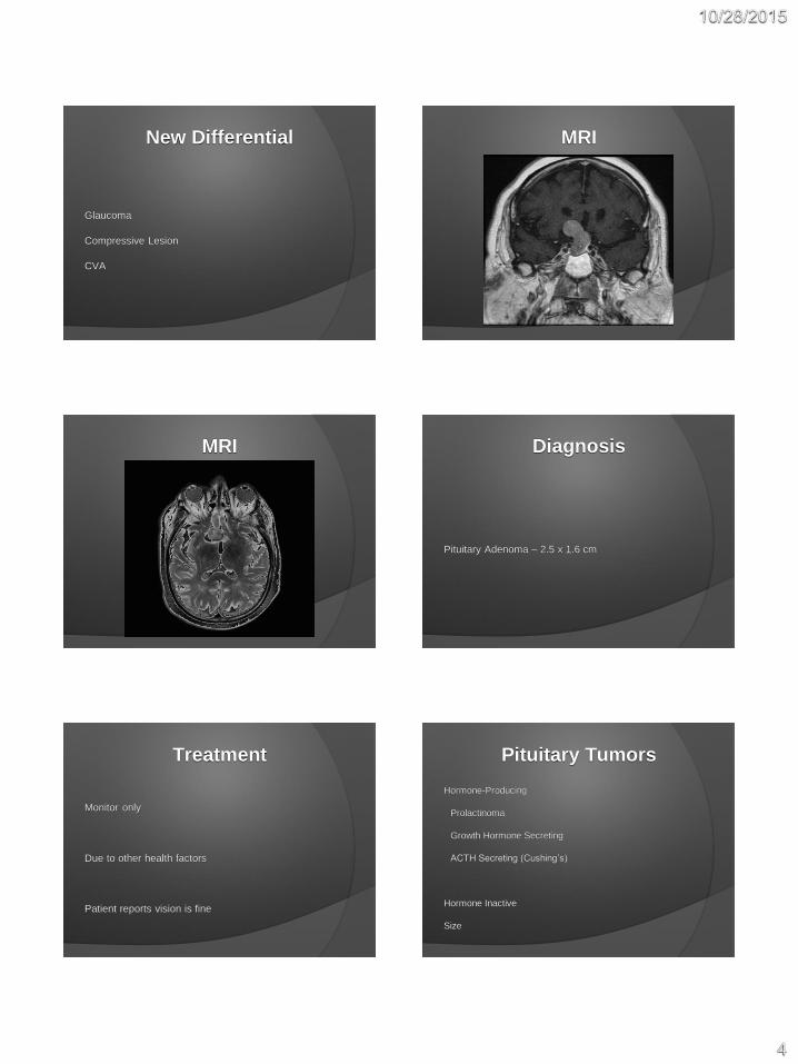
4
New Differential
Glaucoma
Compressive Lesion
CVA
MRI
MRI Diagnosis
Pituitary Adenoma – 2.5 x 1.6 cm
Treatment
Monitor only
Due to other health factors
Patient reports vision is fine
Pituitary Tumors
Hormone-Producing
Prolactinoma
Growth Hormone Secreting
ACTH Secreting (Cushing’s)
Hormone Inactive
Size

10/28/2015
5
Treatment
Goals
Normalize hormone levels
Pituitary gland function
Reduce signs/symptoms of tumor
Treatment
Medication (Micro)
Bromocriptine – Dopamine agonist
Hormone stabilization
Treatment
Surgery (Macro)
Transsphenoidal
Transcranial
Take Home
Check both eyes
Pituitary vs. Glaucoma
Pallor vs. Excavation
Urgency
Patient 2
72 YO Hispanic Male
Blur OD/OS - Worse at night
IOP 16/14
BCVA 20/40, 20/30
Patient 2
Removal of tumor on “Left Optic Nerve” x 1990

10/28/2015
6
Patient 2
Anterior Segment Unremarkable except
2+ NS OU
Patient 2
Anterior Segment Unremarkable except
2+ NS OU
Patient 2 Patient 2
Patient 2 Patient 2

10/28/2015
7
Patient 2
Old or new??
Patient 1.1
Patient 2 Patient 2
Patient 2
Followed by Endocrinology
Tumor resected
Cataract removed
Patient 2
“The best place to find a brain tumor is a
glaucoma clinic”

10/28/2015
8
Patient 3
59 Y/O White Male
Medical HX
HTN, COPD, Arthritis, Kidney Failure, Peripheral
Vascular Dz, Anemia, Carotid Artery Stenosis,
Hyperlipidemia, Amputee – Bilateral
Patient 3
Medications:
HCTZ, Norvasc, Lopid, Metoprolol, Lisinopril,
Simvastatin, Warfarin
Examination
Blur for 1 month
BCVA: 20/100 OD, 20/40 OS
+ APD OD
IOP 6/10
Anterior Segment:
Mild Cataract OS>OD
Posterior Segment
Sclerosed Central Retinal Artery
Mild ONH nerve pallor
Retina appears perfused
Lessons Learned
“Classic” Presentation of CRAO
Giant Cell Arteritis
Consider Ophthalmic Artery
Neovascularization
Risk to Brain and Heart
Have a Heart

10/28/2015
9
Lesson #1
“Classic” Presentation of CRAO
Presentation of CRAO after reperfusion
Differential Diagnosis
Central Retinal Artery Occlusion
Ophthalmic Artery Occlusion
Ocular Ischemic Syndrome
Giant Cell Arteritis
Lesson #2
5% of CRAOs are from Temporal Arteritis
Additional Tests
ESR = 31 (slightly elevated)**
CRP = 0.5 (normal)
CBC = Abnormal RBC, HCT, HGB (Anemic)**
Carotid Doppler
Carotid Angiography
10/28/2015
10
Carotid Doppler Results
Stent in the right distal common carotid artery
Interval occlusion of the common carotid artery
Degree of stenosis in the right internal carotid artery cannot
be measured
Low flow to internal carotid
Follow – Up
1 month later (2 months after start of blur)
Very sluggish pupil and VA Hand Motion
5 month follow-up:
Dense, hypermature cataract
Neovascularization of the Iris
Neovascularization of the Angle 360 degrees
Lesson #3
Consider an Ophthalmic Artery Occlusion/OIS
Lesson #4
CRAO can develop anterior segment NV
Treatment
Avastin for Neovascularization
Didn’t help regress NV
Otherwise, monitor only
Goal of this eye is no pain
Still weighing Risk/Benefit of Cataract Extraction
Lesson #5
Increased risk for CVA/MI
Risk for Heart Attack is greater
10/28/2015
11
Lesson #5
AHA and ASA recommend urgent referral
CRAO/BRAO requires ER visit
Lesson #5
Amaurosis Fugax requires ER visit
Lesson #5
Asymptomatic retinal emboli??
Lesson #5
TIA used to mean timing
TIA now means location
Eye is part of the Central Nervous System
Lesson #6
Compassion
Patient 4
30 year old white female
CC: Headache and vision loss
H/O vision loss OD, diplopia
Improved now

10/28/2015
12
Currently on
Diamox
Butalbital
BCVA 20/40 OD, CF OS
APD OS
Confrontation VF Full OD, Limited OS
Color Vision: 10/10 OD, 1/10 OS
Anterior Segment Unremarkable
Differential
Papilledema
Intracranial Hypertension
Malignant Hypertension
Space Occupying Lesion
Cerebral Venous Thrombosis
Inflammatory
Infectious vs Non-infectious
ONH Drusen
Uveitis
Optic Neuritis??

10/28/2015
13
62 inches (157 cm)
462 lbs (210 kg)
BMI 85
BP: 110/80
MRI Normal
LP – 490 then 310
Normal opening pressure up to 250 mm H2O
in obese patients
1 Month Later…
BCVA 20/20 OD, 20/200 OS
S/P VP shunt x 1 month
On Diamox still
OD swollen, OS swollen and atrophic
8 years later…
no Diamox
20/20 OD - minimal VF loss
20/200 OS - stable VF loss

10/28/2015
14
Why is one eye 20/20 and the other 20/200?
Idiopathic Intracranial
Hypertension
Old names:
Benign Intracranial Hypertension
Pseudotumor Cerebri
Idiopathic Intracranial
Hypertension
1 in 100,000 people
Young women
10% overweight = 13x more likely
Idiopathic Intracranial
Hypertension
Causes are unknown
10/28/2015
15
Idiopathic Intracranial
Hypertension
Symptoms are varied
Asymptomatic
Headache
Blurred vision
Diplopia
Nausea
Idiopathic Intracranial
Hypertension
Diagnosis of exclusion
MRI/MRA/MRV
Lumbar Puncture
Blood Pressure
Idiopathic Intracranial
Hypertension
Treatment
Weight loss
Diamox
VP shunt
Patient 5
74 YO WM
C/O Blur
No pain or discomfort
Patient 5
BCVA 20/40 OD/OS
Anterior Segment normal except
2+ NS OU
Patient 5
Diagnosis: Cataract

10/28/2015
16
Patient 5
But…what about this?
Patient 14
Patient 5
Nevus OD - 2007
Patient 5
Fast forward to 2013
Patient 14

10/28/2015
17
Patient 5
Small Choroidal Melanoma
Patient 5
Risk Factors
1. Tumor thickness greater than 2.0 mm
2. Subretinal fluid
3. Visual symptoms
4. Orange pigment
5. Posterior tumor margin touching the disc
6. Lack of drusen
Patient 5
Shields CL, et al. Risk factors for growth and metastasis of
small choroidal melanocytic lesions. Ophthalmology
1995;102:1351-1361.
The Collaborative Ocular Melanoma Study Group. Factors
predictive of growth and treatment of small choroidal
melanoma. Arch Ophthalmol 1997;115:1537-1544.
Patient 5
Treated with Radioactive Plaque Therapy
Developed Radiation Retinopathy
Optic nerve edema, Macular edema
Treated with Anti-VEGF

10/28/2015
18
COMS
Small Melanoma
< 2.4 mm height
5-16 mm diameter
COMS
Small Melanoma
5-year all cause mortality = 6%
COMS
Medium Melanoma
2.5–10 mm height
≤16 mm diameter
COMS
Medium Melanoma
Mortality with enucleation = 19%
Mortality with brachytherapy = 18%
COMS
Large Melanoma
>10 apical height
>16 mm diameter
COMS
Large Melanoma
Mortality with enucleation = 43%
Mortality with enucleation and pre-op radiation = 38%

10/28/2015
19
COMS
COMS excluded tumors near optic nerve
Meta-analysis
5-year all cause mortality
Large = 53% Medium = 32% Small = 16%
Radiation Retinopathy
Exposure to External Beam Radiation or Brachytherapy
Usually within 6 months to 3 years
Can happen 15 years later
http://www.med.unc.edu
Radiation Retinopathy
Signs
Retinopathy similar to DM
Macular edema
Optic nerve edema
Radiation Retinopathy
Symptoms
Typically asymptomatic
Can develop floaters and/or visual acuity loss

10/28/2015
20
Radiation Retinopathy
Treatment
Monitor
Laser (focal/grid or PRP)
Intravitreal triamcinolone
Intravitreal Anti-VEGF
Patient 6
64 year old WM
CC: Blurred vision OS - present since last year
+DM
BCVA: 20/25 OD, 20/40 OS
Anterior Segment Unremarkable
Impending Lamellar Macular Hole
Will discuss treatment later
Patient 7
74 Y/O Hispanic Male
CC: Blurry vision
+DM, +CATARACT
BCVA: 20/40 OD, 20/25 OS

10/28/2015
21
Patient 7
Lamellar Macular Hole
Patient 8
65 Y/O WM
Progressive blurring
Affecting golf, reading
Patient 8
07/2014
20/20 OD, 20/25 OS
Anterior segment: Mild NS OU
Mild ERM OU
Patient 8
11/2014
20/20 OD, 20/50 OS
Mild ERM OD
Moderate ERM OS
Patient 8
02/2015
20/30 OD, 20/50 OS
Moderate ERM/VMT OD
Moderate ERM OS

10/28/2015
22
Patient 8
08/2015
20/50 OD, 20/200 OS
Moderate ERM/VMT OD
Moderate ERM OS
Patient 9
75 Y/O WM
CC: Blur when reading
BCVA: 20/40 OD, 20/30 OS
PCIOL OU
Anterior segment unremarkable

10/28/2015
23
Patient 9
Recent exam
VMT released w/o treatment
20/20 OD and OS
Patient 10
75 Y/O WM
CC: Blur OU
S/P PCIOL OU
COPD, DM, HTN
Anterior segment unremarkable
Patient 11
BCVA: 20/50 OD, 20/100 OS
Patient 11
Bilateral macular hole
Patient elected to not have treatment
COPD

10/28/2015
24
Discussion
ERM
VMT
Lamellar Macular Hole
Macular Hole
Discussion
Historically diagnosed by combination of:
Fundus Appearance
Visual Acuity
Discussion
OCT changed accuracy of diagnosis
Better able to give prognosis
Better guidance for treatment
Better post-op monitor
ERM
Macular Pucker
Creates traction of retina
Can induce edema (typically cystic)
ERM
Blur
Distortion
Metamorphosis
Range of acuity
How many progress?

10/28/2015
25
ERM
Typically asymptomatic
If acuity reduced to < 20/40, treat
ILM peel
ERM peel
Vitrectomy
**Cataract Surgery
VMT
Vitreous has firm adhesions at
ONH
Macular
Vitreous base (ora serrata)
VMT
If vitreous detachment incomplete, can lead to VMT
Distorted, blurred vision
VMT
Treatment typically includes
Monitor only
Vitrectomy
Ocriplasmin (Jetrea)
VMT
Jetrea
25% of patients improved
10% of placebo improved
Have to weigh this with complications

10/28/2015
26
VMT
Complications
Conjunctival hemorrhage
Photopsia
Vitreous floaters
Blurred vision
Injection-related eye pain
VMT
Complications
6% cataract (9% placebo)
4% increased IOP (5% placebo)
Lamellar Macular Hole
Not full thickness
Atypical borders
“Inverted Anvil”
Acuity typically better
Difficult to treat
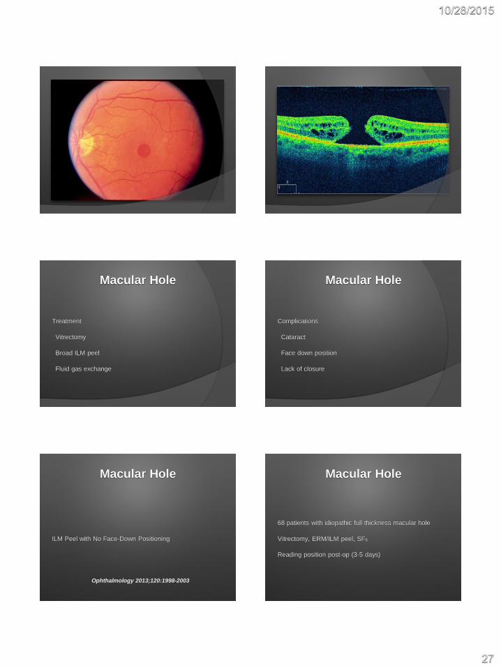
Macular Hole
Full thickness retinal break
Acuity typically 20/100-20/200**
10/28/2015
27
Macular Hole
Treatment
Vitrectomy
Broad ILM peel
Fluid gas exchange
Macular Hole
Complications
Cataract
Face down position
Lack of closure
Macular Hole
ILM Peel with No Face-Down Positioning
Ophthalmology 2013;120:1998-2003
Macular Hole
68 patients with idiopathic full thickness macular hole
Vitrectomy, ERM/ILM peel, SF6
Reading position post-op (3-5 days)

10/28/2015
28
Macular Hole
100% closure
Mean pre-op 20/100
Mean post-op 20/40
No complications
Patient 12
58 year old Black Male
C/O blur at near
Myopic
20/20 OD, 20/25 OS
Anterior Segment Unremarkable
No APD
IOP 17/17

10/28/2015
29
Diagnosis?
Solar Maculopathy
Solar Maculopathy
Thermal Burn
Pscyh Diagnosis
Eclipse
Drugs
Solar Maculopathy
Adjustment Disorder
Alcohol Abuse
Inadequate Housing
Depressive Disorder
Tobacco Dependence

10/28/2015
30
Solar Maculopathy
No ocular treatment - non progressive
Mental health referral?
Patient 13
92 YO WM
CC: “Needs Driver’s Form Filled Out”
Patient 13
Ocular History
Cataract Surgery
Glaucoma Suspect
Patient 13
Medical History
Hypertension
Colon Cancer
Lung Metastasis?

10/28/2015
31
Patient 13
VAcc 20/20, 20/25
Anterior Segment healthy except
Patient 13
Patient 13 Patient 13
~2MM amelanotic cystic growth
Pushing iris back
Gonio: 2 Feeder vessels
Patient 13
Posterior segment normal - no tumor
Large C/D ~ 0.6 OU
IOP 16/18
Patient 13
Diagnosis: Invasive Carcinoadenoma
Metastasis from Colon
Likely Metastasis to lung

10/28/2015
32
Patient 13
Treatment: Hospice
Patient 13
Returned to clinic 2 months
New Hyphema
Stabilized
Goal is no pain
Patient 13
If eye tumor
Need to consider ophthalmic prognosis
Need to evaluate for metastasis
Patient 14
54 YO WM
Blur
Diplopia
Patient 14
BCVA 20/25, 20/20
EOM: Poor abduction OS
Not comitant
Anterior Segment Normal
No APD

10/28/2015
33
Patient 14
Bilateral Optic Nerve Edema
MRI
Lumbar Puncture
Blood Pressure
Patient 14
MRI - Inconclusive
Blood Pressure - 145/80
LP - Opening pressure Day 1 = 43
Day 2 = 36
+WBC
Patient 14
LP Cytology - cancer cells
Diagnosis = Leptomeningeal Carcinomatosis
Patient 14
Metastasis from esophageal cancer (rare)
More common from breast, lymphoma, leukemia
Patient 14
Patient treated with systemic chemotherapy
Died 6 months later

10/28/2015
34
Patient 15
35 YO WM
CC: “Black Sunspot Left Eye"
Flashing lights around sunspot
Acuity better with eccentric viewing
Patient 15
Anterior Segment normal
IOP 16/16
No APD
EOM full
Confrontation Full
Patient 15 Patient 15
Patient 15 Patient 15

10/28/2015
35
Patient 15 White Dot Syndrome
Multiple Evanescent White Dot Syndrome (MEWDS)
Acute Posterior Multifocal Placoid Pigment Epitheliopathy
(APMPEE)
Mulifocal Choroiditis and Panuveitis (MCP)
Punctate Inner Choroiditis (PIC)
Birdshot Retinopathy
MEWDS
MEWDS typically affects young women (15-50)
1/3 have viral prodrome
Photopsias, dyschromatopsia
Temporal or paracentral scotoma
MEWDS
MEWDS different than others because:
Mild vitritis
ONH edema
Granular changes at macula
Patient 15 MEWDS
Treatment: Monitor only
Self-limiting disease
Typically full recovery

10/28/2015
36
Patient 16
42 y/o white male
CC: Spots in vision OS for 7 days. Spots don’t move
No pain or discomfort
No family history
No ocular history
Patient 16
BCVA
20/30
20/400
EOM - Full
Confrontation - Full
P + RXN - No APD
Patient 16
IOP 15/15
Anterior Segment Normal
Retina:
White lesions throughout retina OS>OD
Patient 16
Patient 16 Patient 16

10/28/2015
37
Patient 16 Patient 16
Patient 16 Patient 16
Purtscher’s Retinopathy (or Purtscher’s Like Retinopathy)
In a patient with Severe Alcoholism
Recent Acute Pancreatitis
Patient 16
Refer to:
Retina
Rehab
Purtscher’s Retinopathy
Purtscher’s Retinopathy is rare
Often associated with trauma
60% bilateral

10/28/2015
38
Purtscher’s Retinopathy
Can be associated with other conditions:
Acute pancreatitis (100% bilateral)
Connective tissue disorder
Childbirth
Renal disease
Purtscher’s Retinopathy
Purtscher flecken are pathognomonic findings
Polygonal areas of retinal whitening with a clear
demarcating line
Purtscher’s Retinopathy
No known successful treatment
Often try IV steroids
Purtscher’s Retinopathy
Patient unlikely to recover visual acuity
Especially if [email protected]