10.1 dna physical mapping - the blavatnik school of
TRANSCRIPT

Algorithms for Molecular Biology Fall Semester, 1998
Lecture 10: February 7, 1999Lecturer: Ron Shamir Scribe: Eran Yahav and Gaby Liron
10.1 DNA Physical Mapping
10.1.1 Motivation
Physical mapping is the process of determining the relative position of landmarks along agenome segment. The resulting maps are used as a basis for DNA sequencing, and for theisolation and characterization of individual genes or other DNA regions of interest (e.g.,transcribed regions or regulatory genes). The construction of high resolution sequence-readyphysical maps for human and other organisms is still one of the top priorities of the HumanGenome Project.
Given a long DNA segment it is relatively easy to produce a large group of DNA frag-ments known as clones. The process of creating the clones consists of breaking several copiesof the original DNA sequence at many locations, and then cloning each of the fragments.One of the problems with the cloning process is that the resulting fragments are obtained”out of order”. This means that it is difficult to re-assemble the fragments in order to get amap of the original sequence. Moreover, the cloning process does not ensure that a contin-uous sequence of DNA can be reconstructed from the fragments.
10.1.2 Unique Probe Mapping
An STS (Sequence Tagged Site) probe or STS discriminator is a filter that can uniquelydetermine whether or not a specific short sequence of DNA appears along any given (longer)sequence. The filter can identify the existence of the short sequence but provides no in-formation as to its location. Using a short sequence of 200 - 300 bases ensures that theprobability of an error in recognition is relatively low. Running a number of STS probesagainst numerous clones results in a matrix cell Mi,j with the entry 1 (0) representing apositive (negative) result of probe j against clone i,Figure 10.1 gives an example of orderedclones and corresponding STS probes. Figure 10.2 presents the resulting STS matrix.
1

2 Shamir: Algorithms for Molecular Biology c© Tel Aviv Univ., Fall ’98
Figure 10.1: Ordered clones and several STS probes
Figure 10.2: Resulting STS matrix. 1 in line i column j denotes that clone i contains probej.

DNA Physical Mapping 3
Problem 10.1 The unique probes mapping problem.INPUT: A set of elements U (probes) and a collection of subsets ϕ = {S1, S2, . . . , Sn}, ∀ı :Sı ⊆ UQUESTION: Find the set Π(ϕ) of all permutations over U along which every Sı is contin-uous.
Problem 10.1 is equivalent to the problem of rearranging the columns of the STS resultmatrix so that all 1’s in the rows of the matrix are continuous. This attribute is also knownas the consecutive 1’s property.
The problem of finding the set of permutations Π(ϕ) is a well known problem in computerscience. A linear time algorithm for solving this problem was presented in 1976 by Boothand Lueker [1]. Clearly, an explicit representation of the collection of all the resultingpermutations may require much more than linear space. Therefore, a linear time algorithmrequires a linear space representation of this collection. This linear representation is achievedby PQ-trees which are described in the following section.
10.1.3 PQ-Tree Algorithm [1]
A PQ-Tree is a rooted, ordered tree. We will use a PQ-tree with the elements of U as leaves,and internal nodes of two types: P-nodes and Q-nodes.
A P-node whose sub-nodes are T1, . . . , Tk for k ≥ 2 represents k subsets of U (the leafsets of T1, . . . , Tk), each of which is known to be a consecutive block of elements, but with theorder of the blocks unknown. A Q-node whose sub-nodes are T1, . . . , Tk for k ≥ 3 representsthat the k blocks corresponding to the leaf set of T1, . . . , Tk are known to appear in thisorder, up to a complete reversal (see figure 10.3).
It is therefore clear that in order to have these meanings of the P-nodes and Q-nodes wemust allow the following legal transformations (see figure 10.5 1 - 2).
1. Reordering the sub-nodes of some P-node arbitrarily
2. Reversing the order of the sub-nodes of some Q-node
Definition The frontier of a PQ-tree is the set of all leaves, read in a left-to-right order.
As demonstrated in figure 10.4
Definition Two PQ-trees T and T ′ are said to be equivalent if there exists a set of legal
transformations leading from one tree to the other. In such a case, we write T ≡ T ′.
Definition We denote the set of all frontiers equivalent to T as Consistent(T)Consistent(T ) = {Frontier(T ′)|T ′ ≡ T}

4 Shamir: Algorithms for Molecular Biology c© Tel Aviv Univ., Fall ’98
Figure 10.3: PQ-tree node types: we use circles and bars to denote P-nodes and Q-nodes,respectively.
Figure 10.4: Frontier of a PQ-tree

DNA Physical Mapping 5
Figure 10.5: Permitted transformations of a PQ-tree
Theorem 10.2 Booth-Lueker 1976 [1]
1. For every U,ϕ there exists a PQ-tree T s.t. Consistent(T ) = Π(ϕ)
2. For every given PQ-tree T , there exists U,ϕ s.t. Consistent(T ) = Π(ϕ)
Therefore, the problem of permuting the probes in order to achieve the consecutive 1’sproperty of the STS matrix is equivalent to finding a PQ-tree representing Π(ϕ).
PQ-Tree Algorithm for Unique Probe DNA Mapping:
1. Initialize the tree as a root P-node with all elements of U as sub-nodes (leaves).
2. For i = 1, . . . , n : reduce (T, Si)
The procedure reduce (T, Si) returns a tree for any permutation in consistent(T ) in whichSi is continuous.
Reduce(T, Si)
1. Color all Si leaves.
2. Apply transformations to replace T with an equivalent PQ-tree along whose frontierall of the colored leaves are consecutive.

6 Shamir: Algorithms for Molecular Biology c© Tel Aviv Univ., Fall ’98
Figure 10.6: Permitted transformations of a PQ-tree
DNA Physical Mapping 7
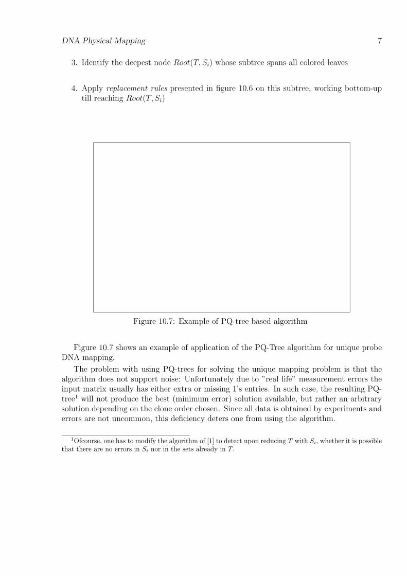
3. Identify the deepest node Root(T, Si) whose subtree spans all colored leaves
4. Apply replacement rules presented in figure 10.6 on this subtree, working bottom-uptill reaching Root(T, Si)
Figure 10.7: Example of PQ-tree based algorithm
Figure 10.7 shows an example of application of the PQ-Tree algorithm for unique probeDNA mapping.
The problem with using PQ-trees for solving the unique mapping problem is that thealgorithm does not support noise: Unfortunately due to ”real life” measurement errors theinput matrix usually has either extra or missing 1’s entries. In such case, the resulting PQ-tree1 will not produce the best (minimum error) solution available, but rather an arbitrarysolution depending on the clone order chosen. Since all data is obtained by experiments anderrors are not uncommon, this deficiency deters one from using the algorithm.
1Ofcourse, one has to modify the algorithm of [1] to detect upon reducing T with Si, whether it is possiblethat there are no errors in Si nor in the sets already in T .
8 Shamir: Algorithms for Molecular Biology c© Tel Aviv Univ., Fall ’98
10.1.4 Solving the Unique Mapping Problem Using Interval Graphs
Create a graph G(V,E) whose vertices are the clones, and its edges are E = {(vi, vj)|there exists a probe p, p(vi) = p(vj)}Definition A graph G(V,E) is said to be an interval graph if every node can be representedas an interval and an edge exists between two vertices if and only if the intervals correspondingto them overlap. The set of such intervals is then called a realization of G.
Problem 10.3 The interval graph problem.INPUT: A graph G(V,E).QUESTION: Determine whether G is an interval graph.
Intuitively, it is clear that the problem of checking if a graph is an interval graph is closelyrelated to 10.1 (finding Π(ϕ)). The problem of recognition of interval graphs can be solvedin polynomial time [1]. The algorithm is based on a following theorem:
Theorem 10.4 Fulkerson - Gross 1965 [3]
A graph G(V,E) is an interval graph iff all of the maximal cliques in the graph can bearranged in linear order so that for every vertex the set of all the cliques containing it iscontinuous.
To solve the problem mentioned above a matrix is created displaying the connectionbetween the maximal cliques and the vertices:
Mi,j =
1 if vertex i is in clique j,
0 otherwise.(10.1)
By using the algorithm of [1] given in section 10.1.3, we try to find a permutation on theclique order satisfying the requirements of theorem 10.4,furthermore, such a permutationallows easy computation of a set of intervals corresponding to the nodes.
Note that construction of matrix M above uses the following property: An interval graphhas O(n) maximal cliques and these cliques can be found in O(n) time.
As mentioned above, solving the unique mapping problem can be done quite easily usingPQ-trees in the absence of noise. In the case of either missing edges (probe not identified) orextra edges (probe identified where it should not have been) the resulting graph might notbe an interval graph. The problem of creating an interval graph from the existing graph isknown as the interval graph editing problem. Slight modifications introduce other variantslike the interval graph sandwich problem.
10.2. PROBABILISTIC MODELS FOR MAPPING 9
10.2 Probabilistic Models for Mapping
Recall the following defintion:
Definition A Poisson process of rate λ is described by:
• A non decreasing function N : R+0 → N where N(t) = number of events until time t
• N(0) = 0
• The number of events in disjoint intervals are independent
P (N(t+ s)−N(s) = n) = e−λt(λt)n
n!, for n = 0, 1, . . . and s ≥ 0
(10.2)
• Distribution of the number of events in an interval is stationary, i.e. depends only onthe length of the interval. The expected number of events in an interval of length t isgiven by E(N(t)) = λt.
Denote:
• Tn = time between event n− 1 and event n
• S0 = 0
• Sı =∑ii=1 Tı
Recall that inter-event times in a Poisson process are i.i.d. random variables, exponen-tially distributed with parameter λ, i.e.,
Pr(Ti > t) = e−λt (10.3)
If it is known that n ≥ 1 events occurred in a Poisson process until time t, then theinter-arrival times {S1, S2, ..., Sn} are distributed uniformly and independently in [0, t].
Assume clone length L, genome length G, and choose N clones at random. What is theexpected fraction of the genome covered by clones?
For a random point b, and an arbitrary clone C the probability of the point b beingincluded in the clone c is given by:
Pr(b ∈ c) =L
G(10.4)

10 Shamir: Algorithms for Molecular Biology c© Tel Aviv Univ., Fall ’98
R Coverage1 0.632 0.8653 0.954 0.985 0.993
Table 10.1: Coverage of genome segment depending on redundancy factor
and therefore, the probability of b being out of all the clones is given by:
Pr(∀c : b /∈ c) = (1− L
G)N = (1− L
G)G
NG ∼ e−
NLG (10.5)
with the last approximation being valid when L� G and N � G.
Definition The fraction
R =NL
G
is said to be the redundancy of the clone set.
Definition The expected fraction of non-covered genome is given by
E(fraction not covered) = e−R (10.6)
where R is the redundancy of the clone setTable 10.2 shows that using redundancy factor of 2 to 5 gives a good coverage of the
genome segment considered.
Define the length by setting clone length = 1, and denote N = number of clones, R =redundancy factor, and assume that the clone starting positions follow a Poisson processwith rate λ. We define a minimal overlap factor θ between clones to identify overlap.
A set of clones covering a continuous segment of the genome, together with their physicaldistances is called a contig. Contigs are sometimes referred to as islands.

Probabilistic Models for Mapping 11
Theorem 10.5 Lander-Waterman 1988 [4]:
1. The expected number of apparent islands is given by
Ne−R(1−θ) (10.7)
2. The expected number of apparent islands with exactly j ≥ 1 clones is given by
Ne−2R(1−θ)(1− e−R(1−θ))(j−1)
(10.8)
3. The expected number of clones in an apparent island is given by
e−R(1−θ) (10.9)
4. The expected length of an apparent island is
e−R(1−θ) − 1
R+ θ (10.10)
Proof: We will prove the first item of the theorem. In order to prove the formula forexpected number of apparent islands, we define J(x) as follows:
J(x) = Pr(two points a, b = a+X are not covered by a common clone)
= Pr(there are no left-end points in the interval [b− 1, a])
Since [b − 1, a] is of length 1 − x, J(x) can be computed from the redundancy factor Ras follows:
J(x) =
e−R(1−x) 0 ≤ x ≤ 1,
1 otherwise.(10.11)
The number of islands is the number of times leaving a clone without detecting an overlap.Let Ec denote the event of a clone c being the right-hand clone of an island. If the right-handside of the island is at a point t, we require that t and t − θ are not covered by a commonclone (other than c) .
The probability of such an event Ec is given by:
P (E) = J(θ) (10.12)
and the expected number of apparent islands is therefore given by:
Exp(number of apparent islands) = N · J(θ) = Ne−R(1−θ) (10.13)

12 Shamir: Algorithms for Molecular Biology c© Tel Aviv Univ., Fall ’98
10.3 Constructing Physical Maps
from Noisy Non-Unique Probes Fingerprints
10.3.1 Introduction
Physical mapping using hybridization fingerprints of short oligonucleotides was first sug-gested by Poustka et al. in 1986 [6]. In this technique short labeled DNA sequences, orprobes, attach, or hybridize, to positions along the target DNA matching their own DNAsequence. The probes are nonunique, i.e., they occur at many points along the genome,and typically hybridize with 10% − 50% of the clones. Overlapping clones can be identi-fied by their similar fingerprints. See figure 10.9 for an illustration of this hybridizationscenario. [6] suggested this method in order to eliminate the need to process individualclones in the restriction digestion technique. They reported preliminary computer simula-tions demonstrating feasibility, and suggested the use of Bayesian inference in data analysis.More detailed strategies were offered by Michiels et al. [5]. A likelihood ratio based on adetailed statistical model was used to make overlap decisions, and a discussion of experimen-tal errors was also included. Craig et al. [2] used short oligonucleotides in the ordering ofcosmid clones covering the Herpes Simplex Virus (HSVI) genome. The clones were orderedmanually. As each probe occurred only once or twice along the short (∼ 140KB) genome,this experiment does not represent the general problem.
Figure 10.8: Clones and non-unique probes. The clones are the horizontal lines. The randomoccurrences of a single nonunique probe are marked by the dotted vertical lines.

Constructing Physical Maps from Noisy Non-Unique Probes Fingerprints 13
Figure 10.9: Physical map example. The short horizontal lines are the clones with their x co-ordinates corresponding to the position on the target genome. The y coordinates correspondto the clone order in the constructed map. Note that each point on the target genome iscovered by many clones. The total length of the clones divided by the length of the genomeis called the clone coverage (10 in this example).

14 Shamir: Algorithms for Molecular Biology c© Tel Aviv Univ., Fall ’98
The location of the clones along the target genome is not directly known to the experi-menters. Mapping data (such as hybridization data) produced by the experiment is used toreconstruct the map. A list assigning every clone its estimated position along the genome isa solution to the mapping problem. According to equation 10.7 in theorem 10.5, with suffi-cient coverage the whole map is usually one contig. A plot of clone order in the constructedmap vs. real clone position (see figure 10.9) provides a visual measure for map quality. Ifthe order of the clones in the constructed map is completely correct then the computed leftendpoints of clones increase as their true value increase (or decrease, if the orders in the trueand constructed maps happen to be reversed, as in the examples of figures 10.8,10.9). Minorordering errors are seen as small deviations from the monotonicity, as in figure 10.8, showthe construction is still essentially correct. Very small errors, which do not change the cloneorder, cannot be observed from the plot. A completely random solution will correspond torandomly placed clones, whereas a nonrandom solution containing several large errors willtranslate into several randomly placed broken contigs with an approximately correct intra –contig order.
10.3.2 The Statistical Model
We will now present the statistical model we use for the above mapping method. We assumethe following:
1. Clones are uniformly and independently distributed along the target genome.
2. Clones are of equal length.
3. Probe occurrences along the genome are modeled by a Poisson process.
4. The Poisson rate is identical for all probes.
5. The noise statistically behaves as follows:
• False positive errors - a Poisson process with parameter β.
• False negative errors independently occur with probability α for each hybridization
The hybridization scenario is shown by figure 10.8. The clones are the horizontal lines.The random occurrences of a single nonunique probe are marked by the dotted vertical lines.We denote by A the probe - clone occurrence matrix: Ai,j = k if probe j occurs k times inclone i. The probe in this example occurs 3 times along this 7 clones genome section, so itscolumn in the occurrence matrix would be (1, 1, 0, 0, 1, 2, 0). The probability of j occurringk times in i is given by:
Pr(Ai,j = k) =(λl)ke−λl
k!(10.14)

Constructing Physical Maps from Noisy Non-Unique Probes Fingerprints 15
We denote by B the probe clone hybridization matrix: Bi,j = 1 or Bi,j = 0 depending
on whether probe j hybridized with clone i or not. The vector−→Bi of the hybridizations of
clone i with all the probes is also called its hybridization fingerprint. In case no noise ispresent hybridization occurs iff there is at least one occurrence of the probe. In this casethe appropriate column of B would be (1, 1, 0, 0, 1, 1, 0). Experimental noise can result inboth false positive hybridizations (Bi,j = 1 when Ai,j = 0), and false negative hybridizations(Bi,j = 0 when Ai,j > 0).
Hybridization fingerprints of intersecting clones are correlated. This fact is used in or-der to estimate the clone pairs overlap. Although noise reduces the correlation betweenfingerprints of overlapping clones, Bayesian inference can still be used to identify overlap,provided a sufficient number of probes is used. It may also be the case that ”soft decision”hybridization signals are available. Such signals provide more information on probe occur-rences than binary signals do. This continuous signal value does not directly correspond tothe hybridization probability, and we have chosen to assume a threshold is used to transformthe hybridization signal into a binary one. We therefore define the hybridization matrix Bto be a binary matrix, such that Bi,j = 1 if probe j has produced a positive hybridizationsignal with clone i. The matrix B is the actual experimental data, which is the input forthe construction algorithm. The matrix contains noise and no information on multiplicities.Using the statistical model we can write the following equation:
Pr(Bi,j = 1|Ai,j) = Pr(false positive) +
(1− Pr(false positive))(1− Pr(false negative|Ai,j))= (1− e−βl) + (1− (1− e−βl))(1− αAi,j)= 1− e−βlααAi,j
10.3.3 Clone Pair Overlap Score
Let Ca and Cb be two clones viewed as intervals of lengths la and lb respectively. Withoutloss of generality, assume la ≤ lb. Define C ′γ = Ca
⋂Cb and lγ = |Cγ|. The overlap score uses
the hybridization vectors−→Ba,−→Bb to produce a vector probabilities for the overlap length lγ.
The relative position of Ca,Cb and C ′γ is shown in figure 10.10.
We first calculate the probability Pr(−→Ba,−→Bb|lγ = t). Let C ′a = Ca\Cb, C ′b = Cb\Ca, and
let Ai,j be the number of occurrences of probe j in Ci. We can thus write the followingequation:

16 Shamir: Algorithms for Molecular Biology c© Tel Aviv Univ., Fall ’98
Figure 10.10: Clone pair overlap score
Pr(Ba,j, Bb,j|lγ = t) =∑K′a
∑K′b
∑Kγ
Pr(Ba,j|Aa,j = K ′a +K ′γ)
·Pr(Bb,j|Ab,j = K ′b +K ′γ)
·Pr(A′a,j = K ′a|lγ = t)Pr(A′b,j = K ′b|lγ = t)
·Pr(A′γ,j = K ′γ|lγ = t)
The calculation of the probabilities inside the summation is straightforward using thestatistical model. Since hybridization is a virtual certainty if a probe occurs many timesinside a clone, we can limit the summation to small values of k (say 0 ≤ k ≤ 5), therebymaking feasible the score’s computation while introducing only a negligible error. Consider-ing each probe as an independent source of information, the conditional probability of thevector pair (
−→Ba,−→Bb) is:
Pr(−→Ba,−→Bb|lγ = t) =
n∏j=1
Pr(Baj, Bbj|lγ = t) (10.15)
Assuming uniform parameters for the probes, the expression Pr(Ba,j, Bb,j|lγ = t) insidethe product is independent of j. Therefore, we can define P t
x,y by P tx,y = Pr(Ba,j = x,Bb,j =
y|t). Denoting by Sx,y(a, b) the set of probes 1 ≤ j ≤ n, such that Ba,j = x and Bb,j = y, wecan write:
Pr(−→Ba,−→Bb|t) =
1∏x=0
1∏y=0
P |Sx,y(a,b)|x,y (10.16)

Constructing Physical Maps from Noisy Non-Unique Probes Fingerprints 17
Having computed Pr(−→Ba,−→Bb|t) we can use Bayes Theorem:
Pr(lγ = t0|−→Ba,−→Bb) =
Pr(−→Ba,−→Bb|lγ = t0)Pr(lγ = t0)∑la
t=0 Pr(−→Ba,−→Bb|lγ = t)Pr(lγ = t)
(10.17)
10.3.4 Problem Statement
Problem 10.6 The noisy non-unique probes mapping problem.INPUT:
• The probe hybridization matrix B (Bi,j = 1 iff probe j has hybridized with clone i).The hybridization matrix contains both false positives (Bi,j = 1 when Ai,j = 0) andfalse negatives (Bi,j = 0 when Ai,j > 0).
• The genome size.
• The length of the probes.
• The length of the clones.
• Estimates of the α, β experimental noise.
QUESTION: Find the relative position of the clones
18 Shamir: Algorithms for Molecular Biology c© Tel Aviv Univ., Fall ’98
10.3.5 The Construction Algorithm
1. Initialize a contig set, so that for each clone there is a corresponding contig consistingof that clone.
2. Calculate P+x,y, and |Sx,y(a, b)| for each pair a, b of contigs.
3. Calculate for all the initial contig pairs and for the two possible relative orientationstheir relative placement probabilities vector. The results are stored in a table.
4. Find for all contig pairs and for their two possible relative orientations their bestrelative placement, and its probability.
5. While more than one contig remains:(a) Find two contigs a, b which have relative orientation and placement that attain thehighest probability.(b) Merge b into a.(c) Change the table calculated in step 3 to reflect the last merger. Only the tableentries for all contig pairs (a, c) need to be changed. The required change is a simplecombination of the previous entries for (a, c) and for (b, c).(d) Find, for contig pairs affected by the merger, their new best relative placement.
Assume that there are n probes and m clones of length d, and the genome has length L.The bottleneck steps in the computation are:
• Step 2 that takes O(m2n) time.
• Steps 3-4 that take O(m2d) time
• Steps 5(c) and 5(d) that take O(mL) time.
Step 5 is repeated m− 1 times giving a total complexity of O(m2(L+ d+ n)).
10.3.6 Map Quality
We have used several criteria to quantify the quality of the constructed map. The majorcriteria included the presence of unacceptable big errors in the constructed map, the averagesize of errors between consecutive clones, and the number of breakpoints in the clone - orderpermutation. We now detail how to estimate the map quality. We measure the distancebetween clones a, b as the number of left clone endpoints between the left endpoints of a andb.
Using the order of the left endpoints of clones in the constructed map, we computethe distance d1 between consecutive clones and compare it with the distance d2 between

Constructing Physical Maps from Noisy Non-Unique Probes Fingerprints 19
the same two clones in the correct map (note that the two clones need not necessarily beconsecutive or even close in the correct map). If the difference |d1 − d2| is more than aclone’s length, we call it a big error. Otherwise, it is called a small error. The averageof the small errors is also calculated and is called the average error. As it is possible thatthe constructed map is oriented in the reverse direction with respect to the correct one, werepeat the calculation with reversed orientations. The correct orientation is assumed to havea longer clone sequence with no big errors. In case this is not a sufficient criterion (thereare no big errors in both orientations, or the first error in both orientations occurs after thesame number of clones) we choose the orientation minimizing average error. If still more bigerrors remain, the process is resumed, starting from the location of the first big error. A 01 variable anybig is defined to be a 1 if the constructed map contains at least one big error.Its average over a number of simulations is used to estimate the probability of a constructedmap to contain big errors.
10.3.7 Main Results
Results presented in this section are for the base scenario, which has the following parameters:
• Length of clones: lc = 40960 base pairs.
• Target genome length: L = 25 clone lengths ( L = 25× 40960 which is approximately1M base pairs).
• Clone coverage: 10
• False negatives probability: α = 0.2
• False positives probability: β = 0.05
• Number of probes: n = 500
• Length of probes: plen = 8
Based on the results of 1000 simulations, the algorithm has a probability of 0.039±0.006of making any big errors in the base scenario. The average error in the constructed map isalso quite small: 1740± 3bp. The average error can be reduced if a finer quantization unit isused (at a linear cost to memory and CPU consumption). A further experiment indicated aprobability of about 0.075 of any mistake that exceeds a clone’s length in the estimation ofthe relative distance between any two (not necessarily adjacent) clones.
20 Shamir: Algorithms for Molecular Biology c© Tel Aviv Univ., Fall ’98
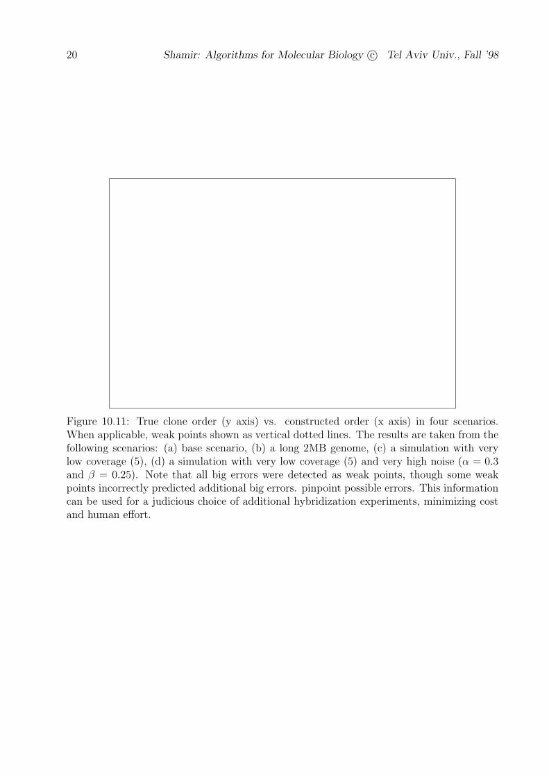
Figure 10.11: True clone order (y axis) vs. constructed order (x axis) in four scenarios.When applicable, weak points shown as vertical dotted lines. The results are taken from thefollowing scenarios: (a) base scenario, (b) a long 2MB genome, (c) a simulation with verylow coverage (5), (d) a simulation with very low coverage (5) and very high noise (α = 0.3and β = 0.25). Note that all big errors were detected as weak points, though some weakpoints incorrectly predicted additional big errors. pinpoint possible errors. This informationcan be used for a judicious choice of additional hybridization experiments, minimizing costand human effort.

Constructing Physical Maps from Noisy Non-Unique Probes Fingerprints 21
Figure 10.12: Influence of various simulation parameters on the probability of having bigerrors. The vertical dotted line indicates the value of the parameter in the base scenario.Note that the effect of a decrease in the number of probes is very similar to that of an increasein the experimental noise. This is because noise decreases the informational content of eachprobe, an effect that can be countered by an increase in the number of probes. It is alsonotable that the probe size has a very significant effect, resulting from its direct influenceon the frequency of probe occurrences, and therefore on the informational content of theexperiment. In contrast, the genome size only moderately effects performance.

22 Shamir: Algorithms for Molecular Biology c© Tel Aviv Univ., Fall ’98
10.3.8 Weak Points Detection
Definition The clone pair energy of a clone pair (Ci, Cj) , with an overlap oi,j = t and
hybridization fingerprints (−→Bi ,−→Bj) is given by Ei,j = − lgPr(
−→Bi ,−→Bj|oi,j = t).
Definition The contig energy E is given by summing over all clones in the contig ,i.e :
E =∑i<j Ei,j.
In analogy to the breaking up of a chemical molecule, the separation of a contig into twononoverlapping parts should increase the energy substantially. However, if the two parts donot overlap in the real map, the separation energy should be quite small or even negative.
Definition A weak point is a point along the contig having separation energy below athreshold (determined experimentally).
Such information can be used by the laboratory in order to pinpoint areas where ad-ditional hybridizations should be performed. We also make use of the weak points in ouralgorithm in order to break up a contig and reassemble it. In case an error was made at anearly stage, this process enables the algorithm to correct its previous error with the benefitof the additional information from other clones added at a later stage.
10.3.9 Results on Real DNA
The next step of the authors was to test the algorithm in simulations involving real DNAsequences. These sequences were taken from a variety of representative organisms: thenematode C.Elegans, bacterium E.Coli, yeast, and human. The lengths of the sequencesranged from about 1.7MB to over 4MB. Non overlapping sections of length 1MB from eachgenome were used for the tests (1MB and 730MB for Homo Sapiens). An additional randomDNA sequence was used for comparison.
With the exception of probes fitting repetitive sequences, the occurrences of most probesalong target genome appear to be uniformly distributed (assumption 3 in section 10.3.2),thus supporting the Poisson model. However, the same rate assumption also predicts aPoisson distribution of the number of probe occurrences. As figure 10.14 demonstrates, thisstronger assumption cannot be sustained. The obvious solution of fitting a separate Poissonmodel for each probe will not do, because most probes occur very infrequently, and willtherefore provide very little information. However, Figure 10.14 leads us to an encouragingobservation: In all the distributions, a significant fraction falls under the graph of the randomDNA, thereby demonstrating that there are fairly many ”good” probes. Because probehybridizations are effort and cost intensive it is impractical to make a large amount ofexperiments and then choose a small subset of ”good” probes.
However, if probes can be chosen before the actual experiment, based on prior knowledgeof the organism’s typical sequences (e.g., from other sequenced parts of its genome), better

Constructing Physical Maps from Noisy Non-Unique Probes Fingerprints 23
results can be achieved.This process is called probe preselection. Figure 10.15 demonstrates that a fairly good
fit with the same rate model can be achieved. One important problem that cannot be seenclearly in figure 10.15 is that the resulting distributions still have very long tails. Thesetails represent probes that either occur very rarely or very frequently, and thus hinder theperformance of the algorithm.
To overcome this problem a procedure of postscreening is used. It uses the hybridizationdata to screen out certain probes that deviate significantly from the same rate model. Onlythose probes which well fit the same rate model are subsequently used by the mappingalgorithm. As hybridization data is obtained after probe pre-selection, most probes alreadyconform with the model, and so there is little loss of usable hybridization information. Thepostscreening process uses the number of actual probe occurrences in the hybridization dataand the noise parameters. This technique works well when the noise estimate is good. If nogood noise estimates are available, one would probably do better by computing a histogramof the number of hybridizations, and keeping only the probes in the central part of thehistogram.
Figure 10.13: Influence of clone length variability. The x axis represents the maximal vari-ability from the average clone sized length. Clone lengths were uniformly distributed in thisrange. The graphs show good performance with a variability of up to about 15000bp, or37.5%.

24 Shamir: Algorithms for Molecular Biology c© Tel Aviv Univ., Fall ’98
Figure 10.14: Histogram of the number of 8 - mer probe occurrences along a genome sectionof length 1MB.

Constructing Physical Maps from Noisy Non-Unique Probes Fingerprints 25
Figure 10.15: Histograms of the number of probe occurrences on a genome section of length1MB, when using only probes with an average number of occurrences (between 0.9 and 1.1of average) as estimated from a different genome section of length 1MB (730KB for human)of the same organism

26 Shamir: Algorithms for Molecular Biology c© Tel Aviv Univ., Fall ’98
organism test type % maps with errorsRandom no postscreening 2.8± 0.7Random PS 500 probes before 2.2± 0.7Random PS 500 probes after 0.0± 0.0
Bacterium no postscreening 60.6± 2.2Bacterium PS 500 probes before 20.6± 1.8Bacterium PS 500 probes after 10.8± 1.4
Yeast no postscreening 20.4± 1.8Yeast PS 500 probes before 4.0± 0.9Yeast PS 500 probes after 1.2± 0.5
C. Elegans no postscreening 34.6± 2.1C. Elegans PS 500 probes before 7.4± 1.2C. Elegans PS 500 probes after 0.0± 0.0
Human no postscreening 88.2± 1.4Human PS 500 probes before 12.6± 1.5Human PS 500 probes after 0.0± 0.0
Table 10.2: Results of the mapping algorithm with probe preselection on real sequence data.The three lines for each organism correspond to: (1) No postscreening, (2) Postscreeningon the 500 original probes (leaving about 300 screened probes), and (3) 500 postscreenedprobes (requiring more to begin with). The screened probes were chosen out of a preselectedsample of probes occurring within 10% of the mean on a different genome section of thesame organism. The postscreened probes are estimated to occur within 10% of the meanfrequency on the target genome section too. Averages and standard deviations are based on1000 simulations in each scenario. PS: postscreening.

Bibliography
[1] K. S. Booth and G. S. Lueker. Testing for the consecutive ones property, interval graphs,and planarity using PQ-tree algorithms. J. Comput. Sys. Sci., 13:335–379, 1976.
[2] A. G. Craig, D. Nizetic, D. Hoheisel, G. Zehetner, and H. Lehrach. Ordering of cos-mid clones covering the herpes simplex virus type I (HSV-I) genome: A test case forfingerprinting by hybridization. Nucleic Acids Research, 18:2653–2660, 1990.
[3] D. R. Fulkerson and O. A. Gross. Incidence matrices and interval graphs. Pacific J.Math., 15:835–855, 1965.
[4] E. S. Lander and M. S. Waterman. Genomic mapping by fingerprinting random clones:A mathematical analysis. Genomics, 2:231–239, 1988.
[5] F. Michiels, A. G. Craig, G. Zehetner, G. P. Smith, and H. Lehrach. Molecular approachesto genome analysis: a strategy for the construction of ordered overlapping clone libraries.CABIOS, 3(3):203–210, 1987.
[6] A. Poustka, T. Pohl, D.P. Barlow, G. Zehetner, A. Craig, F. Michiels, E. Ehrich, A.M.Frischauf, and H. Lehrach. Molecular approaches to mammalian genetics. Cold SpringHarb Symp Quant Biol, 51:131–139, 1986.
27