1. what are the functions of cell membranes? 2. what is ...dstratto/bcor011_handouts/vayda... ·...
TRANSCRIPT

BCOR 011 Lecture 9:
Cell membrane structure
Sept 19, 2005
BCOR 011 Lecture 9:
Cell membrane structure
Sept 19, 2005
Cell membranes1. What are the functions of cell membranes?
2. What is the current model of membrane structure?
3. Evidence supporting the fluid mosaic model
4. How appropriate fluidity is maintained
Membrane: organized arrangement of lipids and proteinsthat encloses and separates the cell from its surroundings
prokaryote eukaryote
Membranes define spaces with distinctive character and function
Membrane Functions
3. transport
4. Signal detection
5. Cell-cellcommunication
2. Localizespecificfunctions
1. boundaries6. Cell-celladhesion

major functions of membrane proteins
Figure 7.9
Transport. (left) A protein that spans the membranemay provide a hydrophilic channel across themembrane that is selective for a particular solute.(right) Other transport proteins shuttle a substancefrom one side to the other by changing shape. Someof these proteins hydrolyze ATP as an energy ssourceto actively pump substances across the membrane.
Enzymatic activity. A protein built into the membranemay be an enzyme with its active site exposed tosubstances in the adjacent solution. In some cases,several enzymes in a membrane are organized asa team that carries out sequential steps of ametabolic pathway.
Signal transduction. A membrane protein may havea binding site with a specific shape that fits the shapeof a chemical messenger, such as a hormone. Theexternal messenger (signal) may cause aconformational change in the protein (receptor) thatrelays the message to the inside of the cell.
(a)
(b)
(c)
ATP
Enzymes
Signal
Receptor
3. transport
2. Localizespecificfunctions
4. Signal detection
Cell-cell recognition. Some glyco-proteins serve as identification tags that are specifically recognized by other cells.
Intercellular joining. Membrane proteins of adjacent cellsmay hook together in various kinds of junctions, such asgap junctions or tight junctions (see Figure 6.31).
Attachment to the cytoskeleton and extracellular matrix(ECM). Microfilaments or other elements of thecytoskeleton may be bonded to membrane proteins,a function that helps maintain cell shape and stabilizesthe location of certain membrane proteins. Proteins thatadhere to the ECM can coordinate extracellular andintracellular changes (see Figure 6.29).
(d)
(e)
(f)
Glyco-protein
Figure 7.9
6. Cell-celladhesion
5. Cell-cellcommunication
1. boundaries
TransportTransport – Lect 10materials across membranes
Cell SignalingCell Signaling – Lect 11external signals trigger internal events
Biochemical functionsBiochemical functions – Lects 16-19Oxidative Phosphor, PhotosynthesisImportance of Membranes in biochemical Rxns
Current Understanding of Membrane Structue: Fluid Mosaic Model
1972 Singer & Nicholson
Familiar features?
Problems ?
Figure 7.3
Phospholipidbilayer
Hydrophobic region of protein
Hydrophobic region of protein
Proteins embedded and floating in a sea of phospholipids

• Integral Membrane proteins
EXTRACELLULARSIDE
Figure 7.8
N-terminus
C-terminus
α HelixCYTOPLASMICSIDE
Span the phospholipid bilayer – usually α-helices
Must present hydrophobic surface
Why do proteins cross membranes as α-helices?
A
B
hydrophobic
hydrophilic
c. Carbohydrates –small amts often linked to proteins or lipids
Sialic acid (SIA)- charge
β-D-galactoseβ-D-mannose
Sugars commonly found on glycoproteins
N-acetyl-β-D-glucosamine
Inside cell
Outside cell
Glycocalyx: “sugar coat”

• Membrane proteins and lipids– Are synthesized in the ER and Golgi
apparatus
ER
Figure 7.10
Transmembraneglycoproteins
Secretoryprotein
Glycolipid
Golgiapparatus
Vesicle
Transmembraneglycoprotein
Membrane glycolipid
Plasma membrane:Cytoplasmic face
Extracellular face
Secretedprotein
4
1
2
3
Membraneproteins
• Integral• Peripheral
• Lipid-anchored
Roles of membrane proteins?
A. Transport – channels and pumpsB. Links to structural proteinsC. Receptors - doorbellsD. Enzymes – localized biochemical rxnsE. Energy Generation – utilize gradient
Hydrophobic region of protein
Hydrophobic region of protein
Fluid Mosaic ModelProteins embedded and floating in a sea
of phospholipids
Evidence?

(a) (b)
Gorter & Grendel – Langmuir troughRed blood cells had enough lipid to twice cover their surface
Conclude lipid is a bilayer – hydrophilic heads facedaqueous environment
Evidence for Phospholipid Evidence for integral membrane proteins:Freeze-Fracture Electron Microscopy
Figure 7.4
A cell is frozen and fractured with a knife. The fracture plane often follows the hydrophobic interior of a membrane, splitting the phospholipid bilayerinto two separated layers. The membrane proteins go wholly with one of the layers.
Extracellular layer Cytoplasmic layer
Extracellularlayer
Proteins
Cytoplasmic layer
Knife
Plasmamembrane
Illustrates: asymmetry of membrane componetsExternal Leaflet Cytoplasmic Leaflet
Fluid Mosaic Model predicts:
A. Membranes are fluid: lipids & proteins move in the plane of the bilayer
B. Proteins and lipids are asymmetricallydistributed in the bilayers
Evidence for protein asymmetry
Lactoperoxidase (LP)
+ I125
LP adds I to proteinLP can’t pass thru
intact membrane
intact
permeable
I125 I125

Evidence for lipid asymmetry?Cut off head groups off of exposed lipids
SM, PC
SM, sphingomyelin; PC, phosphatidylcholine; PE, phosphatidylcholine; PS phosphatidylserine
Results:isolated different types of phospholipids suggesting lipids were distributed differentlyin the inner and out parts of the bilayer
Digested them with phospholipase
Broken red blood cells
Intact red blood cells
PE, PSSM & PC
Mosaic: Lipids are asymmetrically distributed
glycolipid
phosphatidylinositol
phosphatidylethanolamine
phosphatidylserine
cholesterol
sphingomyelin
phosphatidylcholine
Cytosol
Extracellular space
Fluid Mosaic Model predicts:
A. Membranes are fluid: lipids & proteins move in the plane of the bilayer
B. Proteins and lipids are asymmetricallydistributed in the bilayers
The Fluidity of Membranes• Phospholipids can move laterally within the
bilayer
Figure 7.5 A
Lateral movement(~107 times per second)
Flip-flop(~ once per month)
(a) Movement of phospholipids

Movement of membrane phospholipids
2. Lateral exchanges1x107 times/sec.moves several µm/sec
3. Flip-flop – rare<1 time/wk to 1 time/few hrs
“flippases”
1. Rotation about long axis
Evidence for lipid fluidity:Photobleaching
Evidence for membrane protein fluidity?Cell fusion: 1970 D. Frye & M. Edidin
human
mouse
Figure 7.6
•Saturated fatty acids stack nicely
•Unsaturated fatty acids –more fluid; double bond causes kinksStacks poorly
Lipids: critical role in maintainingmembrane fluidity
Shorter chains – stack poorly;More movement
Length & saturation of hydrocarbon tails affect packing & membrane fluidity
stiffer
Morefluid

Figure 7.5 B
Fluid Viscous
Unsaturated hydrocarbontails with kinks
Saturated hydro-Carbon tails
(b) Membrane fluidity
Sterols –affect membrane fluidity
Hopanoid (prokaryotes)
Cholesterol (animal)
OH-
cholesterolcholesterol– At high temperature has a loosening effect– At low temperature has a stiffening effect
Figure 7.5 (c) Cholesterol within the animal cell membrane
Cholesterol
Cholesterol is common inanimal cells
Paradox:a) fluidity at high temp.b) fluidity at low temp.
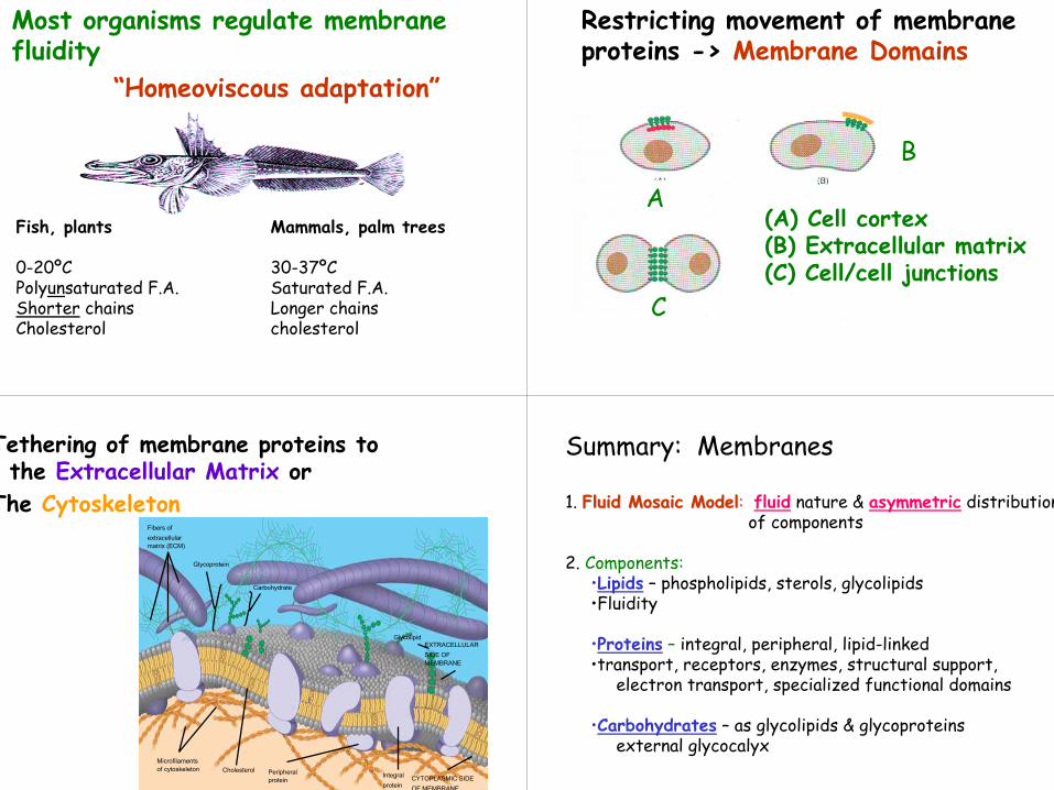
Fish, plants Mammals, palm trees
0-20ºC 30-37ºCPolyunsaturated F.A. Saturated F.A.Shorter chains Longer chainsCholesterol cholesterol
Most organisms regulate membrane fluidity
“Homeoviscous adaptation”
Restricting movement of membrane proteins -> Membrane Domains
(A) Cell cortex(B) Extracellular matrix(C) Cell/cell junctions
A
B
C
Tethering of membrane proteins to the Extracellular Matrix or
The Cytoskeleton
Figure 7.7
Glycoprotein
Carbohydrate
Microfilamentsof cytoskeleton Cholesterol Peripheral
proteinIntegralprotein
CYTOPLASMIC SIDEOF MEMBRANE
EXTRACELLULARSIDE OFMEMBRANE
Glycolipid
Fibers ofextracellularmatrix (ECM)
Summary: Membranes
1. Fluid Mosaic ModelFluid Mosaic Model: fluid nature & asymmetric distribution of components
2. Components:•Lipids – phospholipids, sterols, glycolipids •Fluidity
•Proteins – integral, peripheral, lipid-linked •transport, receptors, enzymes, structural support,
electron transport, specialized functional domains
•Carbohydrates – as glycolipids & glycoproteinsexternal glycocalyx