1 self study outline section title program overview page a.1
TRANSCRIPT
1
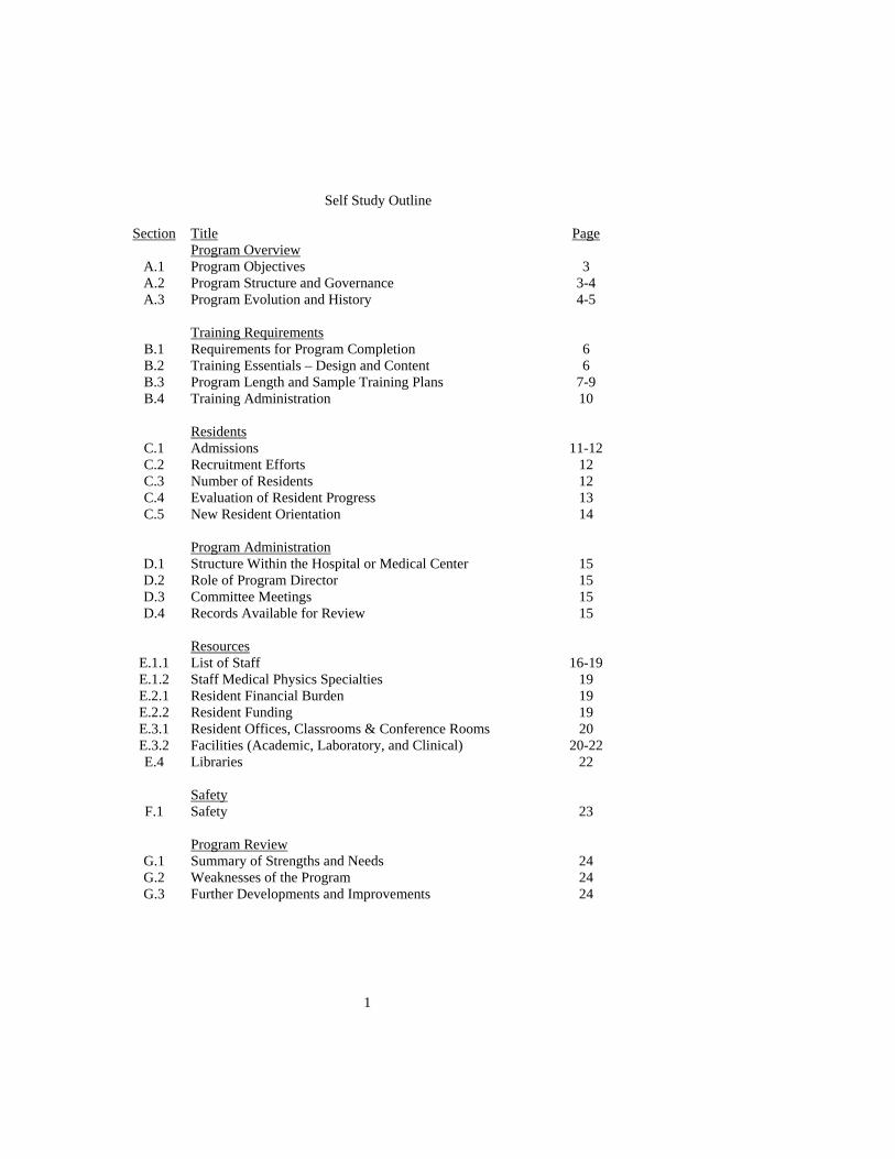
Self Study Outline
Section Title Program Overview
Page
A.1 Program Objectives 3 A.2 Program Structure and Governance 3-4 A.3 Program Evolution and History 4-5
Training Requirements
B.1 Requirements for Program Completion 6 B.2 Training Essentials – Design and Content 6 B.3 Program Length and Sample Training Plans 7-9 B.4 Training Administration 10
Residents
C.1 Admissions 11-12 C.2 Recruitment Efforts 12 C.3 Number of Residents 12 C.4 Evaluation of Resident Progress 13 C.5 New Resident Orientation 14
Program Administration
D.1 Structure Within the Hospital or Medical Center 15 D.2 Role of Program Director 15 D.3 Committee Meetings 15 D.4 Records Available for Review 15
Resources
E.1.1 List of Staff 16-19 E.1.2 Staff Medical Physics Specialties 19 E.2.1 Resident Financial Burden 19 E.2.2 Resident Funding 19 E.3.1 Resident Offices, Classrooms & Conference Rooms 20 E.3.2 Facilities (Academic, Laboratory, and Clinical) 20-22 E.4 Libraries 22
Safety
F.1 Safety 23 Program Review
G.1 Summary of Strengths and Needs 24 G.2 Weaknesses of the Program 24 G.3 Further Developments and Improvements 24

2
Appendices H.1 Letters of Invitations and Institutional Commitment
Dennis E. Hallahan, MD 25 Documentation of Institutional Accreditation 26-27
H.2 Vanderbilt Medical Physics Residents 28
H.3 Clinical Therapy Physics Rotations 29-32 H.4 Didactic Course Titles, Content, and Instructors 33-50 H.5 Residents’ Admissions Data 51 H.6 Introductory Lectures to Incoming Students 52-54 H.7 Faculty and Staff Biographical Sketches 55-95
3
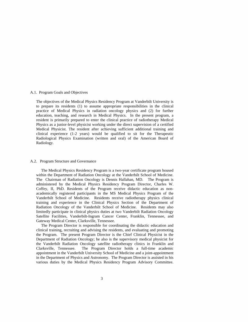
A.1. Program Goals and Objectives
The objectives of the Medical Physics Residency Program at Vanderbilt University is to prepare its residents (1) to assume appropriate responsibilities in the clinical practice of Medical Physics in radiation oncology physics and (2) for further education, teaching, and research in Medical Physics. In the present program, a resident is primarily prepared to enter the clinical practice of radiotherapy Medical Physics as a junior-level physicist working under the direct supervision of a certified Medical Physicist. The resident after achieving sufficient additional training and clinical experience (1-2 years) would be qualified to sit for the Therapeutic Radiological Physics Examination (written and oral) of the American Board of Radiology.
A.2. Program Structure and Governance
The Medical Physics Residency Program is a two-year certificate program housed within the Department of Radiation Oncology at the Vanderbilt School of Medicine. The Chairman of Radiation Oncology is Dennis Hallahan, MD. The Program is administered by the Medical Physics Residency Program Director, Charles W. Coffey, II, PhD. Residents of the Program receive didactic education as non-academically registered participants in the MS Medical Physics Program of the Vanderbilt School of Medicine. Residents receive radiotherapy physics clinical training and experience in the Clinical Physics Section of the Department of Radiation Oncology of the Vanderbilt School of Medicine. Residents may also limitedly participate in clinical physics duties at two Vanderbilt Radiation Oncology Satellite Facilities, Vanderbilt-Ingram Cancer Center, Franklin, Tennessee, and Gateway Medical Center, Clarksville, Tennessee. The Program Director is responsible for coordinating the didactic education and clinical training, recruiting and advising the residents, and evaluating and promoting the Program. The present Program Director is the Chief Clinical Physicist in the Department of Radiation Oncology; he also is the supervisory medical physicist for the Vanderbilt Radiation Oncology satellite radiotherapy clinics in Franklin and Clarksville, Tennessee. The Program Director holds a full-time academic appointment in the Vanderbilt University School of Medicine and a joint-appointment in the Department of Physics and Astronomy. The Program Director is assisted in his various duties by the Medical Physics Residency Program Advisory Committee.

4
Advisory committee members include two PhD physics faculty members, one radiation oncologist faculty member, two MS physics staff members, and one dosimetry staff member from the Department of Radiation Oncology. Resumes of the Advisory Committee members are contained elsewhere in the Appendix. Additionally, resumes of other academic faculty members participating in the didactic education of the residents and clinical faculty and staff participating in the clinical training and experience of the residents are contained elsewhere in the Appendix.
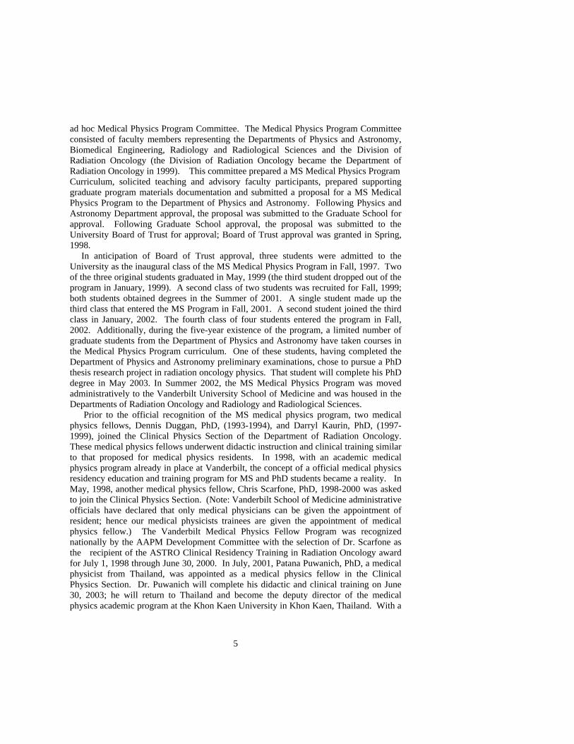
Recruitment of residents is through the Department of Radiation Oncology Educational Office with applicant follow-up coordinated by the Program Director and Educational Office Coordinator. Following official contact by an applicant, the Program Director is notified and a follow-up letter containing a program brochure, resident application, and descriptive medical physics brochures (available from AAPM HQ) are sent by the Program Director. Resident applications are processed through the Education Office in the Department of Radiation Oncology and copies of the application, transcripts, GRE and TOFEL scores, personal statement letter, and letters of reference) are sent to the Program Director. Individual resident applications are reviewed for acceptance, non-acceptance, or postponement by the Medical Physics Residency Program Advisory Committee. The Advisory Committee keeps abreast of resident performance and progression toward successful completion of the program. The Program Director verifies that all requirements are met for completion of the program. A grievance procedure process is in place should the resident have an issue(s) with an academic class or clinical training experience. Briefly, the resident would seek solutions to the issue(s) with the instructor, the Program Director, the Advisory Committee, and finally with the Dean of Biomedical Education and Research of the School of Medicine. A.3. Program Evolution and History Medical Physics at Vanderbilt draws upon a strong heritage in radiation physics and graduate education in medicine and physics. From the 1950s to the early 1970s, 337 students were trained in medical physics and health physics through the Atomic Energy Commission training program that was jointly managed at Vanderbilt by the Departments of Physics and Astronomy and the Nuclear Medicine Division of the Department of Radiology and Radiological Sciences. Of these students, 190 received the MS Degree and 54 received the Ph.D. Degree. Throughout these years, faculty in the Department of Physics and Astronomy with joint appointments in the Department of Radiology taught undergraduate and graduate courses in medical physics and radiological physics and conducted research in nuclear medicine and medical imaging. Although the AEC training program was discontinued, faculty in Radiology continued to teach radiological physics courses in the Department of Physics and Astronomy. In 1996, faculty representatives from the Departments of Physics and Astronomy, Biomedical Engineering, and Radiology and Radiological Sciences formed an ad hoc committee to investigate the creation of an Applied Physics graduate program at Vanderbilt. This collaboration and cooperation resulted in the formation of an additional
5
ad hoc Medical Physics Program Committee. The Medical Physics Program Committee consisted of faculty members representing the Departments of Physics and Astronomy, Biomedical Engineering, Radiology and Radiological Sciences and the Division of Radiation Oncology (the Division of Radiation Oncology became the Department of Radiation Oncology in 1999). This committee prepared a MS Medical Physics Program Curriculum, solicited teaching and advisory faculty participants, prepared supporting graduate program materials documentation and submitted a proposal for a MS Medical Physics Program to the Department of Physics and Astronomy. Following Physics and Astronomy Department approval, the proposal was submitted to the Graduate School for approval. Following Graduate School approval, the proposal was submitted to the University Board of Trust for approval; Board of Trust approval was granted in Spring, 1998. In anticipation of Board of Trust approval, three students were admitted to the University as the inaugural class of the MS Medical Physics Program in Fall, 1997. Two of the three original students graduated in May, 1999 (the third student dropped out of the program in January, 1999). A second class of two students was recruited for Fall, 1999; both students obtained degrees in the Summer of 2001. A single student made up the third class that entered the MS Program in Fall, 2001. A second student joined the third class in January, 2002. The fourth class of four students entered the program in Fall, 2002. Additionally, during the five-year existence of the program, a limited number of graduate students from the Department of Physics and Astronomy have taken courses in the Medical Physics Program curriculum. One of these students, having completed the Department of Physics and Astronomy preliminary examinations, chose to pursue a PhD thesis research project in radiation oncology physics. That student will complete his PhD degree in May 2003. In Summer 2002, the MS Medical Physics Program was moved administratively to the Vanderbilt University School of Medicine and was housed in the Departments of Radiation Oncology and Radiology and Radiological Sciences. Prior to the official recognition of the MS medical physics program, two medical physics fellows, Dennis Duggan, PhD, (1993-1994), and Darryl Kaurin, PhD, (1997-1999), joined the Clinical Physics Section of the Department of Radiation Oncology. These medical physics fellows underwent didactic instruction and clinical training similar to that proposed for medical physics residents. In 1998, with an academic medical physics program already in place at Vanderbilt, the concept of a official medical physics residency education and training program for MS and PhD students became a reality. In May, 1998, another medical physics fellow, Chris Scarfone, PhD, 1998-2000 was asked to join the Clinical Physics Section. (Note: Vanderbilt School of Medicine administrative officials have declared that only medical physicians can be given the appointment of resident; hence our medical physicists trainees are given the appointment of medical physics fellow.) The Vanderbilt Medical Physics Fellow Program was recognized nationally by the AAPM Development Committee with the selection of Dr. Scarfone as the recipient of the ASTRO Clinical Residency Training in Radiation Oncology award for July 1, 1998 through June 30, 2000. In July, 2001, Patana Puwanich, PhD, a medical physicist from Thailand, was appointed as a medical physics fellow in the Clinical Physics Section. Dr. Puwanich will complete his didactic and clinical training on June 30, 2003; he will return to Thailand and become the deputy director of the medical physics academic program at the Khon Kaen University in Khon Kaen, Thailand. With a

6
track record of both didactic and clinical training of four clinical radiotherapy physics residents (fellows) at Vanderbilt, the Medical Physics Residency Advisory Committee and the Department of Radiation Oncology have decided to submit the self-study materials for accreditation by CAMPEP. B.1 Requirements for Program Completion Applicants must have a PhD Degree in Medical Physics, Health Physics, Physics, Biomedical Engineering, or Nuclear Engineering or a MS Degree in Medical Physics. The residency program is twenty-four months in length. The additional didactic medical physics training will be commensurate with the applicant’s prior education according to official college transcript(s). The maximum equivalent course credit hours per semester will not exceed six credits. Excluding the time spent in class attendance per academic semester, the resident candidate will receive clinical training and experience in the Clinical Physics Section of the Department of Radiation Oncology. The normal scheduled workday in the Clinical Physics Section is from 8:00 AM to 5:00 PM; excluding class time, the resident candidate is required to be in the Physics Section. The resident candidate progress will be assessed as he/she performs clinical duties within an assigned clinical sub-specialty. (In the Physics Section at Vanderbilt, a progressive job performance evaluation is in place; all clinical assignments are “signed off” by a second responsible (senior) member of the section before the assignment is assumed completed. Hence, immediate feedback is available for clinical assignments.) Additionally, the Physics Sub-specialty Director may require an oral or written examination for clinical performance assessment. At the discretion of the Program Director, the resident candidate may be assigned a clinical medical physics research project; the project scope would not exceed three-months duration. During the didactic classroom experience, the resident will be asked to participate in the student examination process; hence the examinations will be evaluated (graded) by the course instructor. The resident evaluation process will include the “second signature” policy that is in place in the Clinical Physics Section. The resident will prepare reports of linac commissioning. beam data commissioning in a treatment planning computer, and quality assurance data analysis; these reports will be evaluated by the responsible senior physicist for that sub-specialty rotation. The resident will be responsible to obtain“minimum pass” on each clinic sub-specialty rotation. The graduate will be given a certificate upon completion of the program. The certificate will include the program specialty field completed and the time period spent in the program. The certificate will be co-signed by the Program Director and the Chairman of the Department of Radiation Oncology.
B.2. Training Essentials – Design and Content See Appendix
7
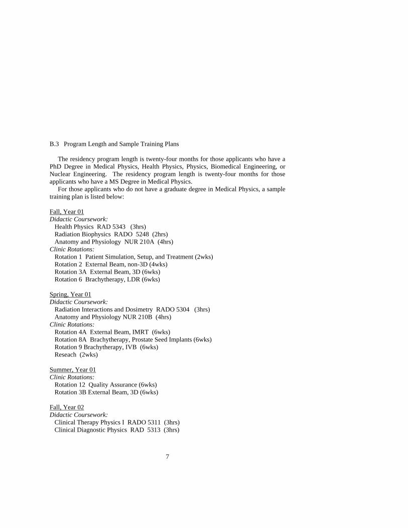
B.3 Program Length and Sample Training Plans The residency program length is twenty-four months for those applicants who have a PhD Degree in Medical Physics, Health Physics, Physics, Biomedical Engineering, or Nuclear Engineering. The residency program length is twenty-four months for those applicants who have a MS Degree in Medical Physics. For those applicants who do not have a graduate degree in Medical Physics, a sample training plan is listed below: Fall, Year 01 Didactic Coursework: Health Physics RAD 5343 (3hrs) Radiation Biophysics RADO 5248 (2hrs) Anatomy and Physiology NUR 210A (4hrs) Clinic Rotations: Rotation 1 Patient Simulation, Setup, and Treatment (2wks) Rotation 2 External Beam, non-3D (4wks) Rotation 3A External Beam, 3D (6wks) Rotation 6 Brachytherapy, LDR (6wks) Spring, Year 01 Didactic Coursework: Radiation Interactions and Dosimetry RADO 5304 (3hrs) Anatomy and Physiology NUR 210B (4hrs) Clinic Rotations: Rotation 4A External Beam, IMRT (6wks) Rotation 8A Brachytherapy, Prostate Seed Implants (6wks) Rotation 9 Brachytherapy, IVB (6wks) Reseach (2wks) Summer, Year 01 Clinic Rotations: Rotation 12 Quality Assurance (6wks) Rotation 3B External Beam, 3D (6wks) Fall, Year 02 Didactic Coursework: Clinical Therapy Physics I RADO 5311 (3hrs) Clinical Diagnostic Physics RAD 5313 (3hrs)

8
Clinic Rotations: Rotation 5 External Beam, Radiosurgery (6wks) Rotation 10 Linac Commissioning (10 wks) Spring, Year 02 Didactic Coursework: Clinical Therapy Physics II RADO 5312 (2hrs) Laboratory in Clinical Therapy Physics RADO 5314 (2hrs) Clinic Rotations: Rotation 7A Brachytherapy, HDR (6wks) Rotation 8B Brachytherapy, Prostate Seed Implants (6wks) Rotation 11 Treatment Planning Computer Commissioning (6 wks) Research (3 wks) Summer, Year 02 Clinic Rotations: Rotation 4B External Beam, IMRT (6 wks) Rotation7B Brachytherapy, HDR (6wks) Research (3wks) For those applicants who have a graduate degree in Medical Physics, a sample training plan is listed below: Fall, Year 01 Didactic Coursework: Clinical Therapy Physics I RADO 5311 (3hrs) Clinical Diagnostic Physics RAD 5313 (3hrs) Clinic Rotations: Rotation 1 Patient Simulation, Setup, and Treatment (2wks) Rotation 2 External Beam, non-3D (4wks) Rotation 3A External Beam, 3D (6wks) Rotation 6 Brachytherapy, LDR (6wks) Spring, Year 01 Didactic Coursework: Clinical Therapy Physics II RADO 5312 (2hrs) Laboratory in Clinical Therapy Physics RADO 5314 (2hrs) Clinic Rotations: Rotation 4A External Beam, IMRT (6wks) Rotation 8A Brachytherapy, Prostate Seed Implants (6wks) Rotation 9 Brachytherapy, IVB (6wks) Research (2wks)

9
Summer, Year 01 Clinic Rotations: Rotation 12 Quality Assurance (6wks) Rotation 3B External Beam, 3D (6wks) Fall, Year 02 Didactic Coursework: Radiation Biophysics RADO 5248 (2hrs) Clinic Rotations: Rotation 5 External Beam, Radiosurgery (6wks) Rotation 10 Linac Commissioning (10 wks) Spring Year 02 Clinic Rotations: Rotation 7A Brachytherapy, HDR (6wks) Rotation 8B Brachytherapy, Prostate Seed Implants (6wks) Rotation 11 Treatment Planning Computer Commissioning (6 wks) Research (3 wks) Summer Year 02 Clinic Rotations: Rotation 4B External Beam, IMRT (6 wks) Rotation 7B Brachytherapy, HDR (6wks) Research (3wks)

10
B.4 Training Administration Training objectives (didactic and clinical experience) will be modified to reflect the specific education and training background of the resident candidate. Thus, the hours spent in the classroom will be modified for individual needs; it is estimated that the classroom equivalent semester credit hours would vary from a minimum of 10 credit hours to a maximum of 26 credit hours. Additionally, individual clinic rotations may vary in length with respect to available patient volumes during the assigned rotation interval and the skills and experience acquired by the individual resident. Didactic classroom evaluation will follow the regular examination(s) and final exam schedule established by the course instructor. The resident should obtain a minimum grade of ‘B’ in each didactic course completed. The resident and specific sub-specialty rotation supervisor will meet periodically throughout the rotation interval to discuss progress and remaining deficiencies. The rotation supervisor will assess performance according to the Vanderbilt Physics Section double check system. Also the rotation supervisor may elect to give an exit rotation written or oral examination. The resident must obtain the supervisor’s signature indicating successful completion of each specific rotation.

11
C.1 Admissions
Inquiries from prospective resident candidates are from contacts resulting from browsing the Vanderbilt School of Medicine and the Department of Radiation Oncology, websites, personal letters including email, and/or phone calls to the Program Director. Once the original contact has been made, the candidate will receive a letter from the Program Director highlighting the Medical Physics Residency Program and Vanderbilt University. The letter further explains that candidates interested in the Medical Physics Residency Program can directly contact the Program Director for additional information. A prospective resident candidate list is generated; from this list a second letter is sent by the Program Director that includes the Vanderbilt Medical Physics Residency Brochure, describing admission requirements, program content, didactic course offerings, clinic rotations and faculty, The Medical Physicist, and The Roles, Responsibilities, and Status of the Clinical Medical Physicist. The resident candidate is encouraged to call or otherwise contact the Program Director for further information. Additionally, the resident is encouraged to complete and return an Admissions Packet to the Radiation Oncology Education Coordinator. Admission requirements to the Vanderbilt Medial Physics Residency Program include a PhD degree in medical physics, health physics, physics, biomedical or nuclear engineering or a MS degree in medical physics. The candidate must however demonstrate a strong undergraduate physics background; as an example of a strong undergraduate physics background the minimum of a physics minor is suggested. The incoming resident candidate should have a graduate GPA of 3.5/4.0 and a combined General GRE score of 1920. In recent months, the General GRE has altered its scoring process. Hence, the incoming student should have a combined score of 1280 on the verbal and quantitative and a score of 5.0 on the written exam. Non-USA trained candidates must, in addition to the above requirements, submit TOEFL and TSE scores of 650 and 375, respectively. The Radiation Oncology Education Coordinator will review the admissions application for completeness and receipt of all supporting materials including letters of recommendation, transcripts, GRE scores, and etc. Copies of the completed application will be sent to the Program Director. The Program Director will contact the resident candidate acknowledging that the completed application has been received and offer the prospective candidate his assistance should further questions or issues arise. The candidate will be informed that the acceptance decision are made and sent to the student by March 15 (September 15). During the months of January through March (July through September), the Program Director will send copies of the resident candidate

12
packets to the members of the Medical Physics Residency Program Advisory Committee. Meetings of the Medical Physics Residency Program Advisory Committee are called by the Program Director as needed to discuss student applications. Action by the Committee may be to accept, reject, or postpone decision concerning the resident’s application. This process is repeated until all the applications are reviewed; those postponed status applications will have final decisions by March 1 (September 1). The Chairman of the Radiation Oncology must grant final approval of resident applications. Candidate acceptance or rejection notices will be sent by March 15 (September 15) with a deadline for candidate response of April 15 (October 15). Consideration of late applications will be at the discretion of the Medical Physics Residency Program Advisory Committee; availability of residency slots and considerations of financial aid will determine the number of late application reviews. Admissions timeline is described below: Receipt of Completed Applications Jan 31 (July 31) Review of Applications Feb 28 (Aug 31) Final Approval of Accepted Applicants March 15 (Sept 15) Letter of Acceptance or Rejection Sent to Candidate March 15 (Sept 15) Candidate Notification of Intent April 15 (October 15) C.2 Recruitment Efforts . During the 1999 Annual Meeting the Vanderbilt Department of Radiation Oncology purchased an exhibit space to promote the MS Medical Physics Program and the Medical Physics Residency Program. The booth was staffed with an information person and literature about the programs. Also Vanderbilt was a cosponsor of the 1999 AAPM Annual Meeting tote bags. Although most of the recruitment efforts to date have been for the MS Program, program administrators consider that any promotional efforts directed toward physics educators and the recruitment of undergraduate students will have positive ramifications for the residency program. Results of program promotional outreach have included contacts at King College in Bristol, Tennessee, Rhodes College, in Memphis, Tennessee, Middle Tennessee State University in Murfreesboro, Tennessee, Belmont College in Nashville, Tennessee, Fisk University in Nashville, Tennessee, Western Kentucky in Bowling Green, Kentucky, and Kentucky Wesleyan College in Owensboro, Kentucky. Future residency positions may be posted on the AAPM Jobs Bulletin website for widespread communication within the medical physics community. C.3 Number of Residents At this time, the program capacity will be financially limited to one medical physics resident. Program administrators hope that the program can be expanded to include two residents, one first-year and one second-year resident candidate. At present, there is one resident in the program. Patana Puwanich, PhD, is an international from Kohn Khen
13
University in Khon Kaen, Thailand. He will complete his twenty-four month program on June 30, 2003. Dr. Puwanich is sponsored by the Thailand government; he will return to Thailand in July, 2003, where he will assume the position of deputy director of the Medical Physics Program at Khon Kaen University. C.4 Evaluation of Resident Progress Didactic Training The Program Director meets with the resident candidate at least once during each semester to assess resident standing in individual classes. Resident progress (quizzes, homework, and examinations) is monitored by the course instructor throughout the semester. Any difficulties will be discussed with the resident in a private faculty/resident meeting. Should the situation warrant, the Program Director will be notified and a Program Director/resident meeting scheduled. The resident is expected to obtain a minimum grade of ‘B’ on the courses completed. Continued resident evaluation of below satisfactory could result in dismissal from the program. Clinical Training The specific clinic rotation supervisor will be responsible for the resident’s evaluation within that rotation. Immediate feedback of the resident’s performance will be determined with the application of the Vanderbilt Physics Section double-check policy. Monitor unit calculations, tumor doses, and treatment beam parameters will be double-checked before the patient receives his/her first treatment. Performance of rotation specific clinical duties will be supervised by the clinic rotation supervisor (or designee). The resident will be allowed to proceed independently with procedures and equipment operation after approval by the rotation supervisor. The resident will keep a notebook of representative treatment plans, clinical procedures, and data for rotation specific assignments; the notebook will be reviewed at least semi-annually by the Program Director. Should the resident’s clinical performance be less than minimally satisfactory, the rotation supervisor will contact the Program Director. The Program Director, rotation supervisor, and the resident will have a joint meeting to discuss the resident’s performance. Should remedial action be required the Program Director and rotation supervisor will determine the magnitude and length of time for which that specific training must be repeated. Continued resident evaluation of below satisfactory could result in dismissal from the program.

14
C.5 New Resident Orientation Following the arrival of a new resident on campus and prior to the beginning of classes and/or clinic rotations, the Program Director calls a meeting with the resident and representative members of the Advisory Committee to present an overview of the program. Although in an informal setting, the Program Director covers three introductory lecture topics: Program Administration including program resources, clinical and laboratory equipment, and funding; Personal and Radiation Safety Issues; and Career Opportunities in Medical Physics. An outline of these lectures can be found in the Appendix. The Program Director also discusses any fees structure (health insurance, student/resident recreation, and parking), and departmental policies and procedures. The meeting format is conducive to resident discussion and interaction. The resident is encouraged to maintain an open dialogue with the Program Director and the Advisory Committee members throughout the residency period. At the conclusion of the introductory meting, the new resident is given a tour of the facilities. During the tour, demonstration of the proper use of dangerous equipment (high voltage) and radiation emitting machines is given. At the conclusion of the tour, the new resident is escorted to the Office of Environmental Health and Safety to obtain individual personnel radiation monitors (film badges).

15
D.1 Structure Within the Medical Center The program is housed in the Department of Radiation Oncology at the Vanderbilt University Medical Center. Didactic instruction and clinical training will be within the Clinical Physics Section of the Radiation Oncology Department. Clinical physics policies, procedures, and administration are under the supervision of the Chief Clinical Physicist who is also the Program Director. Medical policies, procedures, and administration are under the direction of the Chairman of the Radiation Oncology Department. The Vanderbilt Radiation Oncology Department has two satellite radiotherapy facilities located in Franklin, Tennessee, and Clarksville, Tennessee. As with the Department located at Vanderbilt, the Chief Clinical Physicist (Coffey) and the Department Chairman (Hallahan) are responsible for physics and radiotherapy practice policies and procedures at the satellites. On limited occasion the resident candidate may be asked to participate in clinic procedures and treatment planning at the off campus satellite facilities. The total assigned time for a resident to attend an off-campus facility will be limited to less than two months during the two-year residency program. In all administrative and supervisory matters, the resident will be considered an employee of the Department of Radiation Oncology at Vanderbilt Medical Center. D.2 Program Director The Program Director is a Professor within the Department of Radiation Oncology and also serves as the Chief Clinical Physicist. He is boarded in Therapeutic Radiological Physics by the American Board of Radiology (1977) and in Radiation Oncology Physics by the American Board of Medical Physics (1989). D.3 Committees and Meetings The Program Director and members of the Medical Physics Residency Advisory Committee administratively supervise and direct the residency program. Regular meetings of the Advisory Committee are scheduled to assess the resident’s progress and clinic rotation schedule. Direct supervision and implementation of the resident into clinic rotation duties is the responsibility of the rotation supervisor and the Program Director. All members of the Advisory Committee except the radiation oncologist member are also members of the Clinical Physics Section. Regular meetings of the Clinical Physics Section are scheduled to discuss clinical physics issues, work assignment

16
schedules (duty roster) administrative policies, and new technology and /or treatment implementation. D.4 Records Available for Review Due to the informal nature of the past residency program activities few records are available for archival and review. Resident application materials are on file and would be available for review by the site-visit team. E. Resources E.1.1 List of Staff Charles W. Coffey, II, PhD. Professor and Chief of Clinical Physicist Vanderbilt Medical Center Radiation Oncology Department 9.5 years with Vanderbilt ABR (Therapeutic Radiological Physics) and ABMP (Radiation Oncology Physics) 70% clinical effort Dennis M. Duggan, PhD. Associate Professor and Clinical Medical Physicist Vanderbilt Medical Center Radiation Oncology Department 9.5 years with Vanderbilt ABR (Therapeutic Radiological Physics) and ABMP (Radiation Oncology Physics) 70% clinical effort Christopher Scarfone, PhD. Assistant Professor and Clinical Medical Physicist Vanderbilt Medical Center Radiation Oncology Department 2.5 years with Vanderbilt passed Parts I and II of ABR (Therapeutic Radiological Physics) 50% clinical effort Robert Aus, PhD. Instructor and Clinical Medical Physicist Vanderbilt Medical Center Radiation Oncology Department 0.75 year with Vanderbilt 80% clinical effort Wyndee Kirby, MS Clinical Medical Physicist Vanderbilt Medical Center

17
Radiation Oncology Department 7 years with Vanderbilt passed Parts I and II of ABR (Therapeutic Radiological Physics) 100% clinical effort Laura Butler, MS Clinical Medical Physicist Vanderbilt Medical Center Radiation Oncology Department 1.0 year with Vanderbilt 100% clinical effort Mike Beach, MS Clinical Medical Physicist Vanderbilt Ingram Cancer Center, Franklin, and Gateway Medical Center, Clarksville Radiation Oncology Department Start 05/03 with Vanderbilt Radiation Oncology Satellite Network 100% clinical effort Patricia Thompson, RTT, CMD Medical Dosimetrist Vanderbilt Medical Center Radiation Oncology Department 20 years with Vanderbilt 100% clinical effort B.J. Proffitt, BS Assistant Physicist/Dosimetrist Vanderbilt Medical Center Radiation Oncology Department 0.5 years with Vanderbilt 50% clinical effort Wade Bullington, BS Assistant Physicist/Dosimetrist Vanderbilt Medical Center Radiation Oncology Department 0.5 years with Vanderbilt 50% clinical effort Susanne Matthews, BS Assistant Physicist

18
Vanderbilt Medical Center Radiation Oncology Department 0.25 years with Vanderbilt 50% clinical effort Jack Towery, BS Assistant Physicist Vanderbilt Medical Center Radiation Oncology Department 0.25 years with Vanderbilt 50% clinical effort Dennis Hallahan, MD Professor and Chairman Vanderbilt Medical Center Radiation Oncology Department 5.5 years with Vanderbilt 40% clinical effort Hak Choy, MD Professor and Radiation Oncologist Vanderbilt Medical Center Radiation Oncology Department 7.5 years with Vanderbilt 50% clinical effort Ming Teng, MD Assistant Professor and Radiation Oncologist Vanderbilt Medical Center Radiation Oncology Department 6.5 years with Vanderbilt 100% clinical effort Bapsi Chak, MD Assistant Professor and Radiation Oncologist Vanderbilt Medical Center Radiation Oncology Department 5.5 years with Vanderbilt 100% clinical effort Mike Freeman, PhD Associate Professor and Radiation Biologist Vanderbilt Medical Center Radiation Oncology Department 20 years with Vanderbilt 100% research effort

19
(Instructor for RADO 5248) James Patton, PhD Professor Vanderbilt Medical Center Radiology and Radiological Sciences Department 30 years with Vanderbilt (Instructor for RAD 5313) David Pickens, PhD Associate Professor Vanderbilt Medical Center Radiology and Radiological Sciences Department 22 years with Vanderbilt (Instructor for RAD 5313) Mike Stabin, PhD Assistant Professor Vanderbilt Medical Center Radiology and Radiological Sciences Department 2.5 years with Vanderbilt (Instructor for RAD 5343) E.1.2 Staff Medical Physics Specialties See Appendix E.2. Financial E.2.1 Typical Resident Financial Burden to Complete Two-Year Residency
Tuition: waived (resident is considered as a non-registered participant) Books and Supplies: $1250 Resident Health Insurance: $1800 (paid by Vanderbilt) Resident Fees: (ie, resident recreation, parking, etc): $750 Housing, Transportation, and Food: $28,000 E.2.2 Resident Funding Past residency funding has been through departmental clinical or development dollars made available by the Chairman of Radiation Oncology. With CAMPEP residency

20
accreditation, the program will apply for federal government dollars through the Center for Medicare and Medicaid Services resources (Medicare Program Payment for Nursing and Allied Education). Additionally the program will apply for AAPM sponsored residency fellowships. The resident is not required to pay tuition for the didactic training (the resident is considered as a non-registered participant). The residency stipend will be commensurate with other 1st and 2nd year residency salaries within the Vanderbilt Medical Center. Fringe benefits will be provided including FICA and health insurance. E.3 Facilities E.3.1 Resident Offices, Classrooms, and Conference Rooms
1. Classrooms A. Physics and Chemistry Building B. Nursing Building C. Medical Center Complex
a. Radiology Department (Medical Center North) b. Radiation Oncology Department (Vanderbilt Clinic)
2. Resident Offices Radiation Oncology Dept. (Vanderbilt Clinic)
3. Teaching Laboratories* Clinical Therapy Laboratories
a. Radiation Research Labs (Medical Center North) b. Radiation Oncology clinical equipment (Vanderbilt
Clinic) E.3.2. Facilities (Academic, Laboratory, and Clinical) for Resident Use
Major Equipment (Clinical) A. Radiology Department (Imaging)
a. General Electric Discovery LS Combined dedicated PET and multislice x-ray CT scanner
b. Siemans (Computer Technology Industries) 11 MeV negative ion cyclotron and support facilities for FDG production
c. GE LX 3.0 Tesla MR scanner d. GE LX 1.5 Tesla short-bore MR scanner (3) e. 1 Marconi MX-8000 (four slice) f. 2 Philips MX-8000/IDT (16 slice) g. 1 General Electric Light Speed Plus (8 slice) h. GE Millennium VG dual-head, variable angle scintillation
camera equipped for coincidence imaging i. GE Millennium MG dual-head, variable angle scintillation
camera for planar and SPECT imaging

21
j. GE Helix dual-head, fixed 180 degree geometry scintillation camera for planar imaging
B. Radiation Oncology Dept. (Therapy) a. Varian 21EX linear accelerator with 120 MLC collimator and
portal imaging (2) b. Varian 1800 linear accelerator for TBI and TSE c. Varian 4/100 linear accelerator for radiosurgery d. Varian Ximatron EX simulator e. Picker 5000 QC CT/Simulator f. Treatment Planning Workstations
1. Varian Eclipse 2. Varian Eclipse with Helios 3. ADAC Pinnacle (3)
g. Virtual Simulation Workstations 1. Varian (Soma Vision) 2. Picker ACQU Sim 3. GE Advantage Sim
C. Major Equipment (Other) A. Diagnostic Radiology
a. Radiography and fluoroscopy x-ray units b. Cardiac catheterization suites c. Mammography units d. Ultrasound units
B. Radiation Oncology Brachytherapy a. Varian VariSource HDR b. LDR brachytherapy sources
1. Cs-137 (GYN) 2. Ir-192 (Sarcoma and GYN) 3. I-125 (Prostate Seed Implants)
c. Guidant Galileo III intravascular brachytherapy device C. Radiation Biology Laboratory
a. Picker Eldorado 8 Cobalt-60 Unit b. Pantek 300 DXT Orthovoltage x-Ray Unit
D. Ancillary Equipment A. Radiation Detectors
a. Ionization Chambers 1. farmer-type chambers 2. buildup chambers 3. small volume specialty chambers 4. radiation survey meters
b. film and densitometers c. GafChromic media and reader d. Fuji Plate and reader e. TLD and reader
B. Phantoms a. Imaging physics

22
1. GC phantoms 2. anthropomorphic phantoms
b. Therapy physics 1. Wellhofer WP700 water scanner 2. QC phantoms 3. anthropomorphic phantoms
C. Computers a. Image processing workstations b. Treatment planning workstations c. PC computers
*Notes on Resident Use of Clinical Equipment: A. Resident access time on clinical equipment is limited to after clinical hours
activities in order to maintain appropriate patient scheduled examinations and treatment.
B. Direct supervision is provided for resident use of clinical equipment. Once resident has shown competence in the operation and use of equipment, the resident may work with only indirect supervision.
C. Should a problem with use or operation of clinical equipment occur during resident use, the resident must notify his supervisor. The supervisor must notify the appropriate personnel should the equipment need maintenance or repair prior to clinical use the following work day.
E.4. Libraries
1. Sarah Shannon Stevenson Science Library (Physics and Chemistry Building) 2. Eskind Medical Library 3. Departmental Resource
a. Radiology and Radiological Sciences Dept. Library b. Radiation Oncology Dept. Library

23
F.1 Safety On the day of resident orientation, a lecture (and tour) will be given on safety issues within the clinical and laboratory environment of the Radiation Oncology Department. See Appendix for safety lecture outline.

24
G. Program Review G.1 Summary of Strengths and Needs The perceived strengths of the Vanderbilt Medical Physics Residency Program are listed below:
1. Administration: The Medical Physics Residency Program is housed in the Department of Radiation Oncology within the School of Medicine. The Program Director also serves as the Chief of Clinical Radiotherapy Physics.
2. Clinical Rotations/Equipment: The Radiation Oncology Department uses state of the art techniques and equipment for the treatment planning and delivery of radiation therapy including: image fusion, 3D and IMRT radiotherapy, electronic portal imaging, LDR, HDR, and IVB brachytherapy.
3. Didactic Training: Vanderbilt School of Medicine has an active and ongoing MS Medical Physics Program; the residents will be non-registered participants in classes offered in the MS Program.
4. Clinical Training: The Physics Section of the Radiation Oncology Department has a proved tract record in the clinical radiotherapy physics training of both graduate students and post doctorate students/residents.
G.2 Weaknesses of the Program include:
1. Number of Residents: With the present financial resources, the Vanderbilt Program can only support one medical physics resident position per year.
2. Financial Resources: At present the only revenue stream for support of medical physics residency positions is clinical and development funds from the Department of Radiation Oncology.
G.3 Further Developments and Improvements The Vanderbilt Medical Physics Residency Program is making development and improvement plans for the following goals:
1. Number of Residents: With improved financial resources, the Vanderbilt Program would desire two residents per year, one 1st year and one 2nd year.
2. Financial Resources: Departmental administration will seek additional financial support for the medical physics residency program from hospital clinical funds, Center for Medicare and Medicaid Services resources
25
(Medicare Program Payment for Nursing and Allied Education), and extramural training granting including the AAPM sponsored residency fellowships.
H. Appendices H.1 Letter of Invitation and Institutional Commitment
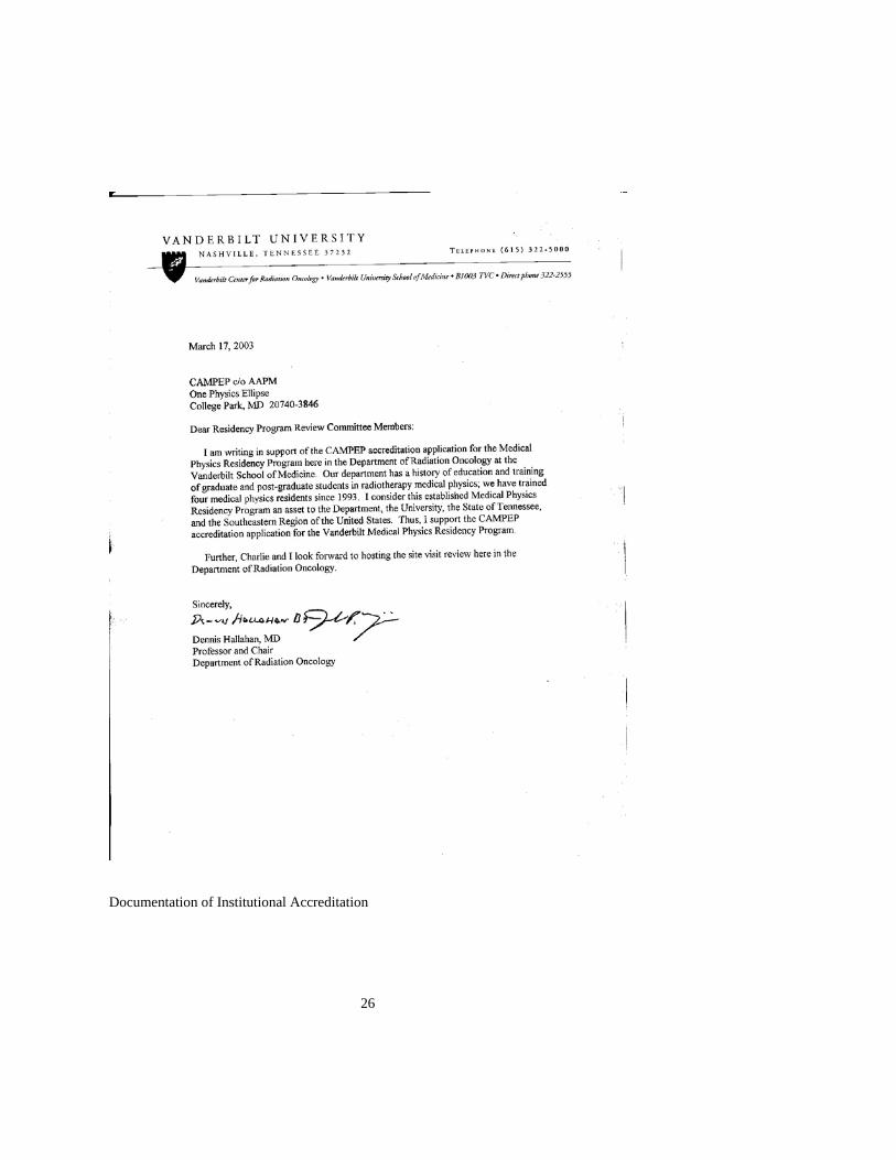
26
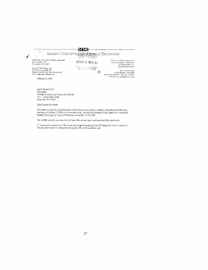
Documentation of Institutional Accreditation
27

28
H.2 Vanderbilt Medical Physics Residents

29
Resident: Patana Puwanich, PhD Date of Completion: June 30, 2003 Length of Time in Program: Two years (7/1/01 – 6/30/03) Medical Physics Specialty: Radiotherapy Physics Current Status: Deputy Director of Medical Physics Academic Program, Khon Kaen University in Khon Kaen, Thailand Board Certification: not applicable Resident: Chris Scarfone, PhD Date of Completion: June 30, 2001 Length of Time in Program: Two years (7/1/98 – 6/30/00) Medical Physics Specialty: Radiotherapy Physics Current Status: Assistant Professor, Department of Radiation Oncology, Vanderbilt University, Nashville, TN Board Certification: passed Parts I and II, Therapeutic Radiological Physics,
American Board of Radiology, will sit for the oral examination in June, 2003
Resident: Darrryl Kaurin, PhD Date of Completion: April 30, 1999 Length of Time in Program: Two years (4/97 – 4/99) Medical Physics Specialty: Radiotherapy Physics Current Status: Assistant Professor, Department of Radiation Oncology, Oregon Health and Sciences University, Portland, OR Board Certification: Therapeutic Radiological Physics, American Board of Radiology Resident: Dennis Duggan, PhD Date of Completion: August 30, 1994 Length of Time in Program: One year (8/93 – 8/94) (Note: Dr. Duggan spent his first in a fellowship position under my direct supervision in the Department of Radiation Medicine at the University of Kentucky Medical Center, in Lexington, KY Current Status: Associate Professor, Department of Radiation Oncology at the Vanderbilt University Medical Center, Nashville, TN Certifications: Therapeutic Radiological Physics, American Board of Radiology Radiation Oncology Physics, American Board of Medical Physics H.3 Clinical Therapy Physics Rotations

30
I. Clinical Rotations 1. Simulator and CT Simulator & Observation of Patient Treatment (2wks) 2. External Beam Treatment Planning (non 3D) (4wks) 3. External Beam Treatment Planning (3D) (12wks) 4. External Beam Treatment Planning (IMRT) (12wks) 5. External Beam Treatment Planning (Radiosurgery) (6 wks) 6. Brachytherapy Treatment Planning (LDR) (6wks) 7. Brachytherapy Treatment Planning (HDR) (12 wks) 8. Brachytherapy Treatment Planning (Prostate Seed Implants) (12wks) 9. Brachytherapy Treatment Planning (Intravascular Brachytherapy) (6wks) 10. Linear Accelerator Commissioning (10wks) 11. Treatment Planning Computer Commissioning (6wks) 12. Quality Assurance (Linac, MLC, Superfical X-ray, & Simulator) (6wks)
II. Training Objectives and Experience Rotation 1: Residents will observe the immbolization and positioning
techniques necessary for conventional and CT simulation. Residents will observe physician colleagues defining treatment fields and outline tumor volumes and adjacent normal tissues. Residents will observe patient immobilization and positioning necessary for daily radiotherapy treatment. Rotation 2: Residents will learn basic treatment planning equations for determination of monitor units and given dose using %DD, TAR, TMR, TPR, and off axis ratio data for applications of single and parallel-opposed radiotherapy fields. Residents will learn the QA associated with patient chart data entry and record keeping. Additionally, residents will learn to input beam parameters into the department’s radiotherapy record and verify computer. Rotation 3: Residents will learn the principles of treatment planning including procedures to achieve a 3D treatment plan (distribution and beam parameters) from a patient 3D data set. Residents will perform hand calculations for determination of monitor units for each treatment field; additionally, a monitor unit check program will used to determine monitor units. Residents will enter beam parameter data into the record and verify computer. Residents learn the principles of image fusion and its applications to 3D treatment planning.

31
Rotation 4: Residents will learn the principles of IMRT optimization, treatment planning, and tumor and normal tissues dose constraints evaluation. Residents will independently determine IMRT treatment field monitor units using a monitor unit check program. Residents will additionally participate in the “in vivo” quality assurance measurements of relative intensity and absolute dose determination for each patient field. Rotation 5: Residents will learn the principles of radiosurgery treatment planning including hand calculation determination of monitor units. Residents will participate in daily QA procedures for radiosurgery patient setup and treatment. Rotation 6: Residents will learn the principles of brachytherapy treatment planning optimization and point dose calculation using hand calculation Methods (Along and Away and point source approximation. Residents will learn the techniques of brachytherapy by understanding the design and use of varying brachytherapy applicators. Residents will participate in QA including source activity and source preparation. Rotation 7: Residents will learn the treatment planning software and hardware associated with an HDR Ir-192 afterloader. Residents will learn the applications of imaging based brachytherapy including orthogonal films and 3D CT images. Residents will learn an independent method to calculate HDR point dose rates. Residents will participate in daily, monthly, and quarterly QA procedures including HDR source calibration determination.
Rotation 8: Residents will learn the procedures and techniques involved in the treatment planning associated with permanent prostate seed implants. Residents will utilize US images for pre-implant
planning and CT images for post implant planning. Residents will participate in the QA procedures including source inventory and calibration. Residents will participant in physics responsibilities during the brachytherapy surgery procedure. Rotation 9: Residents will learn the procedures and techniques involved in intravascular brachytherapy. Residents will participate in daily and monthly QA procedures including IVB source calibration and record keeping. Residents will participate in physics responsibilities during the intravascular surgery procedure. Rotation 10: Residents will participate in the yearly calibration of a high energy linear accelerator. Residents will collect beam data

32
representative of data necessary for linear accelerator commissioning and prepare a calibration report. Rotation 11: Residents will learn the procedures, techniques, and beam data analysis necessary for the commissioning of a clinical radiotherapy beam in a treatment planning computer. Residents will demonstrate competence by entry of test beam data. Rotation 12: Residents will learn procedures for the routine quality assurance of all major radiotherapy equipment including: linear accelerator, superficial x-ray, simulator, MLC, and portal imager. Residents will participate in assigned QA activities and prepare data analysis reports.
III. Clinical Conferences 1. Weekly Patient Chart Conference 2. Weekly Patient Case Presentation Conference
3. Weekly Patient Chart Check Activities 4. Monthly Physics Section Meeting
IV. Medical Physics Teaching Training After adequate medical physics didactic and training experience, residents will be given a limited teaching assignment in the didactic courses of medical physics that are taught in the medical radiotherapy resident and radiation therapist education programs. The Program Director will be present at the lectures to assist and supervise the resident
teaching experience. V. Methods of Resident Evaluation

33
Didactic Training The Program Director meets with the resident candidate at least once during each semester to assess resident standing in individual classes. Resident progress (quizzes, homework, and examinations) is monitored by the course instructor throughout the semester. Any difficulties will be discussed with the resident in a private faculty/resident meeting. Should the situation warrant, the Program Director will be notified and a Program Director/resident meeting scheduled. The resident is expected to obtain a minimum grade of ‘B’ on the courses completed. Continued resident evaluation of below satisfactory could result in dismissal from the program. Clinical Training The specific clinic rotation supervisor will be responsible for the resident’s evaluation within that rotation. Immediate feedback of the resident’s performance will be determined with the application of the Vanderbilt Physics Section double-check policy. Monitor unit calculations, tumor doses, and treatment beam parameters will be double-checked before the patient receives his/her first treatment. Performance of rotation specific clinical duties will be supervised by the clinic rotation supervisor (or designee). The resident will be allowed to proceed independently with procedures and equipment operation after approval by the rotation supervisor. The resident will keep a notebook of representative treatment plans, clinical procedures, and data for rotation specific assignments; the notebook will be reviewed at least semi-annually by the Program Director. Should the resident’s clinical performance be less than minimally satisfactory, the rotation supervisor will contact the Program Director. The Program Director, rotation supervisor, and the resident will have a joint meeting to discuss the resident’s performance. Should remedial action be required the Program Director and rotation supervisor will determine the magnitude and length of time for which that specific training must be repeated. Continued resident evaluation of below satisfactory could result in dismissal from the program. H.4 Didactic Course Titles, Content, and Instructors
34
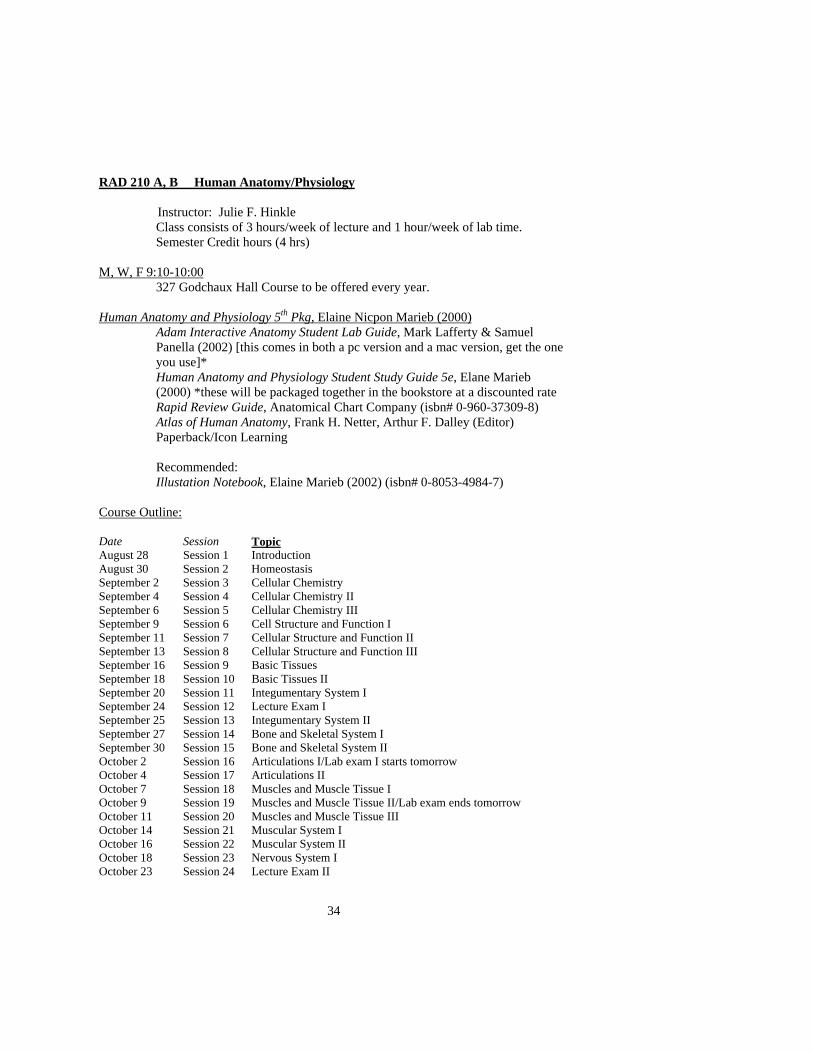
RAD 210 A, B Human Anatomy/Physiology Instructor: Julie F. Hinkle
Class consists of 3 hours/week of lecture and 1 hour/week of lab time. Semester Credit hours (4 hrs)
M, W, F 9:10-10:00 327 Godchaux Hall Course to be offered every year.
Human Anatomy and Physiology 5th Pkg, Elaine Nicpon Marieb (2000)
Adam Interactive Anatomy Student Lab Guide, Mark Lafferty & Samuel Panella (2002) [this comes in both a pc version and a mac version, get the one you use]* Human Anatomy and Physiology Student Study Guide 5e, Elane Marieb (2000) *these will be packaged together in the bookstore at a discounted rate Rapid Review Guide, Anatomical Chart Company (isbn# 0-960-37309-8) Atlas of Human Anatomy, Frank H. Netter, Arthur F. Dalley (Editor) Paperback/Icon Learning Recommended: Illustation Notebook, Elaine Marieb (2002) (isbn# 0-8053-4984-7)
Course Outline:
Date Session Topic August 28 Session 1 Introduction August 30 Session 2 Homeostasis September 2 Session 3 Cellular Chemistry September 4 Session 4 Cellular Chemistry II September 6 Session 5 Cellular Chemistry III September 9 Session 6 Cell Structure and Function I September 11 Session 7 Cellular Structure and Function II September 13 Session 8 Cellular Structure and Function III September 16 Session 9 Basic Tissues September 18 Session 10 Basic Tissues II September 20 Session 11 Integumentary System I September 24 Session 12 Lecture Exam I September 25 Session 13 Integumentary System II September 27 Session 14 Bone and Skeletal System I September 30 Session 15 Bone and Skeletal System II October 2 Session 16 Articulations I/Lab exam I starts tomorrow October 4 Session 17 Articulations II October 7 Session 18 Muscles and Muscle Tissue I October 9 Session 19 Muscles and Muscle Tissue II/Lab exam ends tomorrow October 11 Session 20 Muscles and Muscle Tissue III October 14 Session 21 Muscular System I October 16 Session 22 Muscular System II October 18 Session 23 Nervous System I October 23 Session 24 Lecture Exam II

35
October 25 Session 25 Nervous System II October 28 Session 26 Nervous System III October 30 Session 27 Action Potentials/Lab Exam II starts tomorrow November 1 Session 28 Central Nervous System I November 4 Session 29 Central Nervous System II/Lab Exam ends tomorrow November 6 Session 30 Central Nervous System III November 8 Session 31 Peripheral Nervous System I November 11 Session 32 Peripheral Nervous System II November 13 Session 33 Peripheral Nervous System III November 15 Session 34 Autonomic Nervous System I November 18 Session 35 Lecture Exam III November 20 Session 36 Autonomic Nervous System II December 2 Session 37 Autonomic Nervous System III/Lab Exam Starts Today December 4 Session 38 Neural Integration IDecember 6 Session 39 Neural Integration II December 9 Session 40 Course Review/Lab Exam ends Today December 11 Session 41 Course Review Method of Student Evaluation:
a. Grading Policy:
Grades for each semester of the course are calculated separately. Plus and minus grades are given at the discretion of the instructor
i. Final Grade: 90-100: A 80-89: B 70-79: C
60-69: D Below 60: F
ii. Exams: Three written exams 100 points, each Three laboratory exams 100 points, each Final written exam 200 points Homework 100 points
Final Semester Grade, total Total points/8*
*The lowest score on any single exam, EXCEPT THE FINAL WRITTEN EXAM, will be dropped in the calculation of the final grade.
36
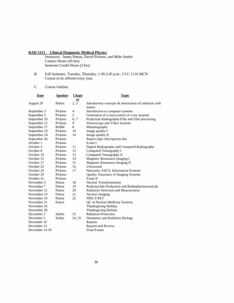
RAD 5313 Clinical Diagnostic Medical Physics Instructor: James Patton, David Pickens, and Mike Stabin Contact Hours (45 hrs) Semester Credit Hours (3 hrs)
B. Fall Semester, Tuesday, Thursday, 1:30-2:45 p.m., CCC-1116 MCN
Course to be offered every year.
C. Course Outline:
Date Speaker Chapter
Topic
August 29 Patton 2, 3 Introductory concepts & interactions of radiation with matter
September 3 Pickens 4 Introduction to computer systems September 5 Pickens 5 Generation of x-rays/control of x-ray systems September 10 Pickens 6, 7 Projection Radiography/Film and Film processing September 12 Pickens 9 Fluoroscopy and Video Systems September 17 Riddle 8 Mammography September 19 Pickens 10 Image quality I September 24 Pickens 10 Image quality II September 26 Pickens Report topic descriptions due October 1 Pickens Exam I October 3 Pickens 11 Digital Radiography and Computed Radiography October 8 Pickens 13 Computed Tomography I October 10 Pickens 13 Computed Tomography II October 15 Pickens 14 Magnetic Resonance Imaging I October 17 Pickens 15 Magnetic Resonance Imaging II October 22 Pickens 16 Ultrasound October 24 Pickens 17 Networks, PACS, Information Systems October 29 Pickens Quality Assurance of Imaging Systems October 31 Pickens Exam II November 5 Patton 18 Nuclear Transformations November 7 Patton 19 Radionuclide Production and Radiopharmaceuticals November 12 Patton 20 Radiation Detection and Measurement November 14 Patton 21 Nuclear Imaging November 19 Patton 22 SPECT/PET November 21 Patton QC of Nuclear Medicine Systems November 26 Thanksgiving Holiday November 28 Thanksgiving Holiday December 3 Stabin 23 Radiation Protection December 5 Stabin 24, 25 Dosimetry and Radiation Biology December 10 Reports December 12 Reports and Review December 14-19 Final Exams

37
D. Method of Student Evaluation:
I. In-depth Report: An area of special interest will be selected by each student. Independent research of the topic will be performed leading to an in-depth review of the area and a written report. Resources should include other texts, journal articles and abstracts, and other materials as appropriate (internet resources, manufacturers’ information, etc.). The reports will be turned in at the time of a presentation of the topic to the class on one of the last two days of classes. Reports are expected to be at least 10 pages with appropriate references. Topics might include direct digital imaging systems for diagnostic or therapy (portal) imaging, CT angiography, functional imaging with emission tomography, etc. The selected area should be written up in the form of single paragraph proposal for review and approval before beginning the research. The instructor can offer suggestions or help with selection of a suitable topic.
II. Grading:
Reports: 20% Exams 1 & 2: 25%, each Exam 3: 30%

38
RADO 5343 Radiology/Health Physics Instructor: Michael Stabin Contact Hours (44 hrs) Semester Credit Hours (3 hrs)
E. Fall Semester, Tuesday, Thursday, 8:10-9:25 a.m.
Course to be offered every year.
F. Text: H. Cember, Introduction to Health Physics, 3rd Edition, McGraw-Hill, New York, 1996
G. Course Outline:
Date Topic Chapter August 29 Atomic and Nuclear Structure 3 September 3 Radioactivity – Transformation Mechanisms 4 September 5 Radioactivity – Transformation Kinetics 4 September 10 Interaction of Radiation with Matter 5 September 12 Interaction of Radiation with Matter 5 September 17 Radiation Dosimetry 6 September 19 Radiation Dosimetry 6 September 24 Biological Effects of Radiation 7 September 26 Biological Effects of Radiation 7 October 1 History of Radiation Regulations 8 October 3 Guidance and Regulatory Bodies 8 October 8 Regulatory Limits 8 October 10 TEST I - October 15 Radiation Instrumentation 9 October 17 Radiation Instrumentation 9 October 22 Counting Statistics 9 October 24 External Protection 10 October 29 External Protection 10 October 31 Internal Protection 11 November 5 Internal Protection 11 November 7 Radioactive Waste Management 11 November 12 Criticality 12 November 14 Protective Measures 13 November 19 Protective Measures 13 November 21 TEST II - November 26 Holiday - November 28 Holiday - December 3 Environmental Monitoring - December 5 Non-ionizing Radiation 14 December 10 Final Exam - *Subject to adjustment as needed
39
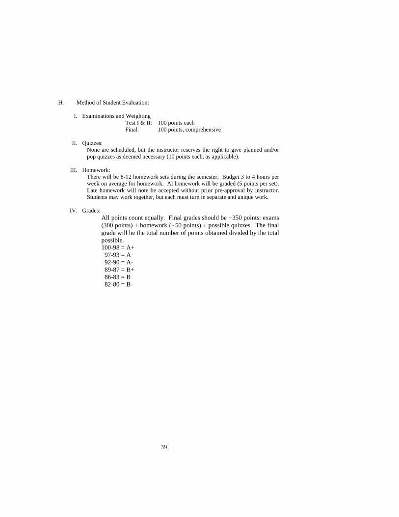
H. Method of Student Evaluation:
I. Examinations and Weighting Test I & II: 100 points each
Final: 100 points, comprehensive
II. Quizzes: None are scheduled, but the instructor reserves the right to give planned and/or pop quizzes as deemed necessary (10 points each, as applicable).
III. Homework: There will be 8-12 homework sets during the semester. Budget 3 to 4 hours per week on average for homework. Al homework will be graded (5 points per set). Late homework will note be accepted without prior pre-approval by instructor. Students may work together, but each must turn in separate and unique work.
IV. Grades: All points count equally. Final grades should be -350 points: exams (300 points) + homework (-50 points) + possible quizzes. The final grade will be the total number of points obtained divided by the total possible. 100-98 = A+ 97-93 = A 92-90 = A- 89-87 = B+ 86-83 = B 82-80 = B-

40
RADO 5248 Radiation Biology Instructor: Mike Freeman, Ph.D.
Contact hours (30 hrs) Semester Credit Hours (2 hrs)
I. Fall Semester
Course to be offered every other year
J. Text: Radiobiology for the Radiobiologist Author: E.J. Hall
*Supplemental information will be provided from the following text: Introduction to Radiobiology by Tubiana Dutreix and Wambersie K. Course Outline (2 lectures per topic):
I. Radiation Physics as it pertains to Radiation Biology A. Electromagnetic and particulate radiation B. Ionization/excitation C. Stopping power, track structure, & LET
II. Radiation Chemistry A. Radiolysis of water B. Direct and indirect effects C. G values D. Radiolysis of DNA
a. Hydration effects, electron & hole transfer b. Base and sugar radicals
E. Generation of ceramides III. Formation and Repair of Damage
A. Formation of breaks (ssb, base, dsb, protein cross links), mutations, and chromosomal damage B. SLD & PLD C. Repairs systems:
a. Genes and enzymes b. Base – NER c. DSB – non-homologous end joining and homologous recombination
D. Cell cycle check points – eg, ATM, p53, p21, etc E. Signaling: eg, NFκB, ceramides, AP-1, Erg-1, etc F. Oncogenes G. Bystander effect
IV. Expression of Cell Death A. apoptosis B. inter-mitotic induced cell death-production of chromosomal aberrations C. quantitation (survival curve analysis –target theory)
V. Modifiers of Radiation Damage A. RBE and LET B. Dose per fraction and dose rate C. Chemical and physical factor – eg, oxygen, Budr, radioprotectors,
radiosensitizers VI. Radiation Effects on Normal Tissues and Organisms

41
A. Organ sensitivity a. TD-tolerance doses as a function of dose, fraction size, volume, dose
rate, multi modalities, late effects and acute effects- predictive assays, cell kinectics
B. Morphologic patterns of radiation injury VII. Radiation Effects on Tumor Tissue
A. Growth fractions, cell loss, cell cycle times B. The 4 Rs of radiobiology C. α/β model for late & acute reactions vs tumor tissures D. accelerated repopulation E. hyperfractionation F. accelerated fractionation G. Strandquist plots and NSD H. TCD50 I. Growth Delay
VIII. The Role of Modalities in Radiation Therapy A. Alternative modalities
a. Physical: neutrons, protons, pi mesons, hyperthermia b. Pharmacological: hypoxic cell sensitizers, halogenated pyrimidines,
radioprotectors c. Hyperthermia d. Gene therapy e. Angiogenic inhibitors
IX. Acutes Effects of Whole Body Irradiation A. Prodromal B. Cerebrovascular C. Gastrointestinal D. Hematopoietic E. LD50
X. Radiation Cataractogenesis A. Cataracts of the Lens B. Lens opacification in experimental animals C. Cataracts in humans D. Degree of opacity E. Latent period F. Dose-response relationships
XI. Carcinogenesis and Non-specific Life Shortening A. Definition of late effects B. Carcinogenesis C. Latent period D. Models of carcinogenesis E. Assessing risk for formation of various tumors F. Malignancy in children G. Molecular mechanisms of radiation induced carcinogenesis H. Nonspecific life shortening
XII. Genetic Changes A. Mutation effects in Drosophila B. Mutation effects in mice C. Mutation effects in human
XIII. Effects of Radiation on the Embryo and Fetus A. Effects in developing embryo

42
B. Experience in humans C. Summary of animal and human data for teratogenesis D. Malignancies associated with irradiation in utero E. Occupational exposure of women of childbearing age F. Pregnant or potentially pregnant patient
XIV. Radiation Protection: Risk vs. Benefit A. Sources of radiation to the human population B. Risk C. Doses from diagnostic radiology D. Doses from nuclear medicine E. Doses from natural background
L. Method of Student Evaluation:
Students are required to submit 3 papers

43
RADO 5301A Radiation Oncology Clinical Oncology Lecture Seminar
Semester Credit Hours (1 hrs)
Fall Semester, Friday, 8:00 a.m. Course to be offered every year.
Handouts Lecture Schedule Date Topic Presenter
July 11 Emergency Overview Anthony Cmelak July 19 Prostate/GYN Overview Ming Teng July 26 Breast Overview Bapsi Chak August 2 Lung I Hak Choy August 16 PEDS Overview Ming Teng August 22 GI Overview Bapsi Chak September 9 Breast I Bapsi Chak September 16 Breast Dosimetry Wyndee Kirby September 23 Breast II Bapsi Chak September 30 Breast Journal Club Bapsi Chak October 7 Head and Neck I Anthony Cmelak October 14 Head and Neck II Anthony Cmelak October 21 Head and Neck Dosimetry Wyndee Kirby October 28 Head and Neck Journal Club Anthony Cmelak November 4 Lung Dosimetry Charles Coffey November 11 Non-Small Cell Lung Cancer Hak Choy November 18 Small Cell Lung Cancer Hak Choy November 25 Lung Journal Club Hak Choy December 2 Cervix Ming Teng December 16 Endometrium Ming Teng

44
RADO 5304 Radiation Interactions and Dosimetry
Instructor: Dennis Duggan Contact Hours (44 hrs)
Semester Credit Hours (3 hrs)
Spring Semester, Monday, Wednesday; 10:20 a.m. Course to be offered every year.
Text: Attix, Introduction to Radiological Physics and Radiation Dosimetry
Wiley-Interscience, 1986 Course Outline:
Lecture 1 Fluence and flux; Fluence, fluence rate, flux, flux density, Energy fluence
Attix, Ch. 1
Lecture 2 Energy Transferred and Kerma; Energy transferred, energy fluence, energy transfer coefficient, and kerma; Average secondary electron energy for a photon beam; Radiation and collisional kerma and fraction of energy lost to bremsttrahlung
Attix, Ch. 2
Lecture 3 Exposure and Air Kerma; Relationship between exposure, W, and collision kerma
Attix, Ch. 2
Lecture 4 Absorbed Dose and Terma; Energy imparted, energy absorption coefficient, energy fluence and absorbed dose (A); Terma and the convolugiton integral for dose (P)
Attix, Ch. 2 Papanikalaou article
Lecture 5-6 Charged Particle Equilibrium; Definition; Absorbed dose and collision kerma under CPE conditions; Exposure rate constant and absorbed dose from brachytherapy source; TG-43 definition of source strength and absorbed dose
Attix, Ch. 4 TG-43 Report
Lecture 7 Transient Charged Particle Equilibrium; Definition of buildup, TCPE, and beta factor; Absorbed dose and collision kerma under TCPE conditions
Attix Ch. 4
Lecture 8-9 Cavity Theory; The three limiting cases: Small cavity, Large cavity, Medium-sized cavity; Bragg-Gray cavity theory: Dose, particle fluence and mass stopping power ratio, Bragg-Gray equation and assumptions behind it
Attix, Ch. 10
Lecture 10 Cavity Theory; Spencer-Attix Cavity Theory; Assumptions in addition to those behind Bragg-Gray equation; Spencer-Attix equation; Restricted mass stopping power
Attix, Ch. 10
Lecture 11-12 Burlin Cavity Theory; Addition assumptions; Burlin equation: approximations for beta, limiting cases
Attix, Ch. 10
Lecture 13-14 Free Air Chamber Attix, Ch. 12 Lecture 15-16 Cavity Chambers; Parallel plate chambers, thimble
chambers, saturation, recombination, polarity effect Attix, Ch. 12
Lecture 17 Midterm Exam --

45
Lecture 18-20 TG-21 Protocol; Rationale for TG-21 Attix, Ch. 13 Med. Phys. 8, 1, 1981 Rogers in Proceedings of 1980 AAPM Summer School, Advanced Med. Pub., 1990 Shulz et al., Med. Phys. 10, 1, 1983
Lecture 21-24 TG-51 Protocol; Rationale for TG-51 Protocol; Practical Implementation of TG-51
Rogers in Proceedings of 1996 AAPM Summer School, Advanced Med. Publ., 1996 AAPM RT TG-51 Report, AAPM Report #67
Lecture 25 Thermoluminescent Dosimeters; LiF TLDs and Burlin Theory
Attix, Ch. 10, 11, 14
Lecture 26 Silicon Diodes Attix, Ch. 16 AAPM RT TG-62 Report
Lecture 27 Silver Halide Film Dosimetry Attix, Ch. 14 Lecture 28 Radiochromic Film Dosimetry AAPM RT TG-55
Report AAPM Report #63
46
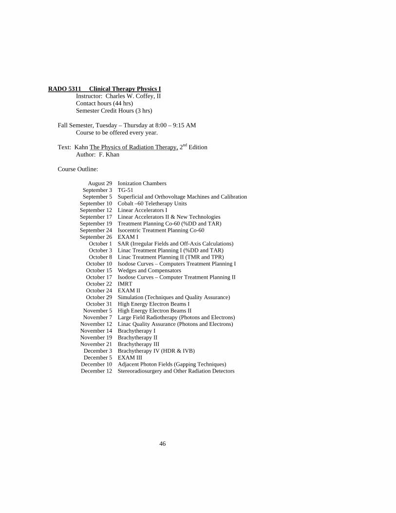
RADO 5311 Clinical Therapy Physics I Instructor: Charles W. Coffey, II
Contact hours (44 hrs) Semester Credit Hours (3 hrs)
Fall Semester, Tuesday – Thursday at 8:00 – 9:15 AM
Course to be offered every year.
Text: Kahn The Physics of Radiation Therapy, 2nd Edition Author: F. Khan
Course Outline:
August 29 Ionization Chambers September 3 TG-51 September 5 Superficial and Orthovoltage Machines and Calibration
September 10 Cobalt –60 Teletherapy Units September 12 Linear Accelerators I September 17 Linear Accelerators II & New Technologies September 19 Treatment Planning Co-60 (%DD and TAR) September 24 Isocentric Treatment Planning Co-60 September 26 EXAM I
October 1 SAR (Irregular Fields and Off-Axis Calculations) October 3 Linac Treatment Planning I (%DD and TAR) October 8 Linac Treatment Planning II (TMR and TPR)
October 10 Isodose Curves – Computers Treatment Planning I October 15 Wedges and Compensators October 17 Isodose Curves – Computer Treatment Planning II October 22 IMRT October 24 EXAM II October 29 Simulation (Techniques and Quality Assurance) October 31 High Energy Electron Beams I
November 5 High Energy Electron Beams II November 7 Large Field Radiotherapy (Photons and Electrons)
November 12 Linac Quality Assurance (Photons and Electrons) November 14 Brachytherapy I November 19 Brachytherapy II November 21 Brachytherapy III
December 3 Brachytherapy IV (HDR & IVB) December 5 EXAM III
December 10 Adjacent Photon Fields (Gapping Techniques) December 12 Stereoradiosurgery and Other Radiation Detectors

47
Method of Student Evaluation:
I. Examinations and Weighting Exam I: 20% Exam II: 20% Exam III: 20% Final: 40%
II. Grading A 89.5 – 100 B+ 86.5 – 89.4, B 82.5 – 86.4, B- 79.5 – 82.4 C 69.5 – 79.4 Failing Below 69.4
III. Exam Policy:
1) Should student knowingly have a conflict prior to day of exam, exam must be taken before scheduled examination date.
2) Should student miss a scheduled exam due to illness, a make-up exam will be given the first day that the student is able to return to class.
48
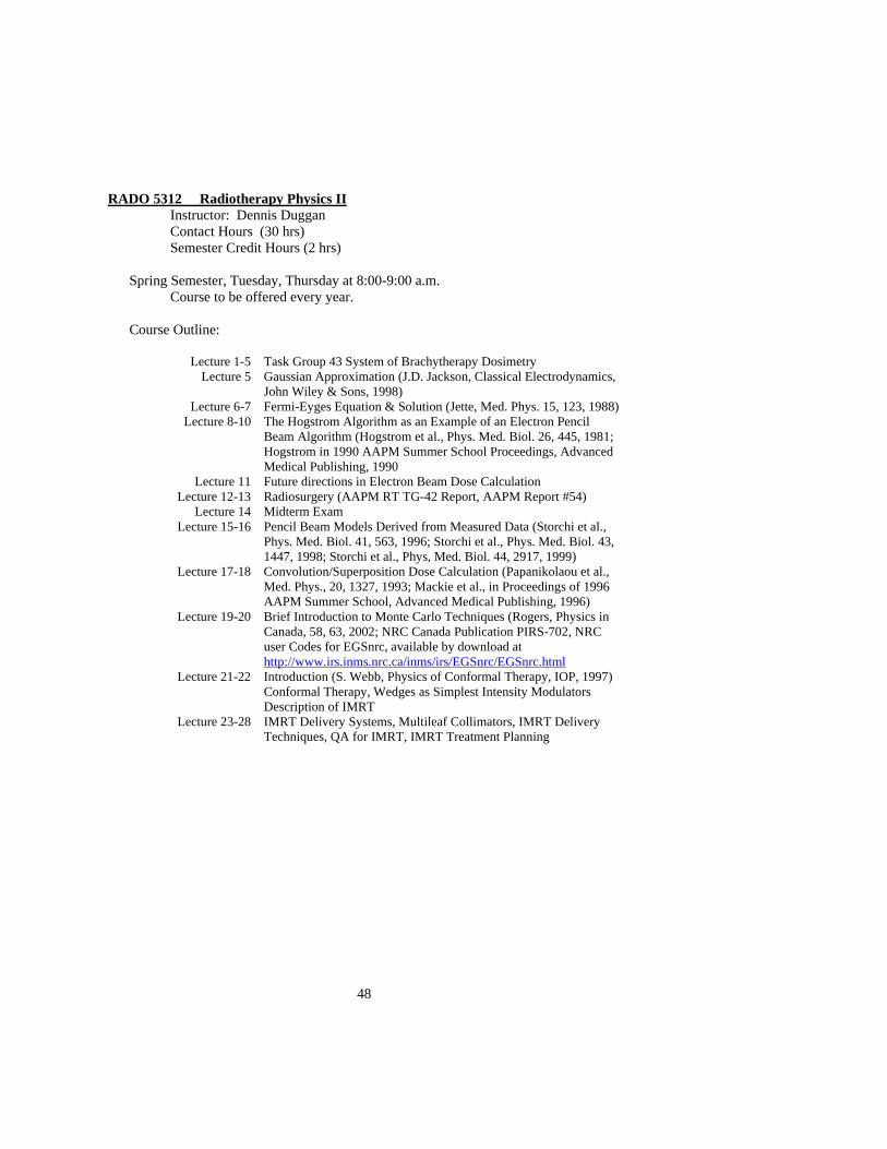
RADO 5312 Radiotherapy Physics II Instructor: Dennis Duggan Contact Hours (30 hrs)
Semester Credit Hours (2 hrs)
Spring Semester, Tuesday, Thursday at 8:00-9:00 a.m. Course to be offered every year.
Course Outline:
Lecture 1-5 Task Group 43 System of Brachytherapy Dosimetry Lecture 5 Gaussian Approximation (J.D. Jackson, Classical Electrodynamics,
John Wiley & Sons, 1998) Lecture 6-7 Fermi-Eyges Equation & Solution (Jette, Med. Phys. 15, 123, 1988)
Lecture 8-10 The Hogstrom Algorithm as an Example of an Electron Pencil Beam Algorithm (Hogstrom et al., Phys. Med. Biol. 26, 445, 1981; Hogstrom in 1990 AAPM Summer School Proceedings, Advanced Medical Publishing, 1990
Lecture 11 Future directions in Electron Beam Dose Calculation Lecture 12-13 Radiosurgery (AAPM RT TG-42 Report, AAPM Report #54)
Lecture 14 Midterm Exam Lecture 15-16 Pencil Beam Models Derived from Measured Data (Storchi et al.,
Phys. Med. Biol. 41, 563, 1996; Storchi et al., Phys. Med. Biol. 43, 1447, 1998; Storchi et al., Phys, Med. Biol. 44, 2917, 1999)
Lecture 17-18 Convolution/Superposition Dose Calculation (Papanikolaou et al., Med. Phys., 20, 1327, 1993; Mackie et al., in Proceedings of 1996 AAPM Summer School, Advanced Medical Publishing, 1996)
Lecture 19-20 Brief Introduction to Monte Carlo Techniques (Rogers, Physics in Canada, 58, 63, 2002; NRC Canada Publication PIRS-702, NRC user Codes for EGSnrc, available by download at http://www.irs.inms.nrc.ca/inms/irs/EGSnrc/EGSnrc.html
Lecture 21-22 Introduction (S. Webb, Physics of Conformal Therapy, IOP, 1997) Conformal Therapy, Wedges as Simplest Intensity Modulators Description of IMRT
Lecture 23-28 IMRT Delivery Systems, Multileaf Collimators, IMRT Delivery Techniques, QA for IMRT, IMRT Treatment Planning

49
RADO 5314 Radiotherapy Physics Laboratory Instructor: Charles W. Coffey, II
Contact hours ( 56 hrs) Semester Credit Hours (2 hrs)
Fall Semester, Monday 1:00-5:00 p.m.
Course to be offered every year.
Course Outline:
January 10 Ion Chamber Measurements & Performance Characteristics Co-60 January 17 Cobalt-60 Beam Parameters Co-60 January 24 Absolute Output, %Depth Dose and Sc/Sp Clinac 4 January 31 TAR and TPR Clinac 4 February 7 TMR, TMR0, and SMR Clinac 4
February 14 Superficial/Orthovoltage X-Ray Beam Parameters X-Ray Unit February 21 Absolute Outputs and %Depth Dose for Electrons Clinac 6/18 February 28 Simulator Quality Assurance Simulator
March 7 SPRING BREAK -- March 14 Adjacent Fields (Gapping Techniques) Simulator/Clinac 4 March 21 CPK Quality Assurance Clinac 4 March 28 Pelvis Phantom and Verification of IMRT Doses in Water Clinac 6/18
April 4 Total Skin Electron Dosimetry Verification Clinac 6/10 April 11 HDR and IVB Quality Assurance HDR & IVB Units April 18 GYN Brachytherapy Dose Verification using HDR HDR Unit
Method of Student Evaluation:
I. Lab Report and Grading: Achievable Objectives: 5 pts Materials and Methods: 15 pts Data: 15 pts Data Analysis and Results: 35 pts Discussion and Conclusions: 25 pts References (2)* : 5 pts
II. Final Grade Assessment:
Lab Reports: 80% Final (2 hours written exam): 20%
III. Other Topics/Rules:
1. Lab Write-Up Deadlines: a. Due at beginning of scheduled lab session 2 weeks following
lab experiment. b. 20% late fee within 24 hours c. 30% late fee 1 day late d. 40% late fee 2 days late e. 50% late fee 3 days late
2. Laboratory report not submitted will receive 0 points.

50
3. Missed labs a. Missed Lab (excused) make-up available b. Missed Lab (not excused) 50% late fee (lab data will be
assigned) 4. General Rules
a. Wear Film Badge b. Don’t plagiarize thy neighbor’s report c. Don’t be tardy d. During laboratory class, if you are not doing something constructive, ask to be involved.
*References must be copied and submitted with lab report.
I. Sample Laboratory Exercise: Experiment #3
Absolute Output, %Depth Dose, and SC/SP
Radiation Source: Clinac 4/100 Equipment:
1. Working ion chamber and electrometer 2. Standard ion chamber and electrometer 3. Water phantom 4. Water proof sheath for ion chamber 5. Buildup cap for ion chamber 6. Solid water phantom 7. Clinical wedge set including 30, 45, and 60 degree wedges.
Objectives
1. Determine absolute output for Clinac 4/100 at 100 cm SSD, at a depth of dmax, for a field size of 10 x 10, using TG#51 formalism.
2. Determine the %DD for field sizes of 5 x 5, 8 x 8, 10 x 10, 12 x 12, 14 x 14, and
18 x 18 for depths of dmax, 3, 5, 8, 10, 12, 15, 18 cm. 3. Determine SC,P for field sizes, 4 x 4, 6 x 6, 8 x 8, 10 x 10, 12 x 12, 16 x 16, 20 x
20, 25 x 25, 30 x 30, and 36 x 36 at depth of dmax, in water.
4. Determine SC for field sizes, 4 x 4, 6 x 6, 8 x 8, 10 x 10, 12 x 12, 16 x 16, 20 x 20, 25 x 25, 30 x 30, and 36 x 36 in air with buildup cap.
5. Compare %DD data with published values.

51
6. Determine SC and SP using Khan formalism.
7. Determine beam symmetry and flatness for 4 MV x-rays for 30 x 30 field size,
depth = 10 cm, and 100 cm SSD for transverse and radial planes.
8. Measure OCR profiles for 30 x 30 field at 100 cm SSD for depths of dmax, 5, 15, and 20 cm.
9. Measure wedge attenuation factors at 10 x 10 field size, depth of 10 cm (solid
water), 100 cm SSD for 30, 45, and 60 degree wedges.
10. Measure wedge attenuation factors for the 30 degree wedge atg 5 x 5 and 15 x 15 field sizes at 100 cm SSD, for depths of dmax, 5, 10, and 15 cm (solid water).
52
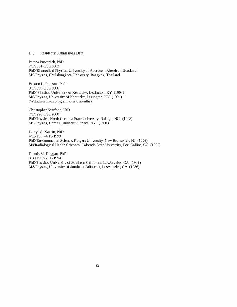
H.5 Residents’ Admissions Data Patana Puwanich, PhD 7/1/2001-6/30/2003 PhD/Biomedical Physics, University of Aberdeen, Aberdeen, Scotland MS/Physics, Chulalongkorn University, Bangkok, Thailand Buxton L. Johnson, PhD 9/1/1999-3/30/2000 PhD/ Physics, University of Kentucky, Lexington, KY (1994) MS/Physics, University of Kentucky, Lexington, KY (1991) (Withdrew from program after 6 months) Christopher Scarfone, PhD 7/1/1998-6/30/2000 PhD/Physics, North Carolina State University, Raleigh, NC (1998) MS/Physics, Cornell University, Ithaca, NY (1991) Darryl G. Kaurin, PhD 4/15/1997-4/15/1999 PhD/Environmental Science, Rutgers University, New Brunswick, NJ (1996) Ms/Radiological Health Sciences, Colorado State University, Fort Collins, CO (1992) Dennis M. Duggan, PhD 8/30/1993-7/30/1994 PhD/Physics, University of Southern California, LosAngeles, CA (1982) MS/Physics, University of Southern California, LosAngeles, CA (1986)
53
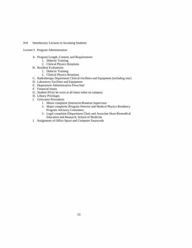
H.6 Introductory Lectures to Incoming Students Lecture I: Program Administration
A. Program Length, Content, and Requirements
1. Didactic Training 2. Clinical Physics Rotations
B. Resident Evaluations 1. Didactic Training 2. Clinical Physics Rotations
C. Radiotherapy Department Clinical Facilities and Equipment (including tour) D. Laboratory Facilities and Equipment E. Department Administration Flowchart F. Financial Issues G. Student ID (to be worn at all times when on campus) H. Library Privileges I. Grievance Procedures
1. Minor complaint (Instructor/Rotation Supervisor 2. Major complaint (Program Director and Medical Physics Residency
Program Advisory Committee 3. Legal complaint (Department Chair and Associate Dean Biomedical
Education and Research, School of Medicine J. Assignment of Office Space and Computer Passwords
54
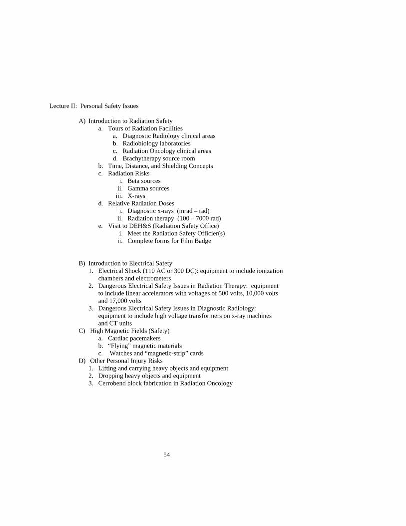
Lecture II: Personal Safety Issues
A) Introduction to Radiation Safety a. Tours of Radiation Facilities
a. Diagnostic Radiology clinical areas b. Radiobiology laboratories c. Radiation Oncology clinical areas d. Brachytherapy source room
b. Time, Distance, and Shielding Concepts c. Radiation Risks
i. Beta sources ii. Gamma sources
iii. X-rays d. Relative Radiation Doses
i. Diagnostic x-rays (mrad – rad) ii. Radiation therapy (100 – 7000 rad)
e. Visit to DEH&S (Radiation Safety Office) i. Meet the Radiation Safety Officier(s)
ii. Complete forms for Film Badge
B) Introduction to Electrical Safety 1. Electrical Shock (110 AC or 300 DC): equipment to include ionization
chambers and electrometers 2. Dangerous Electrical Safety Issues in Radiation Therapy: equipment to include linear accelerators with voltages of 500 volts, 10,000 volts and 17,000 volts
3. Dangerous Electrical Safety Issues in Diagnostic Radiology: equipment to include high voltage transformers on x-ray machines and CT units
C) High Magnetic Fields (Safety) a. Cardiac pacemakers b. “Flying” magnetic materials c. Watches and “magnetic-strip” cards
D) Other Personal Injury Risks 1. Lifting and carrying heavy objects and equipment 2. Dropping heavy objects and equipment 3. Cerrobend block fabrication in Radiation Oncology
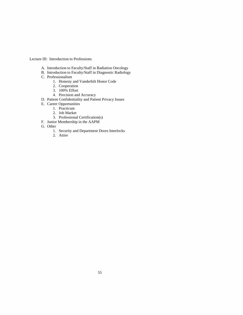
55
Lecture III: Introduction to Professions
A. Introduction to Faculty/Staff in Radiation Oncology B. Introduction to Faculty/Staff in Diagnostic Radiology C. Professionalism
1. Honesty and Vanderbilt Honor Code 2. Cooperation 3. 100% Effort 4. Precision and Accuracy
D. Patient Confidentiality and Patient Privacy Issues E. Career Opportunities
1. Practicum 2. Job Market 3. Professional Certification(s)
F. Junior Membership in the AAPM G. Other
1. Security and Department Doors Interlocks 2. Attire

56
H.7 Faculty Biographical Sketches
Charles Wade Bullington
Biographical Sketch
Job Title: Dosimetrist
Education: B.S. in Physics, University of Central Arkansas, Conway, Arkansas (2000)
Training: On the job training in Clinical Physics Section, May. 2002 to present
Board Certification/Licensure: N/A
Teaching Experience: Taught undergraduate entry level physics labs for 2 years,
Vanderbilt University
Clinical Experience: Employed as Medical Physics Assistant / Dosimetrist since
May, 2002

57
CURRICULUM VITAE
Laura E. Butler, M.S. Clinical Medical Physicist Vanderbilt University Medical Center EDUCATION 1999-2002 M.S. in Medical Physics University of Texas at
Houston Graduate School of Biomedical Sciences
May 1999 B.S. in Physics University of Memphis HONORS AND AWARDS Recipient of Robert J. Shalek Fellowship-University of Texas M.D.Anderson 1999-2000 Mu Alpha Theta Physics Award- University of Memphis, May 1999 PUBLICATIONS “Comparison of two immobilization techniques using portal film and digitally reconstructed radiographs for pediatric patients with brain tumors,” Int. J. Radiat. Oncol. Biol. Phys. 48, 1233-1240 (2000). CLINCIAL EXPERIENCE May 2002-present Vanderbilt University Medical Center Monthly and Annual QA of linear accelerators, stereotactic
radiosurgery, IMRT planning, 3D conformal treatment planning, HDR Brachytherapy planning
RESEARCH 2000-2002 Research Assistant, M.D. Anderson Medical Physics program 1997-1998 Research Assistant, St. Jude Children’s Research Hospital, Memphis, TN

58
PRESENTATIONS Comparison of two immobilization devices for pediatric patients with brain tumors- L. Butler, Y. Zhu, W. Gaber, S. Samant, M. Sontag, T. Merchant. Therapy Poster, American Association of Physicists in Medicine 1999 Annual Conference. Dosimetric Benefits of Respiratory Gating- L.E. Butler, K.M. Forster, C.W. Stevens, S.L. Tucker, G. Starkschall. Therapy Poster, American Association of Physicists in Medicine 2002 Annual Conference. PROFESSIONAL MEMBERSHIP American Association of Physicists in Medicine
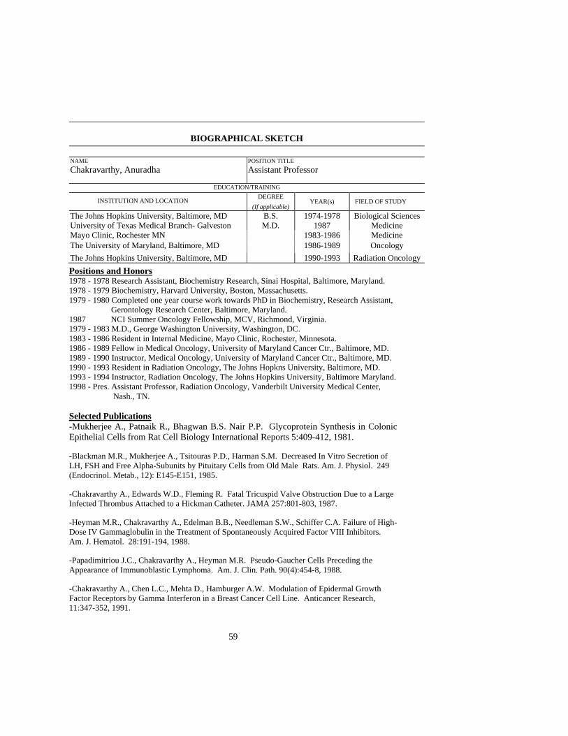
59
BIOGRAPHICAL SKETCH
NAME Chakravarthy, Anuradha
POSITION TITLEAssistant Professor
EDUCATION/TRAINING
INSTITUTION AND LOCATION DEGREE (If applicable)
YEAR(s) FIELD OF STUDY The Johns Hopkins University, Baltimore, MD University of Texas Medical Branch- Galveston
B.S. M.D.
1974-1978 1987
Biological Sciences Medicine
Mayo Clinic, Rochester MN 1983-1986 Medicine The University of Maryland, Baltimore, MD 1986-1989 Oncology The Johns Hopkins University, Baltimore, MD 1990-1993 Radiation Oncology Positions and Honors 1978 - 1978 Research Assistant, Biochemistry Research, Sinai Hospital, Baltimore, Maryland. 1978 - 1979 Biochemistry, Harvard University, Boston, Massachusetts. 1979 - 1980 Completed one year course work towards PhD in Biochemistry, Research Assistant, Gerontology Research Center, Baltimore, Maryland. 1987 NCI Summer Oncology Fellowship, MCV, Richmond, Virginia. 1979 - 1983 M.D., George Washington University, Washington, DC. 1983 - 1986 Resident in Internal Medicine, Mayo Clinic, Rochester, Minnesota. 1986 - 1989 Fellow in Medical Oncology, University of Maryland Cancer Ctr., Baltimore, MD. 1989 - 1990 Instructor, Medical Oncology, University of Maryland Cancer Ctr., Baltimore, MD. 1990 - 1993 Resident in Radiation Oncology, The Johns Hopkns University, Baltimore, MD. 1993 - 1994 Instructor, Radiation Oncology, The Johns Hopkins University, Baltimore Maryland. 1998 - Pres. Assistant Professor, Radiation Oncology, Vanderbilt University Medical Center, Nash., TN. Selected Publications -Mukherjee A., Patnaik R., Bhagwan B.S. Nair P.P. Glycoprotein Synthesis in Colonic Epithelial Cells from Rat Cell Biology International Reports 5:409-412, 1981.
-Blackman M.R., Mukherjee A., Tsitouras P.D., Harman S.M. Decreased In Vitro Secretion of LH, FSH and Free Alpha-Subunits by Pituitary Cells from Old Male Rats. Am. J. Physiol. 249 (Endocrinol. Metab., 12): E145-E151, 1985.
-Chakravarthy A., Edwards W.D., Fleming R. Fatal Tricuspid Valve Obstruction Due to a Large Infected Thrombus Attached to a Hickman Catheter. JAMA 257:801-803, 1987.
-Heyman M.R., Chakravarthy A., Edelman B.B., Needleman S.W., Schiffer C.A. Failure of High-Dose IV Gammaglobulin in the Treatment of Spontaneously Acquired Factor VIII Inhibitors. Am. J. Hematol. 28:191-194, 1988.
-Papadimitriou J.C., Chakravarthy A., Heyman M.R. Pseudo-Gaucher Cells Preceding the Appearance of Immunoblastic Lymphoma. Am. J. Clin. Path. 90(4):454-8, 1988.
-Chakravarthy A., Chen L.C., Mehta D., Hamburger A.W. Modulation of Epidermal Growth Factor Receptors by Gamma Interferon in a Breast Cancer Cell Line. Anticancer Research, 11:347-352, 1991.
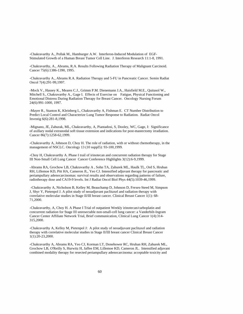
60
-Chakravarthy A., Pollak M., Hamburger A.W. Interferon-Induced Modulation of EGF-Stimulated Growth of a Human Breast Tumor Cell Line. J. Interferon Research 11:1-8, 1991.
-Chakravarthy, A., Abrams, R.A. Results Following Radiation Therapy of Malignant Carcinoid. Cancer 75(6):1386-1390, 1995.
-Chakravarthy A., Abrams R.A. Radiation Therapy and 5-FU in Pancreatic Cancer. Semin Radiat Oncol 7(4):291-99,1997.
-Mock V., Hassey K., Meares C.J., Grimm P.M. Dienemann J.A., Haisfield M.E., Quitasol W., Mitchell S., Chakravarthy A., Gage I. Effects of Exercise on Fatigue, Physical Functioning and Emotional Distress During Radiation Therapy for Breast Cancer. Oncology Nursing Forum 24(6):991-1000, 1997.
-Mayer R., Stanton K, Kleinberg L, Chakravarthy A, Fishman E. CT Number Distribution to Predict Local Control and Characterize Lung Tumor Response to Radiation. Radiat Oncol Investig 6(6):281-8,1998.
-Mignano, JE, Zahurak, ML, Chakravarthy, A, Piantadosi, S, Dooley, WC, Gage, I: Significance of axillary nodal extranodal soft tissue extension and indications for post-mastectomy irradiation. Cancer 86(7):1258-62,1999.
-Chakravarthy A, Johnson D, Choy H. The role of radiation, with or without chemotherapy, in the management of NSCLC. Oncology 13 (10 suppl5): 93-100,1999.
-Choy H, Chakravarthy A. Phase I trail of irinotecan and concurrent radiation therapy for Stage III Non-Small Cell Lung Cancer Cancer Conference Highlights 3(12):6-9,1999.
-Abrams RA, Grochow LB, Chakravarthy A , Sohn TA, Zahurek ML, Haulk TL, Ord S, Hruban RH, Lillemoe KD, Pitt HA, Cameron JL, Yeo CJ. Intensified adjuvant therapy for pancreatic and periampullary adenocarcinomas: survival results and observations regarding patterns of failure, radiotherapy dose and CA19-9 levels. Int J Radiat Oncol Biol Phys 44(5):1039-46,1999.
-Chakravarthy A, Nicholson B, Kelley M, Beauchamp D, Johnson D, Frexes-Steed M, Simpson J, Shyr Y, Pietenpol J. A pilot study of neoadjuvant paclitaxel and radiation therapy with correlative molecular studies in Stage II/III breast cancer. Clinical Breast Cancer 1(1): 68-71,2000.
-Chakravarthy, A, Choy H. A Phase I Trial of outpatient Weekly irinotecan/carboplatin and concurrent radiation for Stage III unresectable non-small-cell lung cancer: a Vanderbilt-Ingram Cancer Center Affiliate Network Trial, Brief communication, Clinical Lung Cancer 1(4):314-315,2000.
-Chakravarthy A, Kelley M, Pietenpol J: A pilot study of neoadjuvant paclitaxel and radiation therapy with correlative molecular studies in Stage II/III breast cancer Clinical Breast Cancer 1(1):20-23,2000.
-Chakravarthy A, Abrams RA, Yeo CJ, Korman LT, Donehower RC, Hruban RH, Zahurek ML, Grochow LB, O'Reilly S, Hurwitz H, Jaffee EM, Lillemoe KD, Cameron JL. Intensified adjuvant combined modality therapy for resected periampullary adenocarcinoma: acceptable toxicity and

61
suggestion of improved 1-year disease-free survival. Int J Radiat Oncol Biol Phys. 2000 Nov 1;48(4):1089-96.
-Choy H, Chakravarthy A, Kim JS. Radiation therapy for non-small cell lung cancer (NSCLC).Cancer Treat Res. 2001;105:121-48.
-Dennis Hallahan, Ling Geng, Anthony Cmelak, A. Chakravarthy, Christopher Scarfone, Albin Gonzalez. Targeting Drug Delivery to Radiation-Induced Neoantigens in Tumor Microvasculature , Controlled Release Journal, J Control Release 2001; 74(1-3):183-91
-Hallahan DE, Qu S, Geng L, Cmelak A, Chakravarthy A, Martin W, Scarfone C, Giorgio T. Radiation-mediated control of drug delivery. Am J Clin Oncol 2001; 24(5):473-80. -Carpenter JS, Wells N, Lambert B, Watson P, Slayton T, Chak B, Hepworth JT, Worthington WB A Pilot study of magnetic therapy for hot flashes after breast cancer. Cancer Nursing 25 (2): 104-109,2002. C. Research Support Chakravarthy(P.I.) 1/18/00-ongoing Clinical Research Grant, Bristol-Myers Squibb A pilot study of neoadjuvant paclitaxel and concurrent radiation with correlative molecular studies in Stage II/III breast cancer The goals of this pilot study were to determine the toxicity of concurrent paclitaxel/radiation in the neoadjuvant setting. R21 CA 89888 (Hallahan, P.I.) 8/1/00-7/31-02 NIH/NCI X-ray guided drug delivery systems by apcitide The objective of this clinical study is to determine the threshold dose and tumor types which radiation activates receptors for drug delivery. 1P50 CA95103-01 (Coffey, P.I.) 8/1/02-4/30/07 NIH/NCI SPORE in GI Cancer Project 3 (Beauchamp D, Chak B) Molecular profiling of rectal cancers to evaluate the role of COX-2

62
BIOGRAPHICAL SKETCH
NAME Choy, Hak
POSITION TITLEProfessor and Vice Chairman, Research Department of Radiation Oncology
EDUCATION/TRAINING
INSTITUTION AND LOCATION DEGREE (If applicable)
YEAR(s) FIELD OF STUDY
University of Texas Medical Branch- San Antonio University of Texas Medical Branch- Galveston
B.S. M.D.
1983 1987
Biology Medicine
Employment/Experience 1991-1992 Assistant Professor, The University of Texas Health Science Center, San Antonio, TX
1992-1995 Clinical Assistant Professor Radiation Medicine, Brown University, Providence, RI 1995-1998 Clinical Dir. and Associate professor for Radiation Oncology Department, Vanderbilt
School of Medicine; Nashville, TN. 1998-present Professor and Vice Chairman, Clinical Director for Radiation Oncology Department,
Vanderbilt School of Medicine, Nashville, TN. Honors And Activities Member of Editorial Board 1998-present Investigational New Drug 2001-present Contemporary Oncology 2001-present ALCASE Lung Cancer Manual National Cancer Institute (NCI) 1997-2000 Head/Neck Intergroup Correlative Sciences Review Committee
1997-2000 Head/Neck Intergroup Clinical Trial Strategy Committee Radiation Therapy Oncology Group (RTOG) 2001-Present Chair, Lung Cancer 1998-Present Corporate Relations Committee 2001-present Research strategy Committee Eastern Cooperative Oncology Group (ECOG) 1997-2000 Co-Chair, Head & Neck Committee 1996-Present Thoracic Core Committee 1997-Present Radiation Oncology Core Committee Publications: Selected recent articles (out of 84)
1. Choy, H., Von Hoff, D.D., Sulak, L., Clark, G.M., Tran, M., Feldmeier, J.J.: Analysis of GSH from Primary Tumors and Its Role in In Vitro Chemosensitivities. Investigational New Drugs. 9:4, 327-328, 1991.
2. Choy, H., Rodriguez, F., Koester, S., Hillsenbeck, S. Investigation of Taxol as a Potental Radiation Sensitizer. Cancer. 71:11, 3774-3778, 1993.
3. Choy, H., Akerley, W., Safran, H., Brown, B., Rege, V., Papa, A., Glantz, M., Capistrano, M., Puthawala, Y., Soderberg C., and Leone, L.: Phase I Trial of Concurrent Weekly Paclitaxel and Concurrent Radiation Therapy for Advanced Non-Small Cell Lung Cancer. Journal of Clinical Oncology. 12:12, 2682-2686, 1994.
4. Glantz, M.J., Choy, H., Kearns, C.M., Mills, P.C., Wahlberg, L.U., Zuhowski, E.G., Calabresi, P., Egorin, MJ. : Paclitaxel Disposition in Plasma and Central Nervous Systems of Humans and Rats with Brain Tumors. Journal of National Cancer Institute. 87:14, 1077-1081, 1995.
5. Choy, H., Yee L., Cole, B.: Combined-Modality Therapy for Advanced Non-Small Cell Lung Cancer: Paclitaxel and Thoracic Irradiation. Seminars in Oncology. 22:6 (Supp. 15), 38-44, 1995.

63
6. Choy, H., Safran, H.: Preliminary Analysis Of A Phase II Weekly Paclitaxel And Concurrent Radiation Therapy For Locally Advanced Non-Small Cell Lung Cancer. Seminars in Oncology. 22:4 (Supp. 9), 55-57, 1995.
7. Choy, H. and Browne, M.: Paclitaxel as a Radiation Sensitizer in Non-Small Cell Lung Cancer. Seminars In Oncology. 22:3, 6, 70-74, 1995.
8. Glantz, M., Choy, H, Kearns, C., Cole, B., Mills, P., Zuhowski, E., Saris, S., Rhodes, C., Stopa, E., Egorin, M.: Phase I Study of Weekly Outpatient Paclitaxel and Concurrent Cranial Irradiation in Adults with Astrocytomas. Journal of Clinical Oncology. 14:2, 600-609, 1996.
9. Akerley, W., Choy, H., Glantz, M.: Phase I Trial of Weekly Paclitaxel in Advanced Lung Cancer. Journal of Clinical Oncology 16:1 153-158, 1998
10. Choy, H., Akerley, W., Glantz, M., Safran, H., Graziano, S., Chung, C.: Concurrent Paclitaxel and Radiation Therapy for Solid Tumors. Cancer Control. 3:4, 310-318, 1996.
11. Choy, H., Safran, H., Akerley, W., Graziano, S., Chung, C., Dores, G., Glantz, M., Cole, B., Clark, J.: Phase II Trial of Weekly Paclitaxel and Concurrent Radiation Therapy for Locally Advanced Non-Small Cell Lung Cancer. Clinical Cancer Research, 4 1931-1936, 1998.
12. Choy, H., Akerley, W., Safran, H., Brown, B., Rege, V., Papa, A., Glantz, M., Capistrano, M., Puthawala, Y., Soderberg C., and Leone, L.: A Phase I Trial of Outpatient Weekly Paclitaxel and Concurrent Radiation Therapy for Advanced Non-Small Cell Lung Cancer. Classic Papers and Current Comments 2:1,167-171, 1997.
13. Choy, H. Akerley, W., DeVore R Paclitaxel in Combined Modality Therapy for Locally Advanced Non-Small Cell Lung Cancer. Advances in Oncology, 13:4, 17-24, 1997.
14. Choy, H., Akerley, W., Safran, H., et.al. Multi-Institutional Phase II Trial of Paclitaxel, Carboplatin, and Concurrent Radiation Therapy for Locally Advanced NSCLC. Journal of Clinical Oncology, 16:10, 1998
15. Amorino, G., Freeman, M.L., Carbone, D.P., Lebwohl, D.E., Choy, H. Radiopotentiation by the oral platinum agent, M216: Role of repair inhibition. Int. J. Radiation Oncology Biol. Phys. 44:2, 399-405, 1999
16. Amorino, GP, Hamilton, VM, Choy, H.: Enhancement of Radiation Effects by Combined Docetaxel and Carboplatin Treatment In Vitro. Radiation Oncology Investigations. 7(6): 343-352, 1999
17. Choy, H., Taxanes in Combined Modality Therapy for Solid Tumors. Oncology. Oct; 13(10 suppl 5): 23-38, 1999.
18. Choy, H., Akerley, W., DeVore, RF. Concurrent paclitaxel, carboplatin, and radiation therapy for locally advanced non-small cell lung cancer. Semin. In Oncol. 26(1 Suppl 2): 36-43, 1999.
19. Choy, H, Chakravarthy, A.: Phase I trial of Irinotecan and concurrent radiation therapy for stage III non-small cell lung cancer. Cancer Conference Highlights, 4(12): 6-7, 1999.
20. Amorino, GP, Freeman, ML, Choy, H.: Enhancement of Radiation Effects In Vitro by the Estrogen Metabolite 2-Methoxyestradiol. Radiation Research. Apr; 153(4): 384-391, 2000.
21. Choy, H: Combination chemotherapy of lung cancer using Gemcitabine: Current data and future prospects. Advances in Lung Cancer 2:3, 2000
22. Choy, H., Shyr, Y., Cmelak, A., Mohr, P., Johnson, D.H. : Pattern of Practice Survey for Non-small Cell Lung Cancer (NSCLC) in US. Cancer. Mar; 88(6): 1336-1346,2000
23. Amorino, GP., Hercules, SK., Mohr, PJ., Pyo, H., Choy, H. : Pre-clinical Evaluation of the Orally Active Camptothecin Analog, RFS-2000 (9-nitro-20(S)-camptothecin) as a Radiation Enhancer. Int J Radiat Oncol Biol Phys. May; 47(2): 503-509, 2000
24. Choy, H., DeVore R.F., Hande, K.R., Arteaga, C.L., Porter, L.L., Rosenblatt, P.A., Slovis, B., LaPorte, K., Teng, M., Shyr, Y., Johnson, D.H. A Phase I Trial of Outpatient Weekly Docetaxel and Concurrent Radiation Therapy for Stage III Unresectable Non-Small-Cell Lung Cancer. Clinical Lung Cancer. 1(1): S27-31,2000
25. Choy, H., DeVore, R.D., Hande, K.R., Porter, L.L., Rosenblatt, P.A., Yunus, F., Schlabach, L., Smith, C., Meshad, M.W., Shyr, Y. Johnson, D.H. . A Phase II Study of Paclitaxel, Carboplatin and Hyperfractionated Radiation Therapy for Locally Advanced Inoperable Non-Small Cell Lung Cancer (NSCLC). Int J Radiat Oncol Biol Phys. Jul; 47(4): 931-937, 2000
26. Choy, H: Combining Taxanes with Radiation for Solid Tumors. Int. J. Cancer (Rad. Onc. Invest). Jun; 90(3):113-127, 2000.

64
27. Choy, H: Combination of Chemoradiotherapy with Gemcitabine: Potential applications for the future. Oncology. Jul; 14(7 Suppl 4): 20-25, 2000.
28. Amorino, GP., Mohr, PM., Hercules, SK., Pyo, H., Choy, H., Combined Effects of Orally-Active Cisplatin (JM216) and Radiation in Antitumor Therapy. Cancer Chemotherapy and Pharmacology. 46(5): 423-426, 2000.
29. Choy, H., Taxanes in Combined Modality Therapy for Solid Tumors. Critical Reviews in Oncology/Hematology. 37: 237-247, 2001.
30. Choy,H., Chakravarthy B., DeVore RF, Jagasia M., Roberts JR., Johnson DH, Yunus F.. Weekly Irinotecan and Concurrent Radiation Therapy for Stage III Unresectable Non-Small Cell Lung Cancer. Oncology. Vol 14 No 7, 43-47, 2000.
31. Pyo, H., Choy, H. Amorino, GP., Kim, J, Cao, Q. Hercules, S., DuBois, RN. A Selective Cyclooxygenase-2 Inhibitor, NS-398, enhances the effect of radiation in vitro and in vivo preferentially on the cells that express Cyclooxygenase-2. Clin Cancer Res Vol 7: 2998-3005, 2001
32. Kim, J., Amorino, GP., Pyo, H., Cao, Q., Price, JO., Choy, H. The Novel Taxane Analogs, BMS-184476 and BMS-188797, Potentiate the Effects of Radiation Therapy in vitro and in vivo Against Human Lung Cancer Cells. Int J Radiat Oncol, Bio, Phys 51(2): 525-534, 2001

65
BIOGRAPHICAL SKETCH
NAME Charles W. Coffey, II
POSITION TITLEChief Clinical Physicist
EDUCATION/TRAINING
INSTITUTION AND LOCATION DEGREE (if applicable)
YEAR(s) FIELD OF STUDY
University of Kentucky B.S. 1971 Physics University of Kentucky M.S. 1972 Medical Dosimetry Purdue University Ph.D. 1975 Bionucleonics
Professional Experience: 1975 - 1977: Clinical Physicist, University of Kentucky Medical Center, Radiation Medicine
Department 1977 - 1978: Acting Chief Clinical Physicist, University of Kentucky Medical Center, Radiation
Medicine Department 1978 - 1993: Chief Clinical Physicist, University of Kentucky Medical Center, Radiation Medicine
Department 1993 - Present: Chief Clinical Physicist, Vanderbilt University Medical Center, Department of Radiation
Oncology Certification: Therapeutic Radiological Physics American Board of Radiology 1979 Radiation Oncology Physics American Board of Medical Physics 1989 Academic Appointments: 2001 – Present Professor Vanderbilt University Medical Center Department of Radiation Oncology 1997 – Present Associate Professor (Secondary Appointment) Vanderbilt University Department of Physics and Astronomy 1993 - 2001 Associate Professor Vanderbilt University Department of Radiation Oncology 1982 – 1983 Associate Professor (Adjunct) University of Kentucky Medical Center Radiation Medicine Department 1976 – 1982 Assistant Professor (Adjunct) University of Kentucky Medical Center Radiation Medicine Department 1975 – 1976 Instructor University of Kentucky Radiation Medicine Department Hospital or Clinical Appointments: Vanderbilt University Medical Center, Department of Radiation Oncology Nashville, Tennessee
Clinical Radiotherapy Physicist, Full Time Vanderbilt-Ingram Cancer Center Nashville, Tennessee Clinical Radiotherapy Physicist, Consultant Administrative Activity and University Service: 1993 – Present Center for Radiation Oncology Supervisors Committee Type: Department 1993 – Present Center for Radiation Oncology Quality Assurance Committee Type: Department 1994 - 1999 Radiation Safety Committee Type: University 1996 - 1998 Applied Physics Steering Committee Type: University 1996 – Present Medical Physics Committee
Comment [JG1]: For any page on which you wish to edit a Header or Footer, select “Unprotect Document” under the Tools menu, double-click in the Header/Footer, and enter text. Re-protect forms: Select “Protect Document for FORMS” under the Tools menu.
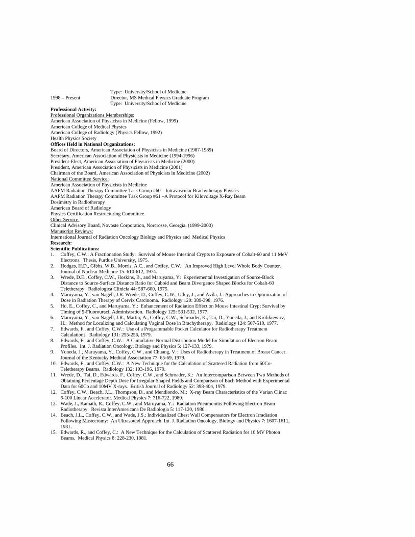
66
Type: University/School of Medicine 1998 – Present Director, MS Medical Physics Graduate Program Type: University/School of Medicine Professional Activity: Professional Organizations Memberships: American Association of Physicists in Medicine (Fellow, 1999) American College of Medical Physics American College of Radiology (Physics Fellow, 1992) Health Physics Society Offices Held in National Organizations: Board of Directors, American Association of Physicists in Medicine (1987-1989) Secretary, American Association of Physicists in Medicine (1994-1996) President-Elect, American Association of Physicists in Medicine (2000) President, American Association of Physicists in Medicine (2001) Chairman of the Board, American Association of Physicists in Medicine (2002) National Committee Service: American Association of Physicists in Medicine AAPM Radiation Therapy Committee Task Group #60 – Intravascular Brachytherapy Physics AAPM Radiation Therapy Committee Task Group #61 –A Protocol for Kilovoltage X-Ray Beam Dosimetry in Radiotherapy American Board of Radiology Physics Certification Restructuring Committee Other Service: Clinical Advisory Board, Novoste Corporation, Norcrosse, Georgia, (1999-2000) Manuscript Reviews: International Journal of Radiation Oncology Biology and Physics and Medical Physics Research: Scientific Publications: 1. Coffey, C.W.; A Fractionation Study: Survival of Mouse Intestinal Crypts to Exposure of Cobalt-60 and 11 MeV
Electrons. Thesis, Purdue University, 1975. 2. Hodges, H.D., Gibbs, W.B., Morris, A.C., and Coffey, C.W.: An Improved High Level Whole Body Counter.
Journal of Nuclear Medicine 15: 610-612, 1974. 3. Wrede, D.E., Coffey, C.W., Hoskins, B., and Maruyama, Y: Experiemental Investigation of Source-Block
Distance to Source-Surface Distance Ratio for Cuboid and Beam Divergence Shaped Blocks for Cobalt-60 Teletherapy. Radiologica Clinicia 44: 587-600, 1975.
4. Maruyama, Y., van Nagell, J.R. Wrede, D., Coffey, C.W., Utley, J., and Avila, J.: Approaches to Optimization of Dose in Radiation Therapy of Cervix Carcinoma. Radiology 120: 389-398, 1976.
5. Ho, E., Coffey, C., and Maruyama, Y.: Enhancement of Radiation Effect on Mouse Intestinal Crypt Survival by Timing of 5-Fluorouracil Administration. Radiology 125: 531-532, 1977.
6. Maruyama, Y., van Nagell, J.R., Martin, A., Coffey, C.W., Schroader, K., Tai, D., Yoneda, J., and Krolikiewicz, H.: Method for Localizing and Calculating Vaginal Dose in Brachytherapy. Radiology 124: 507-510, 1977.
7. Edwards, F., and Coffey, C.W.: Use of a Programmable Pocket Calculator for Radiotherapy Treatment Calculations. Radiology 131: 255-256, 1979.
8. Edwards, F., and Coffey, C.W.: A Cumulative Normal Distribution Model for Simulation of Electron Beam Profiles. Int. J. Radiation Oncology, Biology and Physics 5: 127-133, 1979.
9. Yoneda, J., Maruyama, Y., Coffey, C.W., and Chuang, V.: Uses of Radiotherapy in Treatment of Breast Cancer. Journal of the Kentucky Medical Association 77: 65-69, 1979.
10. Edwards, F., and Coffey, C.W.: A New Technique for the Calculation of Scattered Radiation from 60Co-Teletherapy Beams. Radiology 132: 193-196, 1979.
11. Wrede, D., Tai, D., Edwards, F., Coffey, C.W., and Schroader, K.: An Intercomparison Between Two Methods of Obtaining Percentage Depth Dose for Irregular Shaped Fields and Comparison of Each Method with Experimental Data for 60Co and 10MV X-rays. British Journal of Radiology 52: 398-404, 1979.
12. Coffey, C.W., Beach, J.L., Thompson, D., and Mendiondo, M.: X-ray Beam Characteristics of the Varian Clinac 6-100 Linear Accelerator. Medical Physics 7: 716-722, 1980.
13. Wade, J., Kamath, R., Coffey, C.W., and Maruyama, Y.: Radiation Pneumonitis Following Electron Beam Radiotherapy. Revista InterAmericana De Radiologia 5: 117-120, 1980.
14. Beach, J.L., Coffey, C.W., and Wade, J.S.: Individualized Chest Wall Compensators for Electron Irradiation Following Mastectomy: An Ultrasound Approach. Int. J. Radiation Oncology, Biology and Physics 7: 1607-1611, 1981.
15. Edwards, R., and Coffey, C.: A New Technique for the Calculation of Scattered Radiation for 10 MV Photon Beams. Medical Physics 8: 228-230, 1981.

67
16. Coffey, C.W., Maruyama, Y., Stewart, B.L., and White, G.A.: Electron Beam Irradiation for Mycosis Fungoides Using Variable Energy. Journal of the Kentucky Medical Association 80: 398-404, 1982.
17. Matar., J.R., Coffey, C., and Maruyama, Y.: Rectal Carcinoma: Treatment with Papillon Technique and Fiberoptic-Guided Methods. Radiology 168: 562-564, 1988.
18. Coffey, C.W., Morris, R.S., Martin, J., and Maruyama, Y.: Dosimetric Evaluation of a Variable Energy Superficial X-Ray Machine With Applications for Endocavitary Techniques. Int. J. Radiation Oncology, Biology and Physics 16: 849-855, 1989.
19. Coffey, C.W., Taylor, R., and Umstead, G.: A Slice Geometry Phantom for Cross Sectional Tomographic Imagers. Medical Physics 16: 273-278, 1989.
20. Shih, W.J., Li, C.Y., Coffey, C.W., and Maruyama, Y.: Thoracic Vertebral Photopenia May Predict Fatty Changes of the Corresponding Bone Marrow Following Irradiation. Radiation Medicine 7: 32-35, 1989.
21. Fleming, D.R., Henslee-Downey, P.J., and Coffey, C.W.: Radiation Induced Acute Tumor Lysis Syndrome in the Bone Marrow Transplant Setting. Bone Marrow Transplantation 8: 235-236, 1991.
22. Wierzbicki, J.G., Poteat, G.N., and Coffey, C.W.: Measurements of Relative Strength of Iridium-192 Seeds Assembled in Ribbons. Endocurietherapy Hyperthermia Oncology 9: 229-232, 1993.
23. Coffey, C.W., Sanders, M., Cashon, K., Miller, R., Walsh, J., and Patel, P.: A Tissue Equivalent Phantom for Stereotactic Radiosurgery Localization and Dose Verification. Stereotactic and Functional Neurosurgery 62(SI): 130-141, 1993.
24. Sanders, M., Sayeg, J., Coffey, C.W., Patel, P., and Walsh, J.: Beam Profile Analysis Using Gaf Chromic Films. Stereotactic and Functional Neurosurgery 61 (SI): 124-129, 1993.
25. Fischell, T., Kharma, B.K., Fischell, D.R., Loges, P.G., Coffey, C.W., Duggan, D. M., Naftilan, A.J.: Low-Dose, B-Particle Emission From 'Stent' Wire Results in Complete, Localized Inhibition of Smooth Muscle Cell Proliferation. Circulation 90: 2956-2963, 1994.
26. Duggan, D.M., and Coffey, C.W.: Use of a Micro-Ionization Chamber and an Anthropomorphic Head Phantom in a Quality Assurance Program for Stereotactic Radiosurgery. Medical Physics 23:513-516, 1996.
27. Janicki, C., Duggan, D.M., Coffey, C., Fischell, D., and Fischell,T.: Radiation Dose from a Phosphorous-32 Impregnated Wire Mesh Vascular Stent. Medical Physics 24: 437-445,1997.
28. Duggan, D.M., Coffey, C.W., and Levit, S.: Dose Calculation for a 32P Impregnated Coronary Stent: Comparison of Theoretical Calculations and Measurements with Radiochromic Film. Int. J. Radiation Biology Oncology and Physics 40: 713-720, 1998.
29. Nath, R., Amols, H., Coffey, C., Duggan, D., Jani, S., Li, Z., Schell, M., Soares, C., Whiting, J., Cole, P., Crocker, I., and Schwartz, R.: Intravascular Brachytherapy Physics: Report of AAPM Radiation Therapy Task Group No.60 Medical Physics 26: 119-152, 1999
30. Duggan, D.M. and Coffey, C.W.: Better Narrow Beam Profiles by Exposing a Stack of Radiochromic Films and Averaging the Readings. Medical Physics 26: 489-492, 1999.
31. Duggan, D.M., Coffey, C., Lobdell, J.L., and Schell, M.C.: Radiochromic Film Dosimetry of a High Dose Rate Beta Source for Intravascular Brachytherapy. Medical Physics 26: 2461-2464, 1999.
32. Janicki, C., Duggan, D., Gonzalez, A, Coffey, C.,and Rahdert D: Dose Model for a Beta Emitting Stent in a Realistic Artery Consisting of Soft Tissue and Plaque. Medical Physics 26: 2451-2460, 1999.
33. Ma, C., Coffey, C., DeWerd, L., Liu, C., Nath, R., Seltzer, S., and Seuntgens, J.: AAPM Protocol for 40-300 kV X-Ray Beam Dosimetry in Radiotherapy and Radiobiology: Report of the AAPM Radiation Therapy Committee Task Group No. 61. Medical Physics 28: 868-893, 2001.
Books and/or Chapters: 1. Coffey, C.W., Droege, R.T., and Ekstrand, K.E.: Systems Specifications and Acceptance Testing. In NMR in
Medicine: The Instrumentation and Clinical Applications. Eds. S.R. Thomas and R.L. Dixon. American Association of Physicists in Medicine, Medical Physics Monograph No. 14: 445-475, 1986.
2. Coffey, C.W.: Calibration of Low Energy X-Ray Units. In Advances in Radiation Oncology Physics-Dosimetry Treatment Planning-Brachytherapy. Editor. J. Purdy. American Association of Physicists in Medicine, Medical Physics Monograph No. 19: 134-172, 1991.
3. Coffey, C. and Duggan, D.: Dosimetric Considerations and Dose Measurement Analysis of a P-32 Radioisotope Stent. In Vascular Brachytherapy. Eds. Waksman, King, Crocker, and Mould. Nucletron Publishing, Netherlands, 207- 216, 1996.
4. Coffey, C., Duggan, D., and Reddick, R.: Dosimetric Considerations and Dose Measurement Analysis of Phosphorus-32 Beta-Emitting Stents: A Review.In Vascular Brachytherapy, 2nd Edition. Ed. R. Waksman. Futura Publishing Company, Armonk, NY, 313-332, 1999.
5. Coffey, C. and Duggan, D.: The Calculation and Measurement of Radiation Dose Surrounding Radioactive Stents: The Effects of Radionuclide Selection and Stent Design on Dosimetric Results. 3rd Edition, Ed. R.Waksman. Futura Publishing Company, Armonk, NY, 349-378, 2002.
Patent: 1. Smith, S.L., Wang, P.C., and Coffey, C.W.: Quality Control Phantom for Use In Computed Tomographic
Imaging Instruments and Method of Use. U.S. Patent No. 4,618,826. October 21, 1986.
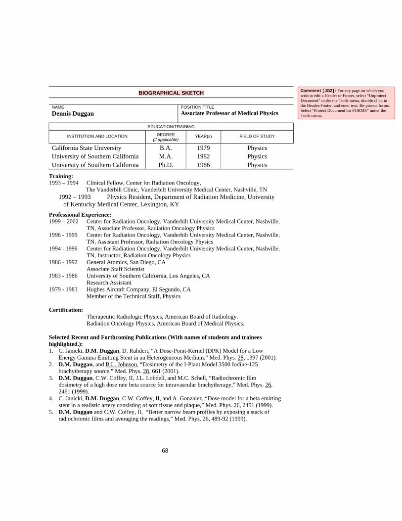
68
BIOGRAPHICAL SKETCH
NAME Dennis Duggan
POSITION TITLEAssociate Professor of Medical Physics
EDUCATION/TRAINING
INSTITUTION AND LOCATION DEGREE (if applicable)
YEAR(s) FIELD OF STUDY
California State University B.A. 1979 Physics University of Southern California M.A. 1982 Physics University of Southern California Ph.D. 1986 Physics
Training: 1993 – 1994 Clinical Fellow, Center for Radiation Oncology,
The Vanderbilt Clinic, Vanderbilt University Medical Center, Nashville, TN 1992 – 1993 Physics Resident, Department of Radiation Medicine, University
of Kentucky Medical Center, Lexington, KY Professional Experience:
1999 – 2002 Center for Radiation Oncology, Vanderbilt University Medical Center, Nashville, TN, Associate Professor, Radiation Oncology Physics
1996 - 1999 Center for Radiation Oncology, Vanderbilt University Medical Center, Nashville, TN, Assistant Professor, Radiation Oncology Physics
1994 - 1996 Center for Radiation Oncology, Vanderbilt University Medical Center, Nashville, TN, Instructor, Radiation Oncology Physics
1986 - 1992 General Atomics, San Diego, CA Associate Staff Scientist 1983 - 1986 University of Southern California, Los Angeles, CA Research Assistant 1979 - 1983 Hughes Aircraft Company, El Segundo, CA Member of the Technical Staff, Physics Certification: Therapeutic Radiologic Physics, American Board of Radiology. Radiation Oncology Physics, American Board of Medical Physics. Selected Recent and Forthcoming Publications (With names of students and trainees highlighted.): 1. C. Janicki, D.M. Duggan, D. Rahdert, “A Dose-Point-Kernel (DPK) Model for a Low
Energy Gamma-Emitting Stent in an Heterogeneous Medium,” Med. Phys. 28, 1397 (2001). 2. D.M. Duggan, and B.L. Johnson, “Dosimetry of the I-Plant Model 3500 Iodine-125
brachytherapy source,” Med. Phys. 28, 661 (2001). 3. D.M. Duggan, C.W. Coffey, II, J.L. Lobdell, and M.C. Schell, “Radiochromic film
dosimetry of a high dose rate beta source for intravascular brachytherapy,” Med. Phys. 26, 2461 (1999).
4. C. Janicki, D.M. Duggan, C.W. Coffey, II, and A. Gonzalez, “Dose model for a beta emitting stent in a realistic artery consisting of soft tissue and plaque,” Med. Phys. 26, 2451 (1999).
5. D.M. Duggan and C.W. Coffey, II, “Better narrow beam profiles by exposing a stack of radiochromic films and averaging the readings,” Med. Phys. 26, 489-92 (1999).
Comment [JG2]: For any page on which you wish to edit a Header or Footer, select “Unprotect Document” under the Tools menu, double-click in the Header/Footer, and enter text. Re-protect forms: Select “Protect Document for FORMS” under the Tools menu.

69
6. R. Nath, H. Amols, C.W. Coffey, II, D.M. Duggan, S. Jani, Z. Li, M. Schell, C. Soares, J. Whiting, P. Cole, I. Crocker, and R. Schwartz, “Intravascular Brachytherapy Physics: Report of AAPM Radiation Therapy Task Group No. 60,” Med. Phys., 26, 119-52 (1999).
7. D.M. Duggan and C.W. Coffey, “Small photon field dosimetry for stereotactic radiosurgery.” Medical Dosimetry 23, 153 (1998).
8. D.M. Duggan, C.W. Coffey, II, and S. Levit, “Dose Distribution for a 32P Impregnated Coronary Stent: Comparison of Theoretical Calculations and Measurements with Radiochromic Film,” Int. J. Radiat. Oncol. Biol. Phys., 40, 713 (1997).
Books and/or Chapters: 1. C. W. Coffey and D.M. Duggan, “Dosimetric Considerations and Dose Measurement
Analysis of a P-32 Radiosotope Stent.” In Vascular Brachytherapy. Eds. Waksman, King, Crocker, and Mould. Nucletron Publishing, Netherlands, 207- 216, 1996.
2. C. W. Coffey, D.M. Duggan, and R. Reddick, “Dosimetric Considerations and Dose Measurement Analysis of Phosphorus-32 Beta-Emitting Stents.” A Review. In Vascular Brachytherapy, 2nd Edition. Ed. R. Waksman. Futura Publishing Company, Armonk, NY, 313-332, 1999
3. C.W. Coffey and D.M. Duggan, “The Calculation and Measurement of Radiation Dose Surrounding Radioactive Stents: The Effects of Radionuclide Selection and Stent Design on Dosimetric Results.” 3rd Edition, Ed. R. Waksman. Futura Publishing Company, Armonk, NY, 349-378, 2002.
Research Interests: Present research interests include dosimetry of small teletherapy beams and brachytherapy, quality assurance for stereotactic radiosurgery, intensity modulated radiotherapy, and other types of conformal radiotherapy, and intravascular brachytherapy for reducing restenosis in coronary arteries.

70
BIOGRAPHICAL SKETCH
MICHAEL LEONARD FREEMAN, Ph.D. Appointments: 9/89 Associate Professor with tenure: Dept. of Radiation Oncology/ Secondary Appointment:
Associate Professor of Radiology and Cancer Biology 10/86 to 12/88 Quality Assurance Director: Radiation Oncology 1/83 -8/89 Assistant Professor: Dept. of Radiology and Radiological Sciences 1979-1982 Research Associate Radiological Research Laboratory College of Physicians and Surgeons Columbia University Education: 1974-1978 Ph.D. in Radiation Biology Colorado State University Advisor: W.C. Dewey, Ph.D. 1972-1974 Colorado State University, B.S. Zoology 1970-1972 Tulane University Areas of Research and/or Teaching Interests: Radiation biology, Heat Shock (Stress) Proteins, Glutathione metabolism Grants Received: 1. PHS grant CA-38079, 7/1/84 - 12/2005 PHS/NGA S10RR1 5896, Awarded 10/1/00 Therapeutic X ray Machine. 2. T32-093240 Training Grant in Radiation Biology, NCI 7/1/02-6/30/07 3. Pilot Project support from P30 ES 00267, April 2000 to March 2001. Pilot Project support from P30 ES 00267 July 1991 to June 1992 4. BSRG grant 523928, awarded 9/5/88. 5. Young Investigator Award for the Seventh International Congress of Radiation Research, July 1983. 6. Young Investigator Award for the Third International Symposium, Cancer Therapy by Hyperthermia, Drugs and Radiation,
June 1980. 7. P.H.S. Trainee in Radiation Biology, Colorado State University, January-August 1977. Editorial Boards Editorial Board “Frontiers in Bioscience”. 2002 to present. Editorial Board “Cell Stress and Chaperones” 2002 to 2005 Professional Memberships: Radiation Research Society American Association for Cancer Research North American Hyperthermia Society Oxygen Society Publications: 1. Freeman ML, Dewey WC, Hopwood LE: Effect of pH on hyperthermic cell killing: Brief communication. J Natl Cancer
Inst 58: 1837-839, 1977. 2. Freeman ML, Raaphorst GP, Dewey WC: The relationship of heat killing and thermal radiosensitization to the duration of
heating at 42EC. Radiation Research 78: 172-175, 1979. 3. Raaphorst GP, Freeman ML, Dewey WC: Radiosensitivity and recovery from radiation damage in cultured CHO cells
exposed to hyperthermia at 42.5E or 45.5E. Radiation Research 79: 390-402, 1979. 4. Raaphorst GP, Sapareto SA, Freeman ML, Dewey WC: Changes in cellular heat and/or radiation sensitivity observed at
various times after trypsinization and plating. Int J Radiat Biol 35: 193-197, 1979. 5. Freeman ML, Raaphorst GP, Hopwood LE, Dewey WC: The effect of pH on cell lethality induced by hyperthermic
treatment. Cancer 45: 2291-2300, 1980. 6. Dewey WC, Freeman ML: Rationale for use of hyperthermia in cancer therapy. Annals of NY Acad of Science 335: 372-
378, 1980. 7. Dewey WC, Freeman ML, Raaphorst GP, Clark EP, Wong RS, Highfield DP, Spiro IJ, Tomasovic SP, Denman DL, Coss,
RA: Cell biology of hyperthermia and radiation. In Radiation Biology in Cancer Research, (Meyn RE, Withers HR, editors). p 589-621, 1980.
8. Freeman ML, Holahan EV, Highfield DP, Raaphorst GP, Spiro IJ, Dewey WC: The effect of pH on hyperthermic and x-ray induced cell killing. Inter J Radiat Oncol Biol Phys 7: 211-216, 1981.
9. Freeman ML, Boone MLM, Ensley BA, and Gillette EL: The influence of environmental pH on the interaction and repair of heat and radiation damage. Int J Radiat Oncol Biol Phys 7: 761-764, 1981.
10. Freeman ML: The transient nature of thermotolerance. J Natl Cancer Inst Monogr. 61: 275-278, 1982.

71
11. Freeman ML, Sierra E, Deitch AD, Cubbon RN: The repair of potentially lethal and sublethal damage in unfed plateau phase cultures irradiated at 0.78 Gy/hr. Radiation Research 92: 596-603, 1982.
12. Goldhagen P, Freeman ML, and Hall EJ: Studies with encapsulated 125I sources II: Apparatus and dosimetry for determination of the relative biological effectiveness. Int J Radiat Oncol Biol Phys 8: 1347-1353, 1982.
13. Freeman ML, Goldhagen P, and Hall EJ: Studies with encapsulated 125I sources III: Determination of the relative biological effectiveness using cultured mammalian cells. Int J Radiat Oncol Biol Phys 8: 1355-1361, 1982.
14. Freeman ML, Sierra E, and Hall EJ: The repair of sublethal damage in diploid human fibroblasts: A comparison between human and rodent cell lines. Radiation Research 95: 382-391, 1983.
15. Freeman ML, Sierra E: An acidic extracellular environment reduces the fixation of radiation damage. Radiation Research 97: 154-161, 1984.
16. Freeman ML, Malcolm AW, Meredith MJ: Decreased intracellular glutathione concentration and increased hyperthermic cytotoxicity in an acid environment.Cancer Res 45: 504-508, 1985.
17. Freeman ML, Malcolm AW, Meredith MJ: Glutathione pool size affects cell survival after hyperthermic treatment. Cell Biol and Toxicol 1: 221-231, 1985.
18. Freeman ML and Malcolm AW: Acid modification of thermal damage and its relationship to nutrient availability. Inter J Radiat Oncol Biol Phys 11: 1823-1826, 1985.
19. Freeman ML, Malcolm AW, and Meredith MJ: Role of Glutathione in Cell Survival after Hyperthermic Treatment of Chinese Hamster Ovary Cells. Cancer Research 45: 6308-6313, 1985.
20. Freeman ML, Scidmore NC, Malcolm AW, and Meredith MJ: Diamide exposure, thermal resistance and synthesis of stress (heat shock) proteins. Biochem Pharm 36: 21-29, 1987.
21. Freeman ML, and Meredith MJ: The effect of GSH depletion on thermal radiosensitization. Int J Radiat Oncol Biol Phys. 13: 1371-1375, 1987.
22. Freeman, ML, Scidmore NC, Meredith MJ: Inhibition of heat shock protein synthesis and thermotolerance by cycloheximide. Radiat. Res. 112: 564-574, 1987.
23. Freeman,ML and Meredith, MJ: Subcellular localization of glutathione and thermal sensitivity. Radiation Res. 115:461-471,1988.
24. Freeman, ML, Meredith, MJ, and Laszlo, A: Depletion of glutathione during HSP synthesis and the development of thermotolerance. Cancer Res. 48:7033-7037, 1988.
25. Freeman, ML and Meredith, MJ: Measurement of protein thiols after heat shock using 3-(-N-maleimido-propionyl) biocytin labeled proteins separated by SDS-PAGE and electroluted onto nitrocellulose: Thiol Blotting. Radiation Res. 117: 326-333,1989.
26. Freeman, ML and Meredith, MJ: Glutathione conjugation and induction of a 32,000 dalton stress protein. Biochem. Pharmacol 38:299-304, 1989.
27. Freeman, ML and Meredith, MJ: Modulation of diamide toxicity in thermotolerant cells by inhibition of protein synthesis. Cancer Res. 49: 4493-4498, 1989.
28. Atsmon J, Freeman ML, Meredith MJ, Sweetman BJ, and Roberts LJ. Enhancement of 9 Deoxy >9,>12- PGD2 antiproliferative effect by glutathione depletion. Cancer Research 50:1879-1885,1990.
29. Freeman ML, Meredith MJ, and Eisert DR. Failure of chronic glutathione elevation to reduce cytotoxicity produced by exposure to cis diamminedichloroplatinum (II), ionizing radiation, or hyperthermia. Cancer Res. 50: 5296-5300, 1990.
30. Freeman ML, Spitz DR, and Meredith MJ, Does heat shock promote oxidative stress? Studies with ferrous and ferric iron. Radiat. Res. 288-293, 1990.
31. Saunders EL, Meredith MJ, Eisert DR, and Freeman ML, Depletion of glutathione after (-irradiation modifies survival. Radiation Res. 125: 267-276, 1991.
32. Saunders EL, Maines MD, Meredith MJ, and Freeman ML, Enhancement of heme oxygenase-1 synthesis by glutathione depletion in Chinese hamster ovary cells. Arch. Biochem. Biophys. 288: 368-373, 1991.
33. Atsom J, Freeman ML, Meredith MJ, Sweetman BJ, Roberts LJ 2nd, Modulation of the antiproliferative activity of 9-deoxy-delta 9, delta 12(E)-PGD2 by conjugation with intracellular glutathione. Adv. Prostaglandin Thromboxame Leukotriene Res. 21B: 887-890, 1991.
34. Sierra-Rivera E, Voorhees GJ and Freeman ML, (-irradiation increases HSP 70 in Chinese hamster ovary cells. Radiation Res. 135: 40-45, 1993.
35. Freeman ML, Sierra-Rivera E, Voorhees GJ, Eisert DR, and Meredith MJ, Synthesis of HSP 70 is enhanced in GSH depleted Hep G2 cells, Radiation Res. 135: 387-393, 1993.
36. Sierra-Rivera E, Meredith MJ, Voorhees, GJ, Oberley LW, Eisert SR, and Freeman ML, The synthesis of heat shock proteins following oxidative challenge: The role of glutathione. Inter. J. Hyperthermia 10: 573-586, 1994.
37. Sierra-Rivera E, Meredith MJ, Summar, ML, Smith MD, Voorhees GJ, Stoffel CM, and Freeman, ML, Genes regulating glutathione concentrations in x-ray transformed rat embryo fibroblasts: Changes in (-glutamylcysteine synthetase and (-glutamyl transpeptidase expression. Carcinogenesis 15: 1301-1307, 1994.
38. Freeman ML, Borrelli MJ, Syed K, Senisterra G, Stafford DM, and Lepock JR. Characterization of a signal generated by oxidation of protein thiols that activates the heat shock transcription factor. J. Cell. Physiol. 164: 356-366, 1995
39. Sierra-Rivera E, Summar ML, Krishnamani MRS, Phillips III JA, and Freeman ML. Assignment of the gene that codes the heavy subunit of (-glutamylcysteine synthetase to human chromosome 6. Cytogenetics and Cell Genetics 70: 278-279, 1995.
40. Sierra-Rivera E, Dasouki M, Summar ML, Krishnamani MRS, Meredith M, Rao PN, Phillips JA, and Freeman ML, Chromosomal localization of the human gene that encodes the regulatory subunit of (-glutamylcysteine synthetase to chromosome 1p21. Cytogenetics and Cell Genetics 72: 252-254, 1996.
41. McDuffee AT, Senisterra GA, Huntley SA, Lepock JR, Senkhar KR, Morrow JD, and Freeman ML, Proteins conctaining non-native disulfide bonds generated by oxidative stress may act as signals for the induction of the heat shock response. J. Cellular Physiology 171: 143-151, 1997
42. Sekhar KR, Long M, Long, J, Xu ZQ, Summar, ML, and Freeman, ML, Alteration of transcriptional and post-transcriptional expression of (-glutamylcysteine synthetase by diethyl maleate. Radiation Res. 147: 592-597, 1997
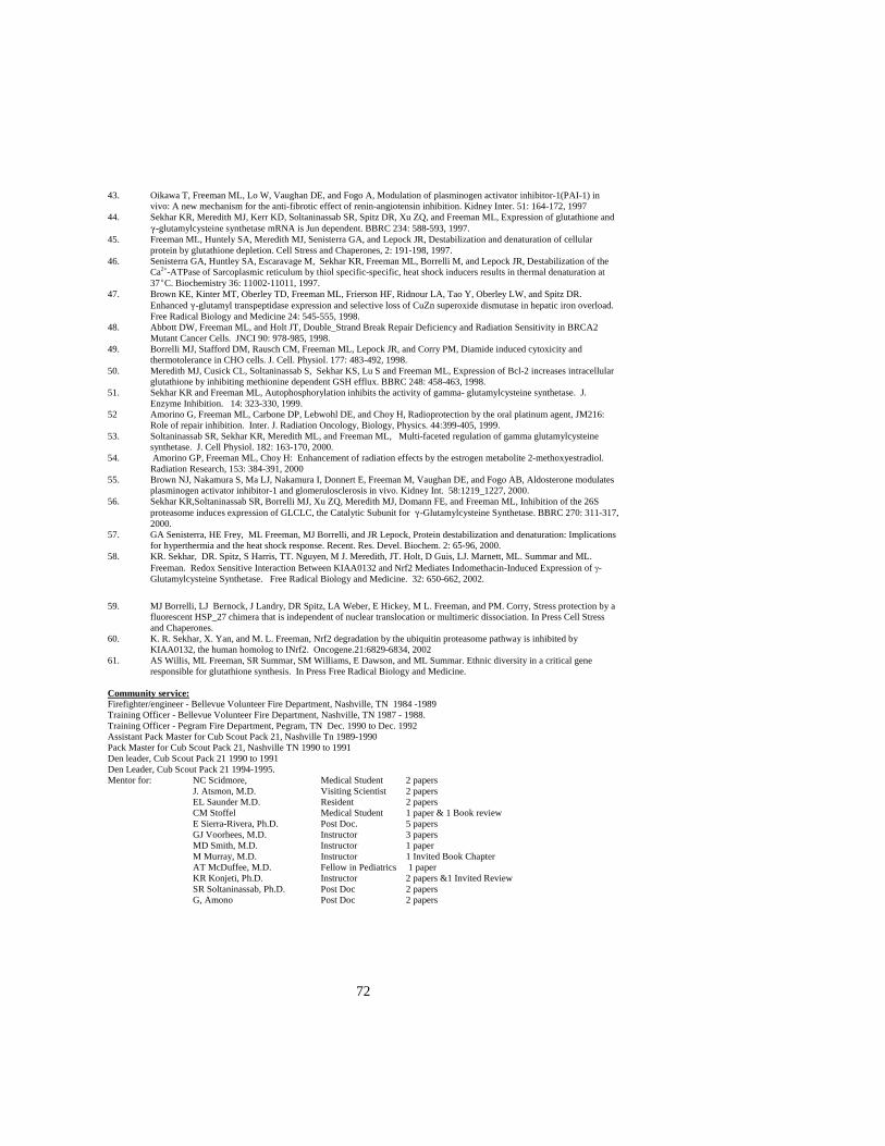
72
43. Oikawa T, Freeman ML, Lo W, Vaughan DE, and Fogo A, Modulation of plasminogen activator inhibitor-1(PAI-1) in vivo: A new mechanism for the anti-fibrotic effect of renin-angiotensin inhibition. Kidney Inter. 51: 164-172, 1997
44. Sekhar KR, Meredith MJ, Kerr KD, Soltaninassab SR, Spitz DR, Xu ZQ, and Freeman ML, Expression of glutathione and (-glutamylcysteine synthetase mRNA is Jun dependent. BBRC 234: 588-593, 1997.
45. Freeman ML, Huntely SA, Meredith MJ, Senisterra GA, and Lepock JR, Destabilization and denaturation of cellular protein by glutathione depletion. Cell Stress and Chaperones, 2: 191-198, 1997.
46. Senisterra GA, Huntley SA, Escaravage M, Sekhar KR, Freeman ML, Borrelli M, and Lepock JR, Destabilization of the Ca2+-ATPase of Sarcoplasmic reticulum by thiol specific-specific, heat shock inducers results in thermal denaturation at 37EC. Biochemistry 36: 11002-11011, 1997.
47. Brown KE, Kinter MT, Oberley TD, Freeman ML, Frierson HF, Ridnour LA, Tao Y, Oberley LW, and Spitz DR. Enhanced (-glutamyl transpeptidase expression and selective loss of CuZn superoxide dismutase in hepatic iron overload. Free Radical Biology and Medicine 24: 545-555, 1998.
48. Abbott DW, Freeman ML, and Holt JT, Double_Strand Break Repair Deficiency and Radiation Sensitivity in BRCA2 Mutant Cancer Cells. JNCI 90: 978-985, 1998.
49. Borrelli MJ, Stafford DM, Rausch CM, Freeman ML, Lepock JR, and Corry PM, Diamide induced cytoxicity and thermotolerance in CHO cells. J. Cell. Physiol. 177: 483-492, 1998.
50. Meredith MJ, Cusick CL, Soltaninassab S, Sekhar KS, Lu S and Freeman ML, Expression of Bcl-2 increases intracellular glutathione by inhibiting methionine dependent GSH efflux. BBRC 248: 458-463, 1998.
51. Sekhar KR and Freeman ML, Autophosphorylation inhibits the activity of gamma- glutamylcysteine synthetase. J. Enzyme Inhibition. 14: 323-330, 1999.
52 Amorino G, Freeman ML, Carbone DP, Lebwohl DE, and Choy H, Radioprotection by the oral platinum agent, JM216: Role of repair inhibition. Inter. J. Radiation Oncology, Biology, Physics. 44:399-405, 1999.
53. Soltaninassab SR, Sekhar KR, Meredith ML, and Freeman ML, Multi-faceted regulation of gamma glutamylcysteine synthetase. J. Cell Physiol. 182: 163-170, 2000.
54. Amorino GP, Freeman ML, Choy H: Enhancement of radiation effects by the estrogen metabolite 2-methoxyestradiol. Radiation Research, 153: 384-391, 2000
55. Brown NJ, Nakamura S, Ma LJ, Nakamura I, Donnert E, Freeman M, Vaughan DE, and Fogo AB, Aldosterone modulates plasminogen activator inhibitor-1 and glomerulosclerosis in vivo. Kidney Int. 58:1219_1227, 2000.
56. Sekhar KR,Soltaninassab SR, Borrelli MJ, Xu ZQ, Meredith MJ, Domann FE, and Freeman ML, Inhibition of the 26S proteasome induces expression of GLCLC, the Catalytic Subunit for (-Glutamylcysteine Synthetase. BBRC 270: 311-317, 2000.
57. GA Senisterra, HE Frey, ML Freeman, MJ Borrelli, and JR Lepock, Protein destabilization and denaturation: Implications for hyperthermia and the heat shock response. Recent. Res. Devel. Biochem. 2: 65-96, 2000.
58. KR. Sekhar, DR. Spitz, S Harris, TT. Nguyen, M J. Meredith, JT. Holt, D Guis, LJ. Marnett, ML. Summar and ML. Freeman. Redox Sensitive Interaction Between KIAA0132 and Nrf2 Mediates Indomethacin-Induced Expression of γ-Glutamylcysteine Synthetase. Free Radical Biology and Medicine. 32: 650-662, 2002.
59. MJ Borrelli, LJ Bernock, J Landry, DR Spitz, LA Weber, E Hickey, M L. Freeman, and PM. Corry, Stress protection by a
fluorescent HSP_27 chimera that is independent of nuclear translocation or multimeric dissociation. In Press Cell Stress and Chaperones.
60. K. R. Sekhar, X. Yan, and M. L. Freeman, Nrf2 degradation by the ubiquitin proteasome pathway is inhibited by KIAA0132, the human homolog to INrf2. Oncogene.21:6829-6834, 2002
61. AS Willis, ML Freeman, SR Summar, SM Williams, E Dawson, and ML Summar. Ethnic diversity in a critical gene responsible for glutathione synthesis. In Press Free Radical Biology and Medicine.
Community service: Firefighter/engineer - Bellevue Volunteer Fire Department, Nashville, TN 1984 -1989 Training Officer - Bellevue Volunteer Fire Department, Nashville, TN 1987 - 1988. Training Officer - Pegram Fire Department, Pegram, TN Dec. 1990 to Dec. 1992 Assistant Pack Master for Cub Scout Pack 21, Nashville Tn 1989-1990 Pack Master for Cub Scout Pack 21, Nashville TN 1990 to 1991 Den leader, Cub Scout Pack 21 1990 to 1991 Den Leader, Cub Scout Pack 21 1994-1995. Mentor for: NC Scidmore, Medical Student 2 papers J. Atsmon, M.D. Visiting Scientist 2 papers EL Saunder M.D. Resident 2 papers CM Stoffel Medical Student 1 paper & 1 Book review E Sierra-Rivera, Ph.D. Post Doc. 5 papers GJ Voorhees, M.D. Instructor 3 papers MD Smith, M.D. Instructor 1 paper M Murray, M.D. Instructor 1 Invited Book Chapter AT McDuffee, M.D. Fellow in Pediatrics 1 paper KR Konjeti, Ph.D. Instructor 2 papers &1 Invited Review SR Soltaninassab, Ph.D. Post Doc 2 papers
G, Amono Post Doc 2 papers

73
BIOGRAPHICAL SKETCH
NAME Dennis E. Hallahan
POSITION TITLEProfessor and Chairman
EDUCATION/TRAINING
INSTITUTION AND LOCATION DEGREE (if applicable)
YEAR(s) FIELD OF STUDY
University of Illinois, Chicago B.S. 1980 Biology Rush Medical College, Chicago M.D. 1984 Medicine
Professional Experience 1984-86 Intern and Resident, Internal Medicine, University of Chicago, Chicago 1986-90 Resident, Radiation Oncology, University of Chicago, IL 1988-90 Molec. Biol. And Rad. Biol., Radiation Oncology, University of Chicago 1990-94 Assistant Professor, Dept Radiation and Cellular Oncology, University of Chicago 1994-97 Associate Professor, Dept Radiation and Cellular Oncology, University of Chicago 1998-present Professor and Chairman, Dept Radiation Oncology, Vanderbilt University 1998-present Professor, Dept Biomedical Engineering, Vanderbilt University Research Projects Ongoing or Completed During the Last 3 Years: “X-ray-guided drug delivery” Principal Investigator: Dennis E. Hallahan, Agency: NIH/NCI, Type: R01 (CA88076-01), Period: 2002-2007 "Flk1 Signaling Protects Tumor Vasculature from Radiation" Principal Investigator: Dennis Hallahan, Agency: NIH/NCI, Type: 1 R01 CA89674-01, Period: 2001 -2005 The objective of this study is to study the mechanisms by which tumor blood vessels respond to ionizing radiation. The general hypothesis is that the FLK-1 receptor mediates resistance of tumor blood vessels to the cytotoxic effects of radiation and participates in repair of radiation-induced injury in tumor blood vessels. “X-Ray-guided drug delivery by apcitide- 99Tc” Principal Investigator: Dennis E. Hallahan, Agency: NIH/NCI, Type: R21 (CA89888-01), Period: 2000-2002 The objective of this clinical study is to determine the threshold dose and tumor types which radiation activates receptors for drug delivery. We are utilizing a peptide that binds to radiation activated receptors. The 99Tc localizes to tumors treated with radiation. This is the prototype for radiation-guided drug delivery. “X-rays Induce Exocytosis of Endothelial P-selectin” Principal Investigator: Dennis E. Hallahan, Agency: NIH/NCI, Type: 2-R01 (CA58508), Period: 1998-2003 The objective of this study is to determine the mechanisms by which ionizing radiation activates exocytosis of storage reservoirs from the endothelium. The components of these storage reservoirs include biologically active molecules. The mechanisms of radiation induced exocytosis include microtubule-dependent transport. The signaling pathway and mechanisms of transport will be studied. “Cancer Center Support Grant” Principal Investigator: Harold Moses, Agency: NIH/NCI, Type: 2-P30 (CA68485-04), Period: 1999-2004 This staff investigator grant supports the percent effort in the Cancer Center. This funding is provided for leadership in the Radiation Biology and Radiation Oncology programs within the Vanderbilt Cancer Center. “Lung Cancer SPORE” Principal Investigator: David Carbone, Agency: NIH/NCI, Type: P50 CA90949, Period: 4/01/01-3/31/06 Clinical trial of SU5416 and radiation in lung cancer patients. “Induction of Cell Adhesion Molecules by Ionizing Radiation” Principal Investigator: Dennis E. Hallahan, Agency: NIH/NCI, Type: R01 (CA70937), Period: 1998-2001
Comment [JG3]: For any page on which you wish to edit a Header or Footer, select “Unprotect Document” under the Tools menu, double-click in the Header/Footer, and enter text. Re-protect forms: Select “Protect Document for FORMS” under the Tools menu.
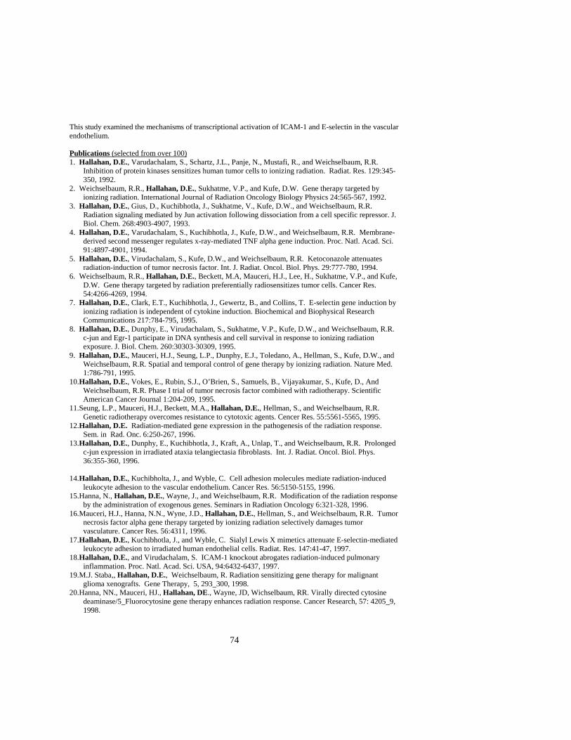
74
This study examined the mechanisms of transcriptional activation of ICAM-1 and E-selectin in the vascular endothelium. Publications (selected from over 100) 1. Hallahan, D.E., Varudachalam, S., Schartz, J.L., Panje, N., Mustafi, R., and Weichselbaum, R.R.
Inhibition of protein kinases sensitizes human tumor cells to ionizing radiation. Radiat. Res. 129:345-350, 1992.
2. Weichselbaum, R.R., Hallahan, D.E., Sukhatme, V.P., and Kufe, D.W. Gene therapy targeted by ionizing radiation. International Journal of Radiation Oncology Biology Physics 24:565-567, 1992.
3. Hallahan, D.E., Gius, D., Kuchibhotla, J., Sukhatme, V., Kufe, D.W., and Weichselbaum, R.R. Radiation signaling mediated by Jun activation following dissociation from a cell specific repressor. J. Biol. Chem. 268:4903-4907, 1993.
4. Hallahan, D.E., Varudachalam, S., Kuchibhotla, J., Kufe, D.W., and Weichselbaum, R.R. Membrane-derived second messenger regulates x-ray-mediated TNF alpha gene induction. Proc. Natl. Acad. Sci. 91:4897-4901, 1994.
5. Hallahan, D.E., Virudachalam, S., Kufe, D.W., and Weichselbaum, R.R. Ketoconazole attenuates radiation-induction of tumor necrosis factor. Int. J. Radiat. Oncol. Biol. Phys. 29:777-780, 1994.
6. Weichselbaum, R.R., Hallahan, D.E., Beckett, M.A, Mauceri, H.J., Lee, H., Sukhatme, V.P., and Kufe, D.W. Gene therapy targeted by radiation preferentially radiosensitizes tumor cells. Cancer Res. 54:4266-4269, 1994.
7. Hallahan, D.E., Clark, E.T., Kuchibhotla, J., Gewertz, B., and Collins, T. E-selectin gene induction by ionizing radiation is independent of cytokine induction. Biochemical and Biophysical Research Communications 217:784-795, 1995.
8. Hallahan, D.E., Dunphy, E., Virudachalam, S., Sukhatme, V.P., Kufe, D.W., and Weichselbaum, R.R. c-jun and Egr-1 participate in DNA synthesis and cell survival in response to ionizing radiation exposure. J. Biol. Chem. 260:30303-30309, 1995.
9. Hallahan, D.E., Mauceri, H.J., Seung, L.P., Dunphy, E.J., Toledano, A., Hellman, S., Kufe, D.W., and Weichselbaum, R.R. Spatial and temporal control of gene therapy by ionizing radiation. Nature Med. 1:786-791, 1995.
10. Hallahan, D.E., Vokes, E., Rubin, S.J., O’Brien, S., Samuels, B., Vijayakumar, S., Kufe, D., And Weichselbaum, R.R. Phase I trial of tumor necrosis factor combined with radiotherapy. Scientific American Cancer Journal 1:204-209, 1995.
11. Seung, L.P., Mauceri, H.J., Beckett, M.A., Hallahan, D.E., Hellman, S., and Weichselbaum, R.R. Genetic radiotherapy overcomes resistance to cytotoxic agents. Cencer Res. 55:5561-5565, 1995.
12. Hallahan, D.E. Radiation-mediated gene expression in the pathogenesis of the radiation response. Sem. in Rad. Onc. 6:250-267, 1996.
13. Hallahan, D.E., Dunphy, E., Kuchibhotla, J., Kraft, A., Unlap, T., and Weichselbaum, R.R. Prolonged c-jun expression in irradiated ataxia telangiectasia fibroblasts. Int. J. Radiat. Oncol. Biol. Phys. 36:355-360, 1996.
14. Hallahan, D.E., Kuchibholta, J., and Wyble, C. Cell adhesion molecules mediate radiation-induced
leukocyte adhesion to the vascular endothelium. Cancer Res. 56:5150-5155, 1996. 15. Hanna, N., Hallahan, D.E., Wayne, J., and Weichselbaum, R.R. Modification of the radiation response
by the administration of exogenous genes. Seminars in Radiation Oncology 6:321-328, 1996. 16. Mauceri, H.J., Hanna, N.N., Wyne, J.D., Hallahan, D.E., Hellman, S., and Weichselbaum, R.R. Tumor
necrosis factor alpha gene therapy targeted by ionizing radiation selectively damages tumor vasculature. Cancer Res. 56:4311, 1996.
17. Hallahan, D.E., Kuchibhotla, J., and Wyble, C. Sialyl Lewis X mimetics attenuate E-selectin-mediated leukocyte adhesion to irradiated human endothelial cells. Radiat. Res. 147:41-47, 1997.
18. Hallahan, D.E., and Virudachalam, S. ICAM-1 knockout abrogates radiation-induced pulmonary inflammation. Proc. Natl. Acad. Sci. USA, 94:6432-6437, 1997.
19. M.J. Staba,, Hallahan, D.E., Weichselbaum, R. Radiation sensitizing gene therapy for malignant glioma xenografts. Gene Therapy, 5, 293_300, 1998.
20. Hanna, NN., Mauceri, HJ., Hallahan, DE., Wayne, JD, Wichselbaum, RR. Virally directed cytosine deaminase/5_Fluorocytosine gene therapy enhances radiation response. Cancer Research, 57: 4205_9, 1998.

75
21. Advani, S., Sibley, G, Hallahan, DE, Roizman, B, Weichselbaum, RR. Enhancement of replication of Genetically Engineered Herpes Simplex Virus by ionizing radiation: a new paradigm for destruction of intractable tumors. Gene Therapy, 5:160_5, 1998.
22. Wyble, CW, Hynes, KL, Kuchibhotla, J, Hallahan, DE, Gewertz, BL. TNF and IL_1 upregulate membrane bound and soluble E_selectin through a common pathway. J. Surg Research, 73(2):107_12, 1998.
23. Hallahan, D.E., Virudachalam, S. Accumulation of P-selectin in the lumen or irradiated blood vessels. Radiation Research, 152(3): 6:13, 1999.
24. Hallahan, D.E., Subbulakshimi Virudachalam, Jaya Kuchibahtla. Inhibitors of transcription factor NfkB abrogate x-ray induction of inflammatory mediators. Cancer Research, 58:5484-5488, 1998.
25. Hallahan, D.E., Virudachalam, S., Kolchinsky, A., Staba, M.J. X-ray induces expression of P-selectin in tumor blood vessels. Cancer Research, 58:5216-5220, 1998.
26. Staba, M.J., Wicham, T.J., Kovesdi, I., Geng, L., Hallahan, D.E. Modifications of the fiber in adenovirus vectors increase tropism for malignant glioma models. Cancer Gene Therapy, 6, 2000.
27. Hallahan, D.E., Cmelak, A., Teng, M., Chen, S., Choy, H. Radiation-Drug Interaction in tumor blood vessels. Oncology, 13:71-77, 1999.
28. Stoekli, M., Hallahan, D.E., Caprioli, R. Molecular imaging of proteins in mammalian brain tissue: application to malignant gliomas. Nature Medicine, In Press, 2001.
29. Dunphy, E.A., Mundt, A., Goldman, S., Nachman, J., Hallahan, D.E. Radiation therapy as consolidation therapy for pediatric sarcoma patients receiving chemo-radiotherapy and stem cell reinfusion. Int J Radiat Oncol Biol Phys, 44:569-78, 1999.
30. Sobeck, R., Daugherty, C., Hallahan, D.E. Phase I dose escalation of total body irradiation in patients with refractory acute leukemia. Bone Marrow Transplantation, 25(8):807-813, 2000.
31. Hallahan, D.E., Geng, L., Cmelak, A., Chakravarthy, A., Scarfone, C., Gonzalez, A. Targeting drug delivery to radiation-induced neoantigens in tumor microvasculature. Controlled Release Journal, In Press, 2001.
32. Hallahan, D., Teng, M. Proteosomes:a molecular target for cancer therapy. Int J Radiat Oncol Biol Phys 47:859-60, 2000.
33. Chen, A., Scruggs, P.B., Geng, L., Rothenberg, M., Hallahan, D.E. P53 and p21 are major cellular determinants for DNA topoisomerase I-mediated radiation sensitization in mammalian cells. Camptothecins. Ed:Leroy Liu, 2000.
34. Donnelly, E.F., Fleischer, A.C., Thirsk, M.I., Hallahan, D.E. Sonographic quantification of changes in tumor vascularity after radiation and/or chemotherapy in an animal domel. Radiology, 219(1):166-70, 2001.
35. Geng, L., Donnelly, E., McMahon, G., Lin, P.C., Sierra-Rivera, E., Oshinka, H., Hallahan, D.E. Inhibition of VEGF receptor leads to reversal of tumor resistance to radiotherapy, Cancer Research, 61(6):2413-9, 2001.
36. Hallahan, D.E., Geng, L. ICAM-1 null mutation attenuates radiation-induces pulmonary fibrosis and subsequent respiratory insufficiency. J. National Cancer Institute, 94: 733-41,2002.
37. Hallahan, Dennis E., Shimian Qu, Ling Geng, Anthony Cmelak, Anuradha Chakravarthy, William Martin, Christopher Scarfone, Todd Giorgio. Radiation Control of Drug Delivery to the fibrinogen receptor on platelets. Am. J. Clinic Oncol. 24: 473-480. 2001
38. Edwards, E., Geng, L., Edwin F. Donnelly, Halina Onishko, P. Charles Lin, Hallahan, Dennis E. Phosphoinositol-3 kinase/ Akt signaling in vascular endothelium in response to ionizing radiation. Cancer Research, 62, 4671-7. 2002
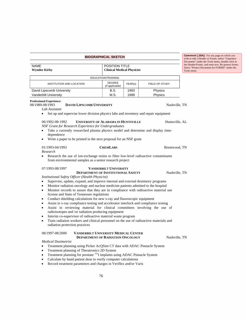
76
BIOGRAPHICAL SKETCH
NAME Wyndee Kirby
POSITION TITLE Clinical Medical Physicist
EDUCATION/TRAINING
INSTITUTION AND LOCATION DEGREE (if applicable)
YEAR(s) FIELD OF STUDY
David Lipscomb University B.S. 1993 Physics Vanderbilt University M.S. 1999 Physics
Professional Experience: 08/1989-08/1993 DAVID LIPSCOMB UNIVERSITY Nashville, TN Lab Assistant • Set up and supervise lower division physics labs and inventory and repair equipment 06/1992-08-1992 UNIVERSITY OF ALABAMA IN HUNTSVILLE Huntsville, AL NSF Grant for Research Experience for Undergraduates • Take a currently researched plasma physics model and determine and display time-
dependence • Write a paper to be printed in the next proposal for an NSF grant 01/1993-04/1993 CHEMLABS Brentwood, TN Research • Research the use of ion-exchange resins to filter low-level radioactive contaminants
from environmental samples as a senior research project 07/1993-08/1997 VANDERBILT UNIVERSITY DEPARTMENT OF INSTITUTIONAL SAFETY Nashville, TN Institutional Safety Officer (Health Physicist) • Supervise, update, expand, and improve internal and external dosimetry programs • Monitor radiation oncology and nuclear medicine patients admitted to the hospital • Monitor records to assure that they are in compliance with radioactive material use
license and State of Tennessee regulations • Conduct shielding calculations for new x-ray and fluoroscopic equipment • Assist in x-ray compliance testing and accelerator interlock and compliance testing • Assist in reviewing material for clinical committees involving the use of
radioisotopes and /or radiation producing equipment • Interim co-supervisor of radioactive material waste program • Train radiation workers and clinical personnel on the use of radioactive materials and
radiation protection practices 08/1997-08/2000 VANDERBILT UNIVERSITY MEDICAL CENTER DEPARTMENT OF RADIATION ONCOLOGY Nashville, TN Medical Dosimetrist • Treatment planning using Picker AcQSim CT data with ADAC Pinnacle System • Treatment planning of Theratronics 2D System • Treatment planning for prostate 131I implants using ADAC Pinnacle System • Calculate by hand patient dose to verify computer calculations • Record treatment parameters and charges in Veriflex and/or Varis
Comment [JG4]: For any page on which you wish to edit a Header or Footer, select “Unprotect Document” under the Tools menu, double-click in the Header/Footer, and enter text. Re-protect forms: Select “Protect Document for FORMS” under the Tools menu.
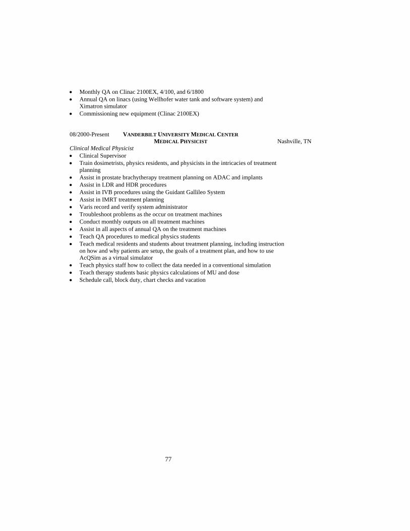
77
• Monthly QA on Clinac 2100EX, 4/100, and 6/1800 • Annual QA on linacs (using Wellhofer water tank and software system) and
Ximatron simulator • Commissioning new equipment (Clinac 2100EX) 08/2000-Present VANDERBILT UNIVERSITY MEDICAL CENTER MEDICAL PHYSICIST Nashville, TN Clinical Medical Physicist • Clinical Supervisor • Train dosimetrists, physics residents, and physicists in the intricacies of treatment
planning • Assist in prostate brachytherapy treatment planning on ADAC and implants • Assist in LDR and HDR procedures • Assist in IVB procedures using the Guidant Gallileo System • Assist in IMRT treatment planning • Varis record and verify system administrator • Troubleshoot problems as the occur on treatment machines • Conduct monthly outputs on all treatment machines • Assist in all aspects of annual QA on the treatment machines • Teach QA procedures to medical physics students • Teach medical residents and students about treatment planning, including instruction
on how and why patients are setup, the goals of a treatment plan, and how to use AcQSim as a virtual simulator
• Teach physics staff how to collect the data needed in a conventional simulation • Teach therapy students basic physics calculations of MU and dose • Schedule call, block duty, chart checks and vacation

78
Aneita Susanne Matthews Biographical Sketch Job Title: Physicist Assistant Education: B.S. in Physics from Cumberland University, Lebanon, Tennessee (2002) Training: On the job training from September, 2002 to present, Vanderbilt University Board Certification or Licensure: NA Teaching Experience: none Clinical Experience: Working in Clinical Physics Section as physicist assistant since September, 2002 to present

79
BIOGRAPHICAL SKETCH
NAME James A. Patton
POSITION TITLEProfessor of Radiology & Physics
EDUCATION/TRAINING
INSTITUTION AND LOCATION DEGREE (if applicable)
YEAR(s) FIELD OF STUDY
Western Kentucky University B.S. 1966 Physics & Math Vanderbilt University, Nashville, TN Ph.D. 1972 Physics
Positions and Employment: Summer 1967: Student Trainee in Health Physics, Oak Ridge National Laboratory, Oak Ridge,
TN 1972-1973: Research Associate, Division of Nuclear Medicine & Biophysics, Vanderbilt
Univ, Nash, TN 1972-1973: Research Associate, Department of Biomedical Eng., Vanderbilt University,
Nash, TN 1973-1975: Instructor in Radiology, Vanderbilt University, Dept of Radiology, Nashville, TN 1975-1979: Assistant Professor of Radiology, Vanderbilt University, Dept of Radiology,
Nashville, TN 1978-Present: Chief of Nuclear Medicine Physics, Vanderbilt University, Dept of Radiology,
Nashville, TN 1979-Present: Adjunct Faculty Member, Austin Peay State University, Clarksville, TN 1979-Present: Program Director and Educational Coordinator of Nuclear Medicine Technology
Training Program, Vanderbilt University, Department of Radiology, Nashville, TN
1979-1988: Associate Professor of Radiology, Vanderbilt University, Nashville, TN 1981-1987: Associate Director of Nuclear Medicine, Vanderbilt University, Nashville, TN 1987-1992: Administrator of Radiology, Vanderbilt University, Nashville, TN 1988-Present: Professor of Radiology, Vanderbilt University, Nashville, TN 1992-Present: Administrator for Radiology Academic Affairs, Vanderbilt University, Nashville, TN 1996-Present: Professor of Physics, Vanderbilt University, Nashville, TN Honors and Professional Memberships: Ogden Trustees Award for Academic Achievement at Western Ky. University, 1966 Scholar of the University Award at Western KY, University, 1966 A.E.C. Health Physics Society Fellowship at Vanderbilt University, 1966-69 Hertz Foundation Fellowship at Vanderbilt University, 1969-71 Visiting Professor in Radiology, University of Arizona, June 1978 Faculty Award, Nuclear Medicine Technology, Vanderbilt University, 1980, 1995, 1997 and 2002 Nuclear Medicine Subcommittee of AAPM, 1994-99 President of Southeastern Chapter of SNM, 1999 Associate Editor, The Journal of Nuclear Medicine, 1999 – Present Member, Board of Delegates of SNM, 2000 – Present. Society of Nuclear Medicine
Comment [JG5]: For any page on which you wish to edit a Header or Footer, select “Unprotect Document” under the Tools menu, double-click in the Header/Footer, and enter text. Re-protect forms: Select “Protect Document for FORMS” under the Tools menu.

80
American Association of Physicists in Medicine American Association for the Advancement of Science Tennessee Radiological Society American Hospital Radiology Administrators Radiology Business Managers Association Nuclear Medicine Committee of American Association of Physicists in Medicine Program Committee of American Association of Physicists in Medicine & Southeastern Chapter of SNM President of Southeastern Chapter of Society of Nuclear Medicine Associate Editor, The Journal of Nuclear Medicine Publications: 1. Martin WH, Delbeke D, Patton JA, Hendrix B, Weinfeld Z, Ohana I, Kessler RM, Sandler
MP. 18FDG-SPECT: Correlation with 18FDG-PET. J Nucl Med 36:988-995, 1995. 2. Delbeke D, Videlefsky S, Patton JA, Campbell MG, Martin WH, Ohana I, Sandler MP. Rest
Myocardial Perfusion/Metabolism Imaging Using Simultaneous Dual Isotope Acquisition SPECT with 99mTc-MIBI/18FDG. J Nucl Med 36:2110-2119, 1995.
3. Sandler MP, Videlefsky S, Delbeke D, Patton JA, Meyerowitz C, Martin WH, Ohana I.
Evaluation of Myocardial Ischemia Using a Rest Metabolism/Stress Perfusion Protocol with 18FDG/99mTc-MIBI and Dual Isotope Simultaneous Acquisition SPECT. J Amer Coll Cardiol 26:870-878, 1995.
4. Patton and Sandler MP. Correction for Downscatter from F-18 in DISA (FDG/MIBI)
SPECT. J Nucl Med 37:31P, 1996. 5. Martin WH, Delbeke D, Patton JA, Sandler MP. FDG SPECT vs. FDG PET for the
Detection of Malignancies. Radiology 198:225-231, 1996. 6. Sandler MP, Patton JA. Fluorine 18-Labeled Fluorodeoxyglucose Myocardial Single-Photon
Emission Computed Tomography: An Alternative for Determining Myocardial Viability. J Nucl Cardiol 3:342-349, 1996.
7. Patton JA, Sandler MP, Ohana I, Weinfeld Z. High Energy (511 keV) Imaging with the
Scintillation Camera. RadioGraphics 16:1183-1194, 1996 8. Sandler MP, Patton JA. Fluorine 18-Labeled Fluorodeoxyglucose Myocardial Single-Photon
Emission Computed Tomography: An Alternative for Determining Myocardial Viability. J Nucl Cardiol 3:342-349, 1996.
9. Patton JA, Hefetz Y, Shone MD, Sandler MP. Measured Coincidence Imaging Parameters of
a Clinical Dual-head Scintillation Camera. J Nucl Med 38:221P, 1997. 10. Sandler MP, Bax JJ, Patton JA, Visser FC, Martin WH, Wijns W. Fluorine-18-
Fluorodeoxyglucose Cardiac Imaging Using a Modified Scintillation Camera: State of the Art. J Nucl Med, 39(12):2035-2043, December 1998.
11. Patton JA. AAPM/RSNA Physics Tutorial for Residents. Introduction to Nuclear Physics.
RadioGraphics 18(4): 995-1007, 1998.

81
12. Patton JA, Turkington TG. Coincidence Imaging with a Dual-Head Scintillation Camera. J Nucl Med, 40:432-441, March 1999.
13. Delbeke D, Patton JA, Martin WH, Sandler MP. FDG PET and dual-head gamma camera
positron coincidence detection imaging using a dual head gamma camera of suspected malignancies and brain disorders. J Nucl Med 1999;40:110-117.
14. Delbeke D, Patton JA, Martin WH, Sandler MP. Comparison of FDG-PET and Positron
Coincidence Detection Imaging Using a Prototype (3/8-Inch-Thick Na(T1) Crystals) Dual Head Gamma Camera (DHC) in Patients with Suspected Body Malignancies and Brain Disorders. J Nucl Med, 40 (1):110-117, January 1999.
15. Boren EL, Delbeke D, Patton JA, Sandler MP. Comparison of FDG PET and Positron
Coincidence Detection Imaging Using a Dual-head Gamma Camera with 5/8-Inch Na1(T1) Crystals in Patients with Suspected Body Malignancies. European Journal of Nuclear Medicine, 26 (4):379-387, April 1999.
16. Patton JA, Delbeke D, Sandler MP. Image fusion using an integrated dual head coincidence
camera with x-ray tube based attenuation maps. J Nucl Med, 41:1364-1368, August 2000. 17. Delbeke D, Patton JA, Martin WH, Sandler MP. Positron imaging in oncology: Present and
future. In Freeman L (ed): "Nuclear Medicine Annual", Williams & Wilkins, Baltimore, MD, 1998, pp 1-49.
18. Patton JA. Instrumentation of Coincidence Imaging with Multihead Scintillation Cameras.
Seminars in Nucl Med, Vol 30, 4 (October), 2000, 239-254. 19. Delbeke D, Martin WH, Patton JA, Sandler MP. Value of Iterative Reconstruction,
Attenuation Correction, and Image Fusion in Interpretation of FDG PET Images with an Integrated Dual-Head Coincidence Camera and X-Ray-Based Attenuation Maps. Radiology, 218(1):163-171, 2001.
20. Delbeke D, Martin WH, Patton JA, Sandler MP. Practical FDG Imaging A Teaching File.
2002, Springer-Verlag New York, Inc. 21. Sandler MP, Coleman, RE, Wackers F. J. Th., Patton JA, Gottschalk. Diagnostic Nuclear
Medicine, 4th edition, Lippincott, Williams & Wilkins, 2002.

82
BIOGRAPHICAL SKETCH
NAME David R. Pickens III
POSITION TITLEAssociate Professor of Radiology
EDUCATION/TRAINING
INSTITUTION AND LOCATION DEGREE (if applicable)
YEAR(s) FIELD OF STUDY
Vanderbilt University Sch of Eng, Nash, TN M.S. 1975 Mechanical Engineering Vanderbilt University Sch of Eng, Nash, TN Ph.D. 1981 Mechanical Engineering
Positions and Employment: 1978-1980: Student Employee under Grant #5 R01-GM-2362, National Institute Health
Evaluation of a Mosaic Germanium Scanner 1980: Research III (Part time), Department of Radiology, Vanderbilt University,
Nashville, TN 1981: Research Assistant III, Department of Radiology, Vanderbilt University,
Nashville, TN 1981-1992: Assistant Professor of Radiology, Vanderbilt University, Nashville, TN 1987-1996: Assistant Professor of Biomedical Engineering, Vanderbilt University, Nashville, TN 1990-Present: Member, The Graduate Faculty, The Graduate School, Vanderbilt University,
Nashville, TN 1996-Present: Associate Professor of Biomedical Engineering, Vanderbilt University,
Nashville, TN 1992-Present: Associate Professor of Radiology, Vanderbilt University, Nashville, TN Other Experience and Professional Memberships: 1978: Tennessee Board of Architectural and Engineer Examiners, Engineer-in-Training
(Cert. #5316) 1991: Member, Review Committee, National Institutes of Health, Division of Research
Grants, Special Study Section on Radiological Imaging 1992-Present: Member, Multidisciplinary Special Emphasis Panel, Surgery, Radiology and
Bioengineering Review Group, National Institutes of Health, Division of Research Grants
1992-Present: Member, Diagnostic Imaging Special Emphasis Panel, Diagnostic Imaging, National Institutes of Health, Division of Research Grants
1993: Chairman, Scientific Program, American Association of Physicists in Medicine, National Meeting, 1993. Responsible for the organization and content of the national meeting
1994-Present: American Board of Radiology, Diagnostic Radiological Physics 1996: Chairman, Physics Session (CT Angiography). Radiological Society of North
America Annual Meeting, December 3, 1996. 1997: Co-Chairman, American Association of Physicists in Medicine, Local
Arrangements Committee, Nashville 1999 Meeting 1999: Chairman, Program Committee, American Association of Physicists in Medicine Selected peer-reviewed publications (in chronological order): 1. Melzer P, Pickens DR, Price RR, Morgan VL, Wall RS, Ebner FF. Reading Braille Activates
Cortical Areas of the Dorsal Stream in the Blind: A fMRI Study. Neuroscience 24:1979, 1998.
Comment [JG6]: For any page on which you wish to edit a Header or Footer, select “Unprotect Document” under the Tools menu, double-click in the Header/Footer, and enter text. Re-protect forms: Select “Protect Document for FORMS” under the Tools menu.

83
2. Melzer P, Pickens DR, Price RR, Morgan VL, Symons FJ, Wall RS. Reading Braille Predominantly Activates Extrastriate Cortex in Persons with Severe Visual Disability: A fMRI Study. Proceedings of the International SMRM, Sixth Scientific Meeting and Exhibition, Sydney, Australia, Vol. 3, p.1514, 1998.
3. Melzer P, Morgan L, Pickens DR, Price RR, Wall RS. Cerebral Activation During Reading Nouns and Text in Braille: A fMRI Study. Radiology 209:243, 1998.
4. Pickens DR, Price RR, Morgan VL, Holburn GE, Parks MH, Martin P. Long Term Repeatability of Cerebellar fMRI Correlated with an Independently Monitored Motor Task. Radiology 209:245, 1998.
5. Morgan VL, Price RR, Holburn GE, Butler M, Pickens DR, Partain CL. Functional MRI of the Human Olfactory Cortex. Radiology 209P:276, 1998.
6. Pickens DR, Price RR, Mertz H, Morgan VL, Tanner G. Bowel Pain Response in Irritable Bowel Syndrome Patients Using fMRI. Radiology, 209(P) 276, 1998.
7. Parks M, Morgan V, Pickens D, Price R, Schlack H, Martin P. Functional MRI Studies of Motor Plasticity During Recovery of Alcohol-Induced Brain Injury with Abstinence. Alcoholism, Clinical and Experimental Research, 23(5):66A, 1999.
8. Parks MH, Morgan VL, Pickens DR, Price RR, Schlack HM, Martin PR. Cerebellar fMRI Studies of Alcohol-Induced Brain Injury. Proceedings of the ISMRM, 2:832, 1999.
9. Fleischer AC, Wojcicki W, Donnelly E, Pickens D, Thirsk G, Thurman G, Hellerqvist Carl: Quantified Color Doppler Sonography of Tumor Vascularity in an Animal Model. J Ultrasound Med 18: 547-552, 1999.
10. Mertz H, Tanner G, Morgan V, Pickens D, Price R, Kessler M. Functional MRI Detects Activation of Anterior Cingulate and Prefrontal Cortex in IBS Patients But Not Controls During Rectal Pain. Gastroenterology, 116:4, Part 2, A-1041, April 1999.
11. Melzer P, Pickens DR, Price RR, Morgan VL, Wall RS and Ebner FF. Timing of Sensory Challenges Affects Cortical Activation during Braille Reading in the Visually Disabled: A fMRI Study. Society of Neuroscience 25:1812, 1999.
12. Morgan VL, Mertz H, Pickens DR, Price RR, Tanner WG and Kessler R. Study of Irritable Bowel Syndrome Using Functional MRI. Proceedings of the ISMRM 2:824, 1999.
13. Price RR, Pickens DR, Morgan VL, Fitzpatrick JM, Dawant B, Brill AB. Online Position Tracking for fMRI Motion Correction. Radiology, Annual Meeting of the Radiology 213(P):233, 1999.
14. Fleischer AC, Wojcicki WE, Donnelly EF, Pickens DR, Thirsk G, Thurman GB, and Hellerqvist CG. Quantified Color Doppler Sonography of Tumor Vascularity in an Animal Model. J Ultrasound in Med 18:547-551, 1999.
15. Grossman ED, Donnelly M, Price R, Morgan V, Pickens D, Neighbor G, Blake R. Brain Areas Involved in Perception of Biological Motion. Journal of Cognitive Neuroscience, 12:711-720, 2000.
16. Mertz H, Morgan V, Tanner G, Pickens D, Price R, Shyr Y, Kessler R. Regional Cerebral Blood Flow in IBS and Controls with Painful and Non-Painful Rectal Distention. Gastroenterology, 2000;118:842-848.
17. Melzer P, Morgan V, Pickens D, Price R, Wall R, Ebner F. Cortical Braille Activation Differs With Visual Experience in the Early Visually Disabled: A Correlational fMRI Study. Human Brain Mapping, 2000; Vol. 14, No. 3, 186-195.
18. Morgan VL, Pickens DR, Hartmann SL, Price RR. Comparison of Functional MRI Realignment Tools Using a Computer Generated Phantom. Presented at the Eighth Scientific Meeting of the International Society for Magnetic Resonance in Medicine 848, 2000.
19. Parks MH, Morgan VL, Price RR, Pickens DR, Dietrich MS, Martin PR. Disrupted Simple Motor Fronto-Cerebellar Circuits in Chronic Alcoholics Demonstrated by fMRI. Alcoholism Clinical and Experimental Research 24(5):126A, 2000.

84
20. Morgan VL, Pickens DR, Hartmann SL, Riddle WR and Price RR. Software Phantom for Assessment of Functional MRI Realignment Algorithms. Radiology 217(P): 311, 2000.
21. Morgan VL, Pickens DR, Hartmann SL, Price RR. Comparison of functional MRI Realignment Tools using a Computer Generated Phantom. Magnetic Resonance in Medicine, 2001; 46(3):510-514.
22. Melzer P, Morgan VL, Pickens DR, Price RR, Wall RS and Ebner FF. Cortical Activation During Braille Reading is Influenced by Early Visual Experience in Subjects with Severe Visual Disability: A Correlation fMRI Study. Human Brain Mapping, 2001; 14:186-195.
Research Support Ongoing Research Support 1R25 CA92043-01 Price (PI) 07/01/01-06/30/06 NIH/NCI Multidisciplinary Research Training in Cancer Imaging The goal of this project is to establish a unique training program in cancer imaging research. The program is designed to train both medical post-doctoral candidates and basic-science post-doctoral candidates. Role: Preceptor Completed Research Support BES-880834 Price (PI) 08/15/98-08/14/00 NSF Position Tracking for Motion Correction in MRI The goal of this project is to develop and demonstrate a system whereby the motion of a subject/patient undergoing a magnetic resonance imaging (MRI) examination is monitored in near realtime and the information is fed back to the MRI scanner to produce “motion-free” images. Role: Investigator 5R21 DK57047-02 Mertz (PI) Regional Cerebral Activation with Visceral Pain in IBS and Controls The goal of this project is to investigate cerebral pain response in Irritable Bowel Syndrome with and without pharmaceutical interaction. Role: Investigator Industry-Pharmaceutical Price (PI) 05/01/98-06/30/99 Evaluation of the Varian Amorphous Silicon (aSi) Sensor To determine the optimum radiographic technique for selected clinical procedures to be used with Varian aSi sensor and to evaluate image quality relative to conventional film/screen systems. Role: Investigator

85
James Benjamin (BJ) Proffitt Biographical Sketch
Job Title: Dosimetrist Education: B.S. in Physics from Belmont University in Nashville, TN (2002) Training: On the job training from April ’02 to present at Vanderbilt University Board Certification or Licensure: none Teaching Experience: none Clinical Experience: Working in dosimetry for 1 year (April ’02 – present

86
BIOGRAPHICAL SKETCH
NAME Christopher Scarfone
POSITION TITLE Assistant Professor and Medical Physicist
EDUCATION/TRAINING
INSTITUTION AND LOCATION DEGREE (if applicable)
YEAR(s) FIELD OF STUDY
University of Vermont, Burlington, Vermont B.S. 1988 Physics Cornell University, Ithaca, NY M.S. 1991 Physics University of North Carolina at North Carolina State, Raleigh, NC
Ph.D. 1998 Physics
Professional Experience and Appointments: 1987 Research Trainee, Stanford University, Stanford Linear Accelerator Center, Stanford, CA 1988-1991 Teaching Assistant, Cornell University, Department of Physics, Ithaca, NY 1992-1993 Lecturer, University of Vermont, Department of Physics, Burlington, VT 1993-1998 Research Assistant, Duke University Medical Center, Department of Radiology, Durham, NC 1995-1998 Research Assistant, Duke University Medical Center, Department of Radiation
Oncology, Durham, NC 1995-1998 Staff Physicist, Data Spectrum Corporation, Hillsborough, NC 1998-05/2000 Medical Physics Resident, Vanderbilt University Medical Center, Department of
Radiation Oncology, Nashville, TN 05/2000-2001 Instructor and Medical Physicist, Vanderbilt University Medical Center, Department of
Radiation Oncology, Nashville, TN 05/2000-2001 Instructor (joint appt.), Vanderbilt University Medical Center, Department of Radiology
and Radiological Sciences, Nashville, TN 07/2001-Pres Assistant Professor and Medical Physicist, Vanderbilt University Medical Center,
Departments of Radiation Oncology, Nashville, TN 07/2001-Pres Assistant Professor (joint appt.), Vanderbilt University Medical Center, Department of
Radiology and Radiological Sciences, Nashville, TN Academic Honors and Awards: 1985 Charles G. Fraiser Award for Physics, University of Vermont 1987 Phi Beta Kappa - University of Vermont 1987 David W. Juenker Award for Physics, University of Vermont 1988 Magna Cum Laude – University of Vermont 1988-1991 Tuition Fellowship, Department of Physics, Cornell University 1997 American Cancer Society Institutional Research Grant Student Award Nominee - Duke
University Medical Center 1999 Winner, Vanderbilt-Ingram Cancer Center Retreat Poster Competition National Award: 1998-2000 American Society of Therapeutic Radiation Oncologists (ASTRO) Clinical Radiotherapy
Physics Resident Award Invited Speaker: June 2000 Functional Treatment Planning, 25th Annual Meeting of the American Association of
Medical Dosimetrists (AAMD), Portland, OR Government Funding and Grant Writing Experience: 1. The Use of Fan-Beam SPECT for Breast Tumor Detection, National Institutes of Health - Breast
Imaging Group, Duke University Medical Center, (Funded 1995-1997) 2. Dynamic Cardiac Phantom for Nuclear Medicine Imaging, National Institutes of Health, Small
Business Innovation Research (SBIR) Grant, Data Spectrum Corporation, (Phase I Funded 1996-1997) Research Projects Ongoing or Completed During the Last Three Years:
Comment [JG7]: For any page on which you wish to edit a Header or Footer, select “Unprotect Document” under the Tools menu, double-click in the Header/Footer, and enter text. Re-protect forms: Select “Protect Document for FORMS” under the Tools menu.

87
1. “Flk1 Signaling Protects Tumor Vasculature from Radiation”, (Study the mechanisms by which tumor blood vessels respond to ionizing radiation) Principal Investigator: Dennis E. Hallahan, M.D. Agency: National Cancer Institute Type: RO1 (CA89674-01) Period: 2001-2005. Role on project: Development and implementation of quantitative molecular imaging techniques.
2. “X-ray Guided drug delivery by apcitide-99mTc”, (Determination of the threshold dose and tumor types and which radiation activates receptors for drug delivery) Principal Investigator: Dennis E. Hallahan, M.D. Agency: National Cancer Institute Type: R21 (CA89888-01) Role on project: Quantitative and qualitative analysis of peptide distributions using SPECT.
BOARD CERTIFICATION: American Board of Radiology - Therapeutic Radiologic Physics, Written exam Parts I and II Passed. Part III (oral exam) scheduled for June 2003 in Louisville, KY. PROFESSIONAL MEMBERSHIPS AND COMMITTEES: Full Member - American Association of Physicists in Medicine (AAPM) Full Member - Society of Nuclear Medicine (SNM) Co-founder and core member – AAPM Science Council Committee: Molecular Imaging in Clinical Radiation Oncology (MICRO) Member – AAPM Science Council Committee: Nuclear Medicine UNIVERSITY COMMITTEES: Member – Radiology Grants Committee Member – Quantitative Nuclear Medicine Committee Professional Activities: Reviewer The Journal of Nuclear Medicine, 1998-present Cancer Biotherapy and Radiopharmaceuticals, 2002-present Teaching Activities: 2001-Pres Radiological Physics, Lecturer, Vanderbilt Center for Radiation Oncology Program in
Radiation Therapy 2000-Pres Therapy Physics Laboratory, Laboratory Assistant, Vanderbilt Department of Radiation
Oncology Masters degree program in Medical Physics Fall 2002 The Physics of Radiology, Physics 228 - Department of Physics, Vanderbilt University, Student and Fellow Supervision: 1. Philip Haberlen, B.S., Candidate for degree of Medical Doctor, Vanderbilt University School of
Medicine, Combination SPECT/CT Imaging Provides a Cost-Effective Alternative to Dedicated SPECT and CT Imaging, Introduction to Biomedical Research meeting presentation, Spring 2001.
2. Alisha Smith, B.S., Certified Nuclear Medicine Technician Program, Vanderbilt University School of Medicine, First Generation Radiation Guided Therapy for Brain Tumors, Tennessee Sate Nuclear Medicine Meeting, Pigeon Forge, TN, Spring 2001.
3. Barth Lynch, undergrad, University of Tennessee. REU student through Biomedical Engineering Department at Vanderbilt. Project: IMRT Film QA, (Summer 2001).
4. Mark D. Anderson, undergrad, University of Miami, Florida. REU student through Biomedical Engineering Department at Vanderbilt. Project: IMRT Film / Treatment Plan Overlay Techniques, (Summer 2002).
5. William “Rusty” Lavely, M.D., Imaging Research Fellow, Department of Radiology and Radiological Sciences. Project: Multiple Image Modality Treatment Planning, (January 2002-present).
6. Kenneth Niermann, M.D., Imaging Research Fellow, Department of Radiology and Radiological Sciences. Project: High-Field Magnetic Resonance Spectroscopy with Applications to Radiation Treatment Planning, (July 2002-present).
Publications: 1. Scarfone, C., Cmelak, A.J., Delbeke, D., Lavely, III, W., Martin, W., and Hallahan, D.E.,
Radiotherapy target volumes for head and neck cancer: a comparison of x-ray CT and PET, (in preparation).

88
2. Scarfone, C., Cmelak, A.J., Delbeke, D., Lavely, III, W., Martin, W., and Hallahan, D.E., Influence of PET image reconstruction technique on radiotherapy target volumes, (in preparation).
3. Hallahan, D.E., Qu, S.M., Geng, L., Cmelak, A., Chakravarthy, A., T., Martin, W., Scarfone, C., and Giorgio, T., Radiation-mediated control of drug delivery, Am J Clinical Onc-Cancer Clinical Trials, 24(5), 473-480, Oct. 2001.
4. Hallahan, D.E., Geng, L., Cmelak, A.J., Chakravarthy, A.B., Martin, W., Scarfone, C., and Gonzalez, A., Targeting drug delivery to radiation-induced neoantigens in tumor microvasculature, J Cont Rel, 74 (1-3), 183-191 (2001).
5. Scarfone, C., Patton, J.A., Cmelak, A.J., and Sandler, M.P., Gamma camera-based PET inverse treatment planning for head and neck cancer using hybrid imaging istrumentation and imrt, in The Scintillating Future of Nuclear Medicine, T. Nishimura, H.W. Strauss and M. Fukuchi, Eds., (in print May 2001).
6. Scarfone, C., A transition to clinical medical physics, to appear in The Society of Physics Students Newsletter, American Institute of Physics, pubs., (Spring 2001).
7. Scarfone, C., Marks, L.B., Munley, M.T., Jaszczak, R.J., and Coleman, R.E., Influence of single photon emission computed tomography (SPECT) reconstruction technique on pulmonary dose-response curves: a phantom and patient study, Int J Rad Onc Biol Phys (in preparation).
8. Scarfone, C., and Coffey, C.W., II, Nuclear medicine imaging for external beam cancer treatment: an overview for the clinical medical physicist, American College of Medical Physics Annual Meeting, Whistler Resort, British Columbia, CA (May 2000).
9. Scarfone, C., Morgan, V., Rohde, G.K., Patton, J.A., and Sandler, M.P., Combined functional and anatomical imaging for conformal radiotherapy, J Nucl Med, 41, P273, (2000).
10. Scarfone, C., Geng, L., Giorgio, T., Clanton, J., and Hallahan, D.E., Tumor-trageted delivery systems guided by radiation, Science (submitted July 2000, under preparation for resubmission).
11. Scarfone, C., Jaszczak, R.J., Munley, M.T., Marks, L.B., Gilland, D.R., Greer, K.L., and Coleman, R.E., Quantitative pulmonary SPECT for radiotherapy applications, Med Phys, 26, 8, 1579-1587 (1999).
12. Scarfone, C., Geng, L., Holburn, G., Brill, A.B., Dugger, J., Patton, J.A., Sandler, M.P., and Hallahan, D.E., Evaluation of radiation guided gene therapy using pinhole imaging and region-of-interest (ROI) analysis, Med Phys, 26, 6, 1159 (1999).
13. Scarfone, C., Geng, L., Brill, A.B., Holburn, G., Clanton, J.A., Dugger, J., Patton, J.A., Sandler, M.P., and Hallahan, D.E., External beam guided drug delivery using radiolabeled microspheres: evaluation with nuclear medicine and autoradiography region-of-interest (ROI) techniques, Med Phys, 26, 6, 1160 (1999).
14. Geng, L., Dugger, J., Fleischer, A., Brill, A.B., Donnelly, E., Sandler, M.P., Clanton, J., Holburn, J., Scarfone, C., and Hallahan, D.E., Noninvasive imaging of the biodistribution of radiation sensitizing gene therapy, Int. J. Radit. Oncol. Biol. Phys., 45, 3, 298 (1999).
15. Jaszczak, R.J., Scarfone, C., Marks, L.B., Munley, M.T., Greer, K.L., and Gilland, D.R., Investigation of SPECT quantification of lung perfusion imaging: a phantom study, Euro J Nucl Med, 25, 8, pp. 911, (1998).
16. Scarfone, C., Jaszczak, R.J., Gilland, D.R., Munley, M.T., Marks, L.B., and Coleman, R.E., Bayesian SPECT Lung Imaging for Visualization and Quantification of Pulmonary Perfusion, IEEE Trans Nucl Sci, 45, 6, 3045-3052 (1998).
17. Munley, M.T., Marks, L.B., Scarfone, C., Patz, Ed, Jaszczak, R.J., Sibley, G.S., and Coleman, R.E., Multimodality nuclear medicine imaging for 3D lung radiotherapy: challenges and prospects, Lung Cancer, 23, 105-114 (1999).
18. Marks, L.B., Munley, M.T., Bentel, G.C., Zhou, SM., Hollis, D., Scarfone, C., Sibley, G.S., Kong, F.M., Jirtle, R., Jaszczak, R.J., Coleman, R.E., Tapson, V., and Anscher, M., Physical and biological predictors of changes in whole lung function following thoracic irradiation, Int. J. Radit. Oncol. Biol. Phys., 39, 3, 563-570 (1997).
19. Scarfone, C., Jaszczak, R.J., Li, J., Soo, M.S., Smith, M.F., Greer, K.L., and Coleman, R.E., Breast tumor imaging using incomplete circular orbit pinhole SPECT: a phantom study, Nuclear Medicine Communications, 18, pp. 1077-1086, (1997).
20. Wang, H., Scarfone, C., Greer, K.L., Coleman, R.E., and Jaszczak, R.J., Prone breast tumor imaging using vertical axis-of-rotation (VAOR) SPECT systems: an initial study, IEEE Trans Nucl Sci, 44, no. 3, pp. 1271-1276, (1997).
89
21. Peter, J., Freyer, R., Smith, M.F., Scarfone, C., Coleman, R.E., and Jaszczak, R.J., Nuclear medicine image segmentation using a connective network, IEEE Trans Nucl Sci, 44, no. 4, pp. 1583-1590, (1997).
22. Scarfone, C., Jaszczak, R.J., Gilland, D.R., Munley, M.T., Marks, L.B., and Coleman, R.E., Bayesian SPECT Lung Imaging for Visualization and Quantification of Pulmonary Perfusion, 1997 Nuclear Science and Medical Imaging Conference, Albuquerque, NM (1997).
23. Peter, J., Smith, M.F., Scarfone, C., Coleman, R.E. and Jaszczak, R.J., Synergetically generalized expectation maximization algorithm for ECT, 1997 Nuclear Science and Medical Imaging Conference, Albuquerque, NM (1997).
24. Wang, H., Scarfone, C., Greer, K.L., Coleman, R.E., and Jaszczak, R.J., Breast cancer detection using vertical axis-of-rotation (VAOR) SPECT with half-cone beam collimation, Society of Nuclear Medicine 44th Annual Meeting (San Antonio, TX 1997).
25. Wang, H., Scarfone, C., Greer, K.L., Coleman, R.E., and Jaszczak, R.J., Prone breast tumor imaging using vertical axis-of-rotation (VAOR) SPECT systems: an initial study, Conference Record of the 1996 Nuclear Science Symposium and Medical Imaging Conference, Anaheim, CA, 3, pp. 1387-1391 , (1997).
26. Peter, J., Freyer, R., Smith, M.F., Scarfone, C., Coleman, R.E., and Jaszczak, R.J., Nuclear medicine image segmentation using a connective network, Conference Record of the 1996 Nuclear Science Symposium and Medical Imaging Conference, Anaheim, CA, 3, pp. 1782 - 1786, (1997).
27. McCormick, J.W., Jaszczak, R.J., Scarfone, C., and Coleman, R.E., The effect of truncation reduction in fan beam transmission for attenuation correction of cardiac SPECT, Conference Record of the 1995 IEEE Nuclear Science Symposium and Medical Imaging Conference, San Francisco, CA, 1252-1256 (1996).
28. Munley, M.T., Scarfone, C., Marks, L.B., Anscher, M., McAdams, H.P., Patz, E.F., Jaszczak, R.J., and Coleman, R.E. Multimodality nuclear medicine imaging in radiation treatment planning for lung cancer: challenges and prospects, American Society for Therapeutic Radiology and Oncology 38th Annual Meeting, Anaheim,
CA (1996). 29. Li, J., Jaszczak, R.J., VanMulkan, A, Scarfone, C., Greer, K.L., and Coleman, R.E., Half-cone beam
collimation for triple-camera SPECT systems. J Nucl Med, 37: pp. 498-502 (1996). 30. Jaszczak, R.J., Gilland, D.R., McCormick, J.W., Scarfone, C., and Coleman, R.E., The effect of
truncation reduction in fan beam transmission for attenuation correction of cardiac SPECT. IEEE Trans Nucl Sci, 43, pp. 2255-2262 (1996).
31. Marks, L..B., Munley, M.T., Bentel, G., Hollis, D., Zhou, S., Jirtle, R., Kong, F.M., Scarfone, C., Antoine, P., Chew, M., Tapson, V., Spencer, D., Sherouse, G., Jaszczak, R.J., Coleman, R.E., and Anscher, M., Physical and biological predictors of radiation-induced whole lung injury: early results of a prospective study, 1996 American Society for Therapeutic Radiology and Oncology 38th Annual Meeting (Anaheim, CA, 1996).
32. Li, J., Scarfone, C., Jaszczak, R.J., Wang, H., and Coleman, R.E. Limited-angular view ML-EM pinhole SPECT for breast tumor detection. J Nucl Med 37: 214P (1996).
33. Wang, H., Scarfone, C., Jaszczak, R.J., and Coleman, R.E. A new breast tumor imaging approach using SPECT systems with non-tiltable cameras: A phantom study. Med Phys 23: pp. 1107 (1996).
34. Jang, S.B., Jaszczak, R.J., Gilland, D.R., Hanson, M.W., Scarfone, C., and Coleman, R.E. Effect of breast size and shape on defect contrast in myocardial perfusion SPECT imaging. J Nucl Med 37: 147P (1996).
35. Norton, M. G., Scarfone, C., Jian, L., Carter, C.B., and Mayer, J.W., Epitaxy of barium titanate thin-films grown on MgO by pulsed-laser ablation. J Mater Res, vol. 6, pp. 2022-2025 (1991).
36. Norton, G., Carter, B.C., Scarfone, C, et. al., Early stages of growth of barium titanate thin-films studied by transmission electron microscopy. Proc XIIth Int Cong for Electron Microscopy 4 (1990).
37. Scarfone, C., Norton, M.G., Carter, C.B., Jian, L., and Mayer, J.W., Characterization of BaTiO3 thin-films deposited by pulsed-laser ablation. Mat Res Soc Symp Proc 201; pp. 183-188 (1990).

90
BIOGRAPHICAL SKETCH
NAME Michael G. Stabin
POSITION TITLEAssistant Professor
EDUCATION/TRAINING
INSTITUTION AND LOCATION DEGREE (if applicable)
YEAR(s) FIELD OF STUDY
University of Florida B.S. 1981 Environmental Engineering University of Florida M.E. 1983 Environmental Engineering University of Tennessee, Knoxville Ph.D. 1996 Nuclear Engineering
Work Experience: 2000-Present: Assistant Professor, Vanderbilt University
Duties include giving classes at the graduate level in radiological protection; performing support duties for the Vanderbilt Hospital and the Department of Radiology, including radiation dose calculations for patients; orienting students in their graduate work; and performing research. Research results are routinely presented at national and international conferences.
1998-2000: Visiting Professor, Universidade Federal de Pernambuco
Duties include giving classes at the graduate level in radiological protection, orienting students in their graduate work, and performing research. Research results are routinely presented at national and international conferences. Some outside teaching duties were performed as well, in short courses, seminars, etc.
1983-1998: Scientist, Radiation Internal Dose Information Center, Oak Ridge Associated Universities, Oak Ridge, Tennessee
Duties include responding to requests for information about problems in internal dosimetry, performing research in internal dosimetry, presenting results of research at scientific meetings and in the open literature, lecturing in training courses in internal dosimetry and radiation safety, and assisting graduate students in their research and in developing and defending their theses/projects. Training course experience has included lecturing in one week courses in Occupational Internal Dosimetry and Radiopharmaceutical Internal Dosimetry, coordinating both courses, giving lectures in one week courses on uses of radionuclides in medicine and in several courses for the Radiation Emergency Assistance Center and Training Site (REAC/TS) in health physics in radiation accidents, health physics for medical personnel and others, mainly on the topic of internal dosimetry. In addition, many lectures have been given, mostly on an invited basis, at short courses at technical meetings including the Health Physics Society (Professional Enrichment Program), the Society of Nuclear Medicine, the American Association of Physicists in Medicine, the Campus Radiation Safety Officers' Meeting, the Drug Information Association, the American Pharmaceutical Association, and the World Congress on Medical Physics and Biomedical Engineering.
1980-1983: Department of Environmental Engineering; Student assistant and graduate assistant
Assisted Dr. Charles Roessler in several projects at the University of Florida involving the radiological impacts of phosphate mining on the environment in Florida. Also assisted Dr. Roessler on several projects for private firms in Florida, also studying environmental impacts of naturally-occurring radionuclides in phosphate and phosphate by-products.
Comment [JG8]: For any page on which you wish to edit a Header or Footer, select “Unprotect Document” under the Tools menu, double-click in the Header/Footer, and enter text. Re-protect forms: Select “Protect Document for FORMS” under the Tools menu.

91
Honors: 1996 Elda E. Anderson Award Recipient, Health Physics Society Certification: Certified Health Physicist – 1988, Recertified – 1992, 1996, 2000 Professional Societies: Health Physics Society - President, Medical Section, 1993-present (first section president) Associate Editor, Health Physics Journal, 1992-present Secretary (1989-1990), Council Member (1986), chair of Public Education Committee (1983-1985), President-Elect (1998), East Tennessee Chapter of the Health Physics Society. Sigma Xi Tau Beta Pi Society of Nuclear Medicine, Associate Editor, Journal of Nuclear Medicine Editorial Board, Cancer Biotherapy & Radiopharmaceuticals, 1998- pres. Committee Activities: Human Subjects Radiation Committee, Vanderbilt University, 2001 - pres. Health Physics Society Electronic Media Committee, 2001 – pres.
Special Task Group of the American Association of Physicists in Medicine on dosimetry for monoclonal antibodies - 1988 - present
Special Task Group of the Society of Nuclear Medicine's Medical Internal Radiation Dose ( MIRD) committee to develop a new model for the urinary bladder - 1985 - 1992
Special Task Group of the Society of Nuclear Medicine's Medical Internal Radiation Dose (MIRD) committee to develop a radiation dosimetry model for adults and children for Tc-99m MAG3 - 1991 - present Special Task Group of the International Commission on Radiological Protection on Dose to Patients from Radiopharmaceuticals - 1991 - present
Publications: Author or Co-author on 118 full articles or book chapters and 62 abstracts. Most Recent: 1. Stabin, M. and Zaidi, H. (2002) Monte Carlo codes for use in therapeutic nuclear medicine.
In Therapeutic applications of Monte Carlo calculations in nuclear medicine. (Eds, Zaide, H. and Sgouros, G.) Institure of Physics Publishing, London, PP. 286-309.
2. Bouchet, L., Bolch, W., Stabin, M., Eckerman, K., Poston, J.W. and Brill, A.B. (2002) Monte Carlo methods and mathematical models for the dosimetry of skeleton and bone marrow. In Therapeutic applications of Monte Carlo calculations in nuclear medicine. (Eds, Zaidi, H. and Sgouros, G.) Institute of Physics Publishing, London, pp. 310-323.
3. Stabin, M. and Duggan, F. (2002) Monte Carlo modeling of dose distributions in intravascular radiation therapy. In Therapeutic applications of Monte Carlo Calculations in nuclear medicine. (Eds, Zaidi, H. and Sgouros, G.) Institute of Physics Publishing, London, pp. 310-323.
4. Stabin, MG, da Luz CQPL. New Decay Data For Internal and External Dose Assessment, Health Phys. 83 (4) :471-475, 2002.
5. M.G. Stabin, K.F. Eckerman, W.E. Bolch, L.G. Bouchet, and P.W. Patton. Evolution and Status of bone and Marrow Dose Models Cancer Biotherapy and Radiophermaceuticals, 17 (4) :427-434, 2002.

92
MIRDOSE Software: I developed and wrote this software package, which has been distributed to over 1200 sites worldwide. It automates the calculation of internal dose estimates, primarily in nuclear medicine. Radsafe Professional Mailing List: When the Radsafe mailing list, which has about 2000 members worldwide and post 10-50 e-mail messages per day on the topic of radiation protection, was moved from the University of Illinois at Urbana-Champaign campus, I had it moved to the Vanderbilt campus and took over as list owner/manager. Students Supervised: 1. David Marshall, Master’s student, University of Texas, 1993. Subject - development of phantom
representing woman at 6 months’ pregnancy. Result - received Master’s degree, completed thesis, thesis became part of publication of ORNL report on 4 phantoms representing the adult female at different stages of pregnancy.
2. Julie Behm, Master’s student, University of Florida, 1994. Supervised on several projects involving
new models for internal dosimetry. Also served on advisory committee for Master’s degree. Result - received Master’s degree, completed thesis.
3. Joy Russell, University of Tennessee, 1995. Subject - use newly developed pregnant female phantoms
to calculate radiation dose estimates at all stages of pregnancy for many radiopharmaceuticals. Result - received Master’s degree, completed thesis, received student award from Health Physics Society, two full articles published in Health Physics Journal, 1997.
4. Michalene Rodriguez, Colorado State University, 1996. Subject - develop kinetic model for kinetics
of iodine in subjects whose thyroids have been removed surgically or with radioiodine. Result - received Master’s degree, completed project report. Publication of work pending, awaiting collaboration with CNEN-IRD, Rio de Janeiro.
5. Helio Yoriyaz, CNEN-IPEN, Sao Paulo, Brazil, 1996-1997. Subject - investigate ability of radiation
transport software to perform patient-specific dosimetry. Result - project has good progress, will be used for PhD dissertation, work ongoing.
6. Gina Cotlet, Ohio State University, 1997. Subject - develop model for bone marrow response in
radioimmunotherapy. Result - student is using results of Master’s thesis (kinetic model for cell division) with data on marrow response to radiation from radioimmunotherapy applications, work ongoing.
7. Lydia Luz, Departamento de Energia Nuclear, 1998- pres. Subject – evaluate existing bone marrow
dose models and investigate how to adapt them to individual patients in nuclear medicine therapy. 8. Fabiana Farias da Lima, Departamento de Energia Nuclear, 1998- 2002. Subject – evaluate methods
for patient-specific imaging and treatment planning for thyroid cancer patient treatment. 9. Eduardo Loureiro, Departamento de Energia Nuclear, 1998- 2002. Subject – adapt voxel-based images
to the calculation of radiation doses from diagnostic radiology in dentistry. 10. Laelia P. Campos, Departamento de Energia Nuclear, 1998- 2000. Subject – calculation of radiation
dose distributions in intravascular brachytherapy.

93
BIOGRAPHICAL SKETCH NAME Teng, Ming
POSITION TITLE Assistant Professor
EDUCATION/TRAINING INSTITUTION AND LOCATION DEGREE
(if applicable) YEAR(s) FIELD OF STUDY Shanghai Medical University Medical College of Wisconsin
MD PhD
1987 1993
Medicine Molecular Immunology
Employment and Experience 1987-88 Fellow, Shanghai Medical University, Shanghai, P.R. China 1993-94 Resident, Internal Medicine, University Hospital of Columbia University, New
York, New York 1994-97 Residence, Radiation Oncology, New York Medical College, Valhalla, New
York 1997-98 Instructor, Radiation Oncology, Vanderbilt University, Nashville, Tennessee 1998-Present Assistant Professor, Radiation Oncology, Vanderbilt University, Nashville, Tennessee Professional Societies: American College of Radiology American College of Radiation Oncology American Society for Therapeutic Radiology and Oncology BIBLIOGRAPHY:
Yan, X., Hallahan DE, Teng M “Monastrol Cytotoxicity and Radiation Sensitization in Three Human Carcinoma Cell Lines.” Submitted to Int J Radiat Oncol Biol Phys
Hallahan DE, Teng M., “The proteasome: a molecular target for cancer therapy.” Int J Radiat Oncol Biol Phys 2000 Jul 1;47(4):859-60
Teng, M., Choy H, Ettinger D “Combined chemoradiation therapy for limited-stage small-cell lung cancer.” Oncology (Huntingt) 1999 Oct;13(10 Suppl 5):107-15
Hallahan DE, Chen AY, Teng, M., Cmelak “AJ Drug-radiation interactions in tumor blood vessels.” Oncology (Huntingt) 1999 Oct;13(10 Suppl 5):71-7
Blanke CD, Teng, M., Choy H “The role of UFT in combined-modality therapy.” Oncology (Huntingt) 1999 Oct;13(10 Suppl 5):47-54
Blanke CD, Choy H, Teng, M., Beauchamp RD, Leach S, Roberts J, Washington K, Johnson DH “Concurrent paclitaxel and thoracic irradiation for locally advanced esophageal cancer.” Semin Radiat Oncol 1999 Apr;9(2 Suppl 1):43-52 Teng, M., H. Choy, R.D. DeVore, K.R. Handee, C.L. Arteaga, L.L. Porter, P.A. Rosenblatt, B. Slovis, K. Laporte, Y. Shyr, D.H. Johnson “Phase I Trial of Outpatient Weekly Docetaxel and Concurrent Radiation Therapy for Stage III Unresectable Non-small Cell Lung Cancer”. ASCO 1998
Guilliams, T. G., Teng, M., Halligan, B. D. “Site directed DNA joining.” Biochimie 79(1):13-221997 Teng, M., Tchelebi, A., Moscatello, A., Moorthy, C. R., Kaufmann, T., Ahmed, T. “Low Dose Radiation in the Treatment of HIV Related Benign Lymphoepithelial Lesions of the Parotid Gland.” Presented in ASCO in 1996.
Halligan, B. D., Teng, M., Guilliams, T.G., Nauert, J. B., and Halligan, L. N. “Cloning of the Murine cDNA Encoding VDJP, a Protein Homologous to the Large Subunit of Replication Factor C and Bacterial DNA Ligases.” Gene 161(2): 217 (1995).

94
Guilliams, T. G., Teng, M., and Halligan, B. D. "Distance and End Configuration Effects on VDJP-mediated DNA Joining.” Biochem. Biophys. Res. Commun. 202(2): 1134 (1994).
Teng, M., Guilliams, T. G., Nauert, J. B., Halligan, N. L. and Halligan, B. D. "Functional Analysis of a cDNA Encoded V(D)J DNA Joining Protein (V(D)JP) with Recombinational Signal Sequence (RSS) Specific DNA Joining Activity." J. Cell. Biochem. 17: 236 (1993). Guilliams, T. G., Teng, M., Nauert, J. B., Halligan, N. L., and Halligan, B. D. "Identification of an in vitro V(D)J Recombinational Signal Sequence Dependent DNA Joining Activity from Lymphoid Nuclear Extracts." J. Cell. Biochem. 17: 231 (1993). Halligan, B. D., Teng, M., Guilliams, T. G., Nauert, J. B., and Halligan, L. N. "Cloning and DNA Sequence Analysis of a cDNA Encoding Nonamer Binding Protein (NBP), A Component of the V(D)J Recombinase System with DNA Strand Transfer Activity." J. Cell. Biochem. 16: 63 (1992).
Halligan, B. D., Halligan, N. L., Andrews, R. and Teng, M. "Cloning and Sequencing of the Gene Encoding the Nonamer Binding Protein (NBP)." J. Cell. Biochem. 14: 221 (1990).
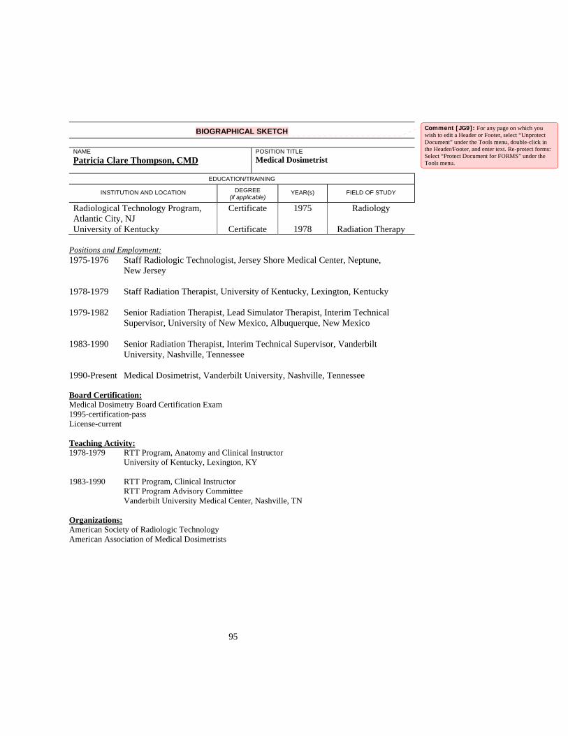
95
BIOGRAPHICAL SKETCH
NAME Patricia Clare Thompson, CMD
POSITION TITLEMedical Dosimetrist
EDUCATION/TRAINING
INSTITUTION AND LOCATION DEGREE (if applicable)
YEAR(s) FIELD OF STUDY
Radiological Technology Program, Atlantic City, NJ
Certificate 1975 Radiology
University of Kentucky Certificate 1978 Radiation Therapy Positions and Employment: 1975-1976 Staff Radiologic Technologist, Jersey Shore Medical Center, Neptune,
New Jersey 1978-1979 Staff Radiation Therapist, University of Kentucky, Lexington, Kentucky 1979-1982 Senior Radiation Therapist, Lead Simulator Therapist, Interim Technical
Supervisor, University of New Mexico, Albuquerque, New Mexico 1983-1990 Senior Radiation Therapist, Interim Technical Supervisor, Vanderbilt
University, Nashville, Tennessee 1990-Present Medical Dosimetrist, Vanderbilt University, Nashville, Tennessee Board Certification: Medical Dosimetry Board Certification Exam 1995-certification-pass License-current Teaching Activity: 1978-1979 RTT Program, Anatomy and Clinical Instructor
University of Kentucky, Lexington, KY 1983-1990 RTT Program, Clinical Instructor
RTT Program Advisory Committee Vanderbilt University Medical Center, Nashville, TN
Organizations: American Society of Radiologic Technology American Association of Medical Dosimetrists
Comment [JG9]: For any page on which you wish to edit a Header or Footer, select “Unprotect Document” under the Tools menu, double-click in the Header/Footer, and enter text. Re-protect forms: Select “Protect Document for FORMS” under the Tools menu.

96
Biography Jack Henry Towery III Job Title: Medical Physicist’s Assistant Education: B.A. in Physics from the University of Colorado at Boulder Training: NA Board Certification or Licensure: None Teaching Experience: None Clinical Experience: Clinical experience as a Medical Physicist’s Assistant from August 2002 – Present (6 months)