1 introduction to ecgs terry white, rn. 2 discussion topics n ecg monitoring basics n standardized...
TRANSCRIPT
1
Introduction to ECGs
Terry White, RN

2
Discussion Topics ECG Monitoring Basics
Standardized Methods & Devices
Components & Measurements of the ECG Complex
ECG Analysis

3
ECG Monitoring
4
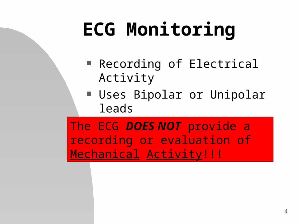
ECG Monitoring
Recording of Electrical Activity Uses Bipolar or Unipolar leads
The ECG DOES NOT provide a recording or evaluation of Mechanical Activity!!!

5
ECG Monitoring
Bipolar Leads 1 positive and 1
negative electrode RA always negative LL always positive
Traditional limb leads are examples of these Lead I Lead II Lead III
Provide a view from a vertical plane

6
ECG Monitoring
Unipolar Leads 1 positive electrode 1 negative “reference point”
calculated by using summation of 2 negative leads
Augmented Limb Leads aVR, aVF, aVL vertical plane
Precordial or Chest Leads V1-V6 horizontal plane
7
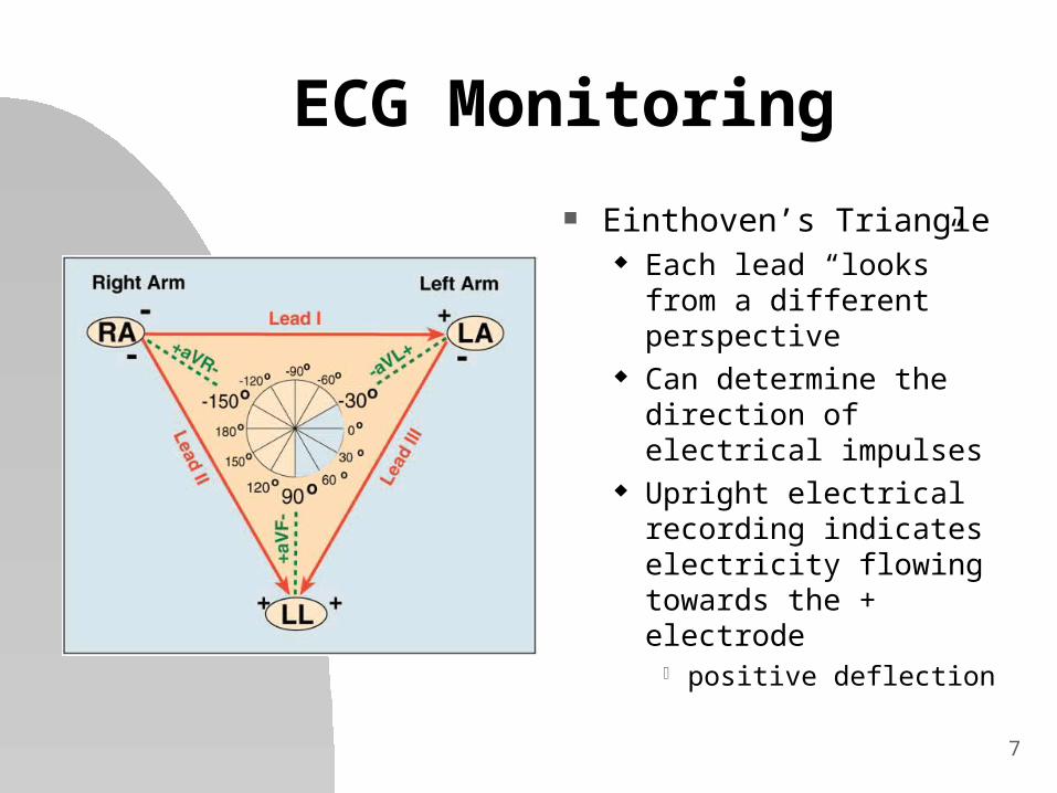
ECG Monitoring
Einthoven’s Triangle Each lead “looks” from a
different perspective Can determine the
direction of electrical impulses
Upright electrical recording indicates electricity flowing towards the + electrode positive deflection

8
Standardized Methods & Devices
9
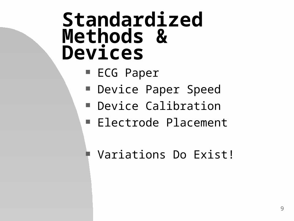
Standardized Methods & Devices
ECG Paper Device Paper Speed Device Calibration Electrode Placement
Variations Do Exist!
10
Standardized Methods & Devices
ECG Graph Paper Vertical axis- voltage
1 small box = 1 mm = 0.1 mV
Horizontal axis - time 1 small box = 1 mm = 0.04 sec.
Every 5 lines (boxes) are bolded
Horizontal axis - 1 and 3 sec marks

11
Standardized Methods & Devices
ECG Paper Examples Vertical Axis
No. of mm in 10 small boxes? No. of small boxes in 2 mm?
Horizontal Axis No. of seconds in 5 small boxes? No. of small boxes in 0.2 second? No. of small boxes in 1 second?
12
Standardized Methods & Devices
Paper Speed & Calibration Paper Speed - 25 mm/sec standard
Calibration of Voltage is Automatic
Both Speed and voltage calibration can be changed on most devices
13
Standardized Methods & Devices Electrode Placement
Standardization improves accuracy of comparison ECGs
3 Lead and 12 Lead Placement are most common
Assure good conduction gel Prep area with alcohol prep Avoid
Bone Large muscles or hairy areas Limb vs. Chest placement

14
Standardized Methods & Devices Electrode Placement
Poor placement or preparation Often results in artifact
Stray energy from other sources can also lead to poor ECG tracings (noise) 60 cycle interference

15
Components of the ECG
16
Components of the ECG Complex
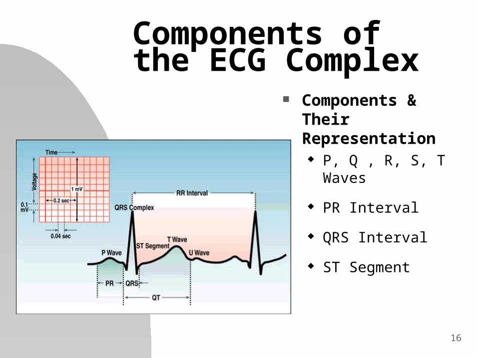
Components & Their Representation P, Q , R, S, T Waves
PR Interval
QRS Interval
ST Segment

17
Components of the ECG Complex
P Wave first upward
deflection represents atrial
depolarization usually 0.10 seconds
or less usually followed by
QRS complex

18
Components of the ECG Complex
QRS Complex Composition of 3
Waves Q, R & S represents ventricular
depolarization much variability
usually < 0.12 sec

19
Components of the ECG Complex
Q Wave first negative
deflection after P wave
depolarization of septum
not always seen
20
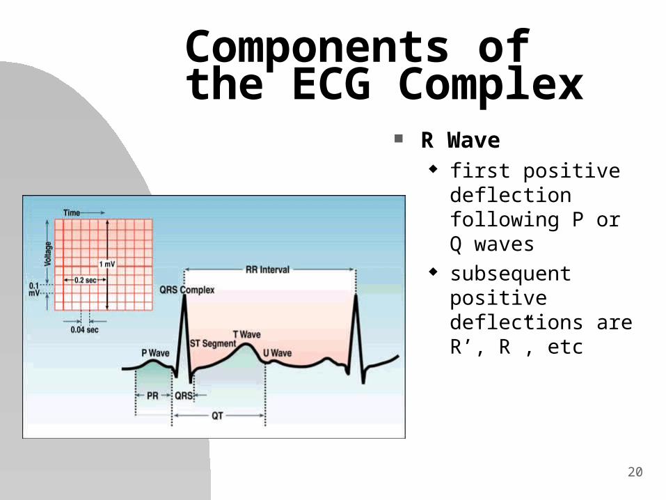
Components of the ECG Complex
R Wave first positive
deflection following P or Q waves
subsequent positive deflections are R’, R”, etc

21
Components of the ECG Complex
S Wave Negative deflection
following R wave subsequent negative
deflections are S’, S”, etc
may be part of QS complex absent R wave in
aberrant conduction

22
Components of the ECG Complex
PR Interval time impulse takes to
move through atria and AV node
from beginning of P wave to next deflection on baseline (beginning of QRS complex)
normally 0.12 - 0.2 sec may be shorter with
faster rates
23
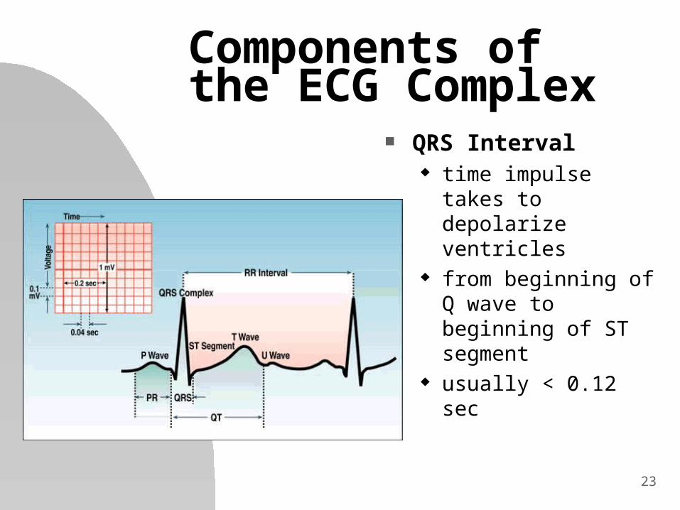
Components of the ECG Complex
QRS Interval time impulse takes to
depolarize ventricles from beginning of Q
wave to beginning of ST segment
usually < 0.12 sec

24
Components of the ECG Complex
J Point point where QRS
complex returns to isoelectric line
beginning of ST segment
critical in measuring ST segment elevation
25
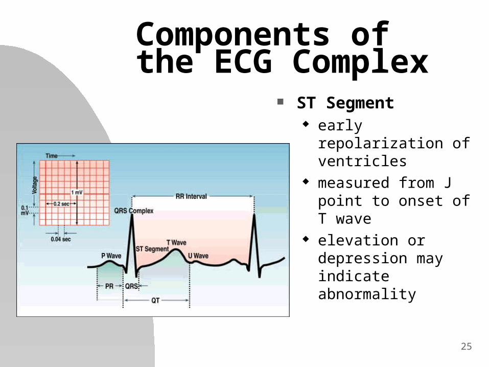
Components of the ECG Complex
ST Segment early repolarization of
ventricles measured from J point
to onset of T wave elevation or
depression may indicate abnormality
26
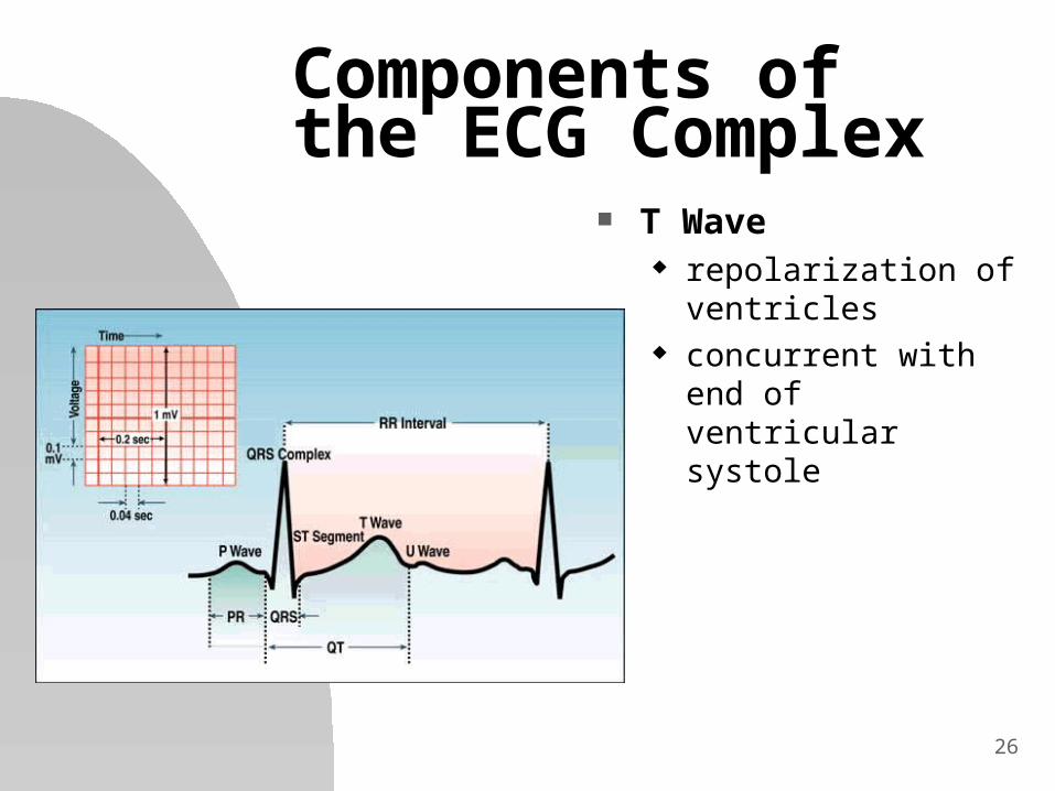
Components of the ECG Complex
T Wave repolarization of
ventricles concurrent with end of
ventricular systole

27
ECG Analysis
28
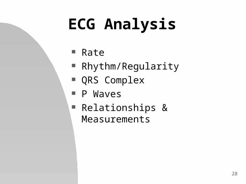
ECG Analysis
Rate Rhythm/Regularity QRS Complex P Waves Relationships & Measurements
29
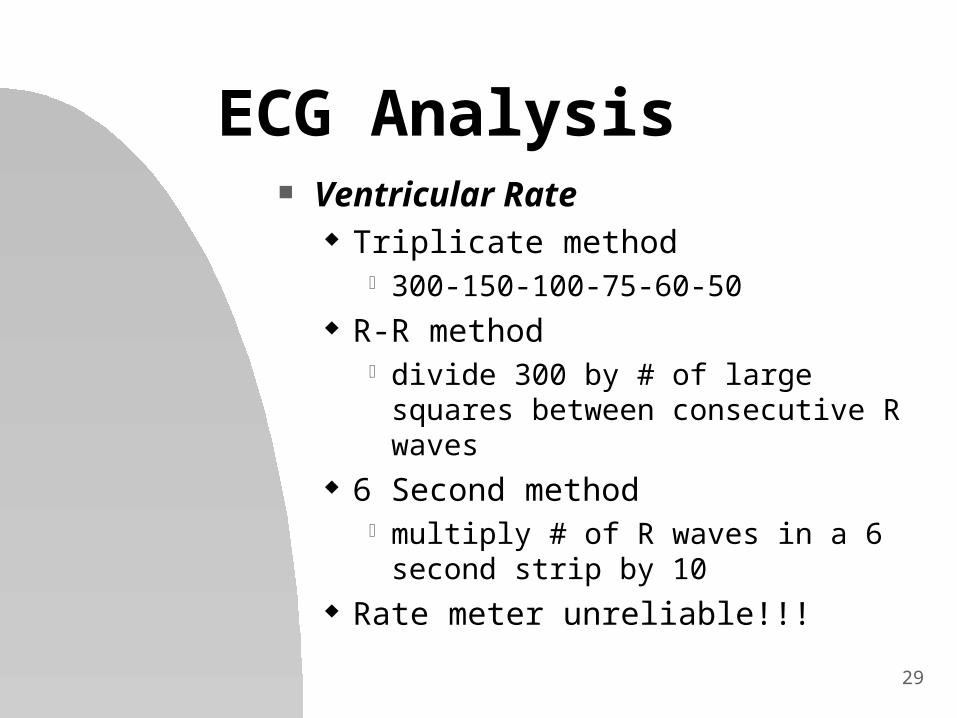
ECG Analysis Ventricular Rate
Triplicate method 300-150-100-75-60-50
R-R method divide 300 by # of large squares
between consecutive R waves 6 Second method
multiply # of R waves in a 6 second strip by 10
Rate meter unreliable!!!
30
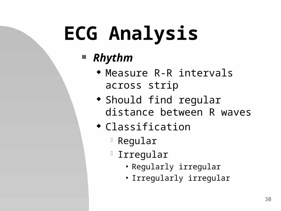
ECG Analysis Rhythm
Measure R-R intervals across strip Should find regular distance
between R waves Classification
Regular Irregular
• Regularly irregular• Irregularly irregular

31
ECG Analysis QRS Complex
Narrow < 0.12 seconds (3 small boxes) is
normal indicates supraventricular origin (AV
node or above) of pacemaker Wide
> 0.12 seconds is wide indicates ventricular or
supraventricular w/aberrant conduction
32
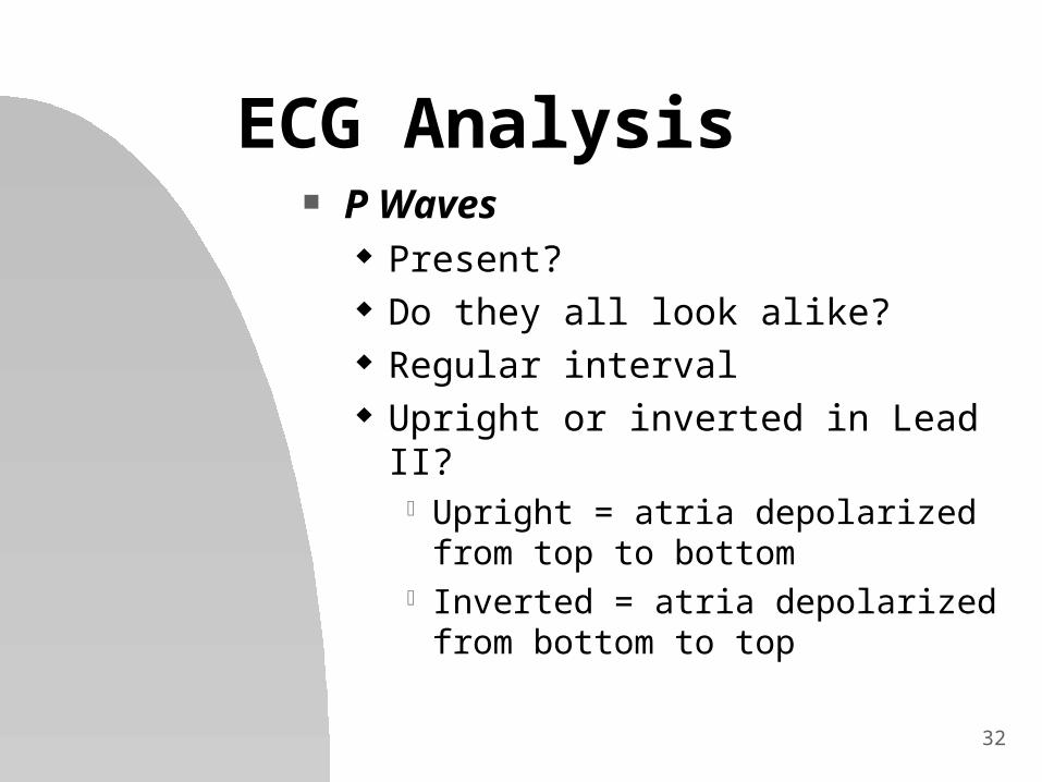
ECG Analysis P Waves
Present? Do they all look alike? Regular interval Upright or inverted in Lead II?
Upright = atria depolarized from top to bottom
Inverted = atria depolarized from bottom to top
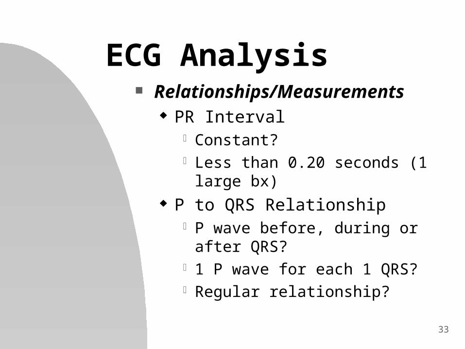
33
ECG Analysis Relationships/Measurements
PR Interval Constant? Less than 0.20 seconds (1 large bx)
P to QRS Relationship P wave before, during or after QRS? 1 P wave for each 1 QRS? Regular relationship?
34
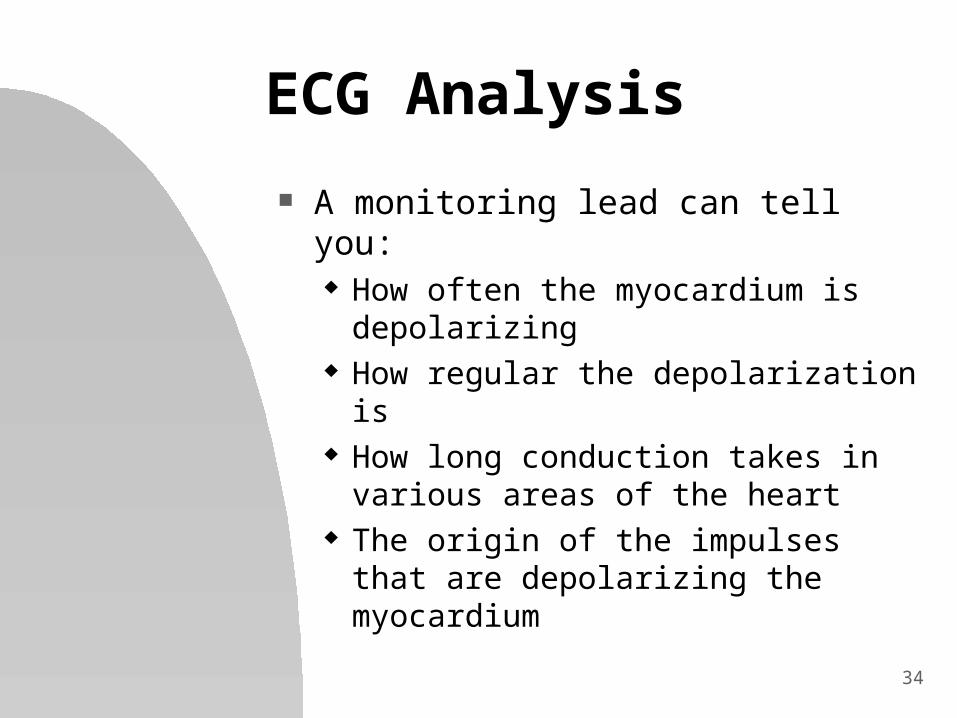
ECG Analysis
A monitoring lead can tell you: How often the myocardium is
depolarizing How regular the depolarization is How long conduction takes in
various areas of the heart The origin of the impulses that are
depolarizing the myocardium
35
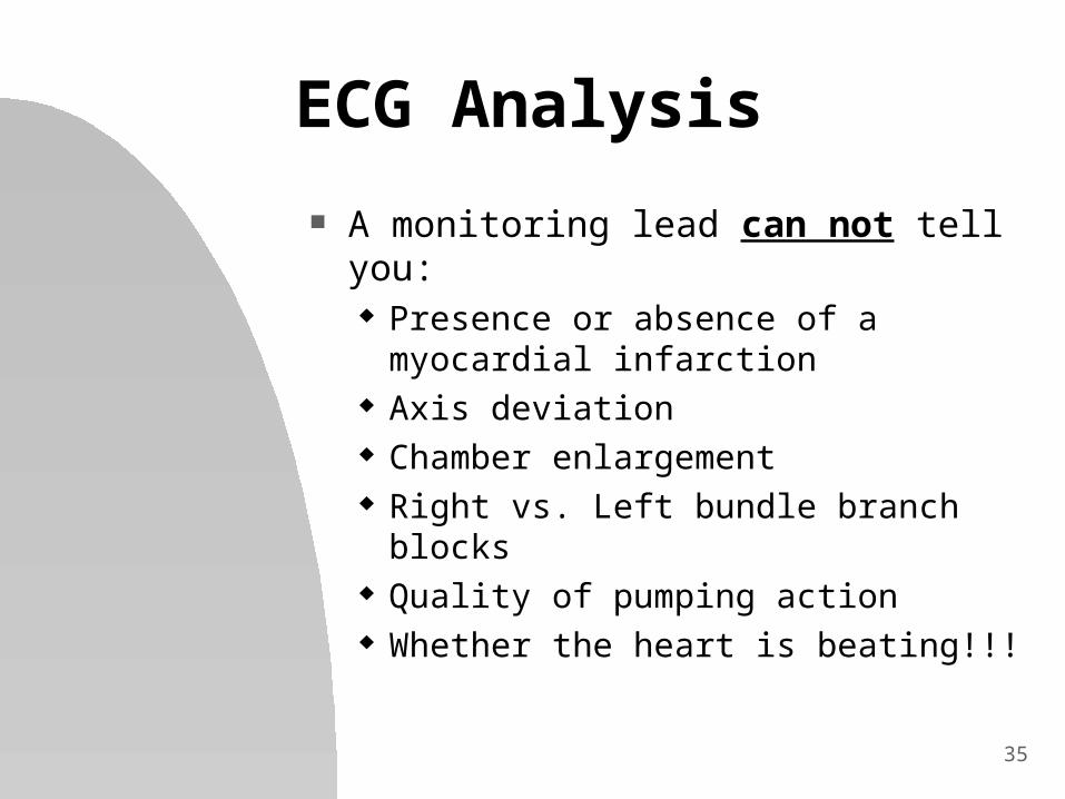
ECG Analysis
A monitoring lead can not tell you: Presence or absence of a
myocardial infarction Axis deviation Chamber enlargement Right vs. Left bundle branch blocks Quality of pumping action Whether the heart is beating!!!

36
ECG Analysis
An ECG is a diagnostic tool, NOT a treatment
No one was ever cured by an ECG!!
Treat the PATIENT not the Monitor!!!