1 chapter 6 skin and the integumentary system. 2 introduction: a.organs are body structures...
TRANSCRIPT

1
Chapter 6Skin and the Integumentary
System
2
Introduction:
A. Organs are body structures composed of two or more different tissues.B. The skin and its accessory organs make up the integumentary system.
Organization p. 122

3

4
6.1 Skin and Its Tissues A. Function of skin:
1. Regulate temperature2. Protective barrier3. Retain body fluids4. Location of sensory receptors5. To synthesize certain chemicals6. Eliminate waste products

5
B. Integument & cutaneous layer1. area 15 – 20 sq ft2. thickness
a. eyelids 1/50 in (<0.5 mm)b. soles of feet 1/8 in (6 mm)
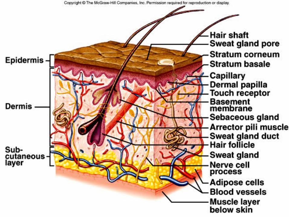
3. Skin is made of two main layers: epidermis & dermis

6
4. Layers are firmly bonded together – basement membrane
a. excessive rubbing can cause layers to separate
b. fluid fills area causing a blister
7

8
C. Epidermis - made up of stratified squamous epithelium and lacks blood vessels.Four or five layers:
1. Outer layer (stratum corneum) made of cells which are kertinized or cornified (cytoplasm replaced by keratin)
a. cells are flattened and scalelike and are constantly being lost b. 1st line of defense (gases & lipid soluble substances pass easily)
9
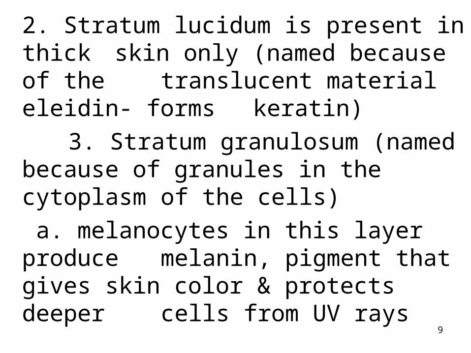
2. Stratum lucidum is present in thick skin only (named because of the translucent material eleidin- forms keratin) 3. Stratum granulosum (named
because of granules in the cytoplasm of the cells)
a. melanocytes in this layer produce melanin,
pigment that gives skin color & protects deeper cells from UV rays

10
Melanin cont.1. 4-6 pairs of genes
2. sunlight increases production
3. pituitary gland hormones can also influence production
4. hemoglobin & cerotene also contribute to skin color
b. Albinism – genetic defect

11
4. Stratum spinosum – flattened cells with spines or processes
5. Stratum germinativum (stratum basale) is made up of germinating cells continually divinding to replace the outer layers.
a. no direct blood supplyb. gets nutrients, water, etc
by diffusion from vessels of the dermis below

12

13
D. Dermis1. The dermis consists of two
layers of connective tissue with collagen and elastic fibers within a gel-like ground substance.
a. papillary layer – composed of loose connective tissue, fine elastic fibers and capillaries
(dermatoglyphics – skin ridges)

14
dermis cont.b. reticular layer – thicker &
made of dense irregular connective tissue giving it strength & elasticity2. The dermis also contains nerve fibers, sensory fibers, hair follicles, sebaceous glands, and sweat glands.
15
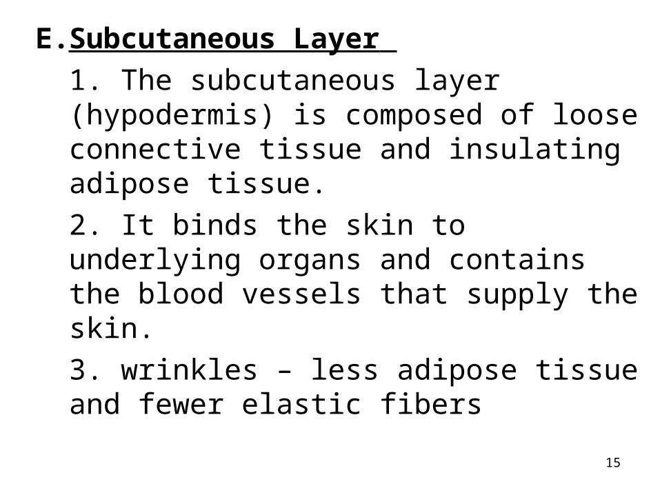
E.Subcutaneous Layer 1. The subcutaneous layer (hypodermis) is composed of loose connective tissue and insulating adipose tissue.
2. It binds the skin to underlying organs and contains the blood vessels that supply the skin.
3. wrinkles – less adipose tissue and fewer elastic fibers

16
6.2 Accessory Organs of the Skin
A. Nails 1. Nails are protective coverings over the ends of fingers and toes.2. Nails consist of stratified squamous epithelial cells overlying the nail bed, with the lunula as the most actively growing region of the nail root.

17
3. As new cells are produced, older ones are pushed outward and
become keratinized.

18
B. Hair Follicles
1. Hair can be found in nearly all regions of the skin.2. Individual hairs develop from cells at the base of the hair follicle, an invagination of the lower epidermis that dips down into the dermis.

19
3. As new cells are formed, old cells are pushed outward and become keratinized, and die forming the hair shaft.

20

21
4. A bundle of smooth muscle cells, called the arrector pili muscle, attaches to each hair follicle. These muscles cause goose bumps when cold or frightened.5. Hair color is determined by genetics; melanin from melanocytes is responsible for most hair colors, but red hair also contains the pigment trichosiderin.

22
C. Glands of the skinThere are two major types of cutaneous glands associated with the skin.
1. Sebaceous glands (holocrine glands)a. Found in the dermis of nearly
all the skin except the palms of the hands and soles of the feet.
b. Ducts open into the upper part of the hair follicles.
23
Sebaceous cont.
c. Produce and oily secretion called sebum that prevents hair from becoming dry and brittle.
~ underactive vs. overactive~ Blackheads

24
25
2. Sweat glands (sudoriferous glands) are two general types:
a. Apocrine glands are associated with hair follicles and are few in number.
1) empty into hair follicles
2) milky, sticky secretion -> bacteria -> odor of sweat 3) respond to body temperature, stress, and sexual arousal

26
Other apocrine glands believed to be related to sweat glands are:
~ ciliary glands of the eyelids
~ ceruminous (wax) glands of the external ear
~ mammary glands

27
b. Eccrine glands open as pores on the surface of the skin and are the most common type in humans.
1) coiled tubes that open onto the surface of the skin
2) secretion is mostly water with some salt
3) respond to body temperature

28

29
6.3 Physiology of the skinA. Protective BarrierB. Temperature Regulation
1. Proper temperature regulation is vital to maintaining metabolic reactions.
2. The skin plays a major role in temperature regulation with the hypothalamus controlling it.

30
3. Active cells, such as those of the heart and skeletal muscle, produce heat.4. The body responds to excessive heat by dilation of dermal blood vessels and sweating.5. The body responds to excessive cooling by constricting dermal blood vessels, inactivating sweat glands, and shivering.

31
C. Sense Receptors1. Free nerve endings
a) touch, pain, & temperature
b) skin, cornea & surround root of the hair follicles
2. Meissner’s corpusclesa) touchb) encapsulated nerve
endings found in hairless portions of the skin

32
3. Ruffini’s corpuscles
a) formerly thought to sense heat ? touch
b) found at the border of dermis and subcutaneous
4. Pacinan corpuscles
a) deep touch & pressure
b) encapsulated nerve endings widely distributed in the subcutaneous tissue
33
5. Krause’s end bulbs a) cold, touchb) encapsulated nerve
endings near the surface of the skinD. UV rays -> Vitamin D (epidermis) -> absorb Calcium -> bone tissue

34

35
6.4 PathologyA. Healing of Wounds
1. Inflammation, in which blood vessels dilate and become more permeable is the body's normal response to injury.~ aids healing by providing more nutrients & oxygen
B. Common Skin Disorders p. 123

36

37
C. Cuts 1. Superficial cuts are filled in by reproducing epithelial cells.2. Deeper cuts are closed off by clots, covered by scabs, and eventually filled in by connective tissue. Blood vessels extend into the area, injured tissues are replaced, and the scab falls off.
38
3. Large wounds leave scars and healing may be accompanied by the formation of granulations (blood vessel & cluster of fibrobasts).
39
D. Burns1. One million people are burned
annually -> 7000 die2. The depth of a burn is measured in degrees.
a. 1st degree burn (superficial partial-thickness)~ reddened as dermal blood vessels dilate~ sunburn -> skin maybe shed
40
b. 2nd degree burn (deep partial-thickness~ Redness and blisters common~ Painful because nerves of the dermis are irritatedc. 3rd degree burn (full-thickness)~ charring of the skin~ healing occurs only by growth
of epithelial cells inward

41
Extensive burning – rule of nines1. autograft (self)
a. unburned region & transplanted to injured region
b. cultured human epithelial -> postage stamp grows to bathmat size then transplanted2. homograft (person to person)
a. temporary – prevent infection b. replaced with autograft

42
3. Othera. amniotic membraneb. artificial membranes
composed of silicone, polyurethane or nylon with collagenous fibersE. Skin Cancer p. 119
1. cutaneous carcinomas2. cutaneous melanomas