1 chapter 3: the cellular level of organization. 2 figure 3–1 cellular organization cell =...
TRANSCRIPT

1
Chapter 3:
The Cellular Level of Organization

2Figure 3–1
Cellular Organization• Cell = smallest living unit• Performs all life functions

3
Two Categories of Cells
• Sex cells (germ cells):– reproductive cells – male sperm– female oocytes (eggs)
• Somatic cells (soma = body):– all body cells except sex cells

4
Cellular Organization• Different cells have different shapes• Unique morphology is related to function• All cells surrounded by plasma membrane:
– Separates cells from the environment
• Plasma membrane “holds in” the cytoplasm• Cytoplasm consists of cytosol (fluid) and
organelles (structures)• Body cells surrounded by interstitial fluid
– Interstitial fluid = fluid outside the membrane

5
Organelle Functions
Table 3–1 (1 of 2)

6
Organelle Functions
Table 3–1 (2 of 2)

7
The structures and functions of the
cell membrane.

8
1. The Plasma (Cell) Membrane
Figure 3–2
- Mostly phospholipid bilayer
- Interface between cell and environment
9
Functions of Plasma (Cell) Membrane
• Physical barrier:– Maintain homeostasis:
• Separates intracellular fluid from extracellular fluid, different conditions in each
• Regulates exchange with environment:– ions and nutrients enter– waste and cellular products released
• Monitors the environment:– extracellular fluid composition– Cell communication and signaling
• Structural support: – anchors cells and tissues

10
Plasma Membrane: Components
• Phospholipid bilayer• Cholesterol: resist osmotic lysis• Carbohydrates• Proteins

11
Plasma Membrane: Components
1. Phospholipid Bilayer:– hydrophilic heads—toward watery
environment, both sides– hydrophobic fatty-acid tails—inside
membrane – barrier to ions and water soluble
compounds
2. Cholesterol: resist osmotic lysis

12
3. Carbohydrates: • Membrane Carbohydrates including:
– Proteoglycans, glycoproteins, and glycolipids•extend outside cell membrane•form sticky carb layer or “sugar coat”
called the glycocalyx
Plasma Membrane: Components
13
Functions of Membrane Carbohydrates
• Lubrication and protection• Anchoring and locomotion• Specificity in binding
– Acts as receptors
• Recognition – Self recognition– immune response

14
Plasma Membrane: Components
4. Protein:– ½ mass of membrane– Integral proteins: span width of
membrane• within the membrane
– Peripheral proteins:• Adhere to inner or outer surface of the
membrane

15
6 Functions of Membrane Proteins
1. Anchoring proteins (stabilizers):– attach to inside or outside structures
2. Recognition proteins (identifiers): – Self identification by immune system
– Label cells normal or abnormal
3. Enzymes: – catalyze reactions in cytosol in extra cellular fluid
4. Receptor proteins:– bind and respond to ligands (ions, hormones) or
signaling, or import/export 5. Carrier proteins:
– transport specific solutes through membrane 6. Channels:
– regulate water flow and solutes through membrane

16
Which component of the cell membrane is primarily responsible for the
membrane’s ability to form a physical barrier between the cell’s internal and
external environments?
A. phospholipid bilayerB. glycocalyxC. peripheral proteinsD. proteoglycans

17
Which type of integral protein allows water and small ions to
pass through the cell membrane?
A. receptor proteinsB. carrier proteinsC. channel proteinsD. recognition proteins

18
How things get in and out of cells.

19
Overcoming the Cell Barrier
• The cell membrane is a barrier, but: – nutrients must get in– products and wastes must get out
• Permeability determines what moves in and out of a cell:
• A membrane that: – lets nothing in or out is impermeable– lets anything pass is freely permeable– restricts movement is selectively
permeable

20
Selective Permeability
• Cell membrane is selectively permeable:– allows some materials to move freely– restricts other materials
• Restricts materials based on:– size– electrical charge– molecular shape– lipid solubility

21
Transport
• Transport through a cell membrane can be:– active (requiring energy and ATP)– passive (no energy required)
• 3 Categories of Transport– Diffusion (passive)– Carrier-mediated transport (passive or
active)– Vesicular transport (active)

22
Solutions
• All molecules are constantly in motion
• Molecules in solution move randomly
• Random motion causes mixing
23
Concentration Gradient• Concentration is the amount of solute
(glucose) in a solvent (e.g. H20)• Concentration gradient:
– more solute in 1 part of a solvent than another
• Function = Diffusion – molecules mix randomly – solute spreads through solvent – eliminates concentration gradient– Solutes move down a concentration gradient
• From high concentration to low concentration

24
Factors Affecting Diffusion Rates
• Distance the particle has to move• Molecule size:
– smaller is faster
• Temperature: – more heat, faster motion
• Gradient size: – the difference between high and low
concentration
• Electrical forces: – opposites attract, like charges repel
25
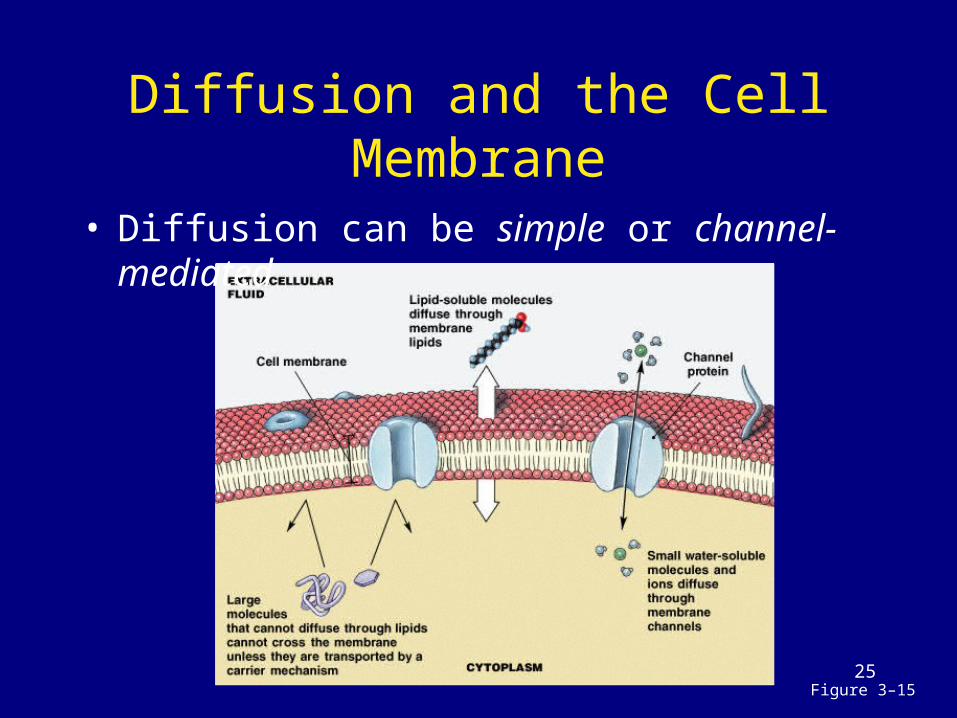
Diffusion and the Cell Membrane
Figure 3–15
• Diffusion can be simple or channel-mediated

26
Simple Diffusion
• Materials which diffuse through cell membrane:– lipid-soluble compounds (alcohols,
fatty acids, and steroids)– dissolved gases (oxygen and carbon
dioxide)

27
Channel-Mediated Diffusion
• Materials which pass through transmembrane proteins (channels):– are water soluble compounds– are ions
• Passage depends on:– size– charge– interaction with the channel
28
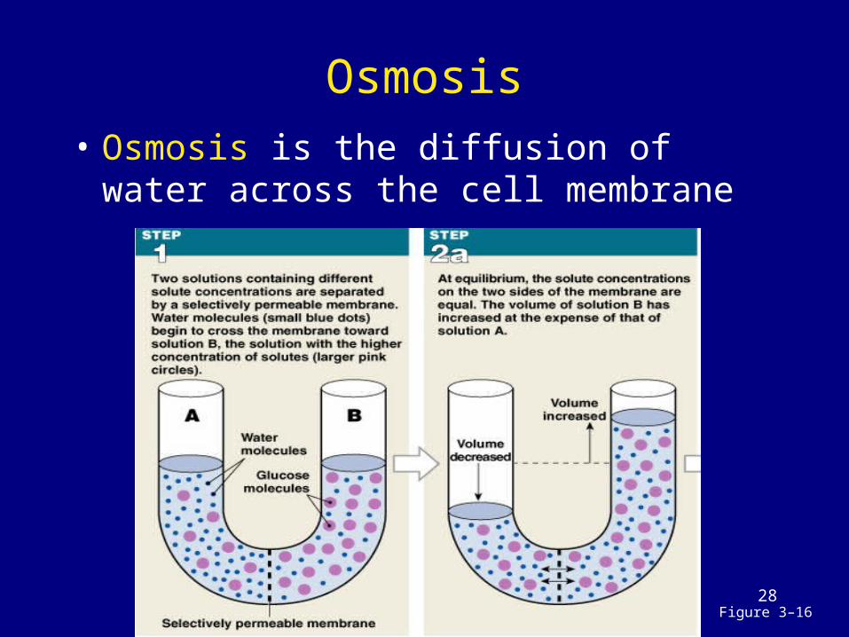
Osmosis
Figure 3–16
• Osmosis is the diffusion of water across the cell membrane

29
How Osmosis Works• More solute molecules, lower concentration of
water molecules • Membrane must be freely permeable to water,
selectively permeable to solutes• Osmosis Water Movement
– Water molecules diffuse across membrane toward solution with more solutes
– Volume increases on the side with more solutes
• Osmotic Pressure– Is the force of a concentration gradient of water– Equals the force (hydrostatic pressure) needed to block
osmosis

30
Osmotic Pressure

31
Isotonic
• A solution that does not cause osmotic flow of water in or out of a cell– iso = same, tonos = tension
• The osmotic effect of a solute on a cell: – 2 fluids may have equal
osmolarity
Figure 3–17a

32
Cells and Hypotonic Solutions
• hypo = below• Has less solutes
– Loses water through osmosis
• A cell in a hypotonic solution:– gains water– ruptures (hemolysis of
red blood cells)
Figure 3–17bLysis

33
Cells and Hypertonic Solutions
• hyper = above • Has more solutes
– Gains water by osmosis • A cell in a hypertonic
solution:– loses water– shrinks (crenation of red
blood cells)
Figure 3–17cCrenation

34
KEY CONCEPT
• Concentration gradients tend to even out
• In the absence of membrane, diffusion eliminates concentration gradients
• When different solute concentrations exist on either side of a selectively permeable membrane, osmosis moves water through the membrane to equalize the concentration gradients

35
How would a decrease in the concentration of oxygen in the lungs affect the diffusion of oxygen into the
blood?
A. decrease in molecule size results in decreased diffusion
B. decrease in distance results in increased diffusion
C. increase in electrical forces results in increased diffusion
D. decrease in gradient size results in decreased speed of diffusion

36
Some pediatricians recommend the use of a 10% salt solution to relieve
congestion for infants with stuffy noses.
What effect would such a solution have on the cells lining the nasal
cavity, and why?A. Cells will lose water because this
is a hypertonic solution.B. Cells will lose water because this
is a hypotonic solution.C. Cells will gain water because
this is a hypertonic solution.D. Cells will gain water because
this is a hypotonic solution.

37
Carrier-Mediated Transport
• Carrier-mediated transport of ions and organic substrates:– facilitated diffusion (No energy
needed)– active transport (Energy is needed)

38
Characteristics of Carrier-Mediated Transport
• Specificity: – 1 transport protein, 1 set of substrates
• Saturation limits: – rate depends on transport proteins, not
substrate (same as enzymatic reactions)
• Regulation: – cofactors such as hormones

39
Carrier-Mediated Transport
• Cotransport– 2 substances move in the same
direction at the same time
• Countertransport – 1 substance moves in while another
moves out

40
Facilitated Diffusion
• Passive, Carrier mediated• Carrier proteins transport molecules too
large to fit through channel proteins (glucose, amino acids):– molecule binds to receptor site on carrier
protein– protein changes shape, molecules pass
through– receptor site is specific to certain molecules
Figure 3–18

41
Active Transport
• Active transport proteins:– move substrates against
concentration gradient– require energy, such as ATP – ion pumps move ions (Na+, K+, Ca+,
Mg2+) – exchange pump countertransports 2
ions at the same time
42
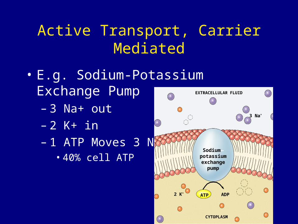
Active Transport, Carrier Mediated
• E.g. Sodium-Potassium Exchange Pump– 3 Na+ out– 2 K+ in– 1 ATP Moves 3 Na+
• 40% cell ATP
EXTRACELLULAR FLUID
2 K+
3 Na+
ADPATP
CYTOPLASM
Sodium—potassiumexchange
pump

43
Secondary Active Transport
Figure 3–20
• Na+ concentration gradient drives glucose transport
• ATP energy pumps Na+ back out
Cotransport Countertransport

44
Transport Vesicles
• Also called bulk transport• Vesicles:
– endocytosis (endo = into) – active transport using ATP:
• receptor-mediated• pinocytosis• phagocytosis
– exocytosis (exo = out of)
45
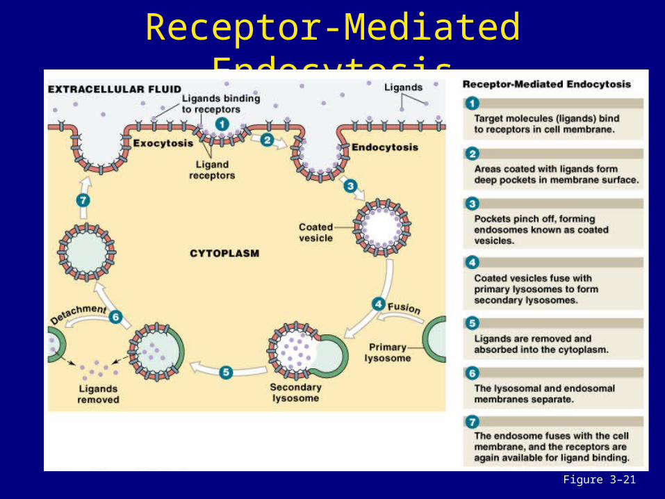
Receptor-Mediated Endocytosis
Figure 3–21

46
Receptor-Mediated Endocytosis
• Receptors (glycoproteins) bind target molecules (ligands)
• Coated vesicle (endosome) carries ligands and receptors into the cell

47Figure 3–22a
Pinocytosis• Pinocytosis (cell drinking) • Endosomes “drink” extracellular fluid
and enclose it in membranous vesicles at the cell surface– Similar to the steps in receptor-
mediated endocytosis, except that ligand binding is not the trigger

48
Phagocytosis
• Phagocytosis (cell eating)– pseudopodia (psuedo =
false, podia = feet) – engulf large objects in
phagosomes
Figure 3–22b

49Figure 3–7b
Exocytosis
• Is the reverse of endocytosis
50
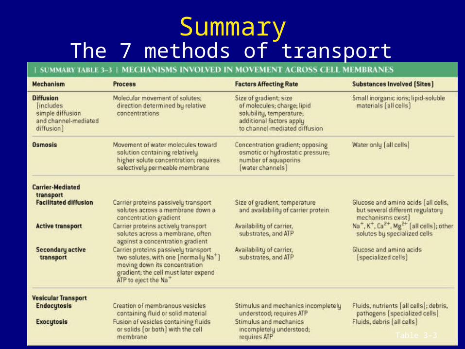
Summary
Table 3–3
The 7 methods of transport

51
Transmembrane potential

52
Electrical Charge
• Selective permeability of membrane allows different concentrations of molecules in/outside cells
• Cell membrane – Inside cell: slightly negative
• due to the abundance of proteins– Outside cell: slightly positive
• due to cations in extracellular fluids• Phospholipids hold charges apart creating a
transmembrane potential– Unequal charge across the cell membrane
• Resting potential ranges from – 10 mV to —100 mV, depending on cell type

53
During digestion in the stomach, the concentration of hydrogen ions (H+)
rises to many times that in cells of the stomach. Which transport process
could be responsible?
A. facilitated diffusionB. osmosisC. active transportD. endocytosis
54
During digestion in the stomach, the concentration of hydrogen ions (H+)
rises to many times that in cells of the stomach. Which transport process
could be responsible?
A. facilitated diffusionB. osmosisC. active transportD. endocytosis

55
If the cell membrane were freely permeable to sodium ions (Na+), how would the transmembrane potential
be affected?
A. it would move closer to zero
B. it would become more positive
C. it would become more negative
D. it would become unstable
56
When they encounter bacteria, certain types of white blood cells
engulf the bacteria and bring them into the cell. What is this process
called?
A. pseudocytosisB. exocytosisC. pinocytosisD. phagocytosis

57
Increase Surface Area: Microvilli
• Surface area of membrane can be increased by microvilli – For absorption or secretion
• Microvilli: ‘fingers’ of cell membrane containing a web of microfilaments and cytoplasm, anchored to cytoskeleton

58
2. Cytoplasm• Material enclosed by plasma membrane• Occupies space between plasma membrane
and nuclear membrane • Components:
– cytosol (fluid): • High K+, low Na+• Colloid Solution: proteins and enzymes• Nutrient Reserves: carbohydrates, lipids, and amino acids
– Inclusions:• Type and number varies with cell• E.g. glycogen, melanin, steroids, etc.
– organelles: • Carry out cellular functions• Each has separate function• Some have membranes• Some free in cytosol

59
Cell organelles and their functions

60
Types of Organelles
• Nonmembranous organelles: – no membrane– direct contact with cytosol
• Membranous organelles: – covered with plasma membrane– isolated from cytosol

61
Nonmembranous Organelles
• 6 types of nonmembranous organelles: – cytoskeleton – Microvilli – centrioles – cilia – ribosomes – proteasomes

62
Figure 3–3a
3. The Cytoskeleton
• Structural proteins for shape and strength (Internal Framework)
• 4 types of filaments– Microfilaments– Intermediate filaments– Thick filaments– Microtubules

63
A. Microfilaments
• Thin filaments (<6nm diameter)• Composed of the protein actin• Usually at periphery of the cell• Functions:
– provide additional strength by attaching the membrane to the cytoplasm
– Attach integral proteins to cytoskeleton– Pairs with thick filaments of myosin for
muscle movement

64
Intermediate Filaments & Thick Filaments
B. Intermediate Filaments:– 7-11 nm diameter
• Mid-sized between microfilaments and thick filaments– Durable, type varies with cell (collagen, elastin,
keratin)– Functions:
• strengthen cell and maintain shape• stabilize position of organelles• stabilize the cell relative to other cells
C. Thick Filaments– 15 nm diameter– Composed of myosin– Muscle cells only– Function
• Interact with actin to produce movement

65
D. Microtubules
• Large (25nm diameter), hollow tubes • Composed of tubulin protein• Originate from centrosome• Functions:
– Foundation of the cytoskeleton– Allows the cell to change shape and assists in mobility– Involved in transport
• Molecular motors travel along microtubule “tracks”• move vesicles within cell
– Makes up the spindle apparatus for nuclear division (mitosis)
– The structural part of some organelles• Centrioles, cilia, flagella

66
4. Centrioles in the Centrosome
Centrioles :form spindle apparatus during cell division
Centrosome: cytoplasm surrounding centriole near the nucleus– Consists of matrix and paired
centrioles– Functions as microtubule
organizing center– Responsible for assembling
spindle apparatus during mitosis Figure 3–4a

67
5. Cilia and Flagella
• Hair like projections• Contain a microtubule
core with cytoplasm covered in plasma membrane
• Anchored in the cytosol by basal bodies
• Cilia: Short, numerous– Function: sweep
substances over cell surface
• Flagella: Long, singular– Function: propel cell
through environment Figure 3–4b,c

68
6. Ribosomes
• Site of protein synthesis (polypeptide formation)
• Two subunits composed of rRNA & protein: – free ribosomes in cytoplasm:
• Manufacture proteins for use in cytoplasm
– fixed ribosomes attached to Endoplasmic reticulum:• Manufacture proteins for export or use in
membrane

69
Cells lining the small intestine have numerous fingerlike projections on their free surface. What are these
structures, and what is their function?
A. microvilli; move substances across cell surface
B. microvilli; increase cell’s surface area and absorptive ability
C. cilia; increase cell’s surface area and absorptive ability
D. cilia; move substances across cell surface

70
Membranous Organelles
• 5 types of membranous organelles:– endoplasmic reticulum (ER)– Golgi apparatus– lysosomes– peroxisomes– mitochondria

71
7. Endoplasmic Reticulum (ER)
Figure 3–5a
Location: - Attached to the Nuclear Envelope

72
Endoplasmic Reticulum (ER)
• endo = within, plasm = cytoplasm, reticulum = network
• Cisternae are storage chambers within membranes
• Function:– Synthesis of proteins, carbohydrates, and lipids– Storage of synthesized molecules and materials– Transport of materials within the ER– Detoxification of drugs or toxins

73
Smooth Endoplasmic Reticulum (SER)
• No ribosomes attached• Tubular Membrane• Functions:
– Lipid metabolism (synthesis, breakdown, transport)
– Synthesis of steroid hormones (reproductive system)
– Detoxification of drugs– Breakdown of glycogen (storage in muscles) to
glucose– Store ions (e.g. Ca2+)

74
Rough Endoplasmic Reticulum (RER)
• Surface covered with ribosomes:• Ribosomes synthesize proteins and
feed them into RER cisternae to be modified – E.g. +carbs = glycoprotein
• Modified proteins are put into transport vesicles to go to Golgi
• These proteins for exocytosis or use in membrane
• Surface covered with ribosomes:• Ribosomes synthesize proteins and
feed them into RER cisternae to be modified – E.g. +carbs = glycoprotein
• Modified proteins are put into transport vesicles to go to Golgi
• These proteins for exocytosis or use in membrane

75
Golgi Apparatus
• Stack of cisternae with associated transport vesicles
• Near nucleus but not attached
• Function:– Modify,
concentrate, and sort export proteins
Figure 3–6a

76
Golgi Apparatus
• Transport vesicles from RER dock on cis (forming) face of golgi and release contents into golgi
• Proteins (and glycoproteins) are modified– Phosphate, carbs, or lipids
attached
• Proteins transit between cisternae via vesicles from cis face (forming) to trans face (maturing)

77
Vesicles of the Golgi Apparatus
• At trans face, proteins are packaged into:– Secretory vesicles:
• modify and package products for exocytosis– Membrane renewal vesicles:
• Carry products to membrane – Lysosomes:
• Membrane bound sacs of digestive enzymes
78Figure 3–7b
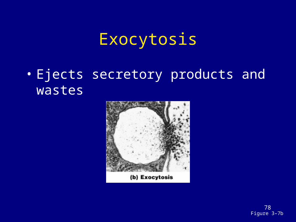
Exocytosis
• Ejects secretory products and wastes

79
9. Lysosomes
Figure 3–8
• Powerful enzyme-containing vesicles: – lyso = dissolve, soma = body
• Digestion centers for large molecules or structures• Endosomes or phagosomes containing endocytosed
things, and organelles targeted for destruction are fused with lysosome and broken down
• Some solutes diffuse into cytoplasm for use, remaining debris are exocytosed
80
Lysosome Structures and Function
• Primary lysosome: – formed by Golgi and inactive enzymes
• Secondary lysosome: – lysosome fused with damaged organelle– digestive enzymes activated– toxic chemicals isolated
• Functions:– Clean up inside cells:
• break down large molecules• Attack bacteria• recycle damaged organelles• ejects wastes by exocytosis

81
Autolysis
• Self-destruction of damaged cells:– auto = self, lysis = break– lysosome membranes break down– digestive enzymes released– cell decomposes– cellular materials recycle

82
Tay Sach’s Disease
• Caused by lysosomes that fail to break down glycolipids in nerve cells
• Accumulation of glycolipids disrupts nerve function
• Progressive mental retardation• Death by age 18 months

83
10. Peroxisomes
• Are enzyme-containing vesicles:– break down fatty acids– Membrane sacs containing oxidases
and catalases to neutralize free radicals that are formed during catabolism of organic molecules• produce hydrogen peroxide (H2O2)
– Peroxisomes not made by golgi • appear to self replicate
84
11. Proteasomes
• Cylindrical structure composed of protein digesting enzymes (proteases)
• Disassemble damaged proteins for recycling– E.g. degrade proteins tagged with
ubiquitin to recycle amino acids
85
KEY CONCEPT
• Cells: basic structural and functional units of life– respond to their environment– maintain homeostasis at the cellular
level– modify structure and function over
time

86
12. Mitochondrion Structure
• Sausage-shaped with double membrane– Outer membrane: Smooth– Inner membrane: folded into cristae– Center: matrix
Figure 3–9a

87
Mitochondrial Function: Power House of the Cell
• Aerobic respiration occurs on surface of cristae– takes chemical energy from food (glucose)– With the use of oxygen, Glucose is catabolized
creating CO2 waste to convert ADP into ATP
• Mitochondria supply most of cell’s energy• Have their own DNA (maternal)• Can replicate independent of the cell
Figure 3–9b
glucose + oxygen + ADP carbon dioxide + water + ATP
88
KEY CONCEPT
• Mitochondria provide cells with energy for life:– require oxygen and organic
substrates– generate carbon dioxide and ATP

89
Certain cells in the ovaries and testes contain large amounts of smooth endoplasmic reticulum
(SER). Why?
A. to produce large amounts of proteins
B. to digest materials quicklyC. to store large amounts of
hormonesD. to produce large
amounts of steroid hormones

90
What does the presence of many mitochondria imply about a cell’s energy requirements?
A. a high demand for energyB. a low demand for energyC. fluctuating energy needs
requiring flexibilityD. number of
mitochondria provides no implication of energy needs

91
How the nucleus controls the cell
92Figure 3–10a
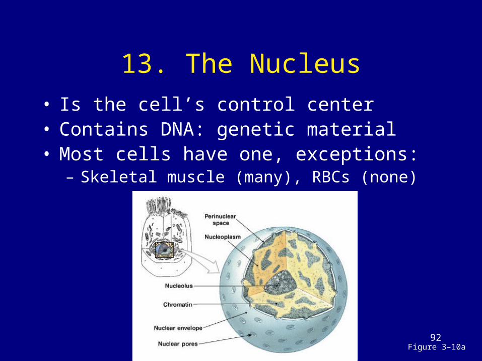
13. The Nucleus• Is the cell’s control center• Contains DNA: genetic material• Most cells have one, exceptions:
– Skeletal muscle (many), RBCs (none)

93
Structure of the Nucleus
• Nucleus:– largest organelle
• Nuclear envelope:– double membrane around the
nucleus, connected to ER • Nuclear pores with regulator
proteins:– Control exchange of materials
between cytoplasm and nucleus

94
Within the Nucleus• Nucleoplasm:
– fluid containing ions, proteins (enzymes), DNA, RNA, and nucleoli
• Nucleoli: Dark areas – site of rRNA synthesis and
packaging into ribosomal subunits
• In non-dividing cells DNA is loose– Called chromatin

95
Organization of DNA• DNA in chromatin is
organized into Nucleosomes:– DNA coiled around
histones
• During Nuclear Division, Chromatin is tightly coiled into visible chromosomes (23 pairs in humans)
• Chromosomes:– tightly coiled DNA
(cells dividing)
Figure 3–11

96
The Genetic Code
97
DNA and Genes• DNA: contains genes
– instructions for every protein in the body• Gene: functional units of heredity
– DNA instructions for a product: RNA or protein• Humans have 30-75 thousand potential genes
(only 1.5% of total DNA)– Remainder is involved with control of genes
or appear to be junk (25%)– Noncoding parts of DNA (non-genes) is highly
variable from one person to the next– Variability allows for identification of an
individual by DNA fingerprinting

98
Gene Activation
• In order for a gene to be expressed (used to make a product) it must be unwound from the histone proteins so it can be read
• Disassembly of the nucleosomes and unwinding of the DNA is called gene activation

99
Genetic Code
• The chemical language of DNA instructions:– Read off a gene in order to assemble a
protein– sequence of bases (A, T, C, G)– triplet code:
• 3 bases of DNA = 1 amino acid (codon)
– A gene = all the codons for all the amino acids in one protein in the correct order

100
Gene Structure and Expression
• Structure
• Expression (original) (copy) (product) DNA RNA Protein Transcription Translation
Open Reading FramePromoter Terminator
Start Codon
Stop Codon

101
KEY CONCEPT
• The nucleus contains chromosomes
• Chromosomes contain DNA• DNA stores genetic instructions for
proteins• Proteins determine cell structure
and function

102
How DNA instructions become proteins

103
Protein Synthesis
• Transcription:– copies instructions from DNA to mRNA (in
nucleus)
• Translation:– ribosome reads code from mRNA (in
cytoplasm)– assembles amino acids into polypeptide
chain
• Processing:– by RER and Golgi apparatus produces
protein

104
mRNA Transcription
• A DNA gene is transcribed to mRNA in 3 steps:– gene activation– DNA to mRNA– RNA processing

105
mRNA Transcription
G
A
A
T
G
A
G
T
A
C
G
G
C
T
C
G
A
T
T A
A
T
C
G
A
G
C
C
G
T
A
C
G C
A
G
C
G
A
C
C
C
G
U
U
A
T
G
A
G
T
A
A
C
C
GC
G
G
C
C
T
C
G
A
T
T
T
C
G
A
A
T
G
G
T
A
A
C
G
G
C
T
G
C
A
T
T
T
T
A
C
C
T
STEP STEP STEP
•
DNA
Gene
Promoter
Triplet 2
Triplet 3
Triplet 4
Triplet 1
Codon1
Codingstrand
Templatestrand
Codon2
Codon3
Codon 4(stop codon)
mRNAstrandRNA
polymerase
Codon1
RNAnucleotide
Adenine
Thymine
Guanine
Cytosine
1
2 2
3
4KEY
Uracil (RNA)

106
Step 1: Gene Activation
• Uncoils DNA, removes histones• Start (promoter) and stop codes on
DNA mark location of gene:– coding strand is code for protein– template strand used by RNA
polymerase molecule

107
Step 2: DNA to mRNA
• Enzyme RNA polymerase transcribes DNA:– binds to promoter (start) sequence– reads DNA code for gene– binds nucleotides to form messenger
RNA (mRNA)– mRNA duplicates DNA coding strand,
uracil replaces thymine

108
Step 3: RNA Processing
• At stop signal, mRNA detaches from DNA molecule:– code is edited (RNA processing)– unnecessary codes (introns) removed– good codes (exons) spliced together– triplet of 3 nucleotides (codon)
represents one amino acid

109
Codons
Table 3–2

110
Key Concept
• The timing of gene activation (transcription) for any gene is controlled by signals from outside the nucleus, either from within the cell or in response to external cues– E.g. Hormones

111
Translation
• Making a protein using the mRNA blueprint
• Occurs in the cytoplasm on free ribosomes or on fixed ribosomes on the RER
• mRNA moves: – from the nucleus– through a nuclear pore
Figure 3–13

112
Translation
A
G
C
U
U A C
STEP STEP
NUCLEUS
mRNA
Adenine
Guanine
Cytosine
Uracil
Smallribosomal
subunit
Amino acid
tRNA
Anticodon
tRNA binding sites
mRNA strandStart codon
Largeribosomalsubunit
The mRNA strand binds to the smallribosomal subunit and is joined at thestart codon by the first tRNA, whichcarries the amino acid methionine.Binding occurs between complementarybase pairs of the codon and anticodon.
The small and large ribosomal subunitsinterlock around the mRNA strand.
11
2
KEY
•tRNA delivers amino
acids to mRNA
113
Translation
AA
A GGG G
GUU C
CCC
A UG G CCC
A
STEP STEP STEP
1 2 12
3
1
2
3
Peptide bond
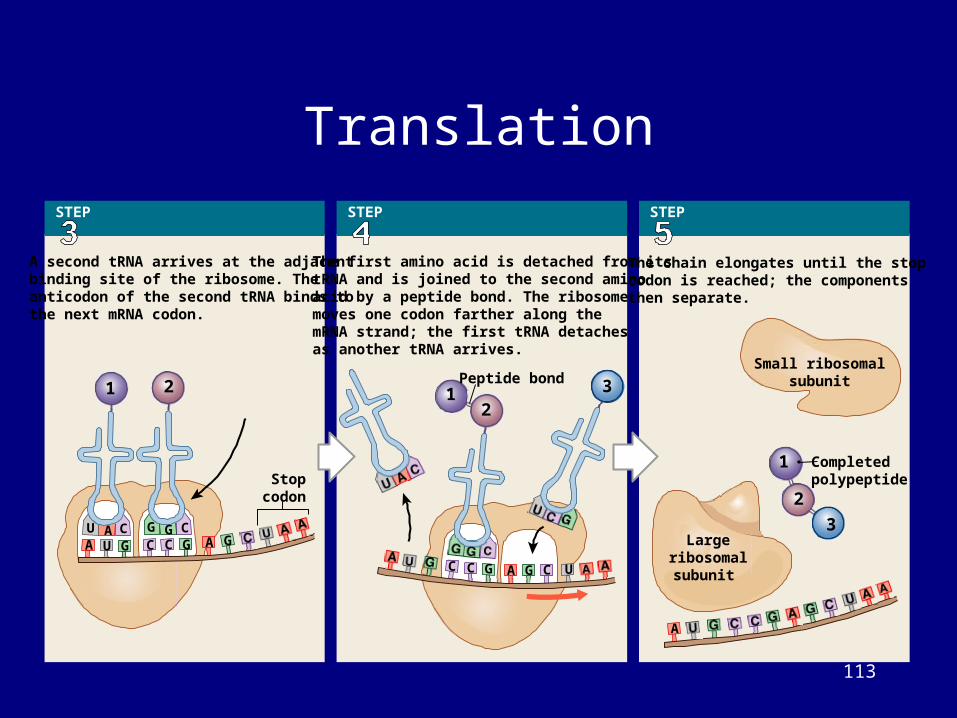
A second tRNA arrives at the adjacentbinding site of the ribosome. Theanticodon of the second tRNA binds tothe next mRNA codon.
The first amino acid is detached from itstRNA and is joined to the second aminoacid by a peptide bond. The ribosomemoves one codon farther along themRNA strand; the first tRNA detachesas another tRNA arrives.
The chain elongates until the stopcodon is reached; the componentsthen separate.
Small ribosomalsubunit
CompletedpolypeptideStop
codon
Largeribosomalsubunit

114
Genetic Code

115
Examples using the Genetic Code:
Coding Strand DNA: ATgCAgTTTACgCAgAAgATCAgTTAgTemplate strand DNA: complement A-T, C-G TACgTCAAATgCgTCTTCTAgTCAATC
Transcription to form mRNA: complementary base pairing to template, U replaces T AUgCAgUUUACgCAgAAgAUCAgUUAg
Translation to form protein: read codons from genetic code
e.g. AUg = Met/Start (start codon) Aug/CAg/UUU/ACg/CAg/AAg/AUC/AgU/UAg Met-Gln-Phe-Thr-Glu-Lys-Ile-SerUAg = stop codon (no tRNA, no amino acid)

116
Mutations• Most non-infectious disease, conditions, and
disorders are due to mutations in the DNA that change the amino acids in the protein – E.g. sickle cell anemia
• Point mutation in DNA: A T• Changes on codon: GAG GTG• Changes one amino acid:
– Glutamic acid (-charge) valine (neutral)• This alters the 3D shape of the whole
hemoglobin protein: globular fibrous• Which changes the shape of the red blood cell:
– Disc crescent• Which prevents the RBC from carrying oxygen,
and causes it to block capillaries

117
Mutations
• Point mutations = change in 1 base of DNA can be a silent mutation if the amino acids is not changed – common at the 3rd base in a codon
• Insertion mutation = addition of a base which changes the reading frame;whole protein after the mutation is wrong
• Deletion Mutation = removal of a base, alter reading frame, protein wrong.

118
KEY CONCEPT• Genes:
– are functional units of DNA – contain instructions for 1 or more proteins
• Protein synthesis requires:– several enzymes– ribosomes– 3 types of RNA
• Mutation is a change in the nucleotide sequence of a gene:– can change gene function
• Causes:– exposure to chemicals– exposure to radiation– mistakes during DNA replication

119
How does the nucleus control the activities of a cell?
A. through nuclear poresB. through the nuclear
matrixC. through DNAD. through RNA

120
What process would be affected by the lack of the enzyme RNA polymerase?
A. nothing would be affected; DNA polymerase would take over
B. cell’s ability to duplicate DNA
C. cell’s ability to translate DNA
D. cell’s ability to transcribe RNA

121
How cells reproduce

122
Cell Life Cycle
• Life span of cell depends on type of cell• All cells eventually die
– Apoptosis: controlled cell death, lysosomes are defused
• Some cells must divide to make cells to replace dying cells; function of stem cells
• To divide, DNA must be replicated and equally distributed between the stem cell and new daughter cell
Figure 3–3

123
Interphase• Most of a cell’s life is spent in a
nondividing state (interphase)– Period of time that a cell performs its normal
functions• The nondividing period:
– G-zero phase—specialized cell functions only• If a cell never divides
• Cells preparing for dividing, will go through 3 stages – G1 phase—cell growth, organelle duplication,
protein synthesis, synthesizes enough cytoplasm for 2 cells
– S phase—DNA replication and histone synthesis
– G2 phase—finishes protein synthesis and centriole replication

124
3 Stages of Cell Division
• Body (somatic) cells divide in 3 stages:– DNA replication duplicates genetic
material exactly– Mitosis divides genetic material
equally – Cytokinesis divides cytoplasm and
organelles into 2 daughter cells

125
DNA Replication

126
DNA Replication
Figure 3–24
• DNA helicases unwind the DNA and separates the strands
• DNA polymerase bind to the DNA and synthesizes complementary antiparallel strands– DNA polymerase only add to the 3’ end of the
molecule• Leading strand: synthesized continuously• Lagging strand: synthesized in pieces called
Okasaki fragments– Okasaki fragments are attached end to end into
one strand by DNA Ligase• DNA rewinds into double helix molecules
– New molecules contains one strand of the original DNA and one newly synthesized strand

127
Overview of Cell Life Cycle
Indefinite period
G0
Specializedcell functions
INTERPHASE
SDNA
replication, synthesis
of histones
G2
Proteinsynthesis
M
MITOSIS(See Figure 3-25)
THECELL
CYCLE
G1 Normal
cell functionsplus cell growth,duplication of organelles,
protein synthesis
Prophase
MetaphaseAnaphase
Telo
ph
ase
CYTOKINESIS

128
Mitosis
• Mitosis (nuclear division) divides duplicated DNA into 2 sets of chromosomes:– DNA coils tightly into chromatids– chromatids connect at a centromere– protein complex around centromere
called the kinetochore
• Followed by cytokinesis:– Separation of the cells

129Figure 3–25 (Stage 1)
Stage 1: Prophase
• Nucleoli disappear • Centriole pairs move
to cell poles• Microtubules (spindle
fibers) extend between centriole pairs
• Nuclear envelope disappears
• Spindle fibers attach to kinetochore

130
Stage 2: Metaphase
• Chromosomes align in a central plane (metaphase plate)
Figure 3–25 (Stage 2)

131
Stage 3: Anaphase
• Microtubules pull chromosomes apart
• Daughter chromosomes groups near centrioles
Figure 3–25 (Stage 3)

132Figure 3–25 (Stage 4, 1 of 2)
Stage 4: Telophase
• Nuclear membranes reform
• Chromosomes uncoil
• Nucleoli reappear• Cell has 2
complete nuclei
133
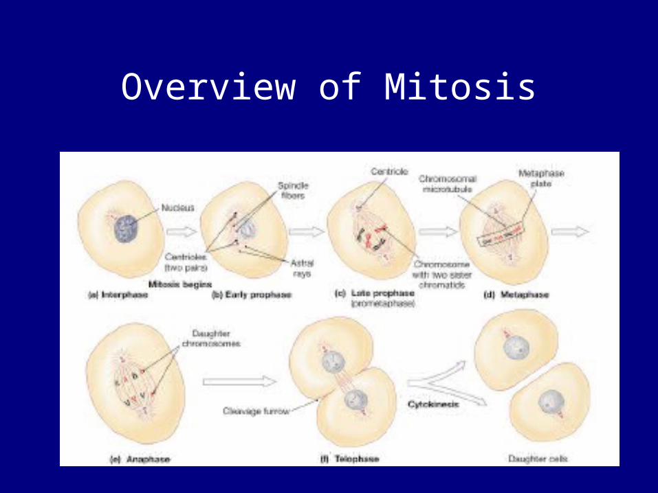
Overview of Mitosis

134
KEY CONCEPT
• Mitosis duplicates chromosomes in the nucleus for cell division
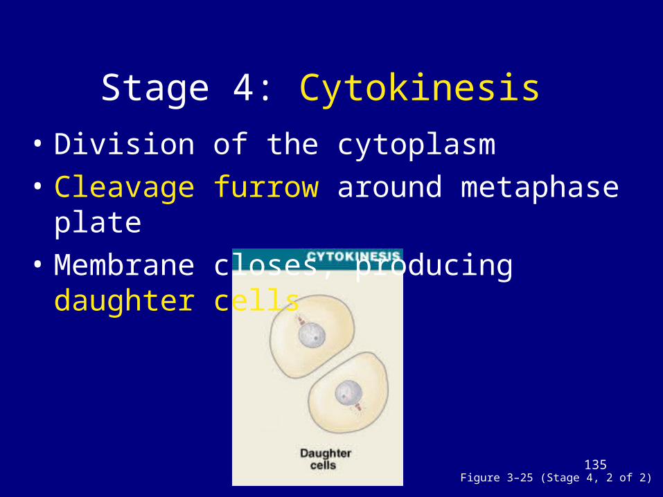
135Figure 3–25 (Stage 4, 2 of 2)
Stage 4: Cytokinesis • Division of the cytoplasm• Cleavage furrow around metaphase
plate• Membrane closes, producing daughter
cells

136
What regulates cell division

137
Mitotic Rate and Life Span
• Rate of cell division:– slower mitotic rate means longer cell
life– cell division requires energy (ATP)
• Cell Life Span:– Muscle cells, neurons rarely divide– Exposed cells (skin and digestive
tract) live only days or hours

138
Regulating Cell Life
• Normally, cell division balances cell loss• Increases cell division:
– internal factors (MPF) – extracellular chemical factors (growth
factors)
• Decreases cell division:– repressor genes (faulty repressors cause
cancers)– worn out telomeres (terminal DNA
segments)

139
Chemicals Controlling Cell Division
Table 3–4

140
A cell is actively manufacturing enough organelles to serve two
functional cells. This cell is probably in which phase of its life
cycle?
A. SB. G1
C. G2
D. M

141
During DNA replication, a nucleotide is deleted from a sequence that
normally codes for a polypeptide. What effect will this deletion have on
the amino acid sequence of the polypeptide?
A. no effect, deletion will be skipped
B. no effect, deletion will be automatically repaired
C. amino acid sequence will disintegrate
D. the amino acid sequence would be altered

142
What would happen if spindle fibers failed to form in a cell
during mitosis?
A. centromeres would not appear
B. nuclear membrane would not disintegrate
C. chromosomes would not separate
D. chromatin would not condense

143
Cancer• Cell division: controlled by internal and
external factors– In adult cell growth = cell death– If growth exceeds death a tumor can
form• Cancer:
– illness that disrupts cellular controls– produces malignant cells

144
Cancer• Benign tumors:
– grow in a connective tissue capsule and remain in one place
• Malignant tumor: ignore growth control mechanisms– spread into surrounding tissues
(invasion)– start new tumors (metastasis)
• Cancer develops in steps:1. abnormal cell 3. metastasis2. primary tumor 4. secondary tumor

145
Cancer• Cancer: caused by mutation in a growth control gene
(oncogene = mutated genes that cause cancer)– 1° tumor: cells grow uncontrolled– 2° tumor: cells metastasize in blood and lymph to establish new
growth elsewhere
• Tumors trigger growth of blood vessels to support the cells– In order for diffusion to bring nutrients and remove wastes all
cells have to be within 125µm of a vessel
• Eventually the tumor will crowd out normal tissues causing organ failure
Figure 3–26

146
KEY CONCEPT
• Mutations disrupt normal controls over cell growth and division
• Cancers often begin where stem cells are dividing rapidly
• More chromosome copies mean greater chance of error

147
Cell Differentiation

148
What is cell differentiation?• Cells specialize or differentiate:
– All somatic cells in the body have the same DNA but different sizes, shapes, and functions
– As cells specialize to become a specific cell type many genes get turned off permanently, cells are considered differentiated
– Differentiated cells only express genes related to their function
– Stem cells are undifferentiated:• Embryonic stem cells can express all of their genes and
become any cell type• Other stem cells can express most of their genes
– All stem cells do not show many specialized functions and can differentiate into many types of tissue

149
KEY CONCEPT
• All body cells, except sex cells, contain the same 46 chromosomes
• Differentiation depends on which genes are active and which are inactive
150
SUMMARY
• Structures and functions of human cells• Structures and functions of
membranous and nonmembranous organelles
• ATP, mitochondria, and the process of aerobic cellular respiration
• Structures and functions of the nucleus:– control functions of nucleic acids– structures and replication of DNA– DNA and RNA in protein synthesis

151
SUMMARY• Structures and chemical activities of the
cell membrane:– diffusion and osmosis – active transport proteins– vesicles in endocytosis and exocytosis– electrical properties of plasma
• Stages and processes of cell division:– DNA replication– mitosis– cytokinesis
• Links between cell division, energy use, and cancer
152
Homework
Lecture• Study Chapter 1, 2, and 3 for Exam
#1• Complete Homework #1Laboratory