1 chapter 28 (genes x) eukaryotic transcription regulation
TRANSCRIPT

1
CHAPTER 28 (Genes X)
EUKARYOTIC TRANSCRIPTION
REGULATION

2
Control of Gene Expression in EukaryotesEukaryotic gene expression is usually controlled at the
level of initiation of transcription.
Control of Gene Expression in EukaryotesEukaryotic gene expression is usually controlled at the
level of initiation of transcription.
GC boxGC boxsilencerssilencers
enhancersenhancers
CCAAT box
(called the CAT box)
CCAAT box
(called the CAT box)
the TATA boxthe TATA box
the promoterthe promoter
Methylation
Hormonal Control

3
Thinking about Gene Regulation
Humans begin life from a single cell; all the genetic information needed to create an adult is in our genome.
Embryonic cells undergo differentiation to produce specific cell types such as muscle, nerve, and blood cells.
Different cell types are the consequence of differential gene expression.
Humans begin life from a single cell; all the genetic information needed to create an adult is in our genome.
Embryonic cells undergo differentiation to produce specific cell types such as muscle, nerve, and blood cells.
Different cell types are the consequence of differential gene expression.

4
A typical differentiated mammalian cell makes about 10,000 proteins from approximately 30,000 genes.
Most of these are housekeeping proteins needed to maintain all cell types.
Certain proteins can only be detected in specific cell types.
How is gene expression regulated?
A typical differentiated mammalian cell makes about 10,000 proteins from approximately 30,000 genes.
Most of these are housekeeping proteins needed to maintain all cell types.
Certain proteins can only be detected in specific cell types.
How is gene expression regulated?

5
COORDINATELY CONTROLLED GENES
• PROCARYOTES– Clustered in operons
– Same regulatory sites
– Single mRNA molecule
– Translated together
• EUCARYOTES– NO operons !
– Genes individually transcribed
– Own promoter
– Specific enhancers for each gene

6

7
(B) In procaryotes the production of mRNA molecules is much simpler. The 5' end of an mRNA molecule is produced by the initiation of transcription by RNA polymerase, and the 3' end is produced by the termination of transcription. Since procaryotic cells lack a nucleus, transcription and translation take place in a common compartment. In fact, translation of a bacterial mRNA often begins before its synthesis has been completed.

8
Genes have regulatory DNA sequences upstream from the initiation site where transcription begins.
The promoter is the RNA polymerase binding site.
Gene regulatory proteins bind to regulatory DNA sequences and can either prevent or enhance RNA polymerase binding.
Co-activators
Activators in enhancer
Activators in distal promoter

9
Independent domains bind DNA and activate transcription
• DNA-binding activity and transcription-activation are carried by independent domains of an activator.
• The role of the DNA-binding domain is to bring the transcription-activation domain into the vicinity of the promoter.

10
Acidic activators: They have multiple negative charges
Yeast activators: GAL4 , GCN4
Herpes Simplex Virus: VP16

11
The modular structure of a gene activator protein. Outline of an experiment that reveals the presence of independent DNA-binding and transcription-activating domains in the yeast gene activator protein Gal4. (A) The normal activation of gene transcription produced by the Gal4 protein. (B) The chimeric gene regulatory protein requires the LexA protein DNA-binding site for its activity.

12

13
Identification of Protein-Protein Interactions by the Yeast 2-Hybrid
System

14
The two hybrid techniques tests the ability of two proteins to interact by incorporating then into hybrid proteins where one has a DNA-binding domain and the other has a transcription-activating domain.

15
Yeast Gal4
Transcription factor• Zinc Finger DNA binding domain (DBD)• Transcriptional Activation Domain (AD)
ADDBD
Gene XON
promoter
Yeast Two Hybrid Assay

16
Yeast Two Hybrid Assay
DBD
Bait
The bait: cDNA of the protein you want to find partners for.
Reporter GeneOFF
promoter
AD Prey
The prey: from random cDNA’s whichencodes for various proteins. Do any interact with the bait?
Reporter GeneOFF
promoter
Reporter GeneON
promoterYeast cells turn blue

17
Yeast Two Hybrid Assay
Uetz, 2001

18
Activators interact with the basal apparatus
•DNA-binding domain determines the specificity of activators for the target promoter or enhancer.
•DNA-binding domain is responsible for localizing a transcription-activating domain in the proximity of the basal appapratus.
•An activator that works directly has a DNA-binding domain and an activating domain.
•An activator does not have an activating domain may work by binding a coactivator that has an activating domain.
•Several factors in the basal apparatus are targets with which activators or coactivators interact.

19
An activator may bind a co-activator that contacts the basal apparatus.

20
Activators may work at different stages of initiation, by contacting the TAFs of TFIID or contacting TFIIB.

21
How does an activator stimulate transcription?
Two models:
1. The recruitment model: Activators sole effect
is to increase the concentration of RNA
polymerases to the promoter.
2. Alternative model: Activator induces some
change in the transcriptional complex, i.e.
conformational change in enzyme, increases its
efficiency.

22
RNA polymerase may be associated with various alternative sets of transcription factors in the form of a holoenzyme complex.

23
A model for the action of some eukaryotic transcriptional activators. The gene activator protein, bound to DNA in the rough vicinity of the promoter, facilitates the assembly of some of the general transcription factors. Although some activator proteins may be dedicated to particular steps in the pathway for transcription initiation many seem to be capable of acting at several steps.

24
Some Promoter Binding Proteins are Repressors
• Repression is usually achieved by affecting
chromatin structure, but there are repressors that act
by binding to specific promoters and function like
trans-acting (such as bacterial repressors) to block
transcription.
• The global repressor NC2/Dr1/DRAP1, is a
heterodimer that binds to TBP to prevent it from
interacting with other components of the basal
apparatus.

25
A transcription complex involves recognition of several elements in the sea urchin H2B promoter in testis. Binding of the CAAT displacement factor (CDP) in embryo prevents the CAAT-binding factor from binding, so an active complex cannot form.
In embryo

26
Take away message:
The function of a protein in binding to a known promoter element
cannot be assumed: It may be activator, a repressor, or even
irrelevant to gene transcription.

27
Transcription factors are activated in several ways
The activators are classified according to their The activity of an inducible activator may be regulated by several ways

28

29
Oncogenes that code for transcription factors have mutations that inactivate transcription (v-erbA and possibly v-rel) or that activate transcription (v-jun and v-fos).

30
Transcription is controlled by proteins binding to regulatory DNA sequences.
Promotor includes
RNA polymerase binding site
initiation site
Regulatory DNA sequences bound by gene regulatory proteins
some short – simple gene switches
some long and complex (eukaryotic)
molecular microprocessors which respond to a variety of signals, integrate them, and determine the rate of transcription.
Expression depends on;
cell type
its environment
its age
extracellular signals
Edge of bases
Gene Regulatory Proteins Bind to DNA

31

32
Gene regulatory proteins insert into the major groove,
contacts and binds (non-covalently) to the edges of the
bases (about 20 interaction), usually without disrupting
the hydrogen bonds.
This binding is very strong and very specific for the
nucleotide sequence. Frequently DNA-binding proteins
contain alpha-helices bind in pairs - dimerization - which
doubles the contact area, increasing the strength and
specificity of the interaction.
Gene Regulatory Proteins Bind to DNA

33

34

35
There are many types of DNA binding domains
The activators are classified according to their DNA binding domain.
• Zinc Finger• Steroid Receptors• Helix-turn-helix motif• Homeodomain• Helix-loop-helix• Leucine zippers
Members of the same group have sequence variations of a specific motif that confer specificity for individual target sites.

36
DNA binding zinc-finger motifs
One or more zinc atoms are added to the structure.
1. It is found in in a cluster with additional zinc fingers, arranged one after another, the helix of each contact the major groove of the DNA, foring a continous strech of a helices along the groove.
2. Two a helices are packed together with zinc atoms. Mostly found in the large family of intracellular receptor proteins (dimers).
• Cys2/His2 Finger
Cys-X2-4-Cys-X3-Phe-X5-Leu-X2-His-X3-His

37
Transcription factor SP1 has a series of three zinc fingers each with a characteristic pattern of cysteine and histidine residues that constitute the zinc-binding site.

38
Zinc fingers may form a-helices that insert into the major groove, associated with b-sheets on the other site.
Cys2/His2 finger
7-8 aa
~23 aa

39
DNA binding by a zinc finger motif: The zinc-finger motif is composed of an alpha helix and a beta sheet.Each zinc-finger is held together by a molecule of zinc.

40

41

42
A dimer of the zinc finger domain of the intracellular receptor family bound bound to its specific DNA sequence. (e.g., glucocorticoid receptor)

43
-sheet can also recognized DNA: i.e. bacterial met repressor. It is a dimeric protein. Each dimers sheet can bind to the major goorve of the DNA.

44
DNA binding by a zinc finger protein: This protein recognizes DNA using three zinc fingers of the cys-cys-his-his type arranged as direct repeats.

45
Sequence specific interactions between different six fingers and their DNA recognition sequences.

46

47
Steroid receptors have several independent domains
All the receptors have independent domains for DNA binding and hormone binding.

48

49

50
Steroid receptors have zinc fingers
The first finger of a steroid receptor controls which DNA sequence is bound (red);The second finger controls spacing between the sequences (blue).

51
Discrimination between GRE and ERE target sequences is determined by two amino acids at the base of the first zinc finger in the receptor.
Cys-X2-Cys-X13-Cys-X2-Cys

52

53
Each receptor recognizes response elements that are related to a consensus.
• These response elements have a characteristic feature:Each consensus consists of two short repeats (or half sites). The receptors bind as a multimer, so that each half of the consensus is contacted by one subunit.
• The response elements for the various receptors may be either palindromes or direct repeats in which the half sites are separated by 0-4 bp whose sequence is irrelevant.

54
Response elements formed from the palindromic half site TGTTCT are recognized by several different receptors depending on the spacing between the half sites.

55
The receptors fall into two groups:
1) GR, MR, AR, PR receptors all form homodimers. They recognize half sites, TGTTCT consensus sequence, arranged as palindormes. The spacing between the elements determins the type of element.
2) T3R, VDR, RAR, 9-cis retinoic acid (RXR) receptors form heterodimers, which recognize half elements with the sequence TGACCT.
1bp - RXR - RXR3bp - VDR - RXR4bp - T3R - RXR5bp - RAR - RXR
They can also form homodimers, which recognize palindromes.

56
Response elements with direct repeats TGACCT are recognized by heterodimers of which one member is RXR.
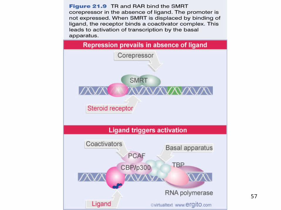
57

58
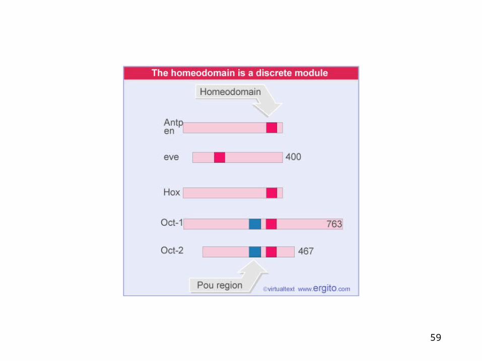
Homeodomain proteins:
Homeotic selector genes play a critical role in
Drosophila development. Several homeotic selector genes contain almost
identical streches of 60 amino acids that defines this
class of proteins called “homeodomain”. Homeodomain proteins are present virtually all
eucaryotic organisms. The heli-turn-helix motif in homeodomains is always
surrounded by the same structure suggesting that the
motif is always presented to DNA in the same way.
59

60

61

62
A homeodomain bound to its specific DNA sequence.In the homeodomain, three alpha helices interact with the DNA. Helix 2 and 3 closely resembles the helix-turn-helix motif.Asparagine in helix number 3 binds to adenine in the DNA

63
Comparison of homeodomains from two organisms separated by more than a billion years of evolution. The Yeast 2 protein (green) and Drosophila engrailed protein (red). Black dots mark sites with identical amino acids. Orange dots indicate the position of a three amino acid insert in the 2 protein.

64
Helix-loop-helix proteins interact by combinatorial association
Helix-loop-helix proteins have a motif of 40-50 amino
acids that comprises two amphipathic -helices of 15-16
residues separated by a loop. The helices are responsible for dimer formation.
bHLH proteins have a basic sequence adjacent to the
HLH motif that is responsible for binding to DNA. Class A bHLH proteins are ubiquitously expressed.
Class B bHLH proteins are tissue specific. A class B protein usually forms a heterodimer with a
class A protein. HLH proteins that lack the basic region prevent a bHLH
partner in a heterodimer from binding to DNA.
HLH proteins form combinatorial associations that may be changed during development by the addition or removal of specific proteins

65
A helix-loop-helix dimer bound to DNA. The two monomers are held together in a four-helix bundle.: each monomer contributes two a helices connected by a felixible loop of protein (red). A specific DNA sequence is bound by the two a helices that are project from the four-helix bundle.
HLH can make both homo and heterodimers
Flexible loop

66
An inhibitory regulation by truncated HLH proteins.HLH motif is responsible both dimerization and DNA binding.It recognizes a symmetric DNA sequence.
Lacks DNA binding domain

67

68
Formation of muscle cells is triggered by several bHLH proteins, including MyoD as a heterodimer MyoD-E12 or MyoD-E47, rather than a MyoD homodimer. Before myogenesis begins, the Id protein may bind to MyoD and or E12 or E47 to form hterodimers that can not bind to DNA. So removal of Id could be the trigger that releases MyoD to initiate myogenesis.
An HLH dimer in which both subunits are of the bHLH type can bind DNA, but a dimer in which one subunit lacks the basic region cannot bind.

69
Leucine zippers are involved in dimer formation
The leucine zipper is an amphipathic helix that
dimerizes.
The zipper is adjacent to a basic region that
binds DNA.
Dimerization forms the bZIP motif in which the
two basic regions symmetrically bind inverted
repeats in DNA.
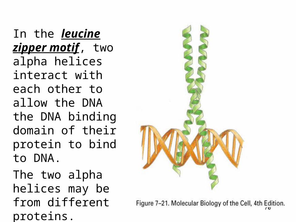
70
In the leucine zipper motif, two alpha helices interact with each other to allow the DNA the DNA binding domain of their protein to bind to DNA.The two alpha helices may be from different proteins.

71

72
Heterodimerization of leucine zipper proteins can alter their DNA-binding soecificity.Two different monomers can combine to form a heterodimer, which now recognizesa hybrid DNA sequence.

73

74
Types of DNA binding proteins
DNA and RNA polymeraserepair enzymes
structural proteinstranscription factors
Types of DNA binding proteins
DNA and RNA polymeraserepair enzymes
structural proteinstranscription factors
DNA binding motifs
zinc fingersleucine zippershelix-turn-helixhelix-loop-helix
DNA binding motifs
zinc fingersleucine zippershelix-turn-helixhelix-loop-helix

75
It is not yet possible to accurately predict the DNA sequences recognized by all gene regulatory proteins.
Is there a simple amino acid-base pair recognition code: e.g., is a G-C base pair always contacted by a particular amino acid side chain?
Although certain amino acid-base interactions appear much frequently than others the answer is NO…
One of the most common protein-DNA interactions
76
Protein surface virtually any shape and chemistry
can be made from just 20 different amino acids, and
a gene regulatory protein uses different
combinations of these to create a surface that is
precisely complementary to a particular DNA
sequence. We know that the same base pair can be
thereby be recognized in many ways depending on
its context.

77
Sequence specific interactions between different six fingers and their DNA recognition sequences.

78

79
HOW GENETIC SWITCHES WORK?

80
An operon = a cluster of bacterial or viral genes transcribed from a single promoter.
A sequence of DNA within the promotor is recognized by regulatory proteins - the operator.

81
Repressor protein switch genes on or off
Is recognized well by RNA polymerase.
Bacteria in your gut after you eat a steak
Regulatory proteins like these are allosteric.
These regulators allow fast response to the environment because they are always present in the cell - constitutively expressed

82
The binding of tryptophan to the tryptophan repressor protein changes the conformation of the repressor. It is an helix-turn-helix protein.

83
Addition of an “inducing” ligand can turn on a gene either by removing a gene repressor protein from the DNA or by causing a gene activator protein to bind.

84
Activator proteins act on promoters that do not bind RNA polymerase on their own. These promoters are made fully functional by the addition of a bound activator protein which is also allosteric, activated by binding another molecule at a site different from the DNA binding site.
Example: CAP has to bind cyclic AMP (an intracellular signaling molecule) to bind DNA. Genes regulated by CAP are switched on in response to increases in cAMP, which is triggered by signals received by cell membrane receptors.

85
A model for gene activation from an enhancer
General transcription factors usually can not efficiently initiate transcription alone.
Enhancers can be thousands of base pairs away, either upstream or downstream and can either increase or decrease transcription.
“Action at a distance”

86
Summary of gene activation and regulation in procaryotes and eucaryotes.

87
The gene control region of a typical eucaryotic gene.

88
Gene Regulatory sequences of a typical eucaryotic cell
Regulated by combinations of proteins over up to 50,000 nucleotide pairs
Combinatorial control can be positive or negative