1 chapter 14 synovial fluid professor a. s. alhomida disclaimer the texts, tables, figures and...
Post on 20-Dec-2015
215 views
TRANSCRIPT

11
Chapter 14 Synovial Fluid
Professor A. S. Alhomida
Chapter 14 Synovial Fluid
Professor A. S. Alhomida
DisclaimerDisclaimer• The texts, tables, figures and images contained in The texts, tables, figures and images contained in
this course presentation (BCH 376) are not my this course presentation (BCH 376) are not my own, they can be found on:own, they can be found on: • References suppliedReferences supplied• Atlases orAtlases or• The webThe web
King Saud University
College of Science
Department of Biochemistry

22
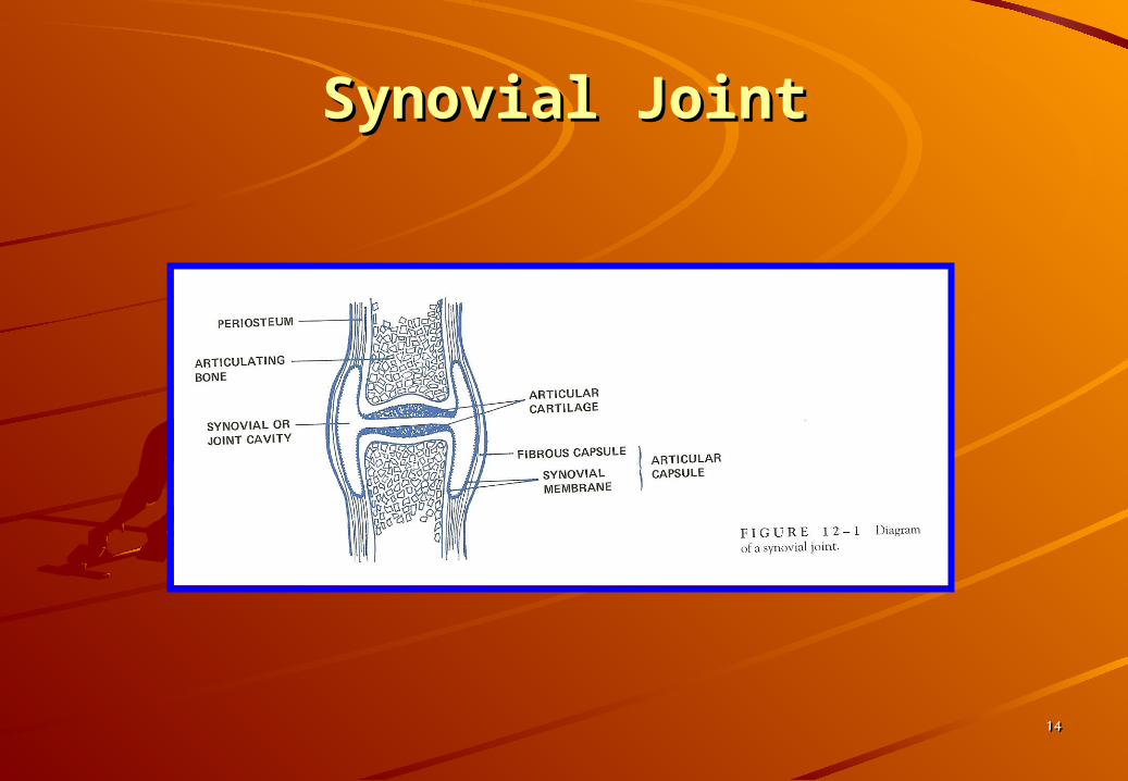
• Movable Joints (diarthroses) Movable Joints (diarthroses) Composed ofComposed of
1.1. Bones lined with articular cartilageBones lined with articular cartilage2.2. Separated by a cavity containing Separated by a cavity containing
synovial fluid enclosed in a synovial synovial fluid enclosed in a synovial membranemembrane
Physiology and CompositionPhysiology and Composition

33
Physiology and Composition, Physiology and Composition, Cont’dCont’d
• Synovial MembraneSynovial Membrane1.1. SynoviocytesSynoviocytes
• Phagocytic: synthesizes degradative enzymes
• Synthesizes hyaluronate
2.2. Connective tissueConnective tissue• Blood vessels, lymphatics and nerves nerves
44
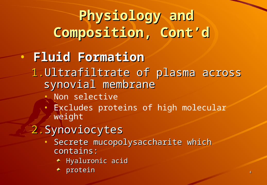
Physiology and Composition, Physiology and Composition, Cont’dCont’d
• Fluid FormationFluid Formation1.1. Ultrafiltrate of plasma across Ultrafiltrate of plasma across
synovial membranesynovial membrane• Non selective• Excludes proteins of high molecular weight
2.2. SynoviocytesSynoviocytes• Secrete mucopolysaccharite which contains:Secrete mucopolysaccharite which contains:
Hyaluronic acidHyaluronic acidproteinprotein

55
• Cartilage and Fluid FunctionCartilage and Fluid Function1.1. Reduce friction between bonesReduce friction between bones2.2. Lubricates jointsLubricates joints3.3. Fluid provides nutrients to cartilageFluid provides nutrients to cartilage4.4. Lessens shock of walking and jogging Lessens shock of walking and jogging
impactimpact
Physiology and Composition, Physiology and Composition, Cont’dCont’d

66
VolumeVolume <3.5 mL<3.5 mLColorColor pale yellowpale yellowClarityClarity clearclearViscosityViscosity forms string 4-6 cm longforms string 4-6 cm longErythrocytesErythrocytes <2000 cells/uL<2000 cells/uLLeukocytesLeukocytes <200 cells/uL<200 cells/uLNeutrophilsNeutrophils <20% of diff.<20% of diff.LymphocytesLymphocytes <15 % of diff.<15 % of diff.Monocytes & macrophagesMonocytes & macrophages 65% of diff.65% of diff.CrystalsCrystals NONENONEGlucoseGlucose <10 mg/dL (< blood <10 mg/dL (< blood glucose)glucose)LactateLactate <250 mg/dL<250 mg/dLTotal proteinTotal protein <3 g/dL<3 g/dLUric acidUric acid = blood value= blood value
Synovial Fluid: Normal ValuesSynovial Fluid: Normal Values

77
• Arthrocentesis1. It is the method for obtaining synovial
fluids by using a needle aspiration of synovial fluid
2. Volume• Normal= 3.5 mL• Diseased and inflamed = up to 25 mL
Specimen CollectionSpecimen Collection

88
3. Collect 2 tubes• Heparin tube: microbiology• Plain top: chemistry and immunology• EDTA (liquid): hematology• Avoid all powdered anticoagulants –
interfere with crystal analysis
Specimen Collection, Cont’dSpecimen Collection, Cont’d
99
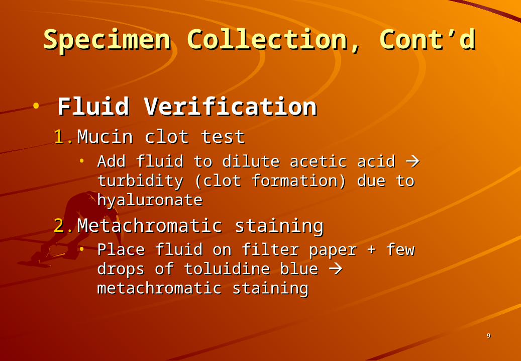
• Fluid VerificationFluid Verification1.1. Mucin clot testMucin clot test
• Add fluid to dilute acetic acid Add fluid to dilute acetic acid turbidity turbidity (clot formation) due to hyaluronate(clot formation) due to hyaluronate
2.2. Metachromatic stainingMetachromatic staining• Place fluid on filter paper + few drops of Place fluid on filter paper + few drops of
toluidine blue toluidine blue metachromatic staining metachromatic staining
Specimen Collection, Cont’dSpecimen Collection, Cont’d
1010
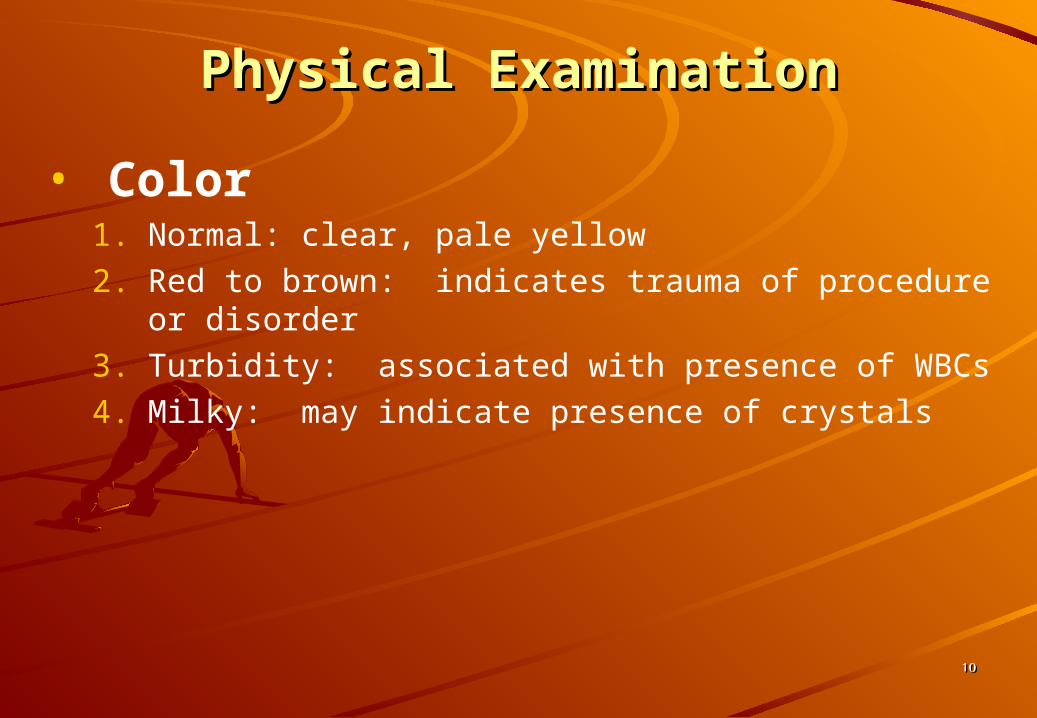
Physical ExaminationPhysical Examination
• Color1. Normal: clear, pale yellow2. Red to brown: indicates trauma of procedure or
disorder3. Turbidity: associated with presence of WBCs4. Milky: may indicate presence of crystals

1111
Physical Examination, Physical Examination, Cont’dCont’d
• Viscosity1. Measured at bedside by ability to form a
string from tip of syringe2. Normal: 4-6 cm

1212
Physical Examination, Physical Examination, Cont’dCont’d
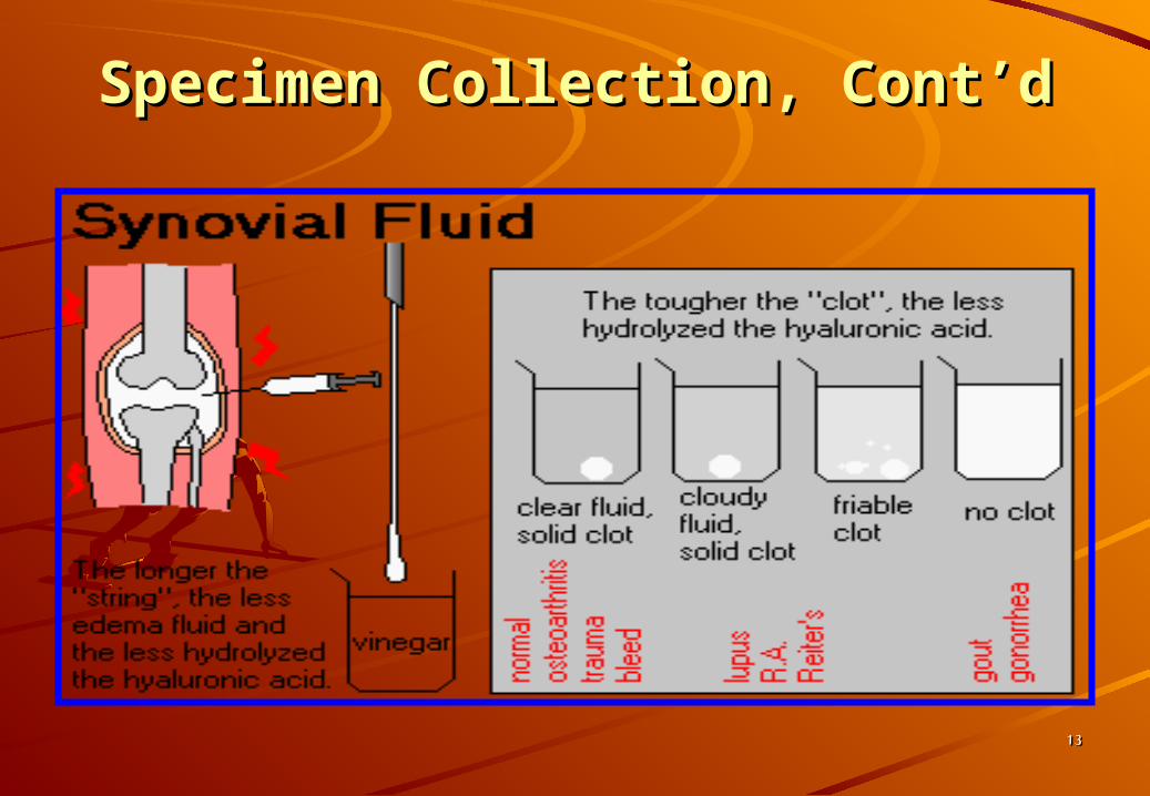
• Ropes Test (Mucin Clot Test)• Measurement of hyaluronate polymerization
1. Fluid forms a clot surrounded by clear fluid when added to acetic acid
2. Clot quality is reported:• Good = solid clot• Fair = soft clot• Poor = friable clot• Very poor = no clot
3. Test is of questionable precision and seldom used
1313
Specimen Collection, Cont’dSpecimen Collection, Cont’d
1414
Synovial JointSynovial Joint

1515
Synovial Joint, Cont’dSynovial Joint, Cont’d

1616
Laboratory Finding in JointsLaboratory Finding in Joints
1717
Microscopic ExaminationMicroscopic Examination
• Cell Count• WBC
• Method1. Use Neubauer counting chamber2. May pretreat viscous fluids with hyaluronidase and
incubate at 37oC for 5 min3. Dilution with hypotonic saline is used to lyse any RBC or4. Dilute with normal saline/methylene blue mixture to
differentiate WBCs from RBC
• Normal = < 200/L

1818
Microscopic Examination, Microscopic Examination, Cont’dCont’d
• Standard Neubauer Calculation Standard Neubauer Calculation Formula used for blood cell countsFormula used for blood cell counts
Lcells
squareofvolumeXcountedsquaresof
dilutionXcountedscellofcellsofNumber
1#
#
1919
Microscopic Examination, Microscopic Examination, Cont’dCont’d
• Differential Count1. Cytocentrifuge specimen and prepare
typical blood smear2. Normal: 60% monocytes, macrophages
• Neutrophils: <20%• Lymphocytes: <15%• (* values vary between texts)
2020
Microscopic Examination, Microscopic Examination, Cont’dCont’d
3. Increased neutrophils: possible septic condition
4. Increased lymphocytes: indicate nonspetic inflammation
2121
Microscopic Examination, Microscopic Examination, Cont’dCont’d
• Other Cell Abnormalities1. Increased eosinophils:rheumatic fever, parasitic
infections, metastatic carcinoma, post radiation therapy or arthrography
2. LE cells: patients with lupus erythematosus3. Reiter cells: macrophages with ingested
neutrophils4. RA cells (ragocytes): precipitated rheumatoid
factor appearing as cytoplasmic granules in neutrophils

2222
Microscopic Examination, Microscopic Examination, Cont’dCont’d
5. Hemosiderin granules: due to hemorrhagic process or cases of pigmented villonodular synovitis
6. Cartilaginous cells: observed in cases of osteoarthritis
7. Rice bodies: found in septic and rheumatoid arthritis and Tuberculosis
8. Fat droplets: indicate traumatic injury

2323
Cells and Inclusions in Cells and Inclusions in Synovial FluidSynovial Fluid

2424
Synovial Lining CellSynovial Lining Cell

2525
Lymphs in Synovial FluidLymphs in Synovial Fluid
2626
Microscopic Examination, Microscopic Examination, Cont’dCont’d
• CrystalsCrystals1. Microscopic examination of synovial fluid
for the presence of crystals is an important diagnostic test in the evaluation of arthritis
2. Crystal formation in a joint frequently results in an acute, painful inflammation

2727
• Crystals Formation• Crystal formation may be due to:
1. Metabolic disorders2. Decreased renal excretion3. Cartilage and bone degeneration4. Medicinal injection (ex: corticosteroids)
Microscopic Examination, Microscopic Examination, Cont’dCont’d
2828
Microscopic Examination, Microscopic Examination, Cont’dCont’d
• Fluid is Examined Using the Wet Preparation Technique1. ASAP examination as pH and temperature
affect observation2. Ideally examined prior to WBC disintegration3. Examine under both direct and compensated
polarizing light4. May also be observed in Wright stain
preparations

2929
Microscopic Examination, Microscopic Examination, Cont’dCont’d
•Under Polarizing Light (Direct Polarization)• Birefringent substances appear as bright objects
on a black background
3030
Microscopic Examination, Microscopic Examination, Cont’dCont’d
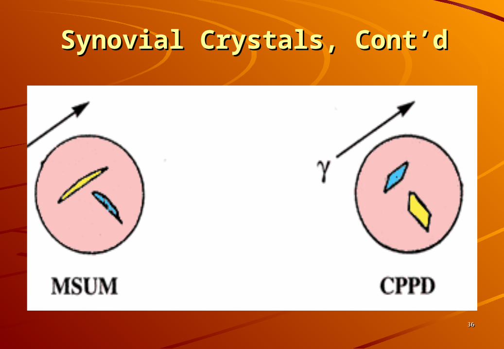
• Under Compensated Under Compensated Polarizing LightPolarizing Light
1. A red compensator plate is placed between the crystal and slide
2. Crystals aligned parallel to the compensator appear yellow (negative birefringence)
3. Crystals aligned perpendicular to the compensator appear blue (positive birefringence)

3131
Polarized LightPolarized Light
3232
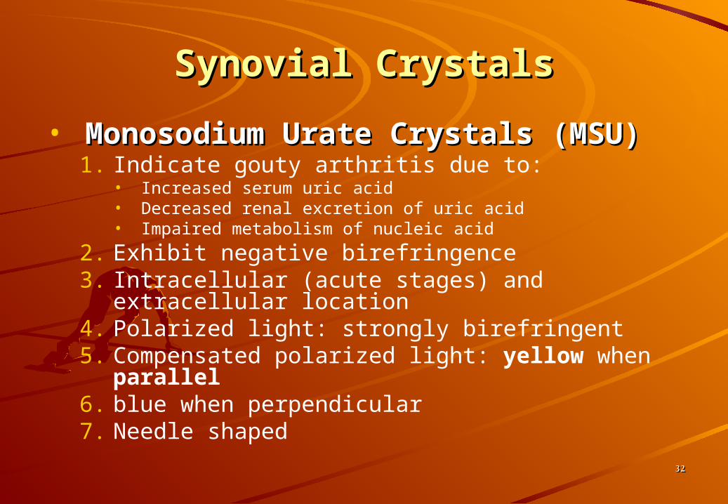
Synovial CrystalsSynovial Crystals
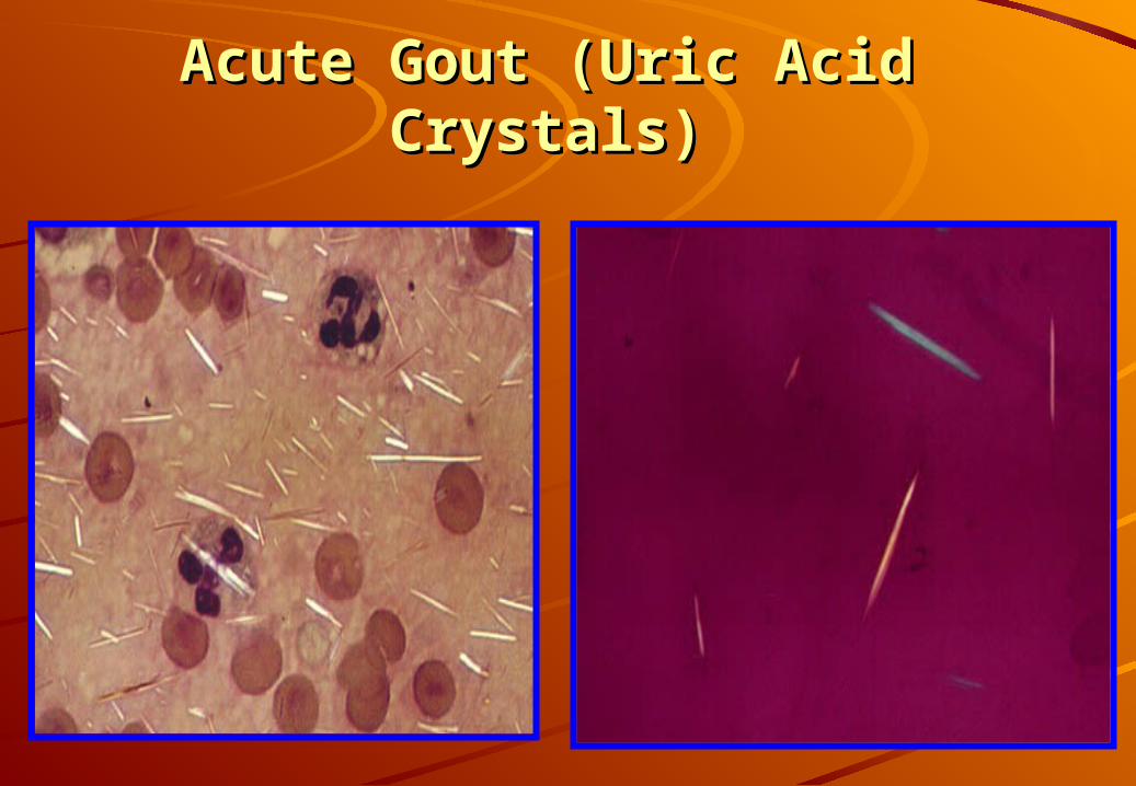
• Monosodium Urate Crystals Monosodium Urate Crystals (MSU)(MSU)1. Indicate gouty arthritis due to:
• Increased serum uric acid• Decreased renal excretion of uric acid• Impaired metabolism of nucleic acid
2. Exhibit negative birefringence3. Intracellular (acute stages) and extracellular
location4. Polarized light: strongly birefringent5. Compensated polarized light: yellow when
parallel6. blue when perpendicular7. Needle shaped

3333
Synovial Crystals, Cont’dSynovial Crystals, Cont’d
• Calcium pyrophosphate Calcium pyrophosphate (CCPD)(CCPD)
1. Indicates pseudogout due to:• Degenerative arthritis• Endocrine disorders with increased serum calcium• Calcification of cartilage
2. Exhibit positive birefringence3. Seen intracellular and extracellularly4. Polarized light: weakly birefringent5. Compensated polarized light: blue when
parallel (yellow when perpendicular)6. Blunt rods or rhombic shapes

3434
Negative and Positive Negative and Positive BirefringenceBirefringence

3535
page 3 of 6
Synovial Crystals, Cont’dSynovial Crystals, Cont’d
3636
Synovial Crystals, Cont’dSynovial Crystals, Cont’d
3737
Acute Gout (Uric Acid Acute Gout (Uric Acid Crystals)Crystals)

3838
• Cholesterol Crystal1. Nonspecific indications
• Associated with chronic inflammation
2. Exhibit negative birefringence (compensated polarized light)
3. Usually seen extracellularly4. Polarized light: strongly birefringence5. Rhombic plates
Synovial Crystals, Cont’dSynovial Crystals, Cont’d

3939
Synovial Crystals, Cont’dSynovial Crystals, Cont’d
• Hydroxyapatite (HA) (Calcium Hydroxyapatite (HA) (Calcium Phosphate) CrystalsPhosphate) Crystals
1. Associated with calcific deposition conditions2. May produce an acute inflammatory reaction3. Intracellular4. Not birefringent5. Require an electron microscope to examine6. Small, needle shaped

4040
Synovial Crystals, Cont’dSynovial Crystals, Cont’d
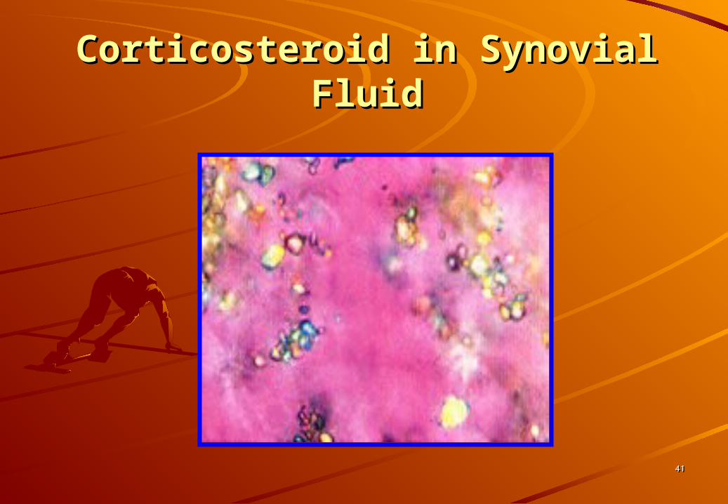
• Corticosteroid CrystalsCorticosteroid Crystals1. Associated with intra-articular injections; NO
clinical significance2. Primarily intracellular3. Exhibit positive and negative birefringence
• Can closely resemble MSU and CCPD
4. Flat, variable shaped plates
4141
Corticosteroid in Synovial Corticosteroid in Synovial FluidFluid
4242
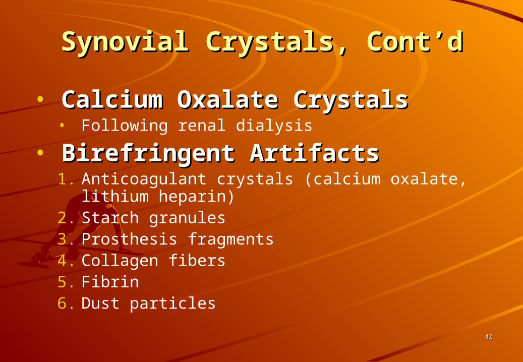
• Calcium Oxalate CrystalsCalcium Oxalate Crystals• Following renal dialysis
• Birefringent ArtifactsBirefringent Artifacts1. Anticoagulant crystals (calcium oxalate, lithium
heparin)2. Starch granules3. Prosthesis fragments4. Collagen fibers5. Fibrin6. Dust particles
Synovial Crystals, Cont’dSynovial Crystals, Cont’d
4343
Biochemistry TestsBiochemistry Tests
1. Because synovial fluid is biochemically an ultrafiltrate of plasma, biochemistry test values are approximately the same of serum values
2. Few biochemistry tests are considered clinically important
3. Most frequently requested test is the glucose determination because markedly decreased values are indicative of inflammatory or septic disorders

4444
• GlucoseGlucose1. Done simultaneously with blood sample
(prefer 8 hour fast)3. Should be run within 1 hour of collection4. Draw in sodium fluoride – prevents
glycolysis
Biochemistry Tests, Cont’dBiochemistry Tests, Cont’d

4545
5. Difference between blood and synovial glucose values is evaluated• Normal = < 10 mg/dL• Inflammatory conditions = > 25mg/dL• Sepsis = >40 mg/dL• Considered low if < ½ serum plasma glucose
value
Biochemistry Tests, Cont’dBiochemistry Tests, Cont’d
4646
Biochemistry Tests, Cont’dBiochemistry Tests, Cont’d
• Total proteinTotal protein1. Not routinely performed2. Normal = < 1/3 of serum value (~3g/dL)
• Large molecule, not easily filtered by membrane
3. Increased protein• Changes in membrane permeability• Increased joint synthesis• Indicates an inflammatory process
4747
Biochemistry Tests, Cont’dBiochemistry Tests, Cont’d
• Uric AcidUric Acid1. Alone, not diagnostic
• May determine gout in conjunction with plasma uric acid, esp. when crystals are undetectable
2. Normal = serum level

4848
Biochemistry Tests, Cont’dBiochemistry Tests, Cont’d
• LactateLactate1. May differentiate between inflammatory
and septic arthritis2. Septic arthritis = >250 mg/dL3. Gonococcal arthritis = normal to low levels4. Production results from:
• Increased demand for energy• Tissue hypoxia• Severe inflammatory conditions
4949
Microbiology TestsMicrobiology Tests
1. Infections may occur as a secondary complication of inflammation
2. Gram stains and cultures are two of the most important tests performed on synovial fluid

5050
Microbiology Tests, Cont’dMicrobiology Tests, Cont’d
• Gram StainGram Stain1. Performed on all specimens2. Most infections are bacterial:
• Staphylococcus• Streptococcus
• S. pyogenes• S. pneumoniae
• Hemophilus• Neisseria gonorrhea
3. Fungal, viral and tubercular agents may also be observed
5151
Microbiology Tests, Cont’dMicrobiology Tests, Cont’d
• CulturesCultures1.1. Routine cultureRoutine culture
2.2. Enrichment medium (chocolate agar)Enrichment medium (chocolate agar)
3.3. Specialty media depending on clinician Specialty media depending on clinician orders and indicationsorders and indications
5252
Serologic TestsSerologic Tests
1. Because of the association of the immune system to the inflammation process, serological testing plays an important role in the diagnosis of joint disorders
2. Majority of the tests are performed on serum with actual analysis of synovial fluid serving as a confirmatory measure in cases that are difficult to diagnose

5353
Serologic Tests, Cont’dSerologic Tests, Cont’d
• Autoantibody Detection Autoantibody Detection (same as found in serum)(same as found in serum)
1.1. Rheumatoid arthritis (RA)Rheumatoid arthritis (RA)
2.2. Lupus erythematosus (LE)Lupus erythematosus (LE)
• Antibody detection in patient’s serumAntibody detection in patient’s serum1.1. Borrelia burgdorferiBorrelia burgdorferi
• Causative agent of Lyme diseaseCausative agent of Lyme disease• Cause of arthritisCause of arthritis

5454
Joint Disorder ClassificationJoint Disorder ClassificationGroup ClassificationGroup Classification Pathological SignificancePathological Significance
11 NoninflammatoryNoninflammatory Degenerative joint disordersDegenerative joint disorders
22 InflammatoryInflammatory Immunologic problems (RA, LE)Immunologic problems (RA, LE)
Gout & pseudogout (rystal induced)Gout & pseudogout (rystal induced)
33 SpecificSpecific Microbial infectionMicrobial infection
44 HemorrhagicHemorrhagic Traumatic injuryTraumatic injury
Coagulation deficiencyCoagulation deficiency
Note: Note: * categories overlap* categories overlap* multiple conditions can occur simultaneously* multiple conditions can occur simultaneously* disease stage can vary laboratory results* disease stage can vary laboratory results
*see text for details of associated abnormal laboratory findings (pages 179-*see text for details of associated abnormal laboratory findings (pages 179-185)185)

5555
THE ENDTHE END
Any questions?