1. brainforebrain cerebral hemispheres cn i diencephalon (epithalamus, thalamus, hypothalamus cn ii...
TRANSCRIPT
1
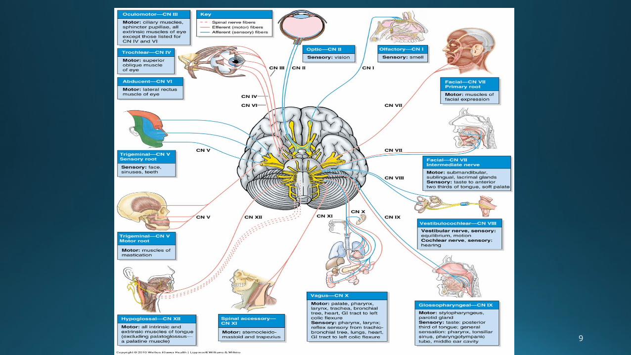
CRANIAL NERVES
• Bundles of sensory or motor fibers that innervate muscles or glands, • Carry impulses from
sensory receptors, or• Have a combo of motor
and sensory fibers.
• Called cranial nerves because they emerge through foramen/ fissures in the cranium, but individual names reflect either their location, distribution or their function.
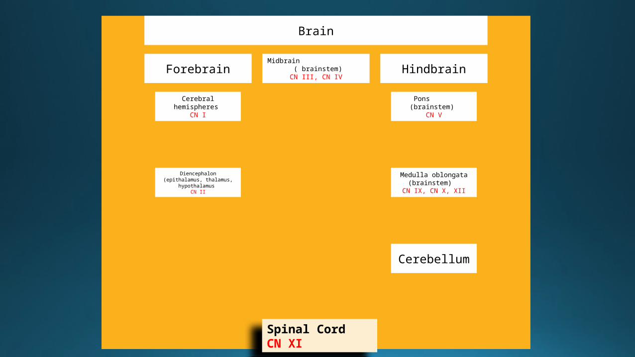
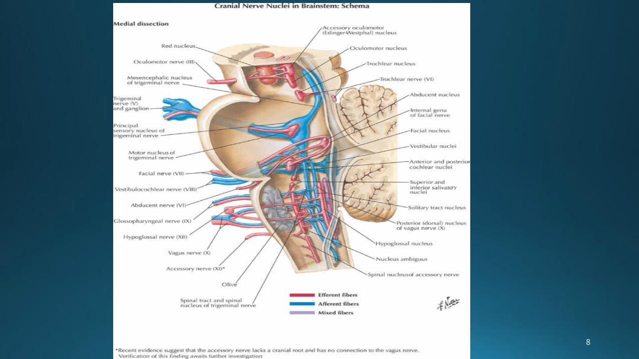
Brain
Forebrain
Cerebral hemispheres
CN I
Diencephalon (epithalamus, thalamus, hypothalamus
CN II
Midbrain ( brainstem) CN III, CN IV
Hindbrain
Pons (brainstem) CN V
Medulla oblongata (brainstem)
CN IX, CN X, XII
Cerebellum
Spinal Cord CN XI
3
4
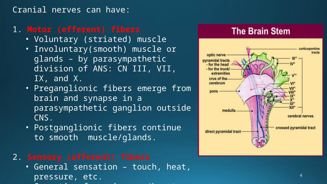
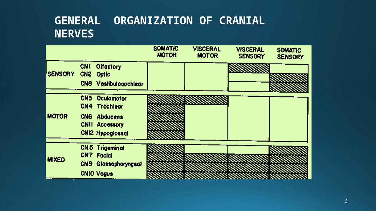
Cranial nerves can have:
1. Motor (efferent) fibers• Voluntary (striated) muscle• Involuntary(smooth) muscle or glands
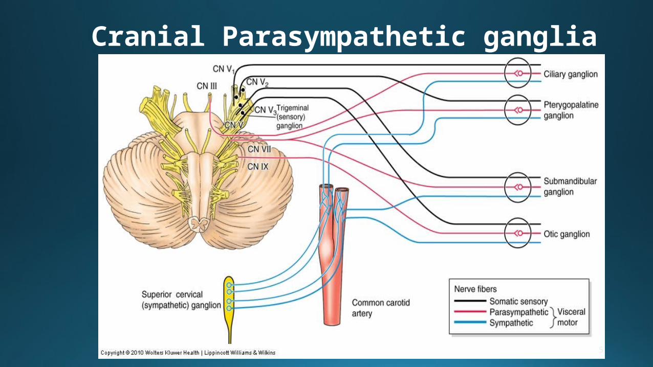
– by parasympathetic division of ANS: CN III, VII, IX, and X.
• Preganglionic fibers emerge from brain and synapse in a parasympathetic ganglion outside CNS.
• Postganglionic fibers continue to smooth muscle/glands.
2. Sensory (afferent) fibers• General sensation – touch, heat,
pressure, etc.• Sensation from viscera (heart, GI
tract, etc.)• Unique sensations – taste, smell, and
those for vision, hearing, balance
5
Cranial Parasympathetic ganglia
6
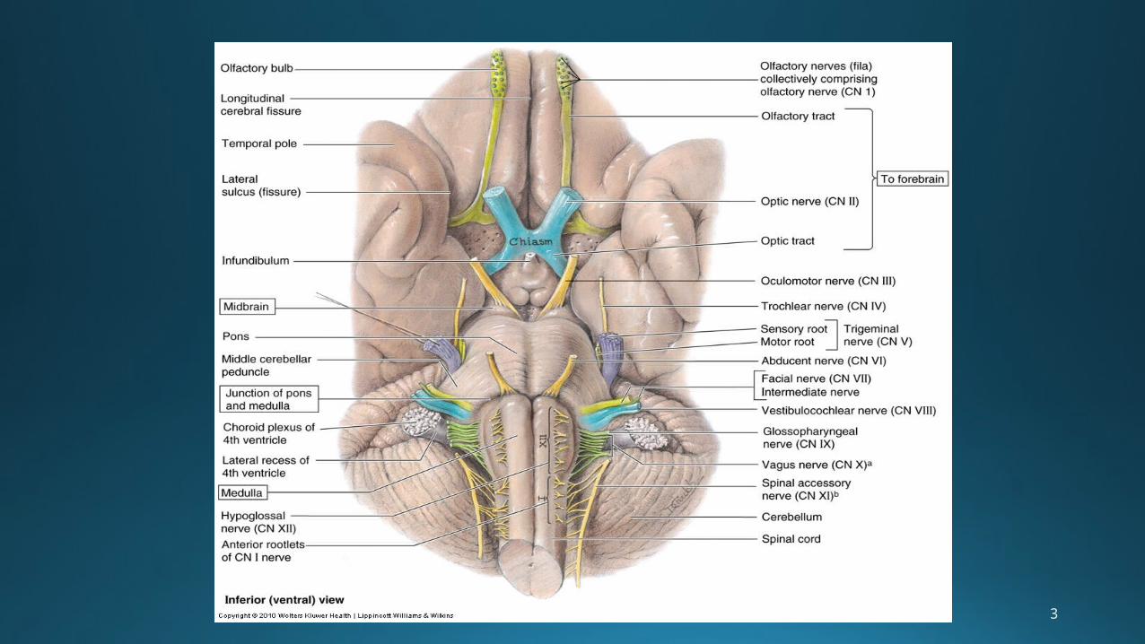
GENERAL ORGANIZATION OF CRANIAL NERVES
7
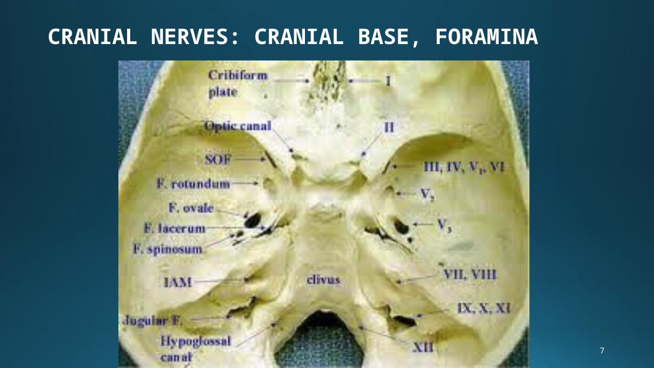
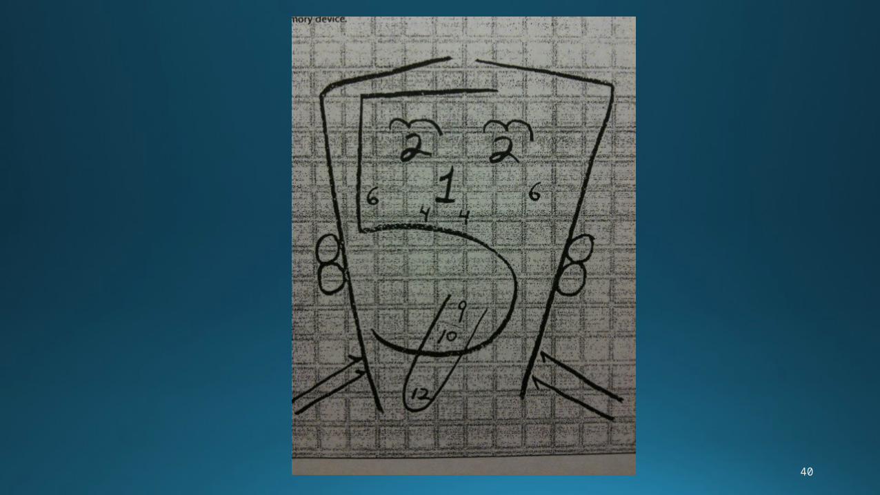
CRANIAL NERVES: CRANIAL BASE, FORAMINA
8
9
10
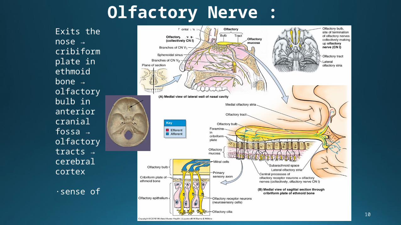
Olfactory Nerve : CN IExits the
nose →cribiform plate in ethmoid bone → olfactory bulb in anterior cranial fossa → olfactory tracts → cerebral cortex
∙sense of smell
11
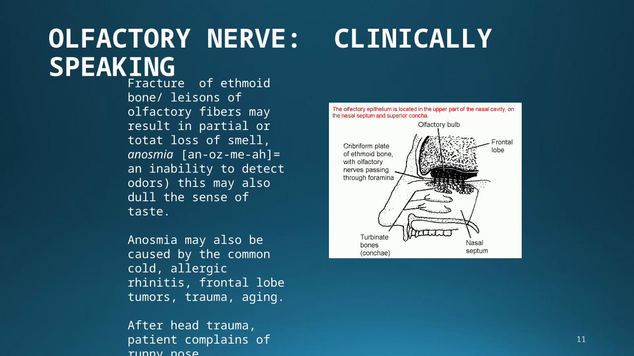
OLFACTORY NERVE: CLINICALLY SPEAKING
Fracture of ethmoid bone/ leisons of olfactory fibers may result in partial or totat loss of smell, anosmia [an-oz-me-ah]= an inability to detect odors) this may also dull the sense of taste.
Anosmia may also be caused by the common cold, allergic rhinitis, frontal lobe tumors, trauma, aging.
After head trauma, patient complains of runny nose (rhinnorhea): test nasal drip with dextrose or urine test strips --- WHY?
12
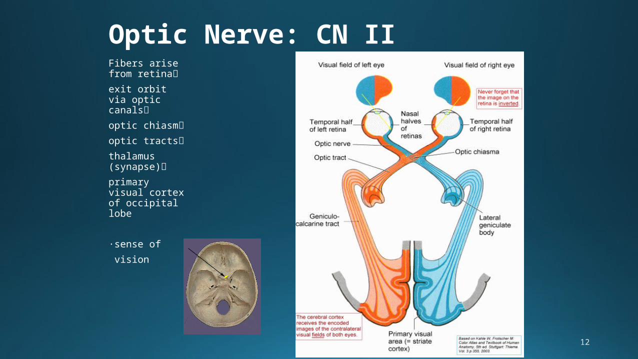
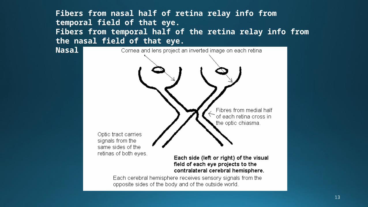
Optic Nerve: CN IIFibers arise from retina
exit orbit via optic canals
optic chiasm
optic tracts
thalamus (synapse)
primary visual cortex of occipital lobe
∙sense of
vision
13
Fibers from nasal half of retina relay info from temporal field of that eye. Fibers from temporal half of the retina relay info from the nasal field of that eye. Nasal fibers cross at chiasm.
14
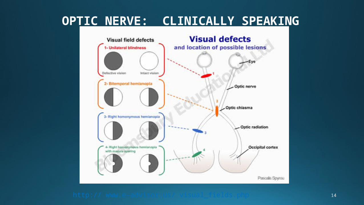
OPTIC NERVE: CLINICALLY SPEAKING
http:// www.e-advisor.us/ visual_fields.php
15
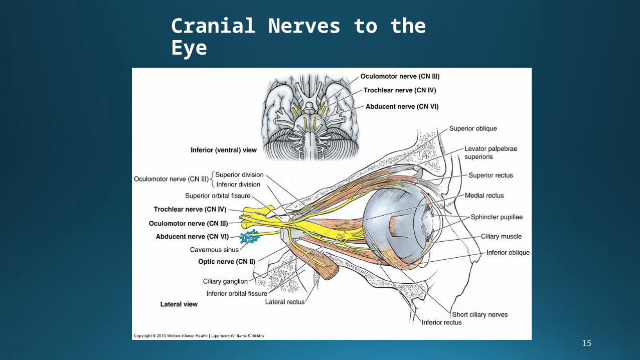
Cranial Nerves to the Eye
16
17
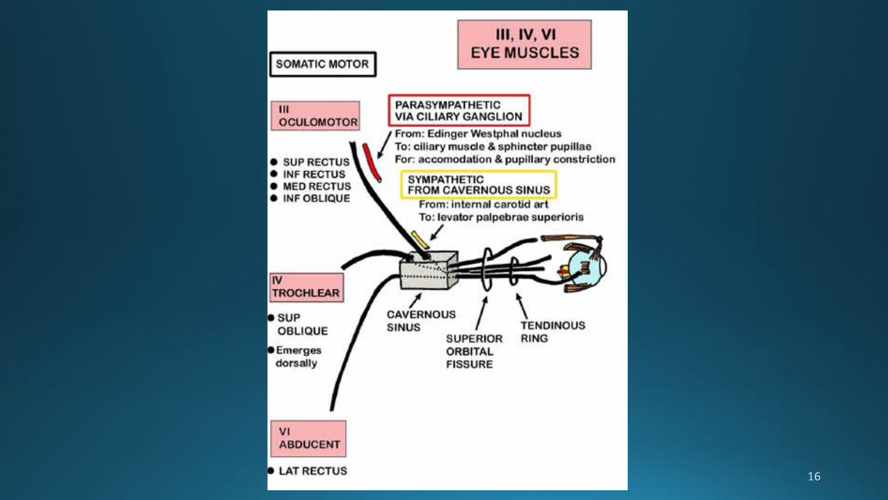
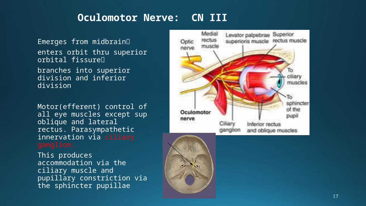
Oculomotor Nerve: CN III
Emerges from midbrain
enters orbit thru superior orbital fissure
branches into superior division and inferior division
Motor(efferent) control of all eye muscles except sup oblique and lateral rectus. Parasympathetic innervation via ciliary ganglion.
This produces accommodation via the ciliary muscle and pupillary constriction via the sphincter pupillae
18
OCULOMOTOR NERVE: CLINICALLY SPEAKING
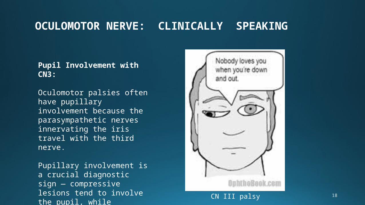
CN III palsy
Pupil Involvement with CN3:
Oculomotor palsies often have pupillary involvement because the parasympathetic nerves innervating the iris travel with the third nerve.
Pupillary involvement is a crucial diagnostic sign — compressive lesions tend to involve the pupil, while vascular lesions actually spare it!
19
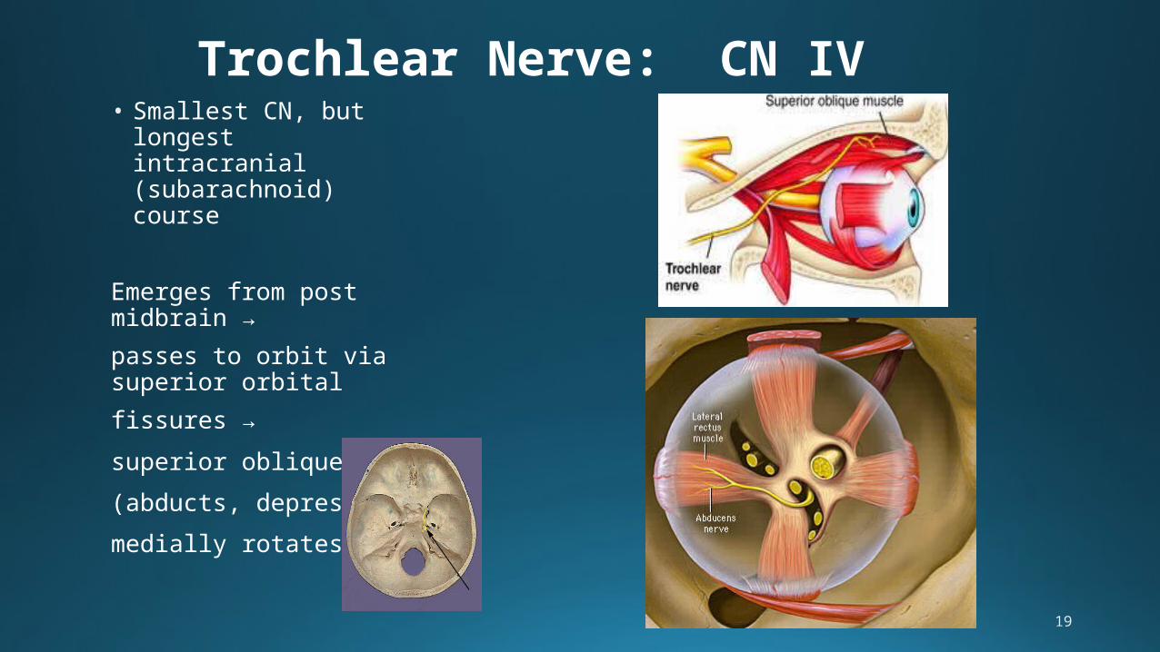
Trochlear Nerve: CN IV• Smallest CN, but
longest intracranial (subarachnoid) course
Emerges from post midbrain →
passes to orbit via superior orbital
fissures →
superior oblique m.
(abducts, depresses,
medially rotates).
20
CN III & CN IV LEISONS: CLINICALLY SPEAKING
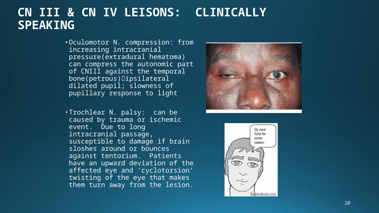
• Oculomotor N. compression: from increasing intracranial pressure(extradural hematoma) can compress the autonomic part of CNIII against the temporal bone(petrous)ipsilateral dilated pupil; slowness of pupillary response to light
• Trochlear N. palsy: can be caused by trauma or ischemic event. Due to long intracranial passage, susceptible to damage if brain sloshes around or bounces against tentorium. Patients have an upward deviation of the affected eye and ‘cyclotorsion’ twisting of the eye that makes them turn away from the lesion.
21
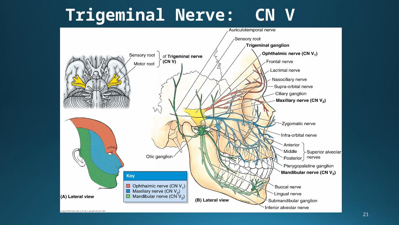
Trigeminal Nerve: CN V
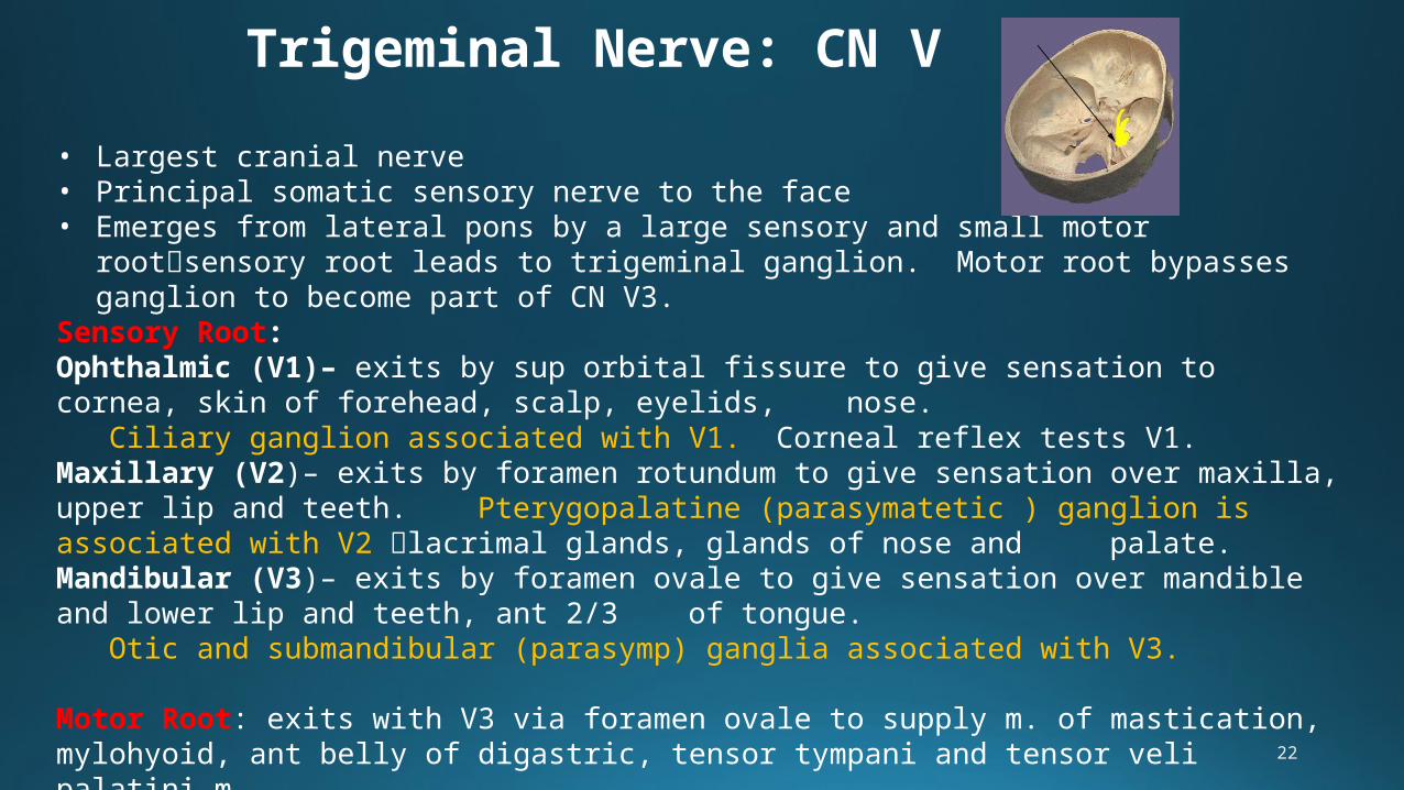
22
Trigeminal Nerve: CN V
• Largest cranial nerve• Principal somatic sensory nerve to the face• Emerges from lateral pons by a large sensory and small motor rootsensory root
leads to trigeminal ganglion. Motor root bypasses ganglion to become part of CN V3.
Sensory Root: Ophthalmic (V1)– exits by sup orbital fissure to give sensation to cornea, skin of forehead, scalp, eyelids, nose.
Ciliary ganglion associated with V1. Corneal reflex tests V1.Maxillary (V2)– exits by foramen rotundum to give sensation over maxilla, upper lip and teeth. Pterygopalatine (parasymatetic ) ganglion is associated with V2 lacrimal glands, glands of nose and palate.Mandibular (V3)– exits by foramen ovale to give sensation over mandible and lower lip and teeth, ant 2/3 of tongue.
Otic and submandibular (parasymp) ganglia associated with V3.
Motor Root: exits with V3 via foramen ovale to supply m. of mastication, mylohyoid, ant belly of digastric, tensor tympani and tensor veli palatini m.

23
TRIGEMINAL NERVE LEISONS: CLINICALLY SPEAKING
• Injury : by trauma (dental, cranial), tumors, aneurysms, infection (herpes zoster ophthalmicus) can all cause numbness
• Paralysis of m of mastication with deviation of mandible toward side of lesion-
Motor branch of V3
24
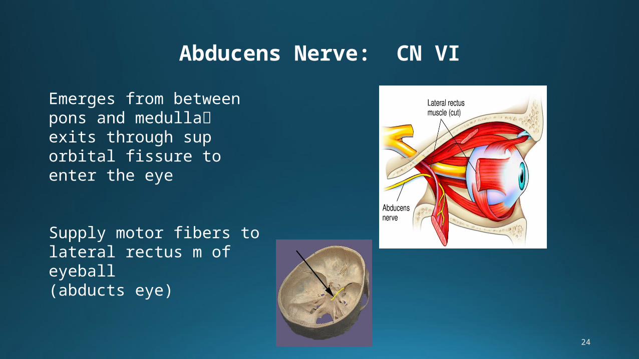
Abducens Nerve: CN VI
Emerges from between pons and medullaexits through sup orbital fissure to enter the eye
Supply motor fibers to lateral rectus m of eyeball (abducts eye)
25
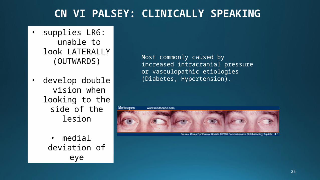
CN VI PALSEY: CLINICALLY SPEAKING
• supplies LR6: unable to look
LATERALLY (OUTWARDS)
• develop double vision when
looking to the side of the lesion
• medial deviation of eye
Most commonly caused by increased intracranial pressure or vasculopathic etiologies (Diabetes, Hypertension).
26
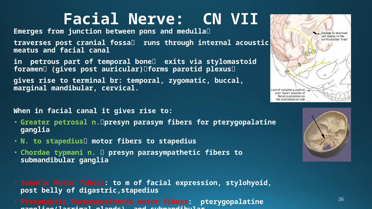
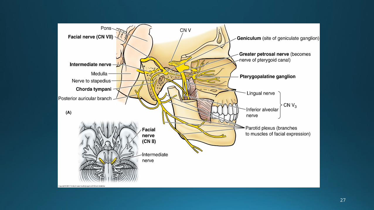
Facial Nerve: CN VIIEmerges from junction between pons and medulla
traverses post cranial fossa runs through internal acoustic meatus and facial canal
in petrous part of temporal bone exits via stylomastoid foramen (gives post auricular)forms parotid plexus
gives rise to terminal br: temporal, zygomatic, buccal, marginal mandibular, cervical.
When in facial canal it gives rise to:
• Greater petrosal n.presyn parasym fibers for pterygopalatine ganglia
• N. to stapedius motor fibers to stapedius
• Chordae typmani n. presyn parasympathetic fibers to submandibular ganglia
• Somatic Motor fibers: to m of facial expression, stylohyoid, post belly of digastric,stapedius
• Presynaptic Parasympathetic motor fibers: pterygopalatine ganglion(lacrimal glands), and submandibular ganglion(sublingual and submandibular salivary glands)
• Special Sensory fibers: taste to ant 2/3 of tongue and soft palate
27
28
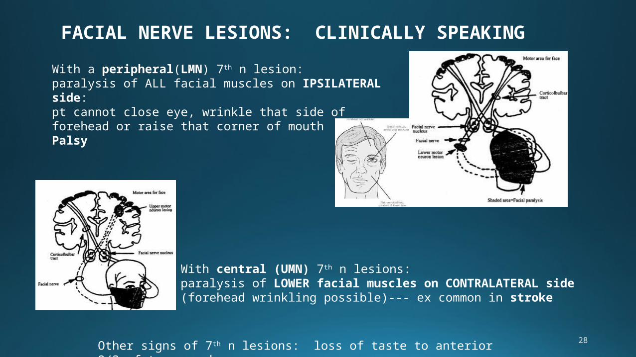
FACIAL NERVE LESIONS: CLINICALLY SPEAKING
With a peripheral(LMN) 7th n lesion: paralysis of ALL facial muscles on IPSILATERAL side: pt cannot close eye, wrinkle that side of forehead or raise that corner of mouth = Bell’s Palsy
With central (UMN) 7th n lesions: paralysis of LOWER facial muscles on CONTRALATERAL side (forehead wrinkling possible)--- ex common in stroke
Other signs of 7th n lesions: loss of taste to anterior 2/3 of tongue; dry cornea
29
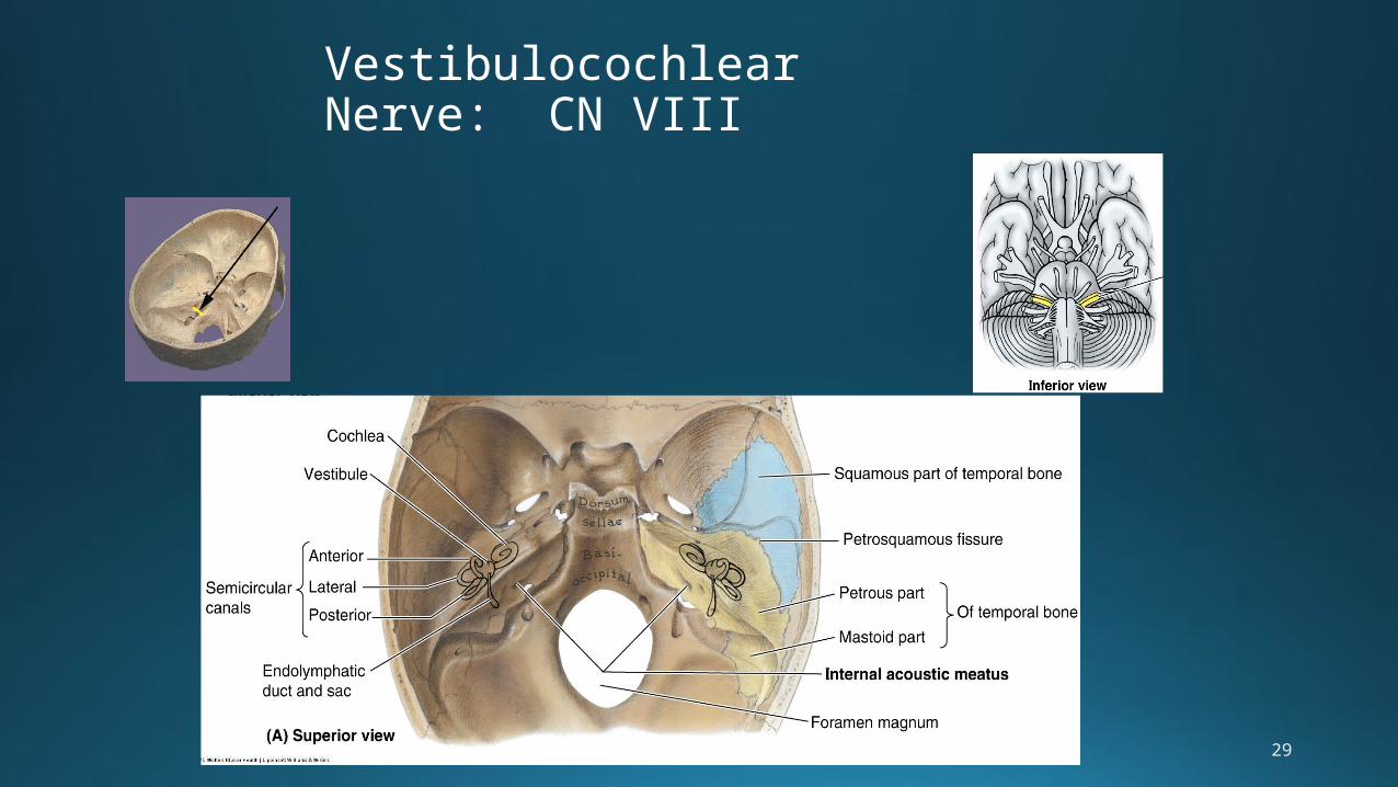
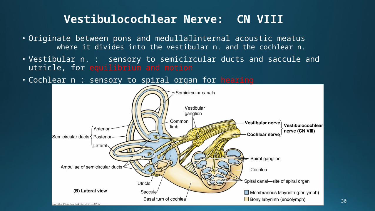
Vestibulocochlear Nerve: CN VIII
30
Vestibulocochlear Nerve: CN VIII
• Originate between pons and medullainternal acoustic meatus where it divides into the vestibular n. and the cochlear n.
• Vestibular n. : sensory to semicircular ducts and saccule and utricle, for equilibrium and motion
• Cochlear n : sensory to spiral organ for hearing
31
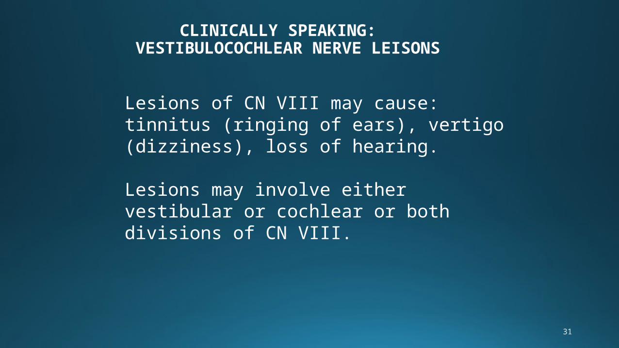
CLINICALLY SPEAKING: VESTIBULOCOCHLEAR NERVE LEISONS
Lesions of CN VIII may cause: tinnitus (ringing of ears), vertigo (dizziness), loss of hearing.
Lesions may involve either vestibular or cochlear or both divisions of CN VIII.
32
33
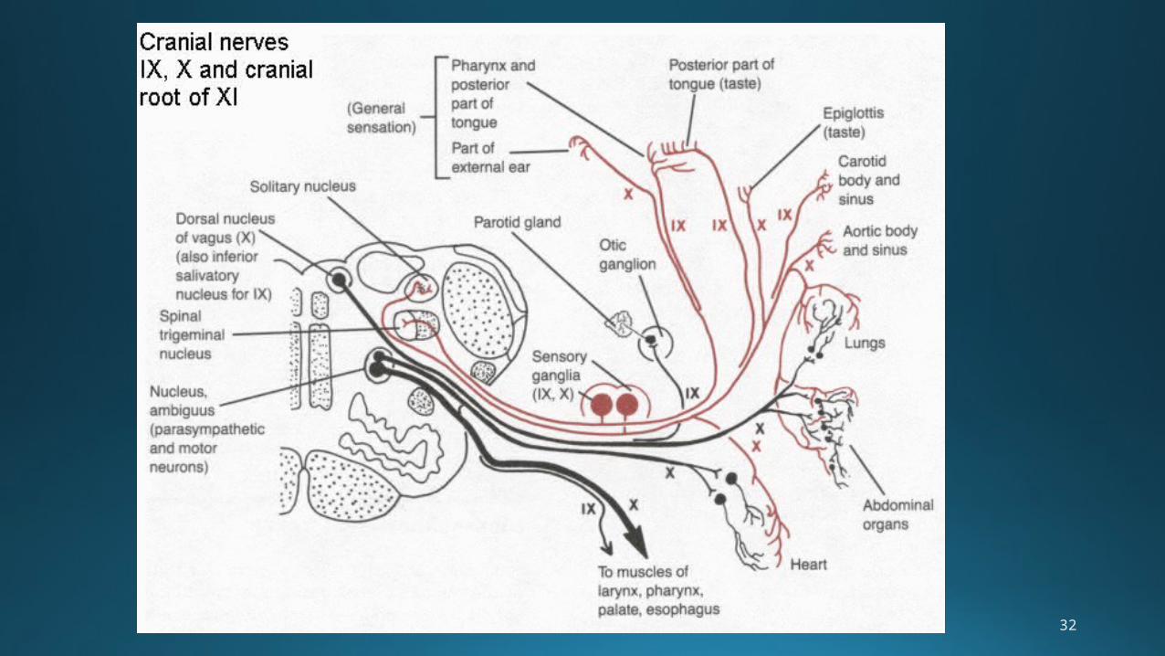
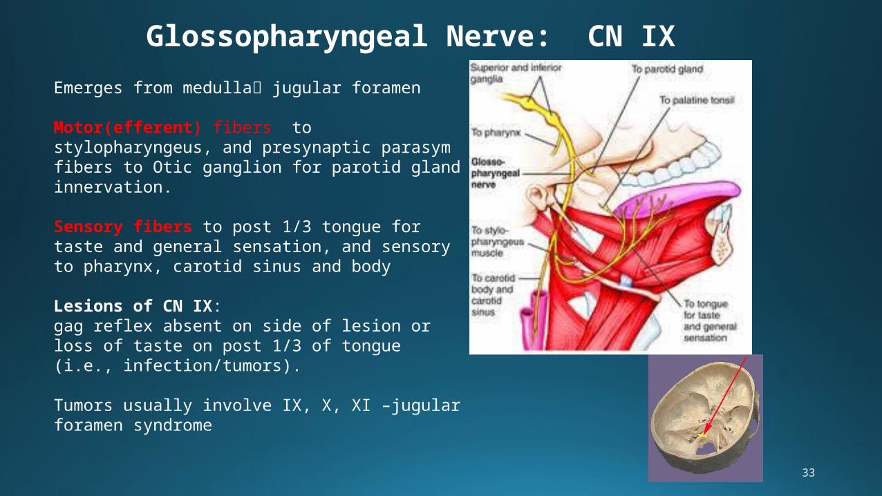
Glossopharyngeal Nerve: CN IX
Emerges from medulla jugular foramen
Motor(efferent) fibers to stylopharyngeus, and presynaptic parasym fibers to Otic ganglion for parotid gland innervation.
Sensory fibers to post 1/3 tongue for taste and general sensation, and sensory to pharynx, carotid sinus and body
Lesions of CN IX: gag reflex absent on side of lesion or loss of taste on post 1/3 of tongue (i.e., infection/tumors).
Tumors usually involve IX, X, XI –jugular foramen syndrome
34
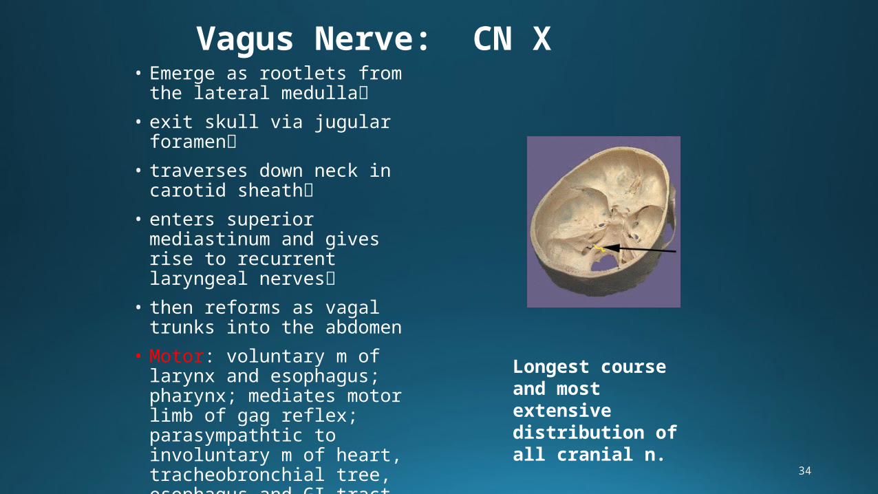
Vagus Nerve: CN X• Emerge as rootlets from the
lateral medulla
• exit skull via jugular foramen
• traverses down neck in carotid sheath
• enters superior mediastinum and gives rise to recurrent laryngeal nerves
• then reforms as vagal trunks into the abdomen
• Motor: voluntary m of larynx and esophagus; pharynx; mediates motor limb of gag reflex; parasympathtic to involuntary m of heart, tracheobronchial tree, esophagus and GI tract
• Sensory: pharynx, larynx
Longest course and most extensive distribution of all cranial n.

35
36
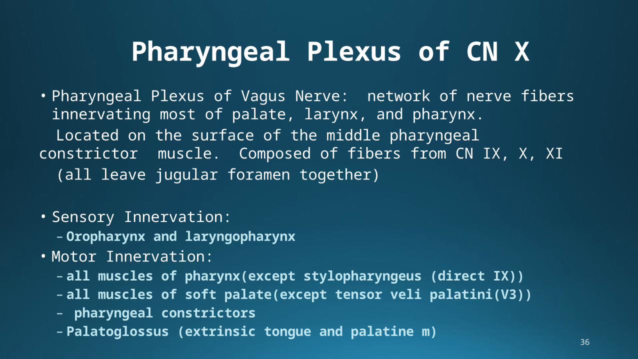
• Pharyngeal Plexus of Vagus Nerve: network of nerve fibers innervating most of palate, larynx, and pharynx.
Located on the surface of the middle pharyngeal constrictor muscle. Composed of fibers from CN IX, X, XI
(all leave jugular foramen together)
• Sensory Innervation:– Oropharynx and laryngopharynx
• Motor Innervation:– all muscles of pharynx(except stylopharyngeus (direct IX)) – all muscles of soft palate(except tensor veli palatini(V3))– pharyngeal constrictors– Palatoglossus (extrinsic tongue and palatine m)
Pharyngeal Plexus of CN X
37
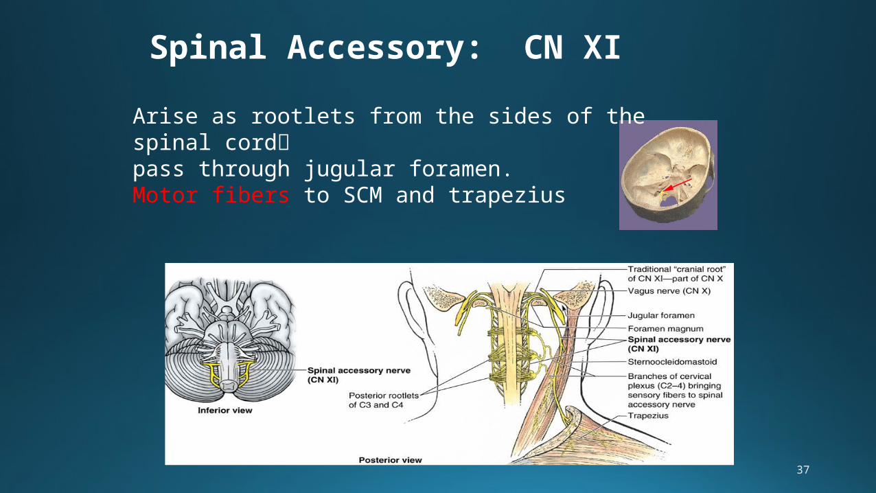
Spinal Accessory: CN XI
Arise as rootlets from the sides of the spinal cordpass through jugular foramen.Motor fibers to SCM and trapezius
38
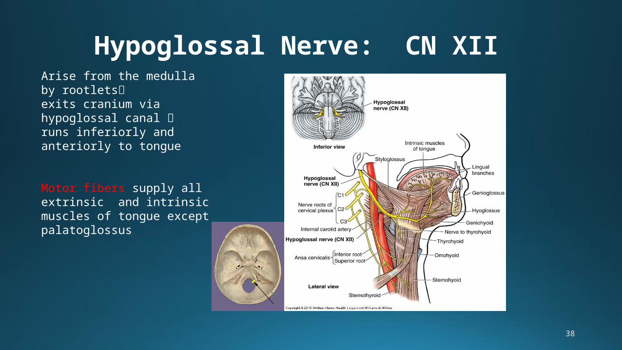
Hypoglossal Nerve: CN XIIArise from the medulla by rootletsexits cranium via hypoglossal canal runs inferiorly and anteriorly to tongue
Motor fibers supply all extrinsic and intrinsic muscles of tongue except palatoglossus
39
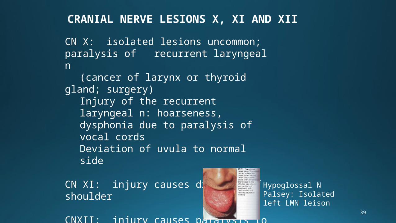
CRANIAL NERVE LESIONS X, XI AND XII
CN X: isolated lesions uncommon; paralysis of recurrent laryngeal n
(cancer of larynx or thyroid gland; surgery)
Injury of the recurrent laryngeal n: hoarseness, dysphonia due to paralysis of vocal cordsDeviation of uvula to normal side
CN XI: injury causes drooping of shoulder
CNXII: injury causes paralysis to ipsilateral side of tongue—tongue deviates toward affected side
Hypoglossal N Palsey: Isolated left LMN leison
40
41
Anatomy of Cranial Nerves and foramina of skull: http://www.youtube.com/watch?v=US6oemgNHGQ
Cranial Nerve and foramina quizzes/tutorials:http://www.gwc.maricopa.edu/class/bio201/cn/cranial.htm