1. biographical sketch - up.ac.za · of the thorax, abdomen and pelvic cavity of these animals for...
TRANSCRIPT

CURRICULUM VITAE of
MARIUS HORNSVELD Senior Lecturer, Department of Anatomy & Physiology
Faculty of Veterinary Science University of Pretoria
P/Bag X 04, Onderstepoort, 0110 Updated: April 2013
EVALUATION DATE: (Office use only): NA.
1. BIOGRAPHICAL SKETCH 1.1 General Information
Surname Hornsveld
First names Marius ID Number UP number
Available on request Available on request
Citizenship South Africa Title Dr Female Male X
Place of birth Pretoria Date of birth 4th June 1952
Population group
African Coloured Indian White Other (Please specify)
NA
Department Anatomy and Physiology Position Senior lecturer, Dept. of Anatomy
Direct Telephone 012 529 8030 Fax: (Dept. of
Physiology) 012 529 8305
E-mail [email protected]
Date of appointment 1st January 1983 Permanent full-
time X
Temporary full-time
1.2 Academic qualifications obtained Degree/
Diploma Field of study Higher education institution Year Distinctions
B.V.Sc.
Vet. Science University of Pretoria 1977 -
Radiology 701 Honours level
Vet. Science Small animals)
University of Pretoria 1989 -
Histology 700
Vet. Science University of Pretoria 1990 -
Embryology 700
Vet. Science University of Pretoria 1990 -
Anatomy 869
Vet. Science University of Pretoria 1990 -
Ph. D. (Anat.)
Vet. Science University of Pretoria 2006 -
Thesis title : The osteology of the cranial and the facial bones of the savannah buffalo Syncerus caffer caffer (Sparrman 1779).

Curriculum Vitae of Dr. M. Hornsveld Page 2 of 22
1.3 Work experience to date Name of employer Capacity and/or type of work Period: mm//yy to mm//yy SA. Reserve Bank Gold clerk Jan 1970 - Aug 1971 SANDF - National Defense Force Infantry training, 4th Infantry Battalion Jan 1970 - Sept 1970 Student work: Wool sorting during studies: National Diploma in wool classing Feb. 1971 - Nov 1977 Dept. of Agriculture State Veterinarian Pretoria March 1978 - Dec 1978 Dept. of Agriculture, South West Africa State Veterinarian Outjo, SWA / Namibia Jan 1979 - Jul 1980 University of Pretoria Senior Lecturer Dept. of Medicine Aug 1980 - Sept 1981Trans-Oranje Cooperation Consulting Veterinarian Oct 1981 - March 1982 National Defense Force Equestrian Centre Veterinarian May 1982 - Nov 1982 University of Pretoria Senior Lecturer, Dept. of Anatomy Jan 1983 - present Self-employed: Locum tenens Small animals & stud cattle (part time) Jan 1981 - Jun 1987
2. TEACHING ACTIVITIES 2.1 Formal academic modules/courses presented Course Level (e.g. second year, Masters) Self-developed (Y / N) Code* : Medicine (Large and Small
animals)
Final years : B.V.Sc. V clinics Final years : Gastro-intestinal tract of
small animals
No No
Code *: Veterinary Clinical Diagnostics
B.V.Sc. III (old system) D. V. N. I
No No
Code * : Anatomy (general)
B.V.Sc. I (old system) No
Code * : Histology (general)
B.V.Sc. I (old system) No
Code * : Embryology (general)
B.V.Sc. I (old system) No
DAF 200: ‘Diere Anatomie en Fisiologie’ - Anatomy component
B.Sc. Biol. Sc. II (Main Campus in so-called III & IV system), Dept. Animal and Wildlife Sciences, Main Campus
Yes
VCA 400: Veterinary Comparative Anatomy - Applied Anatomy Tutorials & Palpations
B.V.Sc. I (so-called III & IV system) Yes
VCA 200: Veterinary Comparative Anatomy - Applied Anatomy Tutorials & Palpations
B.V.Sc. II (lastest system) Yes
WOC 501: Wildlife, Ostrich and Crocodile - Anatomy component
Final year B.V.Sc. students (old system)
Yes
BHP 500: Interdisciplinary Applied Anatomy of the Bovine Herd Health and Production course –
- Anatomy component
B.V.Sc. II (III & IV system) Yes
ECS 601: Interdisciplinary Applied Anatomy of Equine Clinical Studies
- Anatomy component
B.V.Sc. III (III & IV system) Yes
NLB 783: Wildlife Management Honours / ‘Natuur Lewe Bestuur Honeurs’ - Applied Anatomy of selected Wild animal species (Anatomy component of course)
Honours (B.Sc. Vet. Biol.) Dept. Animal and Wildlife Sciences, Main Campus
Yes

Curriculum Vitae of Dr. M. Hornsveld Page 3 of 22
Code *: Capita Selecta for Surgery part of M. Med. Vet. (Chir) Large animals - Anatomy component
Masters (Veterinary Science)
No
Code *: Capita Selecta for Theriogenology part of M. Med. Vet. (Gyn) Large animals - Anatomy component
Masters (Veterinary Science)
No
ANG 774: Capita Selecta for Anesthesiology part of M. Med. Vet. (Anaes) - Anatomy component
Masters (Veterinary Science)
Curriculum completely updated according to modern needs
* Official course codes have changed over the years
2.2 Other presentations Course
Year Institution
NA.
3. TEACHING OUTPUTS
3.1 Educational publications and products
3.1.1 Hornsveld M. 1994. Functional Sketches in the Teaching of the Topographical Anatomy of
the Horse and the Bovine by using three-dimensional sketches, produced in mass at low cost.
Presentation given to Faculty members at the request of the Bureau of Academic Support Services,
Teaching Development Section, University of Pretoria. These sketches form part of the special
presentation and techniques in the approach of teaching the topographical anatomy of the viscera
of the thorax, abdomen and pelvic cavity of these animals for pre- and postgraduate students.
3.1.2 Bezuidenhout A.J, Groenewald H.B., Hornsveld M., Soley J.T., Turner P.H. 1999.
Veterinary Anatomy a study and dissection guide. Volumes 1 - 3. Fourth edition. University of
Pretoria. Subsequent editions appeared based on the original work. I was responsible to compile
the notes for all the Splanchnology and Topography of all the species.
3.1.3 Hornsveld M. 2002. DAF 200 ‘Checklists’: Compiled ‘Checklists’ for the practical sessions,
one for each system of the body that served as crucial learning material (General plan of the body,
Muscular-, Skeletal-, Nervous-, Uro-genital- & Reproductive-, Respiratory-, Cardiovascular-, Skin &
Integument, Endocrinology & Immunology and Digestive systems). The ‘Checklists’ were made
available on the web system of the University (clickUP system) for all students and lecturing
personnel in the Dept. Animal and Wildlife Sciences, Main Campus.

Curriculum Vitae of Dr. M. Hornsveld Page 4 of 22
3.1.4 Hornsveld M. 2002. ‘DAF 200 Theory Notes’: Compiled multiple notes additional to the
textbook for theory and practical parts of the course. A document was also compiled for each
semester addressing all the corrections to the textbook. All matters discussed in class were also
included in this document and together with the theoretical notes; it formed that basis for the
practical component. The notes were made available on the web system of the University (clickUP
system) for all concerned. A portfolio of the work (Checklists, Notes, all illustrations, all old test and
exam papers) was subsequently placed on DVD and handed over to the Departmental Head of that
Department after my term in presenting the course ended.
3.1.5 Hornsveld M. 2002. ‘Procedures and logistics of how to dissect sheep in order to present the
anatomy of the domestic animals in the form of demonstrations on fresh cadavers in a systems
approach’. Approximately 20 ‘Wet Lab’ presentations per year were given for the practical part of
the ‘DAF 200’ course, covering all body systems. The approach was also used to teach lecturers in
the Dept. of Animal and Wildlife studies, Faculty of Natural and Agricultural Sciences, University of
Pretoria. Training additional lecturers were necessary in order to be able to handle the large
number of students (180 – 230 / year) and therefore it eventually had to be done in quadruplicate
form for each practical. Over the period from 2002 to 2009, I had to either do or supervise a total of
approximately 140 such ‘wet-lab’ presentations.
3.1.6 Hornsveld M., Meyer J.A., O’Neill A., Aire T. & Webb E.C. 2006. ‘Videos for DAF 200
Practicals’. The following 19 videos were produced in order to serve as training and educational
material for lecturers and students in the course:
Video 01: Part I: Terminology, Osteology & Joints. Presented by Hornsveld M.
Video 01: Part II: Pelvis as birth canal. Presented by Hornsveld M.
Video 02: General plan of the body. Presented by Hornsveld M.
Video 03: Muscles of Neck, front limb, thorax & abdomen. Neurology & Myology part I. Presented by Hornsveld M.
Video 04: Autonomic Nervous system & Hind limb. Neurology & Myology part II. Presented by Hornsveld M.
Video 05: Central and autonomic nervous system. Neurology & Myology part III. Presented by Hornsveld M.
Video 06: Skin, hoof, claw & udders. Presented by Hornsveld M.
Video 07: Circulatory system. Presented by Hornsveld M. & Meyer J.A.
Video 08: 1st Experimental Farm Practical. Presented by Hornsveld M. & Webb E.C.
Video 09: Respiratory system. Presented by Hornsveld M. & Meyer J.A.
Video 10: Endocrinology. Presented by Hornsveld M. & Webb E.C.
Video 11: Lymphatic vascular system. Presented by Hornsveld M. & Meyer J.A
Video 12: Placentation & fetal membranes. Presented by Hornsveld M. & van der Merwe N.
Video 13:. Fetal circulation. Remnants of fetal circulation. Presented by Hornsveld M. & O’Neill A.
Video 14: Female Uro-genital system. Presented by Hornsveld M. & Webb E.C.
Video 15: Male Uro-genital system. Presented by Hornsveld M., Webb E.C. & Meyer J.A.
Video 16: Avian Anatomy. Presented by Aire T.
Video 17: Splanchnology: Stomachs & Ruminant GIT. Part I. Presented by Hornsveld M.
Video 18: Topography: Sheep Gastro-Intestinal Tract. Part II. Presented by Hornsveld M. & Meyer J.A.
Video 19: Splanchnology: Horse cecum and colon. Presented by Hornsveld M. & Meyer J.A.
The set of videos is sold to students at a very reasonable price.
Curriculum Vitae of Dr. M. Hornsveld Page 5 of 22
3.2 Educational publications and products
Prototype products made and specific investigation for educational use:
Hornsveld M. 2010. ‘Quasi-subaquatic dissection methods’: Three prototypes have been designed
and tested for use as methods to curb carcinogenic and hazardous formaldehyde vapor exposure
to dissectors and researchers.
Hornsveld M. 2011 - 2012. Two types of closed circuit systems investigated to eliminate the
exposure of carcinogenic and hazardous formaldehyde vapors to dissectors and researchers using
full facial masks and filters or compressed air.
4. OTHER TEACHING CONTRIBUTIONS 4.1 Membership of national and international bodies:
Registration at Professional Boards:
South African Veterinary Council (SAVC), Pretoria, South Africa (1977 till present): Full member. Registration number : 77/1380
Namibian Veterinary Council (NVC), Windhoek, Namibia (1979 till present): Full member. Registration number : 86/67
Royal College of Veterinary Surgeons (RCVS), London, England (1995 till March 2012):
Overseas member. Certificate number: 5402;
Membership number: 610078X. Voluntary temporary suspension.
4.2 Visits to local and overseas universities as guest professor or lecturer with regard
to teaching
None. Only the following in a private capacity with regard to teaching:
1993 - 1995. Department of Anatomy, “Universidad de Chile” (Santiago) and “Universidad Austral
de Chile” (Valdivia), Chile, South America.
1994 - 1995. Department of Production Animals, Faculty of Veterinary Medicine, Utrecht,
Netherlands.
4.3 Participation in national and international teaching associations, bodies,
committees
World Veterinary Teachers Association from1996 till about 1997 Member of the Onderstepoort Medicine Club from 1990 till club dissolved.
Curriculum Vitae of Dr. M. Hornsveld Page 6 of 22
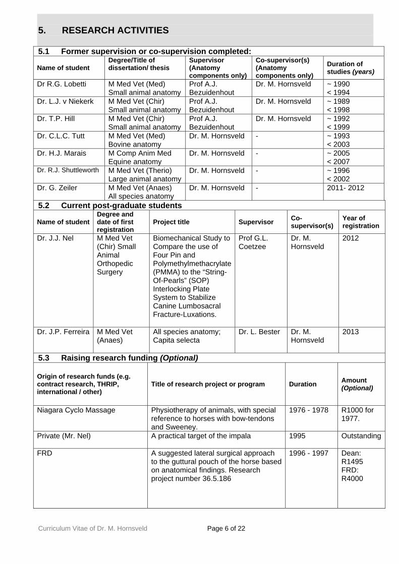
5. RESEARCH ACTIVITIES 5.1 Former supervision or co-supervision completed:
Name of student Degree/Title of dissertation/ thesis
Supervisor (Anatomy components only)
Co-supervisor(s) (Anatomy components only)
Duration of studies (years)
Dr R.G. Lobetti
M Med Vet (Med) Small animal anatomy
Prof A.J. Bezuidenhout
Dr. M. Hornsveld ~ 1990 < 1994
Dr. L.J. v Niekerk M Med Vet (Chir) Small animal anatomy
Prof A.J. Bezuidenhout
Dr. M. Hornsveld ~ 1989 < 1998
Dr. T.P. Hill M Med Vet (Chir) Small animal anatomy
Prof A.J. Bezuidenhout
Dr. M. Hornsveld ~ 1992 < 1999
Dr. C.L.C. Tutt
M Med Vet (Med) Bovine anatomy
Dr. M. Hornsveld - ~ 1993 < 2003
Dr. H.J. Marais
M Comp Anim Med Equine anatomy
Dr. M. Hornsveld - ~ 2005 < 2007
Dr. R.J. Shuttleworth M Med Vet (Therio) Large animal anatomy
Dr. M. Hornsveld - ~ 1996 < 2002
Dr. G. Zeiler M Med Vet (Anaes) All species anatomy
Dr. M. Hornsveld - 2011- 2012
5.2 Current post-graduate students
Name of student Degree and date of first registration
Project title Supervisor Co-supervisor(s)
Year of registration
Dr. J.J. Nel
M Med Vet (Chir) Small Animal Orthopedic Surgery
Biomechanical Study to Compare the use of Four Pin and Polymethylmethacrylate (PMMA) to the “String-Of-Pearls” (SOP) Interlocking Plate System to Stabilize Canine Lumbosacral Fracture-Luxations.
Prof G.L. Coetzee
Dr. M. Hornsveld
2012
Dr. J.P. Ferreira M Med Vet (Anaes)
All species anatomy; Capita selecta
Dr. L. Bester Dr. M. Hornsveld
2013
5.3 Raising research funding (Optional)
Origin of research funds (e.g. contract research, THRIP, international / other)
Title of research project or program Duration Amount (Optional)
Niagara Cyclo Massage
Physiotherapy of animals, with special reference to horses with bow-tendons and Sweeney.
1976 - 1978 R1000 for 1977.
Private (Mr. Nel)
A practical target of the impala 1995 Outstanding
FRD
A suggested lateral surgical approach to the guttural pouch of the horse based on anatomical findings. Research project number 36.5.186
1996 - 1997 Dean: R1495 FRD: R4000

Curriculum Vitae of Dr. M. Hornsveld Page 7 of 22
6. RESEARCH OUTPUTS 6.1 Publications in peer-reviewed or refereed journals
None
6.2 Books and/or chapters in books
Bezuidenhout A.J, Groenewald H.B., Hornsveld M., Soley J.T., Turner P.H. 1999. Veterinary
Anatomy a study and dissection guide. Volumes 1 - 3. Fourth edition. University of Pretoria. I was
responsible to compile the notes for all the Splanchnology and Topography of all the species.
Subsequently, later editions of the guide have appeared with the dog separated from the rest.
Presently, it is in the phase of getting redone again, now combining all the species once again in a
Comparative way as it used to be.
6.3 Published full-length conference papers/keynote addresses
Oral presentations:
Hornsveld M. 1996. Applied anatomical aspects of the African buffalo. Proceeding of a
Symposium on the African Buffalo as a game ranch animal. Wildlife Group of the South African
Veterinary Association.
Hornsveld M. 1998. Postnatal development of the permanent tooth enamel organs in the horse.
Proceedings of a Joint Meeting of The Anatomical Society of Great Britain and Ireland, The
Anatomical Society of Southern Africa, and The Nederlandse Anatomen Vereniging, 15th - 17th
April 1998, Rolduc, Limburg, The Netherlands.
Hornsveld M. & G. Steenkamp 1999. Elephant tusks - Clinical Anatomy/Embryology. Proceedings
of the Sixth World Veterinary Dental Congress, Wrest Point, Hobart, Tasmania, Australia, 17th -
19th May, 1999.
Hornsveld M. 1999. The anatomy in the region of the upper lip and trunk base in the African
elephant Loxodonta africana (Blumenbach, 1797). Proceedings of the 26th World Veterinary
Congress (subsection W.V.A.), Lyon, France, 23rd - 26th September 1999.
Hornsveld M. 1999. Three-dimensional concept formation of the topography of the viscera of the
horse and bovine, using two-dimensional images cheap to reproduce. 15th Congress of the
International Federation of Associations of Anatomists, 11th - 16th September 1999, Rome, Italy.
Poster presentations:
Hornsveld M. 1998. Evidence of an atypical fontanelle in the skull of the savanna buffalo.
Proceedings of a Joint Meeting of The Anatomical Society of Great Britain and Ireland, The

Curriculum Vitae of Dr. M. Hornsveld Page 8 of 22
Anatomical Society of Southern Africa, and The Nederlandse Anatomen Vereniging, 15th - 17th April
1998, Rolduc, Limburg, The Netherlands.
Hornsveld M., Niccol J.K., Keller N. & Guthrie A. 1998. A suggested lateral surgical approach to
the guttural pouch of the horse based on anatomical findings (part one). Proceedings of a Joint
Meeting of The Anatomical Society of Great Britain and Ireland, The Anatomical Society of
Southern Africa, and The Nederlandse Anatomen Vereniging, 15th - 17th April 1998, Rolduc,
Limburg, The Netherlands.
6.4 Non-refereed publications or popular articles
Hornsveld M. 1997. Applied anatomical aspects of the African Buffalo. Presented by invitation
of the S.A. Mint Company at Sabi-Sabi private game reserve at the launch of the gold coin
entitled The African buffalo.
Hornsveld M. & VanderLinde R. 2003. Hunting the African Savannah Buffalo: Techno-
anatomical aspects based on the bony components. Colorado shooting. July - August 2003.
Page 4 - 5 and 30 - 33.
6.5 Patents
None.
6.6 Technical reports
Hornsveld M. 1979. The state of the animal disease control fence in the northwestern parts of
Namibia, Damaraland, Koakoland and the Skeleton coast: Recommendations for maintenance.
Report to the Deputy Director, Veterinary Services, Ausspannplatz, Namibia.
Hornsveld M. 1978. Disease surveillance report for the district Outjo, including the western half
of the Etosha Game reserve, Damaraland, Koakoland and the Skeleton Coast for the period 1st
July 1978 – 30th June 1979. Report to the Deputy Director, Ausspannplatz, Namibia.
Hornsveld M. 1979. Disease surveillance report for the district Outjo, including the western half
of the Etosha Game reserve, Damaraland, Koakoland and the Skeleton Coast for the period 1st
July 1979 – 31st December 1979. Report to the Deputy Director, Ausspannplatz, Namibia.
Hornsveld M. 1983. A mechanical model of the hind limb passive stay mechanism of the horse
using assembled bones. Model used in Introductory lecture for permanent appointment.
Hornsveld M. 1988 - 1992. I served as Departmental coordinator and adviser in a technical
capacity between UP personnel and the different engineers and architects who had the contract
to design and build our Dissection hall, Museum facilities and to revamp our Departmental
offices according to our needs. The total area of the Anatomy complex is 2351 m2 and the

Curriculum Vitae of Dr. M. Hornsveld Page 9 of 22
facilities were eventually inaugurated in March 1992. Despite all the original planning, critical
issues could not be met for financial reasons and therefore the Dissection hall needed a major
upgrade around 2002. That is because the formalin vapour control by the outdated ventilation
system was inadequate as the legal limits for maximum exposure levels got much stricter. Many
years later I designed a system for the ‘DAF 200’ course (vide supra) that was not preservative
based at all. It functioned at a fraction of the cost albeit for a different purpose and standard.
Having had the experience of getting to know the technical issues of both systems, makes me
perhaps one of a few Academic Anatomists in the world that have had the opportunity of
planning both approaches and designs of Veterinary Anatomy Dissection facilities and curricula:
A formalin-based system needs careful planning of the physical facilities and ventilation system
as well as a stringent control over chemicals used and frequent vapor exposure testing.
Conversely, a formalin-free system on un-preserved cadavers needs sound farming
management practices with stringent bio-security control measures. Whichever system is used,
it would demand that personnel are well trained and everyone must adhere to the rules to
maintain safety standards. As of June 2011, formaldehyde has been classified officially as a
human carcinogen on top of being known as a severe allergen. That and other facts may favor
formalin-free systems whether it would be on an un-preserved cadavers or a system where
alternative fixatives are used instead of formalin. The use of masks with chemical filters or in
combination with fresh air - whether in compressed form or not - should be justified for working
on rare specimens for research that has to last for many years in formalin preservation. Ipso
facto, it may mean that three systems (formalin, formalin alternative and formalin free) have to
run simultaneously in order to accommodate all requirements of a modern Vet. Anatomy Dept.
Hornsveld M. 1991. I designed a mobile cabinet for keeping the industrial TV camera for use in
the Dissection hall. The illustration was drawn into a CAD program by the architects and the
cabinet was eventually made as designed.
Hornsveld M. 1999. Report to referring Clinician and Pathologist on a case of polymelia in an
embrio-transferred calf. The report also included a technical explanation on the biomechanics
causing curved bone growth of the parasitized leg.
Hornsveld M. 2000. Report to the referring Gynecologist on a case of hydrocephalus in a foal.
Hornsveld M. 2001. Technical report to the ‘Buro of WAT’ (‘Woordeboek van die Afrikaanse
Taal’) on the meaning of the different mechanical parts of captive bolts for definition purposes.
Hornsveld M. 2003. Report to Private Practitioner on a suspected case of an excessively long
lower mandible in a young bull. The report included an evaluation of the biomechanics of
mastication as well as tooth eruption sequence.
Curriculum Vitae of Dr. M. Hornsveld Page 10 of 22
Hornsveld M. 2006. I designed a technical method to build a three-dimensional model of the
paranasal sinuses of the savannah buffalo without destructing the skull in the process. This
model was used for my Ph. D. studies before CT & MRI scans could analyze such 3-D data.
Hornsveld M. 2009. I was consulted by a well-known international animal food company to
investigate a disease of cattle which has typically been described by others as a ‘regional
arthrosis’. The syndrome has also been investigated by Pathologists and a team of experts both
from Onderstepoort (in which I was part of originally) and the Department of Animal and Wildlife
Sciences in the past. Inconclusive and unconvincing findings prompted the food company to re-
investigate the syndrome from a different angle. Based on dissection findings, retrospectively
interpreted embrylogical development of bones, and a complete survey of the Veterinary and
Human literature on the osteology - and therefore the anticipated Radiological Pathology - I
found that the condition is most likely not caused by nutritional factors as was previously
advocated but is primarily an inherited disease as in man. Based on that, I filed a
comprehensive technical report plus a detailed suggested modus operandi for a comprehensive
study. A protocol then had to be compiled by the food company in order to register it as contract
research at an institute. I would have been responsible for further studies on fetal material in
view of a complete Radiological investigation into the dynamics of pre- and postnatal bone
development. However, no further progress was made since I handed in the report. In the
meantime, the CEO of the company has also resigned from his post during 2011. Given the
economics involved and the possible negative implications for the company, it is unlikely that the
project will ever be started although a geneticists was interested to do that research.
Hornsveld M. 2010. (a) Technical report on ‘Horse liver and kidney morphometric data’ by
request of a Radiologist from the VAH. (b) Technical report on ‘The mass of livers of calves and
adult cattle’ for research use by request of a large animal clinician from the VAH.
Hornsveld M. 2011. Preservation techniques and health hazards of chemicals used in
Veterinary Anatomy. Report to the Department of Anatomy. This comprehensive report included
notes for the new VCA 200 curriculum. There is no place for carcinogenic formalin in Anatomy
dissection halls anymore as there are suitable alternatives (see latest article: Hammer et al.
2012. Ethanol-glycerin fixation with thymol conservation: A potential alternative to formaldehyde
and phenol embalming. Anatomical Sciences Education. 5 (4) pp 225-233).
Hornsveld M. 2011. ‘Technical solution to handling large numbers of students in Anatomy and
at the same time, build up museum specimens as well as collection of videos’. This report was
first given in writing to the Departmental Head by request of the Dean and then - by request of
the Departmental Head – it was presented to all the members of the Department of Anatomy
with a hard copy and e-mail to each. Subject title of email: ‘190 studente logistieke idees
Anatomie: Audiovisuele hulp, meer personeel & koste’

Curriculum Vitae of Dr. M. Hornsveld Page 11 of 22
7. OTHER SCHOLARLY AND RESEARCH-BASED CONTRIBUTIONS 7.1 Participation in conferences, workshops and short courses (specify contribution)
7.1.1 National
Hornsveld M. 1981. Parvo Virus Gastro-enteritis in Dogs. Eastern Cape Branch of the South
African Veterinary Association. Lecture only.
Hornsveld M. 1987. Anatomy of the Head of the African Buffalo with special emphasis on the
splanchnology and topography of the pharyngeal region as related to carriers of Food-and-
Mouth disease . ‘Wet Lab’ and lecture presented for personnel and colleagues at the ‘Foot and
Mouth Disease Laboratory’, Department of Agriculture.
Hornsveld M. 1992. Osteology of the Skull of the African Buffalo. Anatomical Association of
Southern Africa, Sun City. Lecture.
Hornsveld M. 1992. A review of the Anatomy of the Claw of the Bovine. (Wet Lab and lecture.)
Production Animal Continuing Educational Course, Department of Medicine, Faculty of
Veterinary Science, Onderstepoort.
Hornsveld M. 1992. The Abdominal Topography of the Horse. (Wet Lab and lecture.)
Presented at the Equine Colic Short Course, presented by the Department of Surgery, Faculty of
Veterinary Science, Onderstepoort.
Hornsveld M. 1992. Topographical Anatomy of the Bovine Abdomen. Livestock Health and
Production Group of the South African Veterinary Association. Lecture.
Hornsveld M. 1994. The Anatomy of the Teeth, Sinuses and Related Structures. (Wet lab and
lecture.) Proceedings of the Equine Dental Short Course (held together with the Equine Upper
Respiratory Tract Short Course for the ‘Equine Study Group Annual Congress’), Department of
Surgery, Faculty of Veterinary Science, Onderstepoort.
Hornsveld M. 1996. Applied anatomical aspects of the African buffalo. Proceedings of a
Symposium on the African Buffalo as a game ranch animal, Wildlife Group of the South African
Veterinary Association. Lecture.
Hornsveld M. 1998. Anatomical aspects of the teeth of the horse. (Wet Lab and lecture).
Proceedings of the 2nd Equine Dentistry Short course. Department of Surgery, Faculty of
Veterinary Science, University of Pretoria.

Curriculum Vitae of Dr. M. Hornsveld Page 12 of 22
Hornsveld M. & Webb E.C. 2006. The Anatomy (Splanchnology, Topography, Neurology and
Angiology) and Physiology of the Bovine Rumen. Schering Plough Rumen Day. (Wet lab and
lecture - Anatomy component.)
Hornsveld M. 2008. Revision of Anatomical Aspects of the Lower Limb of the Horse. (Wet lab
and lecture.) Equine Endurance Congress, 25 – 27th January 2008, hosted by the Endurance
Ride Association of South Africa’ (‘ERASA’).
7.1.2 International
None.
7.2 Teamwork and collaboration with other researchers, institutions or Industry:
None.
7.3 Membership in national and international bodies, Associations and clubs: S.A. Veterinary Association (S.A.V.A.) from 1978 to present. Full member. Anatomical Association of Southern Africa (A.A.S.A.) from 1983 to present. Life membership. S.A. Hunters' and Game Conservation Association from 1983 to ± 1995. Ordinary member. World Association of Veterinary Anatomists (W.A.V.A.) from 1986 to present. Life member. International Medical Geology Association (I.M.G.A.). Full member.
7.4 Visits to local and overseas universities or research institutes as guest professor
or researcher None. See 4.2
8. ARTISTIC OUTPUTS (if applicable) 8.1 Provide full details of artistic outputs, including public reviews of work,
coordinating reports by experts in the field, publisher, production company etc.
See portfolio of my work regarding the many original and custom made illustrations I made for (a) Lectures that I present such as the Applied Anatomy Tutorials (b) The illustrations I made for my Ph. D. thesis. (c) Illustrations I made for the DAF 200 course. Also see ‘Models made’ under point 9.1 (b): Models made Also see point 12 (see below)

Curriculum Vitae of Dr. M. Hornsveld Page 13 of 22
9. MANAGEMENT AND ADMINISTRATIVE DUTIES 9.1 Involvement in Departmental activities:
(a) administrative functions, faculty committees and other university activities
1984. I accompanied a group of 20 veterinary students of Onderstepoort for an educational tour through Namibia for three weeks. As one of two lecturers who accompanied the students, I was asked to help with the organization of the tour. The reason for that were (a) because of my experience in the desert and semi-desert parts of both the southern and northern parts of Namibia and (b) and because I knew so many people there to arrange for a BBQ sheep per day to eat for the group and place to sleep under the stars. 1984. I took a group of Onderstepoort Veterinary students as part of a Zoötechnology long weekend excursion to the ‘Corbadraai Nature Reserve’ at the ‘Air Force Base’, Pietersburg. Air Force staff presented a lecture on the management of such a specialized reserve, a demonstration of some poisonous snakes of South Africa, and then also took the group on a special educational tour through the reserve. 1985. I accompanied a group of Onderstepoort Veterinary students for an unofficial visit over a long weekend to the ‘Kruger National Park’ for lectures, a rugby match and a sight-seeing drive. 1985 - 1988. I served a 3-year term as ‘House Warden’, Onderstepoort Veterinary Student's Hostel. The hostel then rendered accommodation to approximately 274 resident Veterinary students, governed further by a ‘House Committee’ consisting of veterinary students. This post was over and above lecturing responsibilities at Anatomy. 1990 / 1994 / 1996. Helped the Onderstepoort students' organizing committee with the biannual ‘International Symposium on Wildlife Utilization’. The abbreviation “SYMCO” subsequently became a trade mark of that symposium to which international Veterinary students are invited. 1991. I donated my private collection of preserved snakes, collected in Namibia and Botswana, to the W.E.A.S. group (‘Wild and Exotic Animal Study group’) of Onderstepoort Veterinary students. It formed a basis of a small collection of snakes. One of the leading students in charge of the original group later on became the CEO of the world renowned “Ushaka Marine World” in Durban, Kwazulu Natal. 1993 / 1994. Co-ordinated the yearly exchange visits between our Anatomy students in Veterinary Science, and the Anatomy students in Human Medical Sciences at the Faculties of Medicine, University of the Witwatersrand and Pretoria. 1993 - 1994. I was elected by the Veterinary Student body to serve as Representative Lecturer on the Onderstepoort Veterinary Student Committee (O.P.V.S.C.). 1995. I served in the capacity as consulting Anatomist for a Medical Specialist Oncology Surgeon who - by special invitation of the Production Animal Clinicians - performed a radical excision procedure on a valuable bull. The bull had a suspected malignancy in the deep pharyngeal area. As part of a team effort, it was an ‘in-theatre-operation-by-guided-instruction’ exercise. The procedure was successful but the bull died later due to secondary complications after inhaling grass seeds through the tracheotomy prosthesis. 1995. I cooperated with a team of Human Dentists to form a multi-disciplinary team in an unsuccessful effort to make a synthetic tooth crown for a valuable old savannah buffalo cow. 1995 & 1999. I was consulted by the Department of Pathology to help determine the cause of death of a white- as well as of a black rhinoceros in the two respective years. Both were anticipated legal cases. Evidence was based on post mortem findings by Pathologists as well as my own finding after detailed sculpturing of the skull and mandible were done. My report was based on the findings after I exposed the embedded parts of teeth and the enamel organs of

Curriculum Vitae of Dr. M. Hornsveld Page 14 of 22
permanent teeth still to erupt, and how that fitted in with the history of the animals. 1998. The Provincial Government, Gauteng, Department Agriculture Conservation and Environment, consulted me on the death of ‘Blesbuck’ and ‘Reedbuck’. These animals died in large numbers after a fire swept through the reserve where they were kept. Because the animals burnt to death, only the skulls and mandibles with teeth remained for a forensic investigation, hence referring the case to me. I gave a full written report based on the teeth. 2000. I was consulted by a well-known tooth expert from the National Museum of History, Palaeontology Laboratory, Paris, FRANCE, on fossilized specimens of Equidae teeth (mostly zebra). These archaeological specimens were collected at Elandsfontein, S.A., and showed abnormal gross morphological features for which no explanation existed at the time. I could advise on the possible causes of the malformations based on the embryological development of enamel organs, viral diseases of Equines in Southern Africa, pathological conditions of herbivore teeth and behavior patterns of animals belonging to the horse family. 2001 – 2004. I served as Departmental representative on the Education Innovation (EI) Committee for the Faculty and reported back at monthly Departmental meetings. The committee convened under the leadership of the Deputy Dean who had the responsibility of Education Innovation for the Faculty under his portfolio. Although much has been achieved such as the scanning in of all Faculty slides and photos into digital format on my request, this important committee does not exist anymore. 2005 – current: I serve as Departmental representative on the Continued Education (CE) Committee for the Faculty’s endeavors to be in line with the South African Veterinary Council’s expectations for Continued Professional Development (CPD) of Registered Veterinarians. The activities of this committee diminished in 2010 - 2011 but an effort is put in by the Dean in 2012 to reactivate this committee. 2006. I served a second time in the capacity as consulting Anatomist for another case of ‘in-theatre-operation-by-guided-instruction’ exercise for a Medical Vascular Surgeon. He was requested by the Equine Specialist Surgeon in charge of a horse at the V.A.H. to perform an artificial intra-vascular embolization procedure. The particular horse had intractable epistaxis after treatment by a private practitioner for ‘ethmoidal haematomas’, using intra-lesional formalin. The artificial product is installed intra-vascularly under fluoroscopy guidance. I had to help him find the damaged vessel based on the Anatomy while he was using a special catheter intra-arterially. The product released by the catheter curls up when coming into contact with blood, thereby occluding the artery. The procedure was successful initially, but euthanasia eventually had to be given because the horse turned blind as a sequel to the original treatment. 2007. ‘Facilitation of e-Learning’ course (or ‘FeL course’): This six-week course had both online as well as ‘Face-to-face’ components. Successful completion of the course, certified me as a ‘Facilitator for e-Learning’, using this specific software of the University. The certificate was issued by CE@UP, a business enterprise at the University of Pretoria that handles these types of courses that are also valid for CPD points at the Veterinary Council of South Africa. 2007 - 2009. I served on the Medical Terminology meetings over this period. The Medical Terminology course is presented at Main Campus to a spectrum of students in the medical, veterinary and para-clinical study directions before selection. I represent the Faculty of Veterinary Science at the yearly inter-faculty meetings until the course was officially included in the present curriculum. 2008. By invitation, I attended a ‘Podcasting’ morning exercise called ‘Play around with podcasts’. It was presented by the Education Innovation Department of the University of Pretoria only for those who have attained ‘FeL’ certification. The University was actively promoting the use of alternative electronic media such as podcasting and wireless facilities in- and outside the classroom. 2009. As part of a multi-disciplinary team I was involved in a project involving about 10 experts from various fields. Some of these were Veterinarians (Anatomists like me or from Private

Curriculum Vitae of Dr. M. Hornsveld Page 15 of 22
practice) whereas the Medical Doctors were Human Specialists (Ear Nose and Throat, Radiology as well as Oncology). There were also experts in the military field as well as from the Pharmaceutical Industry. All were chosen for their particular field of expertise and asked to make a contribution in an anticipated project to use the African elephant as a bio-detector, including among other things also the detection of landmines. At the onset, I presented two lectures to the group on the Anatomy and Applied Anatomy of the head and trunk. A so-called ‘proof of concept’ could be established, but in my mind only empirically and therefore I made recommendations as to how to quantify the smelling acuity of elephant on a scientific base, using gross anatomical facts of African elephant. The Director of the ‘Centre for Veterinary Wildlife Studies’ (CVWS, Onderstepoort) and Manager: ‘Trans-Frontier Conservation Areas’ (TFCA) tried unsuccessfully to coordinate the project with all role players and to drive it from the Faculty. 2010. As part of a team of Surgeons and Anesthesiologists form the Onderstepoort VAH, I was involved in my capacity as Anatomist, in the re-suturing of the trunk of an elephant. The trunk was cut half-way off in an accident. I had to orientate the Surgeons on the Gross Anatomy of the Muscles, Arteries, Veins and Nerves of the trunk of the African Elephant before the operation. 2011. I have been consulted by two Equine Clinicians – one of which was busy with further studies - to help them make a dissection and if possible a model on the Dural sinuses of the brain of the horse and in particular that of the dorsal sagittal sinus. They needed to take measurements of the distance of this sinus from the skin and the size of the particular sinus. The reason for that being that they needed to place a special oxygen transducer probe as close to this sinus as possible in order to establish the oxygenation of the brain’s venous drainage during colic operations. The depth of reading of the probe has to be set to the exact position and diameter of the vessel. The hypothesis was that there is a correlation between the level of the brain’s oxygen supply during colic operations and the recovery rate of the horses after Anesthesia. Various anatomical and physiological variables come into play when one considers the Applied Anatomy of the venous drainage of the head that had to be pointed out that affected the original protocol. The preserved head was then sent for CT scanning under my supervision after contrast medium was injected intravenously. Thereafter, I injected colored latex into the venous system and dissected the Dural sinuses of the head. In order to achieve venous filling of the sinuses (whether with contrast agent or with latex), I had to make a catheter myself that I was able to pass beyond all venous valves of the larger veins such as the jugular and the maxillary vein. The two sets of valves in the V. emissaria retroarticularis closest to the dorsal sagittal sinus - before draining into the maxillary vein - could however not be bypassed physically nor by the IV contrast medium or the latex. The contrast medium had to reach the sinus by gravity under scanning conditions which was not ideal. Good CT images have however been obtained, but then only as digital model and not as physical model. Another horse head was destined to be used for that to achieve better results but that did not materialize. 2011. I assisted the Dentist of the Faculty in an exercise to test the effectiveness of custom made instruments and a physical method to remove the tusk of an elephant on a preserved specimen. After the exercise, the site was determined where to do a local nerve block should the procedure have to be done on a life animal. Such a local nerve block is indicated in combination with general anesthesia as well as after the operation as such a procedure is very painful for a long time afterwards.
9.1 Involvement in Departmental activities: (b) collection of specimens of various animals, special dissections and models made for use in the Department and for the museum.
Since 1987, it was my vision to collect material of the wild animals of South Africa in order to do their gross Anatomy. In some cases, the equivalent parts had to be dissected first on domestic animals in order to understand what the situation is in wild animals, and these form part of the collection described below. The technicians from the Department helped with the collection and preservation of most of these although some of the specimens I collected and embalmed myself. Museum specimens and models were dissected by me and put in perspex by the technicians.

Curriculum Vitae of Dr. M. Hornsveld Page 16 of 22
Formalinized specimen collection: The following preserved cadavers or parts of cadavers are kept in the Departmental formalin repository: 47 savannah buffalo heads, all cut at the level of C1 / C2. These were obtained from Skukuza, Kruger National Park after preservation and after clearance from quarantine requirements. Some of these have been used up for my thesis. I use selected specimens and skulls for lectures and practicals on the Applied Anatomy of the savannah buffalo for Honours students of the Department of Animal and Wildlife Sciences (see elsewhere). 4 limbs of a savannah buffalo could be removed from a carcass from Pathology by arrangement. These were individually preserved by request of a clinician. 11 young elephants (complete cadavers were originally fixed) of which most have been at least partially dissected either by myself (3) or by a research team from Austria (unknown number). I do my research on the elephant on these specimens plus on other available material that I collected myself, and I use that material in lectures and practicals on the Applied Anatomy of the African elephant for Honours students of the Department of Animal and Wildlife Sciences. One front limb, one hind limb and the head of a sub-adult African elephant bull was separately preserved from an animal that had to be euthanased because he had the habit of killing rhino. The head was damaged by one clinician who used it to extract a tusk but the spoiled skull could be cleaned and was requested by the particular clinician. The limbs were individually preserved and are kept in the Department by request of another clinician. 1 hyena of which the gastro-intestinal tract was removed for a study by Parasitologists. It was preserved in toto by intra-vascular technique after the GIT was removed. It has not been dissected. 1 ‘blesbuck’ cadaver. I dissected the superficial musculature to serve as a model for a demonstration for Taxidermists and since then I use it in Applied Anatomy lectures for Honours students of the Department of Animal and Wildlife Sciences. A video was made of the public demonstration of my dissection as well as of the sculptor who made the manikin (see 10.2). 11 impala heads cut approximately mid-cervically: Additionally, one complete impala was dissected to make a target-shooting model (see below under models). The dismembered cadaver is available for further use and has indeed been used by Orthopaedic surgeons of the VAH to check on the variations of the hip-joint of valuable black-faced impala. The heads have not been dissected at all. 3 black wildebeest cadavers. These cadavers were obtained during a State Vet. TB investigation among the heard to which they belonged. The cadavers are complete and not yet dissected at all. Roan antelope cadavers: One complete adult cow of which the legs and head were cut off in order to fit into our storage tanks. The carcass of another young cow is complete but with a humerus fracture that does not affect the rest of the carcass at all. A third roan carcass was obtained recently but only the front limbs, head-and-neck and one hind limb with a pelvis could be preserved. 2 Gorilla arms from the shoulders distally, were obtained from Pathology and preserved for later dissection. Lion material collection: I collected the front limbs, hind limbs and heads of two African lions in 2010 as part of a salvaging procedure after the carcasses were used in a surgery experiment. The hind quarters are deep frozen, the one front limb is dissected to show the myology and the other front limb is preserved by intra-vascular perfusion as part of the Departmental collection of preserved wild animal specimens. A complete set of photos were taken. The heads were sent to for CT scans after which one was cleaned in order to get the skull. The other is still used by the resident Veterinary Dentist of the Faculty for future studies. Two more lions have been donated recently and these will hopefully be collected during 2012 and be added to the above specimens. Apart from these lions, the Department also has other lion material collected at another occasion by another member of the Department. Giraffe material collection: I collected one front limb, one hind limb plus the complete head-and-neck of a sub-adult giraffe bull by intra-vascular perfusion. The bull had to be euthanased because it was caught in a snare that damaged the one front limb beyond repair. The head and neck is now dissected by a post graduate student registered at the ‘Centre for Veterinary Wildlife Studies’ (CVWS) as part of a larger project on the Zoological and Physiological aspects of the giraffe. Subsequently, the distal parts of the four limbs of another sub-adult giraffe bull were donated. One front and one hind limb were dissected after which they were preserved together with the non-dissected legs. A complete set of photos were taken.

Curriculum Vitae of Dr. M. Hornsveld Page 17 of 22
Osteology collection: Various bones of different animals forms part of this collection: The complete skeleton of a 26-year-old elephant cow was collected from the elephant abattoir at Skukuza, Kruger National Park in 1989. The carcass so obtained formed part of an approved culling process that was active then. The skeleton had to form the basis of the project on the Anatomy of the African elephant that the Department undertook. The previous Departmental Head and I collected the skeleton, cleaned it from remaining muscles, and then boiled it in a very large cut-open tank under field conditions. A complete set of photos were taken. Once cleared from quarantine, the whole collection was transported to Onderstepoort for further processing and biomedical illustration purposes. Various articles have subsequently been published by other members of the Department on the osteology. A collection of 17 partially complete skeletons of roan antelope that include skulls in some cases. This collection represents approximately 80% of a sub-population of roan that was originally brought in from the Etosha, Namibia. They died out en mass in 2001 and could be collected by myself and one female helper. All the bones were carefully collected in the field at the ‘Percy Fyfe Reserve’ (a breeding establishment of the Department of Agriculture and Environment) at the spots where the animals died within 10 - 14 days after death. A complete set of photos were taken of the inventory of bones. Buffalo skulls: The complete range of skulls that was used for my Ph. D. thesis was kept as is, including all the loose pieces of bone after sculpturing. It also includes the ear ossicles and the hyoid apparatuses of specimens. To understand the complexity of the 3-D nature of many aspects of such a detailed description, often necessitates a re-look at things especially when a concept has to be explained to the uninitiated. It is not suitable to be exhibited in a museum where uncontrolled access is allowed as our present museum facilities are. A complete set of photos were taken. The skull of a white rhino was cleaned and kept for reference purposes. Many white and black rhino skulls have been given to the Department to clean in the last few years by request of a number of clinicians working on rhino since poaching became such a big problem. A complete set of photos were taken.
Teratology collection: Three teratological cases were dissected by me; two of foals and one of a Friesian calf. Another calf with an extraordinary long tongue was also collected. Further detail is as follows: A full term foal with hydrocephalus with severe cranial distention, was dissected in full. Dissection included the thoracic and abdominal viscera and the complete spinal cord. Photos of all abnormalities and the viscera were taken and digitally remastered. Although the complete animal is kept in the general formalin reserve, the head must still be exhibited in a perspex container in the museum. A report was prepared for the clinician that referred the case to me. The cyclopic head of a foal was dissected, radiographed and photographed. The case was discussed with the clinician. The leg of a ‘polymelia’ calf, born as a life embryo-transferred purebred Fries calf, was radiographed and dissected (after euthanasia). It was specifically a case of ‘antebrachymelia paracyticus’ of the left front leg. The leg as well as the other normal leg, have been dissected in full and put in perspex for exhibition in the museum. A report was prepared for the clinician that referred the case to me. An excessively long-tongued calf was obtained, photographed and then preserved after euthanasia. It was one of quite a few such cases found in South Africa over a short period of time. Teeth collection: The teeth in various stages of development of various animals have been collected. The detail is as follows: Buffalo teeth: The complete range of teeth, representing all ages from very young calves to aged buffalo, was collected by special arrangement from the Kruger National Park abattoir. Each specimen was tagged with engraved copper plates for identification, processed and then were dried out. Another set of formalin preserved buffalo heads were dissected to show the enamel organs. One of these disappeared from my collection and was reported as stolen. The wet and the dry specimens were used to study the dentition of the savannah buffalo as incorporated into my Ph. D. thesis as part of the Applied aspects. A complete set of photos were taken. The collection is too rare to be exhibited in a museum such as ours that is also used by students. Horse teeth: The collection on horse teeth includes dissected specimens of enamel organ embryology, skulls with the teeth sculptured, a large collection of marked skulls that are not
Curriculum Vitae of Dr. M. Hornsveld Page 18 of 22
sculptured, a dry specimen showing the wear of the incisors and finally a dry specimen that shows the drainage path of the maxillary sinus complex into the nasal meatus. All the specimens on horses’ teeth formed part of the collection for the two ‘Equine Dental short courses’. A complete set of photos were taken. Further descriptions on each are as follows: Nasal meatus: Drainage of the paranasal sinuses into the middle nasal meatus cannot easily be shown on wet specimens and it is too delicate to be left for handling without supervision. This dry specimen clearly shows it and it is accompanied by graphic illustrations on the topic that formed part of the presentation prepared for the short courses. The specimen is kept in a custom-made perspex container and kept on permanent display in the museum of the Department. Incisor wear and aging: This dry specimen shows the wear of the incisors and how the occlusal surface’s outer shape changes over age. It is on a permanent exhibition in the museum. Embryological Development of Enamel organs: Seven un-preserved heads of horses of various specific ages have been selected for this study and sculptured to show the delicate enamel organs at various stages of development. Six of these have been placed in formalin containing perspex containers, and exhibited in the museum; the seventh specimen disappeared and was reported as stolen. Sculptured skulls: A smaller collection of skulls of specific ages covering the age range from approximately one year to seven years was collected and sculptured. The specimens show tooth development and wear as well as the paranasal sinuses. The collection is reserved and kept behind lock and key, but can be displayed on request. Skulls not sculptured: A large collection of horse and donkey skulls that are marked and grouped but not sculptured is kept under lock and key. This collection can be displayed on request. Together with the sculptured set, it is used for studies on aging of horses. Another aged collection on horse, donkey and mule skulls - collected by an unknown person before my time - is also available. This collection needs a lot of attention but is still useful. Models made: A model of the paranasal sinuses of the savannah buffalo was made inside the skull of a buffalo after which the different parts were removed through small windows without damage to the skull. These were then re-assembled and the final model and the skull were mounted separately as dry specimens above glass and displayed next to each other. The glass helps to show the 3-D aspects of for instance alternative shooting trajectories as it displays the position of the brain under the frontal sinus from any angle. It can be used to teach big game hunters the correct topographical position of the brain in a very visual way. Because of the time, effort and cost spend on this model as part of my Ph. D. thesis, it is kept in a safe place and not exhibited in the museum for logical safety reasons. A complete set of photos were taken. I use the model in my lectures on the Applied Anatomy for Honours students of the Department of Animal and Wildlife Sciences (see elsewhere). I made a reticular groove model by dissecting the complete bovine esophagus which also shows the reticular-, omasal- and abomasal grooves ending at the pylorus. I also made a simple latex tubing model of the same parts to complement it. The parasympathetic nerve supply to the region and the rumen is indicated on the latex model. I use the model and the esophagus dissection together with a specially prepared stuffed rumen with a separate liver in demonstrations. Together, these aids augment all discussions of the ruminant digestive system as it shows schematically the splanchnology, topography and neurology of the region in a logical way. Because students can handle the specimens, it makes understanding the Anatomy as well as the Embryological development of the dilations of the rumen, much easier to comprehend. A complete set of photos were taken. Elephant brain model and brain in formalin in perspex: I dissected out the brain of an elephant while dissecting out the Vomero-nasal organ for a TV programme. I then made an almost exact replica of that brain in fiberglass which can be mounted on a platform on top of a mirror. Subsequent to the dissection, the brain was mounted in a perspex container and forms part of a permanent exhibit in the museum. I use the model of the brain together with a skull of elephants of similar age to help me in discussions about the topographical position of the brain for fatal shots in the Applied Anatomy for Honours students of the Department of Animal and Wildlife Sciences (see elsewhere). A complete set of photos were taken.

Curriculum Vitae of Dr. M. Hornsveld Page 19 of 22
Impala shooting target model: The complete carcass of an impala was preserved specifically to make a two-dimensional life-size replica of an adult impala for use as a practical shooting target. It shows the topography of the vital organs on the reverse side of the target. I use this model in my lectures on the Applied Anatomy of the Impala for Honours students of the Department of Animal and Wildlife Sciences (see elsewhere). A complete set of photos were taken during the whole dissection process and of the end product. Given the high price of roan antelope and buffalo, the value of a collection such as described above is difficult to calculate if it had to be bought. With some more material still to be collected, a wide range of research projects can potentially be undertaken one day on this material.
10. COMMUNITY AND/OR PROFESSIONAL ENGAGEMENT 10.1 Outreach projects (project titles, institutions and communities involved, etc.)
1983 - 1984. Volunteered to provide ‘Pro Gratia’ Veterinary service to the ‘Air Force Base’,
Pietersburg, for their ‘Corbadraai Nature Reserve’. I remained on permanent standby for
Veterinary Game work should any emergency occur, as rhinos could break out. In such a case, I
would have been airlifted with my dart gun and drugs from Pretoria by Air Force plane. Years
ago there were not many Vets in practice that did solely game work to be called for that.
1984. Together with another Veterinarian plus a large team of workers, I assisted in the
translocation - by air - of fourteen Tsessebe animals from South Africa to Namibia. In Namibia, a
third Veterinarian and some more workers joined to form the so-called ‘ground team’. The
animals were exchanged for fourteen Roan antelope from Namibia. The ‘S.A. Air Force’ was
responsible for the logistics of air lifting the animals. This large operation was recorded by the
well-known television program “50/50" and broadcasted by the S.A.B.C. Some of the Roan
antelope were used in a breading establishment on the ‘Percy Fyfe Reserve’ mentioned above.
1984. As Veterinarian in charge of the Veterinary aspect of operations, I served voluntary in a
project by the then “Transvaal Provincial Administration” (T.P.A.), ‘Research & Operations’
section. Radio telemetry collars were fitted on elephants in the ‘Klaserie’ and ‘Timbavati’
reserves bordering the Kruger National Park on the west. All elephants were darted with M99
before the collars could be fitted and I had control over the handling of the scheduled drugs that
were used. The purpose of the project was to monitor the movements of elephants on foot.
Nowadays, the science of monitoring even Trans-boundary movement of elephants and buffalo,
has improved to the point whereby satellites and GPS mapping methods are used in a
continuous process to monitor diseases carried by game.
1985. I served as the Veterinarian in charge of the translocation of some of the wild animals that
were kept on the grounds of the telecommunications group “Telkom” near Onderstepoort. These
animals were sold to various buyers as the animals had to be removed from the farmland as
further development of the property had to take place.
Curriculum Vitae of Dr. M. Hornsveld Page 20 of 22
1984 - 2001. I helped various scholars from time to time with their biology projects that they
handed in at Secondary School. The dissections were all made in our Dissection facilities and
many were placed in perspex containers.
10.2 Professional service performed
(Courses presented, lectures at professional associations/clubs, radio or TV
appearances, outside expert or appointment committee, etc.)
Hornsveld M. 1991. The Anatomical Aspects as seen from a Patient's view on ‘Myalgic
Encephalomyelitis’ (ME). Physiotherapist Seminar, Onderstepoort.
Hornsveld M. 1995. The superficial anatomy of the Blesbuck. Demonstration of a prosection
while a sculptor made a manikin of the head and neck at the same time based on the facial
musculature that was exposed and discussed, all in real time. Annual seminar of the
Taxidermist's Association of South Africa (T.A.S.A.). A video recording was made of the
presentations for use by Taxidermists.
Hornsveld M. 2010. The gross anatomy of the vomero-nasal organ and the olfactory mucosa of
the African elephant and their roles in the ability to smell. TV appearance on the ‘KykNet’
channel: ‘Projek Aardwolf: Die ding omtrent olifante. Deel 1’. A rebroadcast on a related topic of
the potential use of elephant appeared in January 2012.
10.3 Clinical service
(Rank / level of joint appointment, level of clinical service rendering
responsibilities, university administration and academic responsibilities, CPD
involvement, clinical trials involvement, etc.)
1980 – 1981: Clinical duties as Medicine Clinician in the Department of Medicine as part of the
Hospital team. Senior lecturer.
1993 – 2000: Resident Clinical Assistant as part of a multi-disciplinary team at the ‘Veterinary
Academic Hospital (V.A.H.), Onderstepoort. I was in charge of all after-hour emergency cases
when internal year students needed assistance.
Hornsveld M. 1992. A review of the Anatomy of the Claw of the Bovine. (Wet Lab and lecture).
Production Animal Continuing Educational Course, Department of Medicine, Faculty of
Veterinary Science, Onderstepoort.

Curriculum Vitae of Dr. M. Hornsveld Page 21 of 22
Hornsveld M. 1992. The Abdominal Topography of the Horse. (Wet Lab and lecture.)
Presented at the Equine Colic Short Course, presented by the Department of Surgery, Faculty of
Veterinary Science, Onderstepoort
Hornsveld M. 1994. The Anatomy of the Teeth, Sinuses and Related Structures. (Wet lab and
lecture.) Proceedings of the Equine Dental Short Course (held together with the Equine Upper
Respiratory Tract Short Course for the ‘Equine Study Group Annual Congress’), Department of
Surgery, Faculty of Veterinary Science, Onderstepoort.
Hornsveld M. 1998. Anatomical aspects of the teeth of the horse. (Wet Lab and lecture).
Proceedings of the 2nd Equine Dentistry Short course. Department of Surgery, Faculty of
Veterinary Science, University of Pretoria.
Hornsveld M. & Webb E.C. 2006. The Anatomy (Splanchnology, Topography, Neurology and
Angiology) and Physiology of the Bovine Rumen. Schering Plough Rumen Day. (Wet lab and
lecture – Anatomy component.)
Hornsveld M. 2008. Revision of Anatomical Aspects of the Lower Limb of the Horse. (Wet lab
and lecture.) Equine Endurance Congress, 25 – 27th January 2008, hosted by the Endurance
Ride Association of South Africa’ (‘ERASA’). The course was officially accredited for CPD points
for attending Veterinarians.
10.4 Involvement with other universities/scientific institutions as external examiner,
editor of journal, advisory council, CSIR or SA Council for Scientific Professions)
1985. Served as External Examiner for the subject: Hygiene and Ethology, Zoötechnology.
10.5 Referee duties
Koedoe
Invited by the Editor of Koedoe as referee for a scientific article submitted for publication in this
Journal in 1999.
European Journal of Wildlife Research
Invited by the Editor of the European Journal of Wildlife Research as referee for a scientific
article submitted for publication in this Journal in 2005.
African Zoology
Invited by the Editor of African Zoology as referee for a scientific article submitted for
publication in this Journal in 2002.
Curriculum Vitae of Dr. M. Hornsveld Page 22 of 22
South African Society for Animal Science
Invited by the Editor of the South African Society for Animal Science as referee for a
scientific article submitted for publication in this Journal in 2010.
Post-grad student case reports
Invited by Post grad students in Radiology to referee two case reports in 2007.
11. AWARDS AND SCIENTIFIC/SCHOLARLY RECOGNITION 11.1 Evaluation status as scientist/scholar
None.
11.2 Research awards and prizes
None.
11.3 Teaching awards and prizes
Lecturer of the Year awarded by the Faculty of Veterinary Science (1991).
Education Innovation (Laureate) Award (2006).
11.4 Artistic awards and prizes
See point 8
12. ATTACHMENTS
The following are attachments specific to the request of the SAVC for the visitation to our Department
During May 2013. These attachments serve as Teaching Portfolio of my present work.
1. VCA 200 Applied Anatomy: Front limb
2. VCA 200 Applied Anatomy: Head and Neck
3. VCA 200 Applied Anatomy: Thorax
4. VCA 200 Applied Anatomy: Abdomen
5. VCA 200 Applied Anatomy: Hind limb and Pelvis
6. NLB 783 Applied Anatomy: Selected Wildlife species
Dok: CV v MH in UP formaat vs 57 dd 30 Apr 2013 vir SAVC Visitation