1 back and scapular region dr.lubna nazli asst.prof rakmhsu dt.22/10/07
TRANSCRIPT

1
BACK AND SCAPULAR REGION
Dr.Lubna Nazli Asst.Prof RAKMHSU
Dt.22/10/07

2
OBJECTIVES
At the end of the lecture students should know:1. Muscles of the back – layer 1 layer 22. Muscles attached to scapula3. Rotator cuff4. Quadrangular space.5. Triangular spaces.6. Nerves of scapular region.7. Arteries of scapular region.8. Anastomosis around the scapula.9. Applied anatomy.

3
Muscles of the back are arranged in 2 layers
Layer 1 – trapezius
latissimus dorsi
Layer 2 – levator scapulae
rhomboid minor
rhomboid major

4
TRAPEZIUS
• ORIGINMedial third superior nuchal line, ligamentum nuchae, spinous processes and supraspinous ligaments to T12
• INSERTIONUpper fibers to lateral third of posterior border of clavicle; lower to medial acromion and superior lip of spine of scapula.
• ACTIONlaterally rotates, elevates and retracts scapula.
• NERVESpinal accessory nerve (C1-5)

5
LATISSIMUS DORSI• ORIGIN
Spine T7, spinous processes and supraspinous ligaments of all lower thoracic, lumbar and sacral vertebrae, lumbar fascia, posterior third iliac crest, last four ribs and inferior angle of scapula
• INSERTIONFloor of bicipital groove of humerus.
• ACTIONExtends, adducts and medially rotates arm.
• NERVEThoracodorsal nerve (C6, 7, 8) (from posterior cord)
6
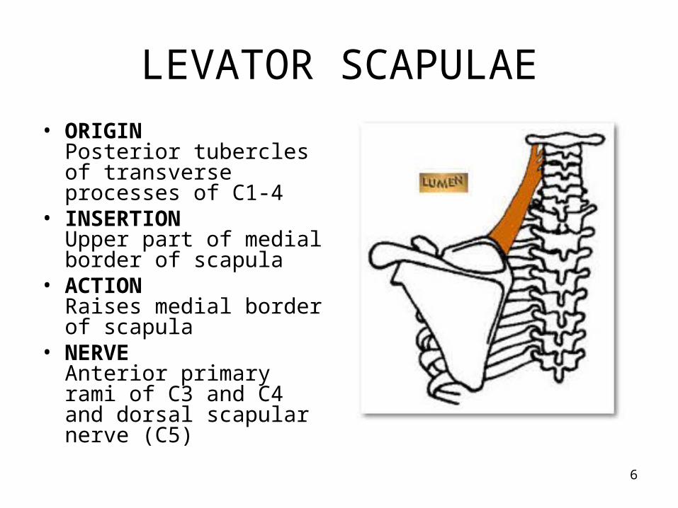
LEVATOR SCAPULAE
• ORIGINPosterior tubercles of transverse processes of C1-4
• INSERTIONUpper part of medial border of scapula
• ACTIONRaises medial border of scapula
• NERVEAnterior primary rami of C3 and C4 and dorsal scapular nerve (C5)

7
RHOMBOID MINOR
• ORIGINLower ligamentum nuchae, spines of C7 and T1
• INSERTIONOn posteromedial border of scapula at level of spine, below levator scapulae
• ACTIONRetracts scapula.
• NERVEDorsal scapular nerve (C5) (from root)

8
RHOMBOID MAJOR
• ORIGINSpines of T2-T5 and supraspinous ligaments
• INSERTIONLower half of posteromedial border of scapula, from angle to upper part of triangular area at base of scapular spine
• ACTIONRetracts scapula.
• NERVEDorsal scapular nerve (C5) (from root )

9
Revise

10
Identify these muscles

11
Muscles attached to scapula
The muscles which join the scapula to the humerus are:
• Deltoid• Supraspinatus• Infraspinatus• Teres minor • Teres major• Subscapularis

12
DELTOID
• ORIGIN
Lateral third of clavicle, acromion, spine of scapula to deltoid tubercle.(multipennate muscle)
• INSERTIONMiddle of lateral surface of humerus (deltoid tuberosity)
• ACTIONAbducts arm, anterior fibers flex and medial rotate, posterior fibers extend and lateral rotate
• NERVEAxillary nerve (C5, 6) (from posterior cord)
• IMPORTANCE Site of intramuscular injection.
13
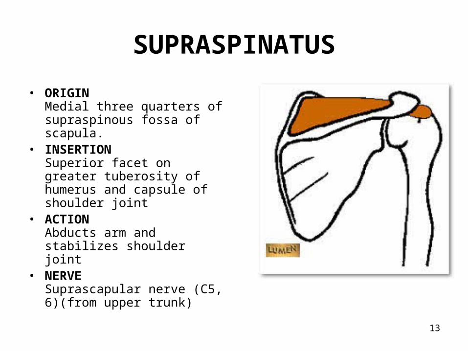
SUPRASPINATUS
• ORIGINMedial three quarters of supraspinous fossa of scapula.
• INSERTIONSuperior facet on greater tuberosity of humerus and capsule of shoulder joint
• ACTIONAbducts arm and stabilizes shoulder joint
• NERVESuprascapular nerve (C5, 6)(from upper trunk)
14
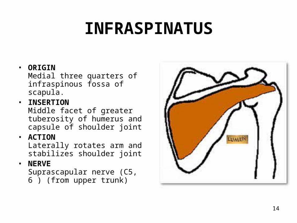
INFRASPINATUS
• ORIGIN
Medial three quarters of infraspinous fossa of scapula.
• INSERTIONMiddle facet of greater tuberosity of humerus and capsule of shoulder joint
• ACTIONLaterally rotates arm and stabilizes shoulder joint
• NERVESuprascapular nerve (C5, 6 ) (from upper trunk)

15
TERES MINOR
• ORIGIN
Middle third lateral border of scapula above teres major
• INSERTIONInferior facet of greater tuberosity of humerus (below infraspinatus) and capsule of shoulder joint
• ACTIONlaterally rotates arm and stabilizes shoulder joint
• NERVEAxillary nerve (C5, 6) (from posterior cord)

16
TERES MAJOR
• ORIGIN
Oval area on lateral side of inferior angle of scapula below teres minor.
• INSERTIONMedial lip of bicipital groove of humerus
• ACTIONMedially rotates and adducts arm. Stabilizes shoulder joint
• NERVELower subscapular nerve (C5, 6) (from posterior cord)
17
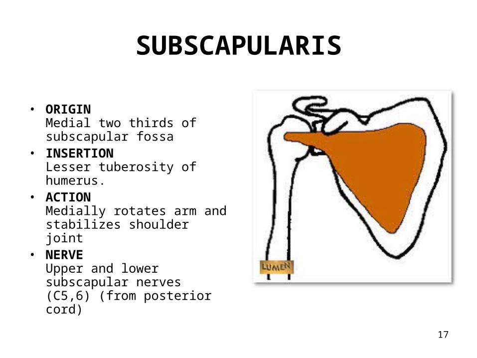
SUBSCAPULARIS
• ORIGIN
Medial two thirds of subscapular fossa
• INSERTIONLesser tuberosity of humerus.
• ACTIONMedially rotates arm and stabilizes shoulder joint
• NERVEUpper and lower subscapular nerves (C5,6) (from posterior cord)

18
Revise :

19
ROTATOR CUFF( Musculotendinous cuff )
The tendons of four muscles form the rotator cuff.
The muscles are:
1. Supraspinatus.
2. Infraspinatus.
3. Teres minor.
4. Subscapularis.

20

21
Intermuscular spaces
Quadrangular spaceTriangular spaces
Boundaries of quadrangular space:Above: teres minor & subscapularisBelow: teres majorMedially: long head of tricepsLaterally: surgical neck of humerus
Transmits: axillary nerve & posterior circumflex humeral vessels.

22

23

24
Triangular space ( upper ) bounded by:Above: teres minorBelow: teres majorLaterally: long head of tricepsTransmits: circumflex scapular vessels
Triangular space ( lower ) bounded by:Above: teres majorMedially: long head of tricepsLaterally: medial head of triceps Transmits: radial nerve and profunda brachii artery
25
Nerves of scapular region
1. Upper subscapular nerve – supplies subcapularis.
2. Lower subscapular nerve – supplies subscapularis and teres major.
3. Suprascapular nerve – supplies supraspinatus & infraspinatus.
4. Axillary nerve – supplies deltoid & teres minor.
26
Arteries of scapular region
The arteries seen are:
1. Transverse cervical artery.
2. Suprascapular artery.

27
Anastomosis around the scapula:
The scapular anastomosis connects subclavian artery and the axillary artery, forming an anastomosis around the scapula. It allows blood to flow past the joint regardless of the position of the arm.
It includes:•transverse cervical artery •transverse scapular artery •branches of subscapular artery •branches of thoracic aorta

28

29
Applied anatomy
• A rotator cuff injury is an injury to 1 or more of the 4 muscles in the shoulder. It can be associated with a fall.
• The type of injury can range from an inflammation of the muscle without any permanent damage, such as tendinitis, to a complete or partial tear of the muscle that might require surgery to fix it.