1 antioxidants to enhance fertility: role of enos and potential
TRANSCRIPT
1
Antioxidants to enhance fertility: Role of eNOS and Potential Benefits
Francesco Visioli, Tory M. Hagen
Laboratory of Functional Foods, IMDEA-Food, Madrid, Spain; Linus Pauling
Institute/Oregon State University, Corvallis, OR, USA.
Correspondence:
Francesco Visioli, PhD
IMDEA-Food
Calle Faraday 7
28049 Madrid
Spain
2
Abstract
The use of antioxidants is now often used as a pharmacological adjunct to limit
infertility. Indeed, the lay public rightly perceives oxidative stress and, thus,
antioxidant treatment as important modulators of infertility. While the direct effects of
antioxidant treatment on the quality of semen and oocytes are still under investigation,
a significant body of evidence points to loss of vascular tone as a root-cause of
erectile dysfunction and, possibly, alterations to female reproduction.
In this article, we will critically review the often neglected link between vascular
dysfunction and infertility. A particular emphasis will be on the potential use of
antioxidants to increase fertility and promote conception.
Keywords: antioxidants, infertility, vascular dysfunction, erectile dysfunction, pre-
eclampsia, reproductive health, nitric oxide
3
1. Introduction
Human reproduction is known to be a highly inefficient process where as much
as 50% of conceptions fail, and ~20% of clinical pregnancies end in spontaneous
abortion [1]. Both male and female reproductive dysfunction is thought contribute to
this high rate of failure, but few defined etiologies have been identified. Despite the
multifactorial nature of human infertility, there is a growing awareness that reactive
oxygen and nitrogen species (ROS/RNS) and associated oxidative damage may be
potent modulators of reproductive health [2,3]. Oxidative and nitrosative stress is
associated with both risk factors for infertility (e.g. smoking, diabetes, hypertension,
and aging), and directly in reproductive disorders as diverse as oocyte implantation,
endometriosis, and pre-eclampsia in women, and erectile dysfunction, sperm
damage and motility in men [2,4]. Because of this recognition, use of antioxidants
and other redox modulatory compounds as nutraceutical/pharmacological treatments
for many forms of infertility has soared as a result [3]. Numerous studies point to the
benefits of dietary antioxidants to limit oxidative stress, thereby potentially
maintaining the quality of semen and oocytes, and also lowering the risk for
endometriosis and other female reproductive disorders. However, clinical trials using
antioxidants as therapies for many reproductive disorders have yielded negative or
conflicting results as to their benefits on reproductive health [5-8]. While the direct
effects of antioxidant treatment are still under investigation, it is interesting to note
that many endpoints for both male and female infertility (e.g. erectile dysfunction in
males, and preeclampsia, oocyte implantation, and endometriosis in women) actually
involve vascular dysfunction, where antioxidants have provided significant benefits.
Herein we review the remarkable roles that vascular dysfunction plays as an
underlying risk for many aspects of infertility as well as potential dietary antioxidant
4
compounds that may improve vasomotor function, thereby limiting risk for infertility
and promote conception.
2. Vascular dysfunction in conditions leading to infertility
The vascular endothelium that lines the lumen of vessels are critically
important in maintaining vascular tone [9]. Endothelial cells are responsive to many
stimuli that either promote vasorelaxation (e.g. shear stress, nitric oxide,
prostacyclins, sphingosine 1-phosphate, acetylcholine, among others) or constriction
(angiotensin II, endothelin-1, thromboxane) [10,11]. Thus, any condition, like
oxidative stress, which impairs vasomotor function, could have profound adverse
consequences on reproductive health.
Endothelial-derived vasorelaxation is gradually lost with age [12].
Consequently, endothelial dysfunction is a primary complication of age-dependent
cardiovascular diseases, including hypertension, atherosclerosis, attendant coronary
artery disease, the metabolic syndrome, and is increasingly recognized as a hallmark
of congestive heart failure [13,14]. Impaired vasomotion is often observed in the
absence of severe atherosclerotic lesions and endothelial dysfunction often occurs
long before symptoms of coronary atherosclerosis become clinically apparent. It is
often at this early stage of cardiovascular disease where clinical studies link
endothelial impairment and infertility. For example, erectile dysfunction is a leading
indicator of CVD [15], and erectile dysfunction patients display significantly impaired
brachial artery flow-mediated dilation without additional cardiovascular risk factors
[16,17].
Aside from erectile dysfunction, endothelial dysfunction also appears to be a
significant factor in preeclampsia (pre-eclampsia), a hypertension syndrome

5
occurring in 3 to 5% of pregnant women that results in reduced placental perfusion
[18]. Pre-eclampsia is a primary event that significantly increases maternal and
neonatal morbidity and mortality with no current therapy other than placental delivery.
A number of studies now show a significantly reduced vasomotor function in pre-
eclampsia patients. For example, Aardema et al. used Doppler analysis to show that
the abnormal flow-mediated dilation of the uterine artery was strongly associated with
pregnancy complications [19,20]. Brodszki and coworkers also showed that uterine
artery flow was strongly impaired in pre-eclampsia with the lowest dilation evident in
women with bilateral uterine artery notches [21]. Interestingly and in keeping with
evidence for erectile dysfunction in men, no significant differences in carotid or aortic
vessel wall stiffness are often evident in women with pre-eclampsia versus healthy
controls. In summary, while pre-eclampsia is not a major cause of infertility,
endothelial dysfunction contributes to this important disorder of pregnancy and
should be adequately addressed.
From the available data, it is clear that infertility is multifactorial and should be
individually addressed based on careful anamnesis [22]. However, infertility in both
males and females appear to be linked by a profound loss in endothelial-dependent
vascular function.
3. Vascular tone is principally regulated by endothelial-derived nitric oxide
As briefly mentioned above, the vascular endothelium governs
vasoresponsiveness by physio-mechanical means, hormonal stimuli, and by
synthesizing and releasing factors that act on the vascular smooth muscle layer [9].
The principal vasodilatory agent is nitric oxide (NO), which induces relaxation by
stimulating cGMP production in vascular smooth muscle cells (VSMC). Often, loss of
6
vasoresponsiveness has been attributed to insufficient endothelial-derived NO as
exogenous NO donors (e.g. nitroprusside) restore adequate vasomotor function in
CVD patients, the elderly, and in animal models [23]. Nitric oxide is synthesized by
endothelial nitric oxide synthase (eNOS), one of a family of heme-containing,
homodimeric enzymes that catalyze the 5 electron oxidation of the guanidinium
moiety of L-arginine to NO and L-citrulline [24]. In endothelial cells, eNOS expression
and activity is tightly governed both at the transcriptional and post-translational levels,
by intracellular Ca+2 concentrations, and protein-protein interactions [25]. Additionally,
its subcellular distribution also markedly affects overall enzyme activity. eNOS exists
in at least two distinct subcellular locales in the endothelium: at caveolae of the
plasma membrane and in the Golgi/perinuclear region of the cell [26]. Plasma
membrane associated eNOS is considered to be more constitutively active and highly
sensitive to agonist-induced intracellular Ca+2 fluxes. Additionally, eNOS at the
plasma membrane is typically phosphorylated at Serine 1176 (rattus sequence) in an
Akt-driven manner, which makes this caveolae-associated enzyme markedly more
active in response to shear stress and other stimuli [27]. In contrast, Golgi-bound
eNOS is less phosphorylated and relatively insensitive to Ca+2-dependent activation;
the latter may be due to inaccessibility of calmodulin to eNOS in this cellular
compartment.
Besides subcellular locale, interactions of eNOS with effector proteins play a
key regulatory role. eNOS in association with caveolin-1, a major protein component
of endothelial caveolae, significantly inhibits eNOS activity as it prevents calmodulin
binding [26,28]. Conversely, active eNOS at the plasma membrane can be co-
immunoprecipitated with Hsp90 and kinases, especially Akt. Together, these
regulatory mechanisms tightly govern eNOS-derived NO availability.

7
4. Protein effectors and the phosphorylation state of eNOS strongly affect its
activity
Recent papers by Smith et al. [29,30] revealed that post-translational
dysregulation of eNOS may play a remarkable role in endothelial dysfunction,
especially in the elderly. These authors showed that aged rats displayed significant
impairment in endothelial-derived vasomotor activity, but total eNOS content did not
change with age. However, aging caused eNOS to disassociate from its activating
proteins (Hsp90 and Akt) while increasing the levels of eNOS associating with
caveolin-1, a protein known to inhibit eNOS activity. Along with this altered
subcellular redistribution, these authors showed that the phosphorylation state of
eNOS was significantly and persistently altered in aging rat aortic endothelia, which is
consistent with its inactivation [29]. Loss of eNOS phosphorylation status was found
to stem from chronically elevated sphingomyelinases (SMases) and resultant
ceramide-driven protein phosphatase 2A (PP2A) activity, which specifically
dephosphorylated S1176. Thus, age-related (and, likely, disease-related) changes in
eNOS phosphorylation and/or its cellular association with other protein effectors may
significantly alter eNOS activity and vascular function.
5. Redox-dependent modifications that affect eNOS activity or NO
bioavailability
Apart from the aforementioned post-translational regulatory mechanisms
governing eNOS function, redox changes in the endothelium can substantially modify
both enzyme activity as well as the enzyme’s product, NO, thereby adversely
affecting vasomotion. For the latter, eNOS synthetic activity is dependent on
8
maintaining tetrahydrobiopterin (BH4) in a highly reduced state. When the
tetrahydrobiopterin/biopterin ratio is high eNOS readily produces NO; however, when
the BH4 redox ratio declines, the internal electron transport chain of eNOS becomes
uncoupled, which actually generates superoxide instead of NO. Thus, in a pro-
oxidative milieu, eNOS may not only become a target of oxidants, but also may
exacerbate oxidative stress and endothelial dysfunction. In keeping with this concept,
incubating endothelial cells with oxidized LDL (oxLDL) constitutively downregulates
eNOS activity and causes loss of eNOS in the plasma membrane [31]. Thus, a pro-
oxidant imbalance in the vasculature may affect both eNOS activity, thereby
contributing to lower synthesis of endothelial-derived NO.
In addition to eNOS uncoupling, reactive oxygen species (ROS) may limit NO
bioavailability by converting it into non-vasoactive reactive nitrogen species (RNS)
[32]. Indeed, production of ROS in excess to the antioxidant defense mechanisms
plays an important role in endothelial dysfunction. Thus far, the three most widely
studied sources of vascular ROS are catalyzed by xanthine oxidoreductase,
NADH/NADPH oxidase, and NO synthase (NOS) [33]. For the vascular endothelium,
substantial evidence shows that NADH/NADPH oxidase is quantitatively the
predominant producer of superoxide, a relatively innocuous but ubiquitous type of
ROS (Fig 1) (see below for the role of NADPH oxidase in spermatozoa). However,
despite its relatively benign nature, superoxide readily reacts with NO at rates
approaching diffusion limitations to produce peroxynitrite (OONO-), a powerful
reactive nitrogen species which would not only limit NO bioactivity, but increase
endothelial oxidative stress and damage [23,34]. Chronic limiting levels of low
molecular weight antioxidants, which would promote sustained increases in
superoxide would be expected to exacerbate peroxynitrite formation. Thus,
9
conversion of NO to deleterious reactive nitrogen species represents a plausible
means for limiting NO bioavailability and its contribution to the altered vessel function
in infertility.
6. Vascular dysfunction: potential therapeutic agents
Because oxidative stress appears to be an underlying factor in endothelial-
dependent decline in vasoactivity, numerous studies have attempted to ameliorate
lower endothelial redox state, and hence improve vascular responsiveness through
increasing antioxidant levels. We will now briefly review many of the antioxidants
used for these studies and their potential benefits both to vascular health and
infertility.
6.1 Vitamin C
Vasomotor dysfunction in cardiovascular patients can be reversed by vitamin C
treatment, which appears to facilitate endothelial-derived NO (EDNO) bioavailability
[35-37]. Ascorbic acid stimulates EDNO synthesis through increasing intracellular
levels of BH4 and its reduced redox state, thereby maintening eNOS coupling [38,39].
Data from Visioli et al. is instructive as to the effectiveness of ascorbate to maintain
eNOS synthetic activity [40]. This study showed that ascorbic acid did not work
through maintaining glutathione (GSH) levels or its reduced redox state in HAEC,
indicating that it is unlikely that ascorbic acid increased EDNO synthesis simply by
markedly improving the intracellular redox environment. Rather, maintenance or
improvement of intracellular BH4 levels (by reducing dihydro- or trihydrobiopterin to
BH4) appeared to be a more likely mechanism. These results were in keeping with
previously published studies showing ascorbic acid can maintain biopterin in a highly
10
reduced state [38,41]. As such, ascorbic acid (500 mg/d) is considered a useful
addition to the pharmacological therapy of hypertension [42] and might be explored
as a potential adjunct to the treatment of impaired vasomotion and associated
erectile dysfunction.
6.2 Glutathione (GSH) analogs and GSH synthesis inducers
Glutathione (GSH) is a major low molecular weight cellular antioxidant that directly
terminates ROS, or acts as a substrate for GSH-dependent antioxidant enzymes (e.g.
GSH-dependent peroxidases, glutaredoxin, and GSH-S transferases) [43]. Even
though GSH is not readily taken up by endothelial cells directly, a number of
compounds increase intracellular levels of GSH either by increasing cysteine
availability (e.g. N-acetyl-cysteine), which is the rate-limiting substrate for GSH
synthesis, or by inducing expression of GSH synthesis genes (e.g. lipoic acid,
sulforaphane, and other Phase II detoxication gene inducers) [44-46]. As mentioned,
GSH does not appear to directly maintain biopterin in a highly reduced and active
state, but does markedly improve EDNO bioavailability. One purported mechanism
for this increased availability may be through formation of GSH-NO conjugates, which
can enhance VSMC cGMP production and vasorelaxation [47,48].
Additionally, endothelial GSH levels may strongly influence eNOS activity by
maintaining phosphorylation status, especially at the S1176 residue. As previously
alluded, oxidative stress induced by pro-inflammatory cytokines (TNFα) [49], redox
cycling agents (doxorubicin), or by ROS [10] lowers GSH levels and activates PP2A
via induction of sphingomyelinases and concomitant increases in ceramide levels
[50-52]. As PP2A specifically dephosphorylates the S1176 site of eNOS, oxidative
stress may thus lead to inactivation of the enzyme. In this regard, the importance of
11
maintaining cellular GSH status is exemplified in in vitro studies where pretreating
MCF7 cells with GSH, GSH-methylester, or N-acetylcysteine prior to TNFα prevented
cellular GSH loss and also limited sphingomyelinase-mediated ceramide generation
[53]. We followed up on this indirect evidence and showed that by improving tissue
and cellular GSH levels, age-associated impaired vascular compliance could be
reversed and eNOS phosphorylation state maintained [44,54-58]. This reversal was
achieved by using (R)-α-lipoic acid (LA), a dithiol compound that is readily absorbed
from the diet and increases cellular GSH by increasing the expression of GSH
synthesis genes. Specifically, aortic EDNO-dependent vasomotor function was
markedly improved 12 hrs following intraperitoneal administration of LA (40 mg/kg),
and this improvement was achieved through enhanced eNOS and Akt
phosphorylation [59]. Thus, pharmacological agents that maintain or improve
endothelial GSH status may markedly improve EDNO bioavailability and vascular
tone through preserving eNOS phosphorylation and activation. In particular, LA
appears to be a promising pharmacological agent for this purpose with little clinical
side-effects [60] as it would induce a long-term elevation in GSH synthetic capacity
rather than an acute, limited effect supplied by compounds that transiently raise
cysteine availability. While the use of LA in the treatment of vascular dysfunction,
specifically related to conditions leading to infertility, requires ad-hoc investigations,
the vasomodulating properties of LA provide solid bases for future clinical trials.
In summary, there appears to be a link between inflammation, oxidative stress,
and generation of ceramide, which can be governed by thiols, namely GSH, status.
Hence, modulation of GSH status may be employed to reverse the age- or disease-
related decline in cellular GSH.
12
6.3 Polyphenols
Polyphenols are being actively investigated as vasomodulators. In particular,
administration of flavonoids, especially from tea and cocoa, ameliorates endothelial
dysfunction and restores proper vasomotion [41]. This is likely as a consequence of
increased endothelial NO synthase (eNOS) activity. The association between
flavonoid intake and maintenance of vascular tone is strongest in the Mediterranean
area, where the traditional diet is rich in plant-based foods and in wild vegetables [61].
It is noteworthy that, based on scientific evidence and targeted marketing, several
polyphenol-containing supplements are being sold to infertile men and women;
hence, the need to clarify, in vivo, the role of these molecules in ameliorating fertility.
Diets rich in plant foods are associated with better vascular compliance [61].
As an example, recent studies using isolated rat vessels showed that Cynara
cardunculus (wild artichoke) exerts vasorelaxant effects. In particular, feeding studies
- using doses of polyphenols that are compatible with human consumption -
confirmed that Cynara c. can restore proper vascular function in aged Fisher 344 x
BN rats fed the extract (10 mg polyphenols/Kg) for five days [62]. These data confirm
in vivo the results that were observed in vitro [63] and thus supply the basis for
formulating nutraceuticals aimed at maintaining vascular motility, especially in the
elderly. Also, this adds further experimental evidence to the body of basic research
supporting the epidemiological evidence of a lower CHD incidence in the
Mediterranean area. In particular, these data suggest that maintenance of correct
vasomotion by bioactive components from plants in various Mediterranean regions
(Lucania - Southern Italy, Crete, Segura Valley – Southern Spain [61]) might
contribute to its cardioprotective properties. The precise mechanism(s) of action
responsible for the enhancement of NO production are still elusive. However, in vitro

13
data exclude any effect on eNOS levels [63] and are in agreement with other data
that suggest how water-soluble antioxidants and thiol agents maintain an intracellular
reduced environment and preserve cofactors such as BH4, hence preventing eNOS
uncoupling [33] and facilitating its activity [64]. Finally, facilitated hydroxylation of
eNOS, which is the first step in NO production by this enzyme [9], might also explain
the potentiating activities of wild plant extracts, which are rich in ortho-diphenols (as
an example, we have identified luteolin-7-glicoside, apigenin, rosmaric acid, and
ursolic acid in wild artichoke [63]).
Other polyphenols that are attracting considerable research are those
contained in cocoa and in tea.
Indeed, cocoa and chocolate products have a much higher content – on a per-
weight basis - of polyphenols and total antioxidant activity than other polyphenol-
containing food and beverages such as red wine, green tea and black tea [65]. There
are interventional studies with cocoa in humans, that demonstrated significant
improvements of arterial dilatation, very likely as consequence of cocoa polyphenols’
ability to enhance the bioactivity of NO [66]. At the cellular level, the use of platelets
as a tool to clarify the role of cocoa’s polyphenols on NO, Pignatelli et al. observed
that polyphenols are able to both reduce the production of superoxide anion and to
increase platelet NO; an inverse correlation between these two effects was also
observed [67]. Mechanistically, these effects have been attributed to the ability of
polyphenols to inhibit the activation of NADPH oxidase [67,68]. The same group
underlined the synergism among the various cocoa polyphenols in exerting an
antioxidant effect [68]. From a potential therapy viewpoint, these findings shed some
light on the antioxidant effect of polyphenol-rich beverages (including those
containing cocoa) because several studies documented blood concentrations of
14
single polyphenols < 1 µM, i.e. much lower than those necessary to exert an
antioxidant effect in vitro. In synthesis, a mixture of polyphenols is able to reduce
superoxide anion and to increase NO generation, in turn suggesting that in vivo a
combination of more than one polyphenols (such as that present in cocoa) is likely
responsible for the beneficial effect on arterial dilatation observed with polyphenol-
rich foods.
More recently, by using noninvasive ultrasound measurements of flow
mediated vasodilation (FMD), Hermann et al. [69] showed that dark chocolate, but
not white chocolate improves endothelial function and platelet function in young
healthy smokers, a population known to have impaired endothelial function and
platelet hyper-reactivity. Smoking is also a risk factor for erectile dysfunction [70].
Flow-mediated dilation by high-sensitive ultrasonography of the brachial artery, shear
stress-dependent platelet adhesion, and total antioxidant status were assessed in 25
persons before and after ingestion of 40 gr of either dark (74% cocoa) or white
chocolate (4% cocoa). Two hours after intake of dark chocolate, FMD (and plasma
total antioxidant capacity) improved significantly. The beneficial effect of dark
chocolate on FMD lasted for eight hours. On the other hand, white chocolate
ingestion did not have any effect neither on FMD nor on platelet function.
The other beverage that is currently being investigated for its vasomodulating
activities is tea [65]. Starting from the landmark experiments of Duffy et al. [71], the
role of tea flavonoids – in particular epigallocatechin gallate - in maintaining proper
vasomotion are being clarified. In addition to their vasomodulating activities, tea
catechins exert other biological activities that might be relevant to fertility/infertility
and their associated risk factors. Among such activities, there are those on the
prostate [72] and those on plasma lipids. The latter suggest a salubrious role of
15
catechins in limiting the progression of the metabolic syndrome [73], which brings
about vascular alterations that have being associated with erectile dysfunction [74].
In synthesis, regular tea drinking is associated with improvements of vascular
reactivity and plasma lipid profile, which might ameliorate erectile function and, in turn,
fertility.
Summing up, polyphenolic compounds are a promising class of antioxidant
nutrients, which has prospects to limiting cardiovascular oxidative stress and thereby
limiting risks for certain forms of infertility. While it is still controversial as to the
bioavailability of flavonoids, accruing evidence points to sufficient absorption to affect
endothelial antioxidant profiles, especially in conditions that limit endogenous
antioxidant defenses. In this regard, cocoa and chocolate polyphenols may have high
potential to lower oxidant-induced infertility because of their high content, palatability,
and their aforementioned effectiveness in improving flow-mediated vascular dilation.
Thus, clinical studies using polyphenol compounds, either specific flavonoids or foods
high in polyphenol content, are warranted if we want to prove their benefits to
improve conditions such as erectile dysfunction, pre-eclampsia, and potentially sperm
motility.
In synthesis, we propose EDNO as a major target to treat infertility, given the
role that a correct vascular response plays both in men and women, via ensuring
erectile and tissutal functions.
7. Antioxidant uses in infertility outside of that associated with vascular
dysfunction: focus on semen
Spermatozoa are very rich in polyunsaturated fatty acids, namely
docosahexaenoic acid (DHA). As such, they are susceptible to oxidation, even
16
though the human semen is rich in vitamin C and omega 3 fatty acids exert cellular
anti- rather than pro-oxidant actions [75,76]. Indeed, there is evidence of decreased
sperm viability associated with increased lipid peroxidation; this phenomenon
becomes more relevant with age. It is noteworthy that – at least in developed
countries – more and more men postpone fatherhood, increasing chances of having
oxidatively-damaged semen. It is also noteworthy that spermatozoa per se produce
elevated fluxes of ROS, which need to be counterbalanced by an appropriate
antioxidant network to avoid damage to macromolecules, namely lipids, proteins, and
nucleic acids [77]. The discovery of a NADPH oxidase in sperm [78] opens a
potential field of research aimed at reducing ROS generation by sperm itself [79]. In
this respect, provision of DHA (an inhibitor of NADPH oxidase in endothelial cells [76]
might prove beneficial both because it provides essential fatty acids indispensable for
sperm survival and because it would lower production of ROS once sperms are
produced. Accordingly, it is of note that infertile men had lower concentrations of
omega-3 fatty acids in spermatozoa than fertile men [80].
Among the antioxidant machinery that is currently being investigated in semen
as potential target for therapy, glutathione peroxidases (GPX) are attracting much
attention [81]. Indeed, several GPX have been found to be present on and around
epididymal transiting sperm cells and the precise localization of the various GPXs in,
on and around sperm cells argues in favor of specific roles for these enzymes [82]. In
particular, GPXs could function as hydrogen peroxide sensors to regulate its
concentration and to find a proper balance between the physiological actions of ROS
on sperm cells and their detrimental activities on cell physiology [82]. It must be
underlined that the mere antioxidant role played by GPX in mature sperm has been
questioned [83] and it has been proposed that mature spermatozoa depend on
17
GPX as a structural protein, to maintain the proper integrity and stiffness of the
midpiece [83]. In synthesis, the nature and precise role(s) of GPX in maintaining
“healthful” spermatozoa is still to be fully clarified [84], but the field is moving rapidly
and in the near future some opportunities for therapy, e.g. with selenium
supplementation [85], might arise.
The use of vitamin C has also been advocated as a tool to ameliorate semen
quality [86]. The rationale is that some investigators found lower ascorbate
concentrations in samples from infertile men, as compared with fertile individuals [87].
To the best of our knowledge, there is a limited number of trials that tested the effects
of vitamin C supplementation on semen quality [88,89]. Hence, while adequate
consumption of vitamin C are recommended and exert positive actions on the
vascular system, its direct effect on spermatozoa viability are suggestive, but as yet
to be fully established. It is also noteworthy that the addition of vitamin C to semen
decreases freezing-thawing damage induced by cryopreservation [90].
Other antioxidant molecules or plants extracts that have been proposed to
increase semen quality via their antioxidant actions include pycnogenol® [91] and
vitamin E [92]. Both await confirmation from randomized and double blind trials.
The final compound that bears discussion is lycopene. In fact, several lines of
evidence (mostly focused on chemoprevention) indicate that lycopene plays a role in
maintaining prostate function and health, mostly as an anti-inflammatory agent [93].
However, conclusive evidence that it works solely through this means is inconclusive
[94]. Based on the purported activities of lycopene on the prostate, some researchers
are starting to investigate its role on quality of semen [95]. Indeed, lycopene
possesses protective properties with respect to DNA damage and the rationale
18
behind testing its activities on spermatozoa is sound. However, the evidence is once
again very limited and human trials are needed to confirm or disprove this hypothesis.
8. Concluding remarks
As briefly reviewed herein, many forms of infertility are associated with
oxidative stress, and many types (e.g. erectile dysfunction, pre-eclampsia) are also
caused by or exacerbated from significant impairment of vascular endothelial function.
Thus, there is a compelling theoretical rationale to limit oxidative stress associated
with these conditions by the use of antioxidant therapies or pharmacological agents,
which induce endogenous antioxidant defenses. While the promise for antioxidants to
limit conditions of infertility is great, most evidence to date showing therapeutic
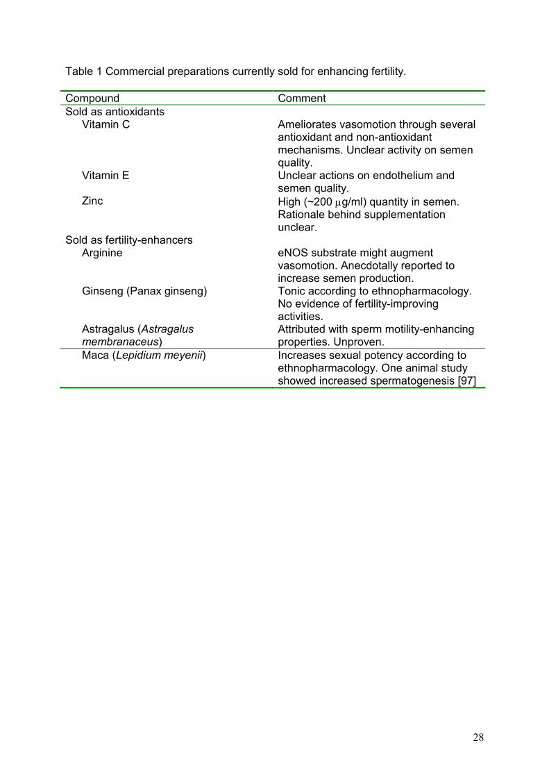
efficacy of antioxidants are from experimental animal models or in vitro studies. Table
1 reports the most common commercial preparations (antioxidants and others) that
are being sold accompanied by various claims to treat infertility. Where small-scale
clinical trials have been performed [96], the outcomes have largely proven
inconclusive. While this may seemingly limit enthusiasm for antioxidant therapies for
infertility, there is an equally good argument for the design and implementation of
clinical trials using specific antioxidants to treat underlying vascular dysfunction
associated with many types of infertility. In this regard, “non-traditional” antioxidants,
such as lipoic acid or polyphenols, may hold significant promise. These compounds
may not only directly improve EDNO, but also have additional long-term benefits to
enhance antioxidant defenses, thereby providing potent means of lowering vascular
dysfunction and attendant infertility. While current evidence is still controversial,
future trials will help clarify this issue and might provide scientific bases for the use of
antioxidants in the treatment of the various facets of infertility.
19
Acknowledgments
This paper stems from a lecture given at the “1st World Congress on Oxidative
Stress & Infertility: Recent Advances and Controversies”, where part of the data
discussed here were presented.
References
(1) Gupta S, Agarwal A, Banerjee J, Alvarez JG. The role of oxidative stress in
spontaneous abortion and recurrent pregnancy loss: a systematic review. Obstet Gynecol Surv 2007;62:335-47.
(2) Desai N, Sabanegh E Jr, Kim T, Agarwal A. Free radical theory of aging: implications in male infertility. Urology 2010;75:14-9.
(3) Lanzafame FM, La Vignera S, Vicari E, Calogero AE. Oxidative stress and medical antioxidant treatment in male infertility. Reprod Biomed Online 2009;19:638-59.
(4) Sikka SC. Role of oxidative stress and antioxidants in andrology and assisted reproductive technology. J Androl 2004;25:5-18.
(5) Zhang Q, Radisavljevic ZM, Siroky MB, Azadzoi KM. Dietary antioxidants improve arteriogenic erectile dysfunction. Int J Androl 2010.
(6) Polyzos NP, Mauri D, Tsappi M, Tzioras S, Kamposioras K, Cortinovis I et al. Combined vitamin C and E supplementation during pregnancy for preeclampsia prevention: a systematic review. Obstet Gynecol Surv 2007;62:202-6.
(7) Fiore G, Capasso A. Effects of vitamin E and C on placental oxidative stress: an in vitro evidence for the potential therapeutic or prophylactic treatment of preeclampsia. Med Chem 2008;4:526-30.
(8) Rumbold A, Duley L, Crowther CA, Haslam RR. Antioxidants for preventing pre-eclampsia. Cochrane Database Syst Rev 2008;CD004227.
(9) Brune B. Nitric oxide: a short lived molecule stays alive. Pharmacol Res 2010;61:265-8.
(10) Pavoine C, Pecker F. Sphingomyelinases: their regulation and roles in cardiovascular pathophysiology. Cardiovasc Res 2009;82:175-83.
20
(11) Feng J, Damrauer SM, Lee M, Sellke FW, Ferran C, Abid MR. Endothelium-dependent coronary vasodilatation requires NADPH oxidase-derived reactive oxygen species. Arterioscler Thromb Vasc Biol 2010;30:1703-10.
(12) Taddei S, Virdis A, Mattei P, Ghiadoni L, Gennari A, Fasolo CB et al. Aging and endothelial function in normotensive subjects and patients with essential hypertension. Circulation 1995;91:1981-7.
(13) Kubo SH, Rector TS, Bank AJ, Williams RE, Heifetz SM. Endothelium-dependent vasodilation is attenuated in patients with heart failure. Circulation 1991;84:1589-96.
(14) Drexler H, Hayoz D, Munzel T, Hornig B, Just H, Brunner HR et al. Endothelial function in chronic congestive heart failure. Am J Cardiol 1992;69:1596-601.
(15) Kaiser DR, Billups K, Mason C, Wetterling R, Lundberg JL, Bank AJ. Impaired brachial artery endothelium-dependent and -independent vasodilation in men with erectile dysfunction and no other clinical cardiovascular disease. J Am Coll Cardiol 2004;43:179-84.
(16) Guay AT. ED2: erectile dysfunction = endothelial dysfunction. Endocrinol Metab Clin North Am 2007;36:453-63.
(17) Palumbo PJ. Metabolic risk factors, endothelial dysfunction, and erectile dysfunction in men with diabetes. Am J Med Sci 2007;334:466-80.
(18) Wallis AB, Saftlas AF, Hsia J, Atrash HK. Secular trends in the rates of preeclampsia, eclampsia, and gestational hypertension, United States, 1987-2004. Am J Hypertens 2008;21:521-6.
(19) Aardema MW, Oosterhof H, Timmer A, van R, I, Aarnoudse JG. Uterine artery Doppler flow and uteroplacental vascular pathology in normal pregnancies and pregnancies complicated by pre-eclampsia and small for gestational age fetuses. Placenta 2001;22:405-11.
(20) Aardema MW, Saro MC, Lander M, De Wolf BT, Oosterhof H, Aarnoudse JG. Second trimester Doppler ultrasound screening of the uterine arteries differentiates between subsequent normal and poor outcomes of hypertensive pregnancy: two different pathophysiological entities? Clin Sci (Lond) 2004;106:377-82.
(21) Brodszki J, Lanne T, Laurini R, Strevens H, Wide-Swensson D, Marsal K. Vascular mechanical properties and endothelial function in pre-eclampsia with special reference to bilateral uterine artery notch. Acta Obstet Gynecol Scand 2008;87:154-62.
(22) Aitken RJ, De Iuliis GN, Finnie JM, Hedges A, McLachlan RI. Analysis of the relationships between oxidative stress, DNA damage and sperm vitality in a patient population: development of diagnostic criteria. Hum Reprod 2010;25:2415-26.
21
(23) van der LB, Labugger R, Skepper JN, Bachschmid M, Kilo J, Powell JM et al. Enhanced peroxynitrite formation is associated with vascular aging. J Exp Med 2000;192:1731-44.
(24) Alderton WK, Cooper CE, Knowles RG. Nitric oxide synthases: structure, function and inhibition. Biochem J 2001;357:593-615.
(25) Kone BC. Protein-protein interactions controlling nitric oxide synthases. Acta Physiol Scand 2000;168:27-31.
(26) Minshall RD, Sessa WC, Stan RV, Anderson RG, Malik AB. Caveolin regulation of endothelial function. Am J Physiol Lung Cell Mol Physiol 2003;285:L1179-L1183.
(27) Fulton D, Gratton JP, McCabe TJ, Fontana J, Fujio Y, Walsh K et al. Regulation of endothelium-derived nitric oxide production by the protein kinase Akt. Nature 1999;399:597-601.
(28) Fulton D, Babbitt R, Zoellner S, Fontana J, Acevedo L, McCabe TJ et al. Targeting of endothelial nitric-oxide synthase to the cytoplasmic face of the Golgi complex or plasma membrane regulates Akt- versus calcium-dependent mechanisms for nitric oxide release. J Biol Chem 2004;279:30349-57.
(29) Smith AR, Visioli F, Hagen TM. Plasma membrane-associated endothelial nitric oxide synthase and activity in aging rat aortic vascular endothelia markedly decline with age. Arch Biochem Biophys 2006;454:100-5.
(30) Smith AR, Visioli F, Frei B, Hagen TM. Age-related changes in endothelial nitric oxide synthase phosphorylation and nitric oxide dependent vasodilation: evidence for a novel mechanism involving sphingomyelinase and ceramide-activated phosphatase 2A. Aging Cell 2006;5:391-400.
(31) Zhu Y, Liao HL, Niu XL, Yuan Y, Lin T, Verna L et al. Low density lipoprotein induces eNOS translocation to membrane caveolae: the role of RhoA activation and stress fiber formation. Biochim Biophys Acta 2003;1635:117-26.
(32) Ullrich V, Bachschmid M. Superoxide as a messenger of endothelial function. Biochem Biophys Res Commun 2000;278:1-8.
(33) Cai H, Harrison DG. Endothelial dysfunction in cardiovascular diseases: the role of oxidant stress. Circ Res 2000;87:840-4.
(34) Drew B, Leeuwenburgh C. Aging and the role of reactive nitrogen species. Ann N Y Acad Sci 2002;959:66-81.
(35) Levine GN, Frei B, Koulouris SN, Gerhard MD, Keaney JF, Jr., Vita JA. Ascorbic acid reverses endothelial vasomotor dysfunction in patients with coronary artery disease. Circulation 1996;93:1107-13.
(36) Taddei S, Virdis A, Ghiadoni L, Magagna A, Salvetti A. Vitamin C improves endothelium-dependent vasodilation by restoring nitric oxide activity in essential hypertension. Circulation 1998;97:2222-9.
22
(37) Gokce N, Keaney JF, Jr., Frei B, Holbrook M, Olesiak M, Zachariah BJ et al. Long-term ascorbic acid administration reverses endothelial vasomotor dysfunction in patients with coronary artery disease. Circulation 1999;99:3234-40.
(38) Huang A, Vita JA, Venema RC, Keaney JF, Jr. Ascorbic acid enhances endothelial nitric-oxide synthase activity by increasing intracellular tetrahydrobiopterin. J Biol Chem 2000;275:17399-406.
(39) Heller R, Munscher-Paulig F, Grabner R, Till U. L-Ascorbic acid potentiates nitric oxide synthesis in endothelial cells. J Biol Chem 1999;274:8254-60.
(40) Visioli F, Smith A, Zhang W, Keaney JF, Jr., Hagen T, Frei B. Lipoic acid and vitamin C potentiate nitric oxide synthesis in human aortic endothelial cells independently of cellular glutathione status. Redox Rep 2002;7:223-7.
(41) Carr A, Frei B. The role of natural antioxidants in preserving the biological activity of endothelium-derived nitric oxide. Free Radic Biol Med 2000;28:1806-14.
(42) Duffy SJ, Gokce N, Holbrook M, Huang A, Frei B, Keaney JF, Jr. et al. Treatment of hypertension with ascorbic acid. Lancet 1999;354:2048-9.
(43) Forman HJ, Zhang H, Rinna A. Glutathione: overview of its protective roles, measurement, and biosynthesis. Mol Aspects Med 2009;30:1-12.
(44) Suh JH, Shenvi SV, Dixon BM, Liu H, Jaiswal AK, Liu RM et al. Decline in transcriptional activity of Nrf2 causes age-related loss of glutathione synthesis, which is reversible with lipoic acid. Proc Natl Acad Sci U S A 2004;101:3381-6.
(45) Lavoie S, Chen Y, Dalton TP, Gysin R, Cuenod M, Steullet P et al. Curcumin, quercetin, and tBHQ modulate glutathione levels in astrocytes and neurons: importance of the glutamate cysteine ligase modifier subunit. J Neurochem 2009;108:1410-22.
(46) Shenvi SV, Smith EJ, Hagen TM. Transcriptional regulation of rat gamma-glutamate cysteine ligase catalytic subunit gene is mediated through a distal antioxidant response element. Pharmacol Res 2009;60:229-36.
(47) Feelisch M, Kotsonis P, Siebe J, Clement B, Schmidt HH. The soluble guanylyl cyclase inhibitor 1H-[1,2,4]oxadiazolo[4,3,-a] quinoxalin-1-one is a nonselective heme protein inhibitor of nitric oxide synthase and other cytochrome P-450 enzymes involved in nitric oxide donor bioactivation. Mol Pharmacol 1999;56:243-53.
(48) Ceron PI, Cremonez DC, Bendhack LM, Tedesco AC. The relaxation induced by S-nitroso-glutathione and S-nitroso-N-acetylcysteine in rat aorta is not related to nitric oxide production. J Pharmacol Exp Ther 2001;298:686-94.
(49) Cornell TT, Hinkovska-Galcheva V, Sun L, Cai Q, Hershenson MB, Vanway S et al. Ceramide-dependent PP2A regulation of TNFalpha-induced IL-8
23
production in respiratory epithelial cells. Am J Physiol Lung Cell Mol Physiol 2009;296:L849-L856.
(50) Rutkute K, Asmis RH, Nikolova-Karakashian MN. Regulation of neutral sphingomyelinase-2 by GSH: a new insight to the role of oxidative stress in aging-associated inflammation. J Lipid Res 2007;48:2443-52.
(51) Martin SF, Sawai H, Villalba JM, Hannun YA. Redox regulation of neutral sphingomyelinase-1 activity in HEK293 cells through a GSH-dependent mechanism. Arch Biochem Biophys 2007;459:295-300.
(52) Marchesini N, Hannun YA. Acid and neutral sphingomyelinases: roles and mechanisms of regulation. Biochem Cell Biol 2004;82:27-44.
(53) Liu B, Andrieu-Abadie N, Levade T, Zhang P, Obeid LM, Hannun YA. Glutathione regulation of neutral sphingomyelinase in tumor necrosis factor-alpha-induced cell death. J Biol Chem 1998;273:11313-20.
(54) Sen CK, Roy S, Han D, Packer L. Regulation of cellular thiols in human lymphocytes by alpha-lipoic acid: a flow cytometric analysis. Free Radic Biol Med 1997;22:1241-57.
(55) Packer L. alpha-Lipoic acid: a metabolic antioxidant which regulates NF-kappa B signal transduction and protects against oxidative injury. Drug Metab Rev 1998;30:245-75.
(56) Hagen TM, Vinarsky V, Wehr CM, Ames BN. (R)-alpha-lipoic acid reverses the age-associated increase in susceptibility of hepatocytes to tert-butylhydroperoxide both in vitro and in vivo. Antioxid Redox Signal 2000;2:473-83.
(57) Bharat S, Cochran BC, Hsu M, Liu J, Ames BN, Andersen JK. Pre-treatment with R-lipoic acid alleviates the effects of GSH depletion in PC12 cells: implications for Parkinson's disease therapy. Neurotoxicology 2002;23:479-86.
(58) Moini H, Tirosh O, Park YC, Cho KJ, Packer L. R-alpha-lipoic acid action on cell redox status, the insulin receptor, and glucose uptake in 3T3-L1 adipocytes. Arch Biochem Biophys 2002;397:384-91.
(59) Smith AR, Visioli F, Frei B, Hagen TM. Lipoic acid significantly restores, in rats, the age-related decline in vasomotion. Br J Pharmacol 2008;153:1615-22.
(60) Ziegler D, Reljanovic M, Mehnert H, Gries FA. Alpha-lipoic acid in the treatment of diabetic polyneuropathy in Germany: current evidence from clinical trials. Exp Clin Endocrinol Diabetes 1999;107:421-30.
(61) The Local Food Nutraceutical Consortium. Understanding local Mediterranean diets: a multidisciplinary pharmacological and ethnobotanical approach. Pharmacol Res 2005;52:353-66.
(62) Rossoni G, Grande S, Galli C, Visioli F. Wild artichoke prevents the age-associated loss of vasomotor function. J Agric Food Chem 2005;53:10291-6.
24
(63) Grande S, Bogani P, de Saizieu A, Schueler G, Galli C, Visioli F. Vasomodulating potential of mediterranean wild plant extracts. J Agric Food Chem 2004;52:5021-6.
(64) Heller R, Unbehaun A, Schellenberg B, Mayer B, Werner-Felmayer G, Werner ER. L-ascorbic acid potentiates endothelial nitric oxide synthesis via a chemical stabilization of tetrahydrobiopterin. J Biol Chem 2001;276:40-7.
(65) Deka A, Vita JA. Tea and cardiovascular disease. Pharmacol Res 2011;64:136-45.
(66) Heiss C, Kleinbongard P, Dejam A, Perre S, Schroeter H, Sies H et al. Acute consumption of flavanol-rich cocoa and the reversal of endothelial dysfunction in smokers. J Am Coll Cardiol 2005;46:1276-83.
(67) Pignatelli P, Di Santo S, Buchetti B, Sanguigni V, Brunelli A, Violi F. Polyphenols enhance platelet nitric oxide by inhibiting protein kinase C-dependent NADPH oxidase activation: effect on platelet recruitment. FASEB J 2006;20:1082-9.
(68) Pignatelli P, Ghiselli A, Buchetti B, Carnevale R, Natella F, Germano G et al. Polyphenols synergistically inhibit oxidative stress in subjects given red and white wine. Atherosclerosis 2006;188:77-83.
(69) Hermann F, Spieker LE, Ruschitzka F, Sudano I, Hermann M, Binggeli C et al. Dark chocolate improves endothelial and platelet function. Heart 2006;92:119-20.
(70) Heidelbaugh JJ. Management of erectile dysfunction. Am Fam Physician 2010;81:305-12.
(71) Duffy SJ, Keaney JF, Jr., Holbrook M, Gokce N, Swerdloff PL, Frei B et al. Short- and long-term black tea consumption reverses endothelial dysfunction in patients with coronary artery disease. Circulation 2001;104:151-6.
(72) Bettuzzi S, Brausi M, Rizzi F, Castagnetti G, Peracchia G, Corti A. Chemoprevention of human prostate cancer by oral administration of green tea catechins in volunteers with high-grade prostate intraepithelial neoplasia: a preliminary report from a one-year proof-of-principle study. Cancer Res 2006;66:1234-40.
(73) Richard D, Kefi K, Barbe U, Poli A, Bausero P, Visioli F. Weight and plasma lipid control by decaffeinated green tea. Pharmacol Res 2009;59:351-4.
(74) Koca O, Caliskan S, Ozturk MI, Gunes M, Kilicoglu G, Karaman MI. Vasculogenic Erectile Dysfunction and Metabolic Syndrome. J Sex Med 2010.
(75) Richard D, Kefi K, Barbe U, Bausero P, Visioli F. Polyunsaturated fatty acids as antioxidants. Pharmacol Res 2008;57:451-5.
25
(76) Richard D, Wolf C, Barbe U, Kefi K, Bausero P, Visioli F. Docosahexaenoic acid down-regulates endothelial Nox 4 through a sPLA2 signalling pathway. Biochem Biophys Res Commun 2009;389:516-22.
(77) Agarwal A, Said TM. Oxidative stress, DNA damage and apoptosis in male infertility: a clinical approach. BJU Int 2005;95:503-7.
(78) Banfi B, Molnar G, Maturana A, Steger K, Hegedus B, Demaurex N et al. A Ca(2+)-activated NADPH oxidase in testis, spleen, and lymph nodes. J Biol Chem 2001;276:37594-601.
(79) Agarwal A, Prabakaran SA, Said TM. Prevention of oxidative stress injury to sperm. J Androl 2005;26:654-60.
(80) Safarinejad MR, Hosseini SY, Dadkhah F, Asgari MA. Relationship of omega-3 and omega-6 fatty acids with semen characteristics, and anti-oxidant status of seminal plasma: a comparison between fertile and infertile men. Clin Nutr 2010;29:100-5.
(81) Schneider M, Forster H, Boersma A, Seiler A, Wehnes H, Sinowatz F et al. Mitochondrial glutathione peroxidase 4 disruption causes male infertility. FASEB J 2009;23:3233-42.
(82) Drevet JR. The antioxidant glutathione peroxidase family and spermatozoa: a complex story. Mol Cell Endocrinol 2006;250:70-9.
(83) Ursini F, Heim S, Kiess M, Maiorino M, Roveri A, Wissing J et al. Dual function of the selenoprotein PHGPx during sperm maturation. Science 1999;285:1393-6.
(84) Chabory E, Damon C, Lenoir A, Henry-Berger J, Vernet P, Cadet R et al. Mammalian glutathione peroxidases control acquisition and maintenance of spermatozoa integrity. J Anim Sci 2010;88:1321-31.
(85) Conrad M, Schweizer U. Unveiling the molecular mechanisms behind selenium-related diseases through knockout mouse studies. Antioxid Redox Signal 2010;12:851-65.
(86) Song GJ, Norkus EP, Lewis V. Relationship between seminal ascorbic acid and sperm DNA integrity in infertile men. Int J Androl 2006;29:569-75.
(87) Colagar AH, Marzony ET. Ascorbic Acid in human seminal plasma: determination and its relationship to sperm quality. J Clin Biochem Nutr 2009;45:144-9.
(88) Dawson EB, Harris WA, Rankin WE, Charpentier LA, McGanity WJ. Effect of ascorbic acid on male fertility. Ann N Y Acad Sci 1987;498:312-23.
(89) Fraga CG, Motchnik PA, Shigenaga MK, Helbock HJ, Jacob RA, Ames BN. Ascorbic acid protects against endogenous oxidative DNA damage in human sperm. Proc Natl Acad Sci U S A 1991;88:11003-6.
26
(90) Bansal AK, Bilaspuri GS. Impacts of oxidative stress and antioxidants on semen functions. Vet Med Int 2010;2010.
(91) Roseff SJ. Improvement in sperm quality and function with French maritime pine tree bark extract. J Reprod Med 2002;47:821-4.
(92) Keskes-Ammar L, Feki-Chakroun N, Rebai T, Sahnoun Z, Ghozzi H, Hammami S et al. Sperm oxidative stress and the effect of an oral vitamin E and selenium supplement on semen quality in infertile men. Arch Androl 2003;49:83-94.
(93) Basu A, Imrhan V. Tomatoes versus lycopene in oxidative stress and carcinogenesis: conclusions from clinical trials. Eur J Clin Nutr 2007;61:295-303.
(94) Kavanaugh CJ, Trumbo PR, Ellwood KC. The U.S. Food and Drug Administration's evidence-based review for qualified health claims: tomatoes, lycopene, and cancer. J Natl Cancer Inst 2007;99:1074-85.
(95) Goyal A, Chopra M, Lwaleed BA, Birch B, Cooper AJ. The effects of dietary lycopene supplementation on human seminal plasma. BJU Int 2007;99:1456-60.
(96) Gharagozloo P, Aitken RJ. The role of sperm oxidative stress in male infertility and the significance of oral antioxidant therapy. Hum Reprod 2011.
(97) Gonzales GF, Ruiz A, Gonzales C, Villegas L, Cordova A. Effect of Lepidium meyenii (maca) roots on spermatogenesis of male rats. Asian J Androl 2001;3:231-3.
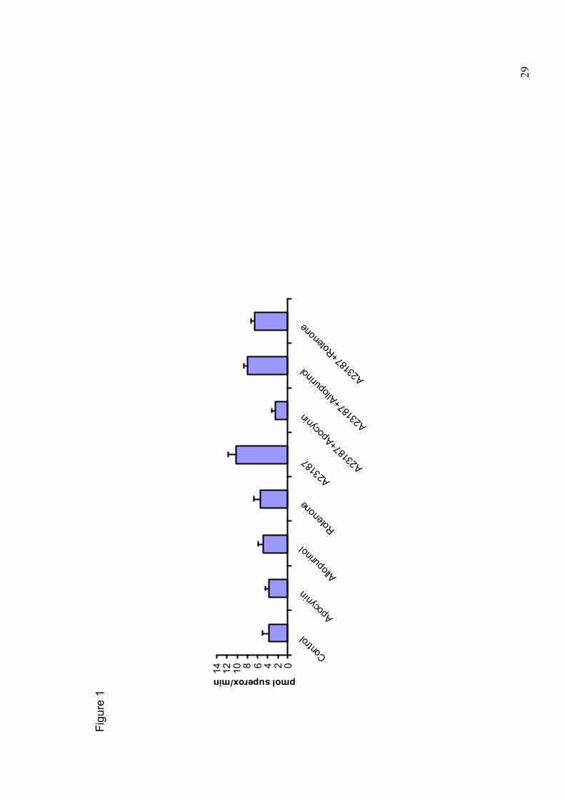
Figure legends
Figure 1. Mitochondria are the major contributors to free radical production by
Human Aortic Endothelial Cells (HAEC). Confluent HAEC were challenged with the
calcium ionophore A23187 in the presence or absence of apocyanin (an inhibitor of
NADPH oxidase), allopurinol (an inhibitor of xanthine oxidase), or rotenone (an
inhibitor of mitochindrial complex I). Superoxide production was continuously
assessed by the SOD-inhibitable cytochrome c reduction assay, using a plate reader
(Tecan, Mannedorf, Switzerland). Absorbance was read at 550 nm and superoxide

27
production was determined using the extinction coefficient of cytochrome c at 550 nm
(ε: 21 mM-1cm-1). Unpublished data.
28
Table 1 Commercial preparations currently sold for enhancing fertility.
Compound Comment
Sold as antioxidants Vitamin C Ameliorates vasomotion through several
antioxidant and non-antioxidant mechanisms. Unclear activity on semen quality.
Vitamin E Unclear actions on endothelium and semen quality.
Zinc High (~200 µg/ml) quantity in semen. Rationale behind supplementation unclear.
Sold as fertility-enhancers Arginine eNOS substrate might augment
vasomotion. Anecdotally reported to increase semen production.
Ginseng (Panax ginseng) Tonic according to ethnopharmacology. No evidence of fertility-improving activities.
Astragalus (Astragalus membranaceus)
Attributed with sperm motility-enhancing properties. Unproven.
Maca (Lepidium meyenii) Increases sexual potency according to ethnopharmacology. One animal study showed increased spermatogenesis [97]
29
Figure 1
02468101214
Control
Apocynin
Allopurinol
Rotenone
A23187 A
23187+Apocynin
A23187+Allopurinol
A23187+Rotenone
pmolsuperox/min