1 a unique and conserved neutralization epitope in h5n1
TRANSCRIPT
1
A Unique and Conserved Neutralization Epitope in H5N1 Influenza Viruses 1
Identified by a Murine Antibody against the A/goose/Guangdong/1/96 2
Hemagglutinin 3
Running title: H5N1 HAs with a broadly Neutralizing Antibody 4
5
Xueyong Zhu,a Yong-Hui Guo,
b Tao Jiang,
c Ya-Di Wang,
b Kwok-Hung Chan,
d 6
Xiao-Feng Li,c Wenli Yu,
a Ryan McBride,
e James C. Paulson,
e,f Kwok-Yung Yuen,
d 7
Cheng-Feng Qin,c Xiao-Yan Che,
b and Ian A. Wilson
a,g 8
9
Department of Integrative Structural and Computational Biology, The Scripps Research 10
Institute, 10550 North Torrey Pines Road, La Jolla, CA 92037, USAa; Center for Clinical 11
Laboratory, Zhujiang Hospital, Southern Medical University, Guangzhou, 510515, 12
Chinab; State Key Laboratory of Pathogen and Biosecurity, Beijing Institute of 13
Microbiology and Epidemiology, Beijing, 100071, Chinac; Department of Microbiology, 14
The University of Hong Kong, Hong Kong Special Administrative Region, Chinad; 15
Department of Chemical Physiology,e Department of Cell and Molecular Biology,f 16
Skaggs Institute for Chemical Biology,g The Scripps Research Institute, 10550 North 17
Torrey Pines Road, La Jolla, CA 92037, USA 18
19
Address correspondence to Ian A. Wilson, [email protected], or Xiao-Yan Che, 20
Xueyong Zhu, Yong-Hui Guo and Tao Jiang contributed equally to this article. 22
Ian A. Wilson, Xiao-Yan Che and Cheng-Feng Qin are co-senior authors. 23 24
JVI Accepts, published online ahead of print on 18 September 2013J. Virol. doi:10.1128/JVI.01577-13Copyright © 2013, American Society for Microbiology. All Rights Reserved.
on April 8, 2018 by guest
http://jvi.asm.org/
Dow
nloaded from
2
Abstract 25
Despite substantial efforts to control and contain influenza H5N1 viruses, ‘bird flu’ 26
viruses continue to spread and evolve. Neutralizing antibodies against conserved epitopes 27
on the viral hemagglutinin (HA) could confer immunity to the diverse H5N1 influenza 28
virus strains and provide information for effective vaccine design. Here, we report on 29
characterization of a broadly neutralizing murine monoclonal antibody H5M9 to most 30
H5N1 clades and sub-clades that was elicited by immunization with viral HA of 31
A/goose/Guangdong/1/96 (H5N1), the immediate precursor of the current dominant 32
strains of H5N1 viruses. Crystal structures of Fab´ H5M9 with H5 HAs of 33
A/Vietnam/1203/2004 and A/goose/Guangdong/1/96 reveal a conserved epitope in the 34
HA1 vestigial esterase subdomain that is some distance from the receptor binding site, 35
and partially overlaps antigenic site C of H3 HA. Further epitope characterization by 36
selection of escape mutant and epitope mapping by flow cytometry analysis of site-37
directed mutagenesis of HA with yeast cell surface display identified four residues that 38
are critical for H5M9 binding. D53, Y274, E83a and N276 are all conserved in H5N1 39
HAs and are not in H5 epitopes identified by other mouse or human antibodies. Antibody 40
H5M9 is effective in protection of H5N1 virus both prophylactically and therapeutically 41
and appears to neutralize by blocking both virus receptor binding and post-attachment 42
steps. Thus, the H5M9 epitope identified here should provide valuable insights into 43
H5N1 vaccine design and improvement, as well as antibody-based therapies for treatment 44
of H5N1 infection. 45
on April 8, 2018 by guest
http://jvi.asm.org/
Dow
nloaded from

3
Keywords: H5N1 influenza virus, epitope, broadly neutralizing murine antibody, crystal 46
structure, escape mutant, site-directed mutagenesis, vaccine design, passive 47
immunotherapy. 48
49
on April 8, 2018 by guest
http://jvi.asm.org/
Dow
nloaded from

4
Introduction 50
The highly pathogenic H5N1 influenza viruses continue to evolve and cause 51
poultry and occasional human infections. In 1996, an avian H5N1 virus, 52
A/goose/Guangdong/1/96 (GD1), was first isolated from a sick farmed goose in 53
Guangdong Province, China (62), and is believed to be the immediate precursor of the 54
current dominant strain of H5N1 virus that is spreading globally. Hemagglutinin (HA) is 55
the surface glycoprotein responsible for viral binding to host cell, internalization of the 56
virus, and subsequent membrane fusion of the viral and host cell membrane within the 57
endosomal pathway inside the infected cell. HA is also the major antigen on the viral 58
surface and provides the primary neutralizing epitopes for antibodies. The HA genes of 59
the subsequent H5N1 viruses are all related to those of GD1 or similar viruses (38), and 60
considerable genetic variation of HA genes have evolved the viruses into over ten distinct 61
phylogenetic clades (numbers 0-9) and second, third and fourth order subclades 62
(http://www.who.int/influenza/gisrs_laboratory/201101_h5fulltree.pdf), but only four 63
clades have been isolated from humans strains (clades 0, 1, 2, and 7) (1). H5N1 virus 64
infection is considered an avian disease, although there is some very limited evidence for 65
direct human-to-human transmission (55). Since 1997, the H5N1 viruses were 66
transmitted to humans mainly by direct contact with sick poultry with a very high fatality 67
rate of about 60% (http://www.who.int). Although the human H5N1 infection is sporadic 68
and rare, there is still great concern about a future H5N1 pandemic due to its high 69
virulence and lethality, its increasing avian reservoir, and the continued evolution and 70
potential reassortment with other human viruses (29, 42). 71
72
on April 8, 2018 by guest
http://jvi.asm.org/
Dow
nloaded from

5
Current strategies against influenza include antiviral treatment and vaccination. 73
Two classes of small molecule drugs, neuraminidase inhibitors and M2 ion-channel 74
blockers, have been used for prophylaxis and treatment of influenza. Neuraminidase is 75
the only other viral surface glycoprotein and cleaves terminal sialic acid moieties from 76
newly formed virions and host cell receptors. Oseltamivir phosphate (Tamiflu), zanamivir 77
(Relenza), and some other NA inhibitors (17) inhibit the NA activity and prevent the 78
budding of new viruses from infected cells, and are effective against both influenza A 79
and B viruses, including H5N1 viruses. These two NA inhibitors are currently used to 80
treat influenza viruses, but resistance to both drugs are emerging, including resistance of 81
H5N1 viruses to treatment by oseltamivir phosphate (33). Amantadine and rimantadine 82
inhibit viral entry and replication by blocking an ion channel formed by the M2 protein, 83
but both drugs are not currently recommended by the Centers for Disease Control and 84
Prevention for treatment of influenza A viruses because of resistance derived from 85
amino-acid substitutions in M2 proteins 86
(http://www.cdc.gov/flu/professionals/antivirals/antiviral-drug-resistance.htm). 87
From a population and global health perspective, vaccination remains the most 88
effective countermeasure against influenza virus. The ideal influenza vaccine would 89
induce cross-protective cellular and humoral responses, and would be safe and 90
immunogenic in all age groups of the population. In 2007, the first H5N1 human vaccine 91
derived from the A/Vietnam/1203/2004 (VN1203) was approved by the U.S. Food and 92
Drug Administration, and a number of other egg-dependent and egg-independent 93
vaccines are at various stages of development (50). Vaccination is also a major strategy 94
to control H5N1 influenza virus in poultry, such as in China, an inactivated vaccine 95
on April 8, 2018 by guest
http://jvi.asm.org/
Dow
nloaded from

6
containing the HA and NA genes of the GD1 virus and internal genes from the A/Puerto 96
Rico/8/34 (H1N1) has been used for domestic poultry since 2004 (35). This GD1 virus-97
based vaccine was demonstrated to be effective against different H5N1 viruses isolated in 98
China, excepted for the recently isolated, low-pathogenic, A/chicken/Shanxi/2/06–like 99
viruses (clade 7.2) (35). 100
A panel of 15 monoclonal antibodies specific for H5N1 viruses was generated by 101
immunizing mice with viral or recombinant HA of the GD1 virus (34). Among them, one 102
antibody H5M9, elicited in mice from H5 HA that was concentrated from GD1 H5N1 103
virus, had broad neutralizing activity against different clades (effective for all tested 104
clades 0, 1, 2.3.4, and 7) of H5N1 influenza viruses isolated from 1997 to 2008 (listed in 105
Table 1 and in our previous report (34)), indicating the presence of a conserved 106
neutralizing epitope in the H5 HA protein recognized by H5M9. Antibody epitope 107
mapping of H5 HA using yeast cell surface display showed that H5M9 binds to the HA1 108
region (34). 109
To define the H5M9 epitope at the atomic level, we determined crystal structures 110
of the H5M9 Fab´ in complex with the VN1203 HA and GD1 HA ectodomains. The 111
H5M9 antibody primarily recognizes a conserved and previously uncharacterized epitope 112
region in the vestigial esterase subdomain of HA1 (22) between the receptor binding site 113
head region and the HA fusion subdomain. The epitope was further refined by selection 114
of escape mutants and flow cytometry analyses of H5M9 binding to HA and by site-115
directed mutagenesis of key HA binding residues. We previously documented that 116
H5M9 is an inhibitor of hemagglutination as it directly blocks virus binding to cellular 117
receptors (34). We show here that H5M9 is also an inhibitor of the pH-induced 118
on April 8, 2018 by guest
http://jvi.asm.org/
Dow
nloaded from
7
conformational change required for virus fusion activity. These properties can be 119
translated into virus neutralization in vivo since H5M9 is also found to be protective 120
against a lethal viral challenge in a mouse model, both prophylactically and 121
therapeutically. 122
123
MATERIALS AND METHODS 124
Ethics statement. The animal experiments with BALB/c mice were carried out in strict 125
accordance with the guidelines of the Animal Experiment Committee of the State Key 126
Laboratory of Pathogen and Biosecurity, Ministry of Science and Technology of the 127
People’s Republic of China, and were approved by the Animal Experiment Committee of 128
the State Key Laboratory of Pathogen and Biosecurity, Beijing Institute of Microbiology 129
and Epidemiology, Beijing, China. 130
Cloning, expression and purification of the HAs. The ectodomains of VN1203 131
and GD1 HAs were expressed in the baculovirus system essentially as previously 132
described (15, 52). Briefly, HA0 cDNAs corresponding to residues 11-327 of HA1 and 1-133
174 of HA2 (H3 numbering, 17-504 in H5 numbering) of the VN1203 HA (GenBank 134
accession number AY818135) and GD1 HA (GenBank accession number AF144305) 135
were inserted into a baculovirus transfer vector, pFastbacHT-A (Invitrogen) with a N-136
terminal gp67 signal peptide, a C-terminal trimerization domain and a 6-His tag, and a 137
thrombin cleavage site incorporated to separate the HA ectodomain and the trimerization 138
domain and His tag. The constructed plasmids were used to transform DH10bac 139
competent bacterial cells by site-specific transposition (Tn-7 mediated) to form a 140
recombinant Bacmid with allowed blue-white selection. The purified recombinant 141
on April 8, 2018 by guest
http://jvi.asm.org/
Dow
nloaded from

8
VN1203 HA and GD1 HA Bacmids were used to transfect Sf9 insect cells for 142
overexpression. HA protein was produced by infecting suspension cultures of Hi5 cells 143
with recombinant baculovirus at an MOI of 5-10 and incubation at 28ºC shaking at 110 144
RPM. After 72 hours, Hi5 cells were removed by centrifugation and supernatants 145
containing secreted, soluble HA proteins were concentrated and buffer-exchanged into 146
1xPBS, pH 7.4. The VN1203 and GD1 HAs consisted of a mixture of uncleaved HA0 147
and cleaved HA1/HA2, and were recovered from the cell supernatants by metal affinity 148
chromatography using Ni-NTA resin. The HAs were digested with thrombin to remove 149
the trimerization domain and His-tag. The cleaved VN1203 HA or GD1 HA was purified 150
further by size exclusion chromatography on a Hiload 16/90 Superdex 200 column (GE 151
healthcare) in 10 mM Tris pH 8.0, 50 mM NaCl, and 0.02% (v/v) NaN3. 152
H5M9 Fab´ preparation and purification. Antibody H5M9 was elicited by 153
immunization of mice with HA from A/goose/Guangdong/1/96 as described (34). The 154
Fab´ fragment of antibody H5M9 (IgG1ț) was produced by standard protocols (25). The 155
intact H5M9 IgG was digested to (Fab´)2 with 1% (w/w) pepsin for four hours and 156
followed by reduction to Fab´ by 15 mM 2-mercaptoethylamine (MEA) for two hours. 157
The protein was purified to homogeneity by a combination of protein A and protein G 158
affinity chromatography, as well as ion-exchange chromatography (Mono-Q column, 159
Pharmacia) and gel filtration (Superdex 200 column, GE healthcare). 160
Purification of H5M9 Fab´-VN1203 HA and H5M9 Fab´-GD1 HA complexes. 161
The H5M9 Fab´-VN1203 HA and H5M9 Fab´-GD1 HA complexes were generated by 162
incubating the two purified components with an optimal ratio of Fab´ to HA that was 163
estimated by gel-shift using Blue Native PAGE (Invitrogen). H5M9 Fab´ at 1 mg/ml was 164
on April 8, 2018 by guest
http://jvi.asm.org/
Dow
nloaded from
9
titrated into 5 µg of VN1203 HA and GD1 HA in a total volume of 10 µl. The mixtures 165
were incubated overnight at 4ºC before Blue Native PAGE analysis. Following 166
determination of the optimal molar ratio (roughly 3 Fabs per trimer), H5M9 Fab´ and 167
purified VN1203 HA or GD1 HA in the same buffer (10 mM Tris, pH 8.0, 50 mM NaCl) 168
were mixed with that ratio. The mixture solution was incubated overnight at 4ºC before 169
further purification by gel filtration (Superdex 200 column) to remove unbound Fab´ and 170
HAs. 171
Crystallization and structural determination of H5M9 Fab´-VN1203 HA 172
complex. Crystallization experiments were set up using the sitting drop vapor diffusion 173
method. Initial crystallization conditions for the H5M9-VN1203 HA complex were 174
obtained from robotic crystallization trials using the automated Rigaku Crystalmation 175
system at the Joint Center for Structural Genomics (JCSG). Following optimization, 176
diffraction quality crystals were obtained by mixing 0.5 µl of the concentrated protein in 177
6.8 mg/ml in 10 mM Tris, pH 8.0, 50 mM NaCl with 0.5 µl of a reservoir solution 178
containing 0.1 M Hepes, pH 7.5, 2% (w/v) PEG 4000, 2.2 M (NH4)2SO4 at 22ºC. The 179
crystals were flash-cooled in liquid nitrogen using 30% saturated malonate in the mother 180
liquor as cryoprotectant. Diffraction data of the complex crystals were collected at 100K 181
at beamline 23ID-B, Advanced Photon Source (APS), Argonne National Laboratory. 182
HKL2000 (HKL Research, Inc) was used to integrate and scale diffraction data. The 183
crystals diffract to 3.6 Å resolution and the diffraction data were indexed in space group 184
C2 with a Matthews’ coefficient (Vm) of 6.1 Å3/Da and 80% solvent content (Table 2). 185
The structure was determined by molecular replacement using the program Phaser 186
(39). The initial model for H5M9 was constructed from a light chain variable domain 187
on April 8, 2018 by guest
http://jvi.asm.org/
Dow
nloaded from

10
from PDB model 2VL5 and a heavy chain variable domain the Fab´ constant domain 188
from PDB model 1P7K which share high sequence similarity (with 88% and 74% 189
identities, respectively) with no sequence gaps. The previously determined structure of 190
VN1203 HA (PDB codes 2FK0, 3GBM) was used as the HA model in the complex. One 191
HA trimer and three Fabs were found in the asymmetric unit. The initial rigid body 192
refinement and restrained refinement were performed in program REFMAC5 (41). 193
Despite the modest resolution, the data-to-atom ratio is still reasonable in the refinement 194
due to high solvent content (Table 2). The electron density maps fit the structure model 195
very well with continuous density for all main-chain atoms and good density for most 196
side chains. Additional positive electron density was observed near all 6 potential N-197
glycosylation sites in each HA monomer (18 glycosylation sites per HA trimer). 198
Interestingly, strong positive density was also observed near mouse H5M9 Fab´ AsnL63 199
possessing an Asn-Gly-Ser motif, and a GlcNAc residue was built at AsnL63 in all three 200
Fabs in the asymmetric unit. Structural refinement was completed with program Buster 201
(5) and Phenix (2). Final refinement statistics are summarized in Table 2. 202
Crystallization and structural determination of GD1 HA and H5M9 Fab´-203
GD1 HA complex. The purified GD1 HA in 10 mM Tris, pH 8.0, 50 mM NaCl was 204
concentrated to 8.0 mg/ml and subjected to crystallization trials using the Mosquito 205
crystal liquid handler (TTP LabTech). Initial crystal hits were obtained from Initial 206
Screening Reagents CP-CUSTOM-IV from Axygen Biosciences. The crystals for data 207
collection were grown by the sitting drop vapor diffusion method with a reservoir 208
solution containing 0.1 M Tris, pH 8.2, 21% (w/v) MPEG 2000 at 22ºC. The crystals 209
were flash-cooled in liquid nitrogen using 25% ethylene glycol (v/v) in mother liquor as 210
on April 8, 2018 by guest
http://jvi.asm.org/
Dow
nloaded from

11
cryoprotectant. Diffraction data were collected at 100K at beamline 9-2, Stanford 211
Synchrotron Radiation Lightsource. The crystals diffract to 2.6 Å resolution and the 212
diffraction data were indexed in space group P21 with a Matthews’ coefficient (Vm) of 213
3.4 Å3/Da and 64% solvent content (Table 2). 214
Initial crystallization conditions for the H5M9 Fab´-GD1 HA complex were 215
obtained from the automated Rigaku Crystalmation system at the JCSG. Following 216
optimization, diffraction quality crystals were obtained by mixing 0.5 µl of the 217
concentrated protein in 9.7 mg/ml in 10 mM Tris, pH 8.0, 50 mM NaCl with 0.5 µl of a 218
reservoir solution containing 0.1 M Hepes, pH 7.5, 2% (w/v) PEG 4000, 2.0 M 219
(NH4)2SO4 at 22ºC. The crystals were flash-cooled in liquid nitrogen with cryoprotectant 220
40% saturated malonate added to mother liquor. Diffraction data were collected at 100K 221
at beamline 11-1, Stanford Synchrotron Radiation Lightsource. HKL2000 (HKL 222
Research, Inc) was used to integrate and scale diffraction data. The crystals diffract to 7.0 223
Å resolution and the diffraction data were indexed in space group P3121 with a 224
Matthews’ coefficient (Vm) of 4.3 Å3/Da and 71% solvent content (Table 2). 225
GD1 HA and H5M9-GD1 HA structures were both determined by molecular 226
replacement with the program Phaser (39). The unliganded GD1 HA structure was 227
determined with the VN1203 HA structure (PDB codes 3GBM) as a model. The H5M9 228
Fab´-GD1 HA complex structure was subsequently determined using the refined GD1 229
HA structure and the refined H5M9 Fab´ structure from H5M9-VN1203 HA complex as 230
input models. For GD1 HA structure, the initial rigid body refinement and restrained 231
refinement were performed with program REFMAC5 (41). Additional positive electron 232
density was observed near all 5 potential N-glycosylation sites in the various HA 233
on April 8, 2018 by guest
http://jvi.asm.org/
Dow
nloaded from

12
monomers (for a total of 32 sites observed out of 45 possible sites for three HA trimers). 234
The structural refinement for GD1 HA was completed with program Buster (5) and 235
Phenix (2). For H5M9-GD1 HA complex, the molecular replacement solution resulted in 236
two HA trimers and only one H5M9 Fab´. The other five Fab´ molecules were modeled 237
into electron density by superimposing the relevant HA molecules, and further refinement 238
reduced the R values. Due to the low resolution, only rigid-body refinement was 239
performed with program REFMAC5 (41). Final refinement statistics are summarized in 240
Table 2. 241
The quality of the structures described here was analyzed using the JCSG 242
validation suite (www.jcsg.org) including MolProbity, WHAT IF, Resolve and Procheck. 243
All figures were generated with Pymol (www.pymol.org). 244
KD determination. KD’s were determined by bio-layer interferometry using an 245
Octet Red instrument (ForteBio, Inc.). For VN1203 HA and GD1 HA binding with 246
H5M9 Fab´ and IgG, VN1203 HA or GD1 HA at 20 µg/ml in 1x kinetics buffer (1x PBS, 247
pH 7.4, 0.01% BSA, and 0.002% Tween 20) was loaded onto Ni-NTA coated biosensors 248
and incubated with varying concentrations of VN1203 HA or GD1 HA in 1x kinetics 249
buffer. All binding data were measured at 30 ºC. Five or six concentrations in 1:2 serial 250
dilutions of H5M9 Fab´ or H5M9 IgG were used, with the highest concentration being 25 251
nM, except for 100 nM for H5M9 Fab´ in binding with GD1 HA. The KD reported here 252
was determined from the ratio of koff to kon. All binding traces and curves used for fitting 253
are reported in Figure S1. 254
HA glycan microarray receptor binding assay. Protocols for microarray HA 255
analysis and the list of glycans on the array (Fig. S2) were as previously described (6, 256
on April 8, 2018 by guest
http://jvi.asm.org/
Dow
nloaded from

13
63). Briefly, HA-antibody complexes were prepared by mixing HA, mouse anti-His 257
Alexa Fluor 488 and goat anti-mouse IgG Alexa Fluor, and the complex mixture was 258
then added directly to the surface of the array and allowed to incubate for 1 hour at room 259
temperature (~ 22 ºC). After washing with 1x PBS and Tween, the array slides were dried 260
and scanned for fluorescence signal. 261
Cells and Virus. MDCK cells were maintained in DMEM (Life technologies) 262
supplemented with 10% FBS at 37°C in 5% CO2. After infection with influenza virus, the 263
MDCK cells were maintained in DMEM containing 0.2% (w/v) BSA and 0.5 ȝg/ml 264
TPCK-trypsin (Sigma-Aldrich). All the viruses in this study were propagated in 10-day-265
old embryonated eggs and titered by hemagglutination tests and plaque forming assays. 266
Neutralization assay. Virus neutralization titers of the antibody were determined 267
as described previously (34, 46). 268
Selection of neutralization escape mutants. 103 PFU of H5N1 virus strain 269
A/Vietnam/1194/2004 (H5N1) (VN1194) was mixed with purified H5M9 and incubated 270
at 37 °C for 1 h. The virus-antibody mixture was added to the MDCK cell monolayer and 271
incubated at 37 °C for another 1 h. Following removal of the inoculum, the plate was 272
rinsed twice with PBS, then 3 ml of DMEM containing 2 ug/ml TPCK-trypsin and 0.5 273
ȝg/ml of H5M9 were added. After 4 days of incubation in the presence of H5M9, 274
medium containing potential escape viruses was harvested. After three passages in the 275
presence of H5M9, escape mutants were isolated by plaque-to-plaque purification and 276
further amplified in MDCK cells. The HA segments of escape mutants obtained from 277
each passage were sequenced and analyzed. 278
on April 8, 2018 by guest
http://jvi.asm.org/
Dow
nloaded from
14
Plaque reduction neutralization test. Antibody H5M9 was diluted to 500 ȝg/ml, 279
and then serially 10-fold diluted in DMEM. H5M9 IgG was added with an equal volume 280
of approximately 100 PFU of wild-type H5N1 virus or escape mutant. The antibody-virus 281
mixture was incubated at 37˚C for 1 h, and then added to MDCK monolayers in a 6-well 282
plate in duplicate and incubated for another 1 h at 35˚C. The supernatant was removed, 283
and 3 ml of 1.0% (w/v) LMP agarose (Promega) in DMEM plus TPCK-trypsin (2 ȝg/ml) 284
was layered onto the infected cells. After further incubation at 35˚C for another 4 days, 285
the overlays were removed and the cells were stained with 1% (w/v) crystal violet 286
dissolved in 4% (v/v) formaldehyde to visualize the plaques. The percentage of plaque 287
reduction was calculated accordingly. 288
Epitope mapping of H5M9 by yeast cell surface display. The DNA fragment 289
(49–954 nt) coding the HA protein was amplified from a cDNA clone of VN1194 HA by 290
PCR with BamHI andXhoI sites and ligated into the pYD1 vector with the Xpress epitope 291
tag (DLYDDDDK) (Invitrogen), producing the recombinant plasmid pYD1-HA. The HA 292
protein was expressed in the Saccharomyces cerevisiae strain EBY100 as described in 293
our previous report (34). The potential key HA contacting residues within H5M9 binding 294
site were mutated using the QuikChange® Lightning Site-Directed Mutagenesis Kit 295
(Stratagene, CA, USA). Yeasts expressing wild-type HA protein and mutant HA proteins 296
were identified by sequencing. The expression of mutant proteins on the EBY100 yeast 297
cell surface and fluorescent labeling of yeast surface displayed proteins were performed 298
according to the method described previously (34). Briefly, yeast cells were washed with 299
PBS and incubated with purified antibodies at a concentration of 10 ȝg/ml with 1 mg/ml 300
BSA in PBS. After a 30-minute incubation on ice, yeast were washed in PBS and then 301
on April 8, 2018 by guest
http://jvi.asm.org/
Dow
nloaded from

15
incubated with a goat anti-mouse IgG secondary antibody conjugated to FITC (Sigma) at 302
a dilution of 1:150 with 1 mg/ml BSA in PBS. After fixation with 2% (w/v) 303
paraformaldehyde in PBS, yeast cells were analyzed on a Accuri C6 flow cytometer 304
(Becton-Dickinson) using software Flowjo (www.flowjo.com). 305
Protease susceptibility assay. Protocols for trypsin susceptibility analysis were 306
as previously described (15). For VN1203 HA, each reaction contained ~2.5 ȝg of the 307
HA or ~2.5 ȝg of the HA and a two-fold molar excess of H5M9 Fab´ (2 Fabs per HA 308
protomer). Reactions were incubated at 37 °C for one hour at pH values 4.9 and 8.0, 309
respectively. After incubation, the reaction pH was neutralized to pH 8.4. Trypsin was 310
then added to all samples except controls at a final ratio of 1:20 (wt/wt) of trypsin to the 311
HA, and reactions were incubated overnight at 22°C. Samples were then analyzed by 312
reducing SDS-PAGE. 313
Prophylactic and therapeutic efficacy of H5M9 in mice. In the prophylactic 314
model, groups of 4 week-old female BALB/c mice (n=6) were i.p. injected with a dose of 315
2 or 20 mg/kg of H5M9 one day prior to intranasal challenge with 10 MLD50 of VN1194. 316
In the therapeutic model, mice received the same dose of H5M9 one day after the lethal 317
challenge of 10 MLD50 of VN1194. The mice were monitored daily for survival and 318
weight loss until day 14 post-infection (p.i.). All manipulations involving live H5N1 319
virus were carried out in the Biosafety Level 3 (BSL-3) or Animal Biosafety Level 3 320
(ABSL-3) containment facility in Beijing Institute of Microbiology and Epidemiology. 321
Statistical Analysis. Survival curves were generated by the Kaplan-Meier method 322
and analyzed with Log-rank test. P<0.05 is considered significant. 323
on April 8, 2018 by guest
http://jvi.asm.org/
Dow
nloaded from
16
Accession numbers. The atomic coordinates and structure factors are deposited 324
in the Protein Data Bank under accession codes 4MHH for the H5M9 Fab´-VN1203 HA 325
complex, 4MHI for the unliganded GD1 HA, and 4MHJ for the H5M9 Fab´-GD1 HA 326
complex. Nucleotide sequences for the H5M9 IgG have been deposited in GenBank 327
under accession numbers KF499999 for the light chain and KF500000 for the heavy 328
chain. 329
330
RESULTS 331
The overall structure of H5M9 Fab´-VN1203 HA complex. VN1203 virus was 332
originally isolated from a 10-year old Vietnamese boy who died of bird flu, and this virus 333
is among the most pathogenic H5N1 isolates studied to date in mammalian models (37). 334
The VN1203 HA ectodomain was expressed in a baculovirus system, and H5M9 Fab´ 335
was digested and purified from the mouse antibody (34). The H5M9 binds to the 336
VN1203 HA with high affinity with Kd values of 1 nM and 0.008 nM for binding of the 337
immobilized HA to the monovalent Fab´ and bivalent IgG, respectively (Table 3, Fig. 338
S1). The crystal structure of the H5M9 Fab´-VN1203 HA complex was determined to 3.6 339
Å resolution with good refinement statistics for this moderate resolution, which might in 340
part be due to high solvent content in the crystals (Table 2). The mature HA is a 341
homotrimer with multiple glycosylation sites (Fig. 1A). Each HA polypeptide is 342
proteolytically cleaved by host proteases into two disulfide-linked subunits HA1 and 343
HA2. As in the unliganded VN1203 HA (52), the overall fold of the VN1203 HA trimer 344
in the complex is very similar to other published HAs, with HA1 containing primarily the 345
receptor binding subdomain and vestigial esterase subdomain, and HA2 constituting most 346
on April 8, 2018 by guest
http://jvi.asm.org/
Dow
nloaded from
17
of the core fusion machinery in the stalk region (Figs 1A and B). Interpretable density is 347
observed for VN1203 HA1 (residues 11-324) and HA2 (1-174) (H3 numbering), and 348
H5M9 Fab´. In the complex, the VN1203 HA trimer is in its prefusion state with the N-349
terminal fusion peptide in HA2 embedded in the hydrophobic core between the trimer 350
HA2 subunits, with one H5M9 Fab´ binding to each HA monomer. Significantly, unlike 351
most of other structurally defined neutralizing antibodies, H5M9 binds in the vestigial 352
esterase subdomain (22) of HA1 at the base of the membrane distal domain, at a distance 353
from the receptor binding site (Figs. 1A and B). The antibody H5M9 and VN1203 HA 354
paratope and epitope surfaces interact well with a shape complementarity of 0.48, and as 355
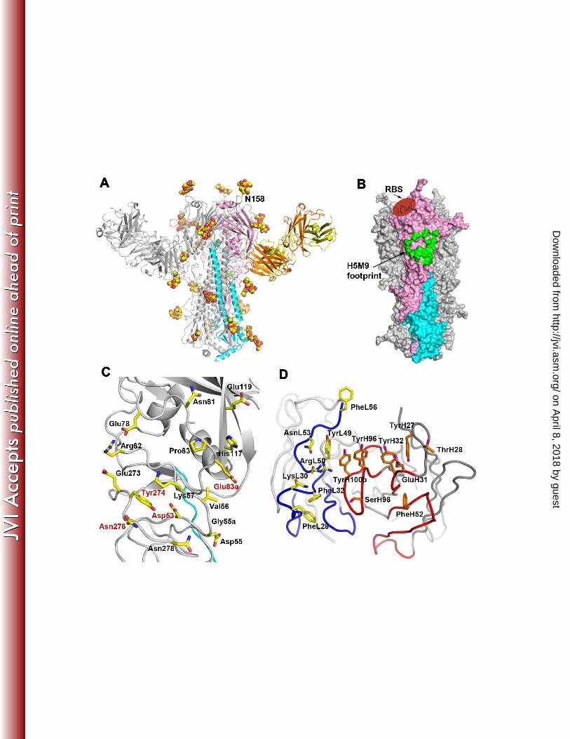
expected, no glycosylation sites are found within the epitope (Fig. 1A). 356
A unique and conserved H5N1 HA epitope from the H5M9 Fab´-VN1203 HA 357
complex. Upon complex formation, one H5M9 Fab´ binds to each H5 HA1 protomer of 358
the trimer (three Fab´s per trimer), burying ~ 820 Å2 of HA protein surface with typical, 359
heavy chain-dominant binding, where 60% of the binding surface arises from the heavy 360
chain. A total of 16 residues from each VN1203 HA monomer participate in the 361
intermolecular contacts. 362
The epitope is conformational and consists of mostly HA1 polar residues from 363
fragments 53 to 62, 78 to 83a, 117 to 119, and 273 to 278 in the vestigial esterase 364
domain, as previously defined by Ha et al. (22). The key HA contacting residues are 365
shown in Figure 1C. Five of the six H5M9 complementarity determining regions (CDRs), 366
i.e. not CDR L3, are involved in HA binding (Fig. 1D). The heavy-chain residues 367
contribute 3 out of 4 hydrogen bonds (H-bonds) or salt bridges, as analyzed by HBPLUS 368
(40), and about 60% of the 110 total van der Waals’ interactions. The light chain of 369
on April 8, 2018 by guest
http://jvi.asm.org/
Dow
nloaded from
18
H5M9 interacts with VN1203 HA1 residues with mainly nonpolar contacts, as well as a 370
salt bridge between ArgL50 and Glu83a. The heavy chain of H5M9 makes mainly 371
nonpolar interactions and three H-bonds with VN1203 HA1, including one H-bond each 372
between SerH98 (OȖ) and Asp53 (Oį2), ThrH28 (OȖ1) and Arg62 (NȘ1), as well as 373
ThrH28 (N) and Glu78 (Oİ2). 374
No major changes are observed in the overall structure of VN1203 HA upon 375
complex formation except for a few side-chain rotamers, such as for Asn278 to avoid a 376
steric clash with the antibody. The CĮ root mean square deviation (rmsd) is 1.3 Å 377
between the VN1203 HA in H5M9 Fab´ complex and the uncomplexed VN1203 HA 378
(PDB code 2FK0), while the CĮ rmsd is only 0.5 Å between the VN1203 HA in the 379
H5M9 Fab´ complex and the VN1203 HA in a complex with a stem binding antibody 380
CR6261 (PDB code 3GBM), where the stem epitope is distant from H5M9 binding site 381
(15). Thus, no large structural changes are required for neutralization to occur, as 382
observed in other influenza neutralizing antibodies, such as the H3 virus neutralizing 383
antibodies HC19 and HC45 (19). 384
Antibody H5M9 binds the same epitope in GD1 HA. As the GD1 H5N1 virus 385
is believed to be the immediate precursor of the dominant strains of H5N1 viruses, it was 386
important to compare a crystal structure of GD1 HA with the VN1203 HA in the 387
VN1203 HA-H5M9 complex to establish the conserved nature of the epitope recognized 388
by H5M9. Thus, we determined the crystal structure of apo-form GD1 HA ectodomain to 389
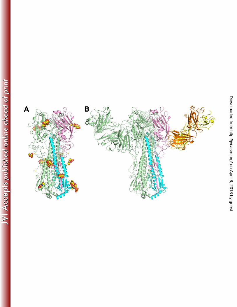
2.6 Å resolution (Table 2). Three GD1 HA protomers form the canonical trimer (Fig. 2A) 390
with a CĮ rmsd of 0.6 Å between GD1 and VN1203 HA protomers, and a CĮ rmsd of 0.5 391
Å between HA1 domains with a total of 16 amino acid differences (Table 4). The 392
on April 8, 2018 by guest
http://jvi.asm.org/
Dow
nloaded from
19
receptor-binding sites (RBS) of GD1 HA and VN1203 HA are almost identical with 393
conserved key residues except for a Ser221 to Pro221 switch in HA1 from VN1203 HA 394
to GD1 HA at its far edge of the RBS (Fig. 3A), implying the similar receptor binding 395
activity and specificity between two viruses. Indeed, direct analysis by glycan microarray 396
showed that both HAs are highly specific for Į2-3 sialosides (Figs. 4 and S2), with the 397
exception that VN1203 HA acquired additional binding to fucosylated Į2-3 linked 398
glycans. 399
The H5M9 binding sites in VN1203 HA and GD1 HA are well conserved (Fig. 400
3B), which is consistent with the similar Kd values of 0.9 nM for the Fab´ binding to GD1 401
HA and 1.0 nM for VN1203 HA, although the Kd of 0.04 nM for IgG binding to GD1 HA 402
is ten-fold higher that the highly potent 0.008 nM binding to VN1203 HA (Table 3, Fig. 403
S1). A conservative variation at position 55 in HA1 (Asp in VN1203 HA and Asn in 404
GD1 HA) is the only difference within the H5M9 footprint between these two H5N1 405
viruses (Fig. 3B, Table 4) and these changes do not appear to affect H5M9 neutralization 406
as H5N1 viruses with Asp55 or Asn55 in HA can all be neutralized by H5M9 (Tables 1 407
and 5). Asp55 of VN1203 HA lies on the edge of H5M9 epitope and makes van der 408
Waals contacts with both PheL28 and LysL30 of CDR L1. Asn55 of GD1 HA is also on 409
the periphery of the epitope and makes contacts with CDR L1. The low-resolution crystal 410
structure of GD1 HA in complex with H5M9 Fab´ (Table 2) also clearly indicates that 411
H5M9 Fab´ binds within the same site in GD1 HA as that of VN1203 HA (Fig. 2B). 412
Sequence analysis and glycosylation prediction (11) indicates that up to seven 413
glycosylation sites can be present in H5N1 HAs, of which six sites are over 95% 414
conserved (N21, N33, N169 and N289 in HA1, and N154 and N213 in HA2); the other 415
on April 8, 2018 by guest
http://jvi.asm.org/
Dow
nloaded from
20
glycosylation site at N158 in HA1 is 47% conserved. In our H5 HA construct (from 416
residue 11 in HA1 to residue 173 in HA2) expressed in a baculovirus expression system 417
in insect cells, all potential glycosylation sites were found to be glycosylated in both 418
VN1203 HA (Fig. 1A) and GD1 HA (Fig. 2A). By comparison, the GD1 HA has five 419
conserved glycosylation sites (N21, N33, N169 and N289 in HA1, and N154 in HA2), 420
while the VN1203 HA has an additional glycosylation site at N158 that is close to the 421
RBS (Fig. 1A). The loss of glycosylation at N158 in recent H5N1 strains has been 422
associated with increased potential to acquire a receptor specificity that supports aerosol 423
transmission in ferrets (45, 51). However, loss of this glycan alone is not sufficient to 424
change receptor specificity since GD1 exhibits strong binding to α2-3 sialosides that are 425
characteristic of avian virus receptor specificity (Fig. 4). 426
Selection and sequence analysis of escape mutants. The HA epitope was further 427
probed by generating and characterizing escape mutants. Neutralization escape mutants 428
were generated by culturing H5N1 virus, A/Vietnam/1194/2004 (H5N1) (VN1194) in the 429
presence of H5M9. VN1194 HA has only one amino-acid substitution T46K compared to 430
VN1203 HA and this change is distant from the epitope (Table 4). After three rounds of 431
passages, escape variants selected by H5M9 in VN1194 virus exhibited mutations in 432
HA1 D53N (with single G-A change at nucleotide position 177), close to the H5M9 433
epitope center (Fig. 1C). The D53N mutant generated similar plaque morphology and 434
peak titers to that of wild-type VN1194 in MDCK cells. Plaque reduction neutralization 435
tests (PRNT) were then performed to compare the relative infectivity of VN1194 and 436
D53N mutant viruses (Fig. 5A). Wild-type VN1194 virus was neutralized by H5M9 at a 437
PRNT50 value of 0.12 µg/ml. In clear contrast, the D53N mutant virus was not 438
on April 8, 2018 by guest
http://jvi.asm.org/
Dow
nloaded from
21
neutralized by H5M9 even at the highest tested concentration of 500 µg/ml (Fig. 5A). 439
The D53N mutation would change the overall electrostatic potential of the HA surface 440
around position 53 from weakly acid to near neutral and reduce the interactions with 441
H5M9 which is weakly basic around D53 binding site (Figs. 5B to D). Otherwise, a 442
similar hydrogen bond is made with Asp/Asn 53 to SerH98. 443
Identification of key epitope residues by yeast cell surface display. To further 444
explore the key epitope residues for H5M9, a panel of HA single mutants of nine H5 445
contacting residues contributing most of the interface interactions was interrogated for 446
specific binding of antibodies by yeast cell surface display. Alanine scanning 447
mutagenesis was carried out for H5M9 epitope residues 53, 57, 62, 78, 83A, 117, 273, 448
274 and 276 on VN1194 HA with only one amino-acid substitution T46K to VN1203 HA 449
(Table 4). Since the amino acids of these residues on VN1194 HA (or VN1203 HA) are 450
among the most frequently found at these positions in all H5N1 HAs (> 85.9% 451
conservation) from our extensive survey of all non-redundant H5N1 HA sequences in the 452
National Center for Biotechnology Information (NCBI) influenza database (Table 6), 453
nine polar mutations for these residues were also made based on the second most-454
frequent amino acid in H5N1 HAs: D53N, K57R, R62K, E78K, E83aK, H117R, E273K 455
and N276D, as well as Y274F (Table 6). Thus, a total of 18 HA mutants and one wild-456
type control were generated from yeast cell surface display and analyzed by FACS of 457
flow cytometry (Fig. 6). Several of these mutants conferred considerable loss of binding 458
to H5M9. Alanine mutants, D53A, E83aA and Y274A abolished and N276A 459
substantially reduced binding of H5M9 (Fig. 6), indicating the importance of these side 460
chains in H5M9 binding. Furthermore, for other mutants at these four positions, D53N, 461
on April 8, 2018 by guest
http://jvi.asm.org/
Dow
nloaded from

22
E83aK abolished binding of H5M9, while Y274F and N276D showed no obvious effect 462
(Fig. 6). Interestingly, although binding of mutant E78A was unchanged, mutant E78K 463
significantly reduced binding of H5M9 (Fig. 6). Alanine scanning mutants at K57, R62, 464
H117 and E273, as well as other mutants at these positions, K57R, R62K, H117R and 465
E273K, cause no obvious loss of H5M9 binding, indicating these positions are not critical 466
for binding to H5M9. Taken together, FACS analysis revealed that D53 and E83a, as 467
well as Y274 and N276 to a lesser extent, constituted critical contact residues for H5M9 468
binding (Fig. 1C). The escape mutant D53N was confirmed once more to be critical and 469
mutations E83aA and E83aK abolish the salt bridge between E83a to ArgL50 of H5M9. 470
In addition, mutation Y274A would eliminate interactions with GlyH97 and SerH98, 471
while mutation N276A abolishes interactions with ThrH33, PheH52 and SerH98 of 472
H5M9. 473
Mechanism of antibody H5M9 neutralization. We previously reported that 474
antibody H5M9 blocked viral entry in a hemagglutination inhibition test, although the 475
inhibition titer is weaker than those of some other antibodies elicited from the same 476
mouse immunization as H5M9 (34). This lower HAI inhibition for H5M might due to the 477
epitope being distant from the receptor binding site and, hence, the antibody only 478
partially blocking receptor binding (Fig. 1). During virus infection, the HA protein is 479
responsible for viral attachment and viral fusion. To further explore the H5M9 480
neutralization mechanism, trypsin digestion of VN1203 HA was performed in which the 481
HA protein was exposed to a low pH (pH 4.9) to trigger the pH-induced conformational 482
changes and acquire sensitivity to cleavage by trypsin (Fig. 7). Convincingly, the low-pH 483
treated HA could be completely digested in its HA1 domain by trypsin in the absence of 484
on April 8, 2018 by guest
http://jvi.asm.org/
Dow
nloaded from
23
H5M9 Fab´ (lane 1 in Fig. 7). However, H5M9 Fab´ was able to protect the HA from 485
degradation by trypsin when treated at pH 4.9 (lane 3 in Fig. 7). These results suggest the 486
binding of H5M9 somehow inhibits the pH-induced conformational changes in HA, 487
indicating its function in preventing virus fusion. 488
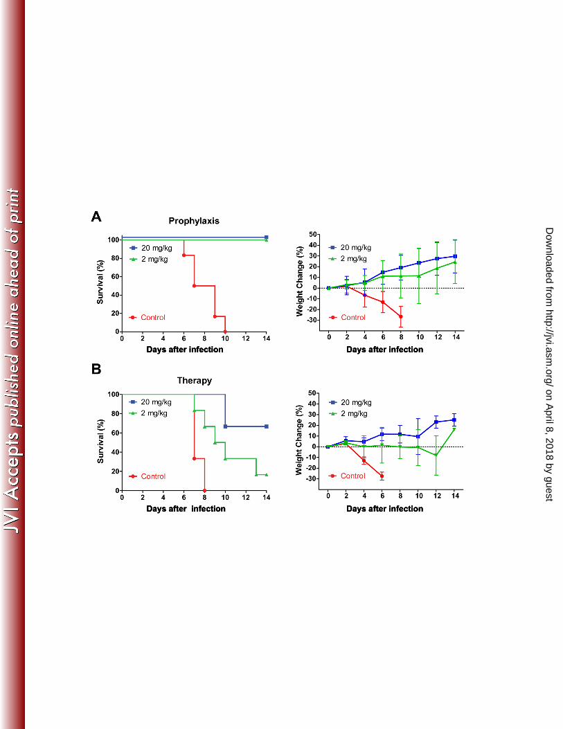
Prophylactic and therapeutic efficacy of antibody H5M9. The in vivo 489
protective effects of H5M9 against H5N1 viral infection were tested in mice using 490
prophylactic and therapeutic models, respectively. In the prophylactic model, female 491
BALB/c mice were i.p. injected with a dose of 2 or 20 mg/kg of H5M9 one day prior to 492
intranasal challenge with 10 MLD50 (50% mouse lethal dose) of VN1194 virus. A single 493
treatment of H5M9 provided full protection against lethal challenge of VN1194 virus in 494
mice (Fig. 8A). All H5M9-treated mice showed increase in body weight without any 495
signs of respiratory distress throughout the study. As expected, all mice that received 496
control PBS treatment showed signs of respiratory distress, rapid weight loss, and 497
succumbed to viral infection (Fig. 8A). In the therapeutic model, a single treatment with 498
20 mg/kg of H5M9 one-day post 10 MLD50 of VN1194 challenge provided 66.7% 499
protection, while 16.7% of mice treated with 2 mg/kg of H5M9 survived (Fig. 8B). 500
Moreover, all surviving mice appeared healthy and showed recovery in body weight after 501
initial infection. Overall, H5M9 treatment significantly increased the mean survival time 502
of infected mice from 7.3 ± 0.5 days (PBS control group) to 12.7 ± 1.9 days (20 mg/kg) 503
and 10.2 ± 2.5 days (2 mg/kg), respectively (Fig. 8B). 504
505
506
507
on April 8, 2018 by guest
http://jvi.asm.org/
Dow
nloaded from

24
DISCUSSION 508
Highly pathogenic avian influenza H5N1 virus can cause morbidity and mortality 509
in humans. So far, the H5N1 virus is mainly panzootic in avian species and leads to 510
occasional human infection through close contact with infected birds. However, recent 511
reports have shown that the H5N1 viruses, including VN1203, could become 512
transmissible through airborne droplets by mutations in H5 HAs so that the HA proteins 513
recognize receptors in the upper airways of mammals, particularly ferrets (9, 26, 28). It 514
is, therefore, necessary to prepare for a potential H5N1 human pandemic. Current 515
strategies against influenza include vaccination and antiviral drug treatment. Due to the 516
existence of multiple clades and subclades of the H5N1 virus, it is difficult to predict the 517
major strain that might cause the next pandemic. In this study, from HA-antibody crystal 518
structures, escape mutant analysis and fine epitope mapping by yeast cell surface display, 519
we identified a unique and conserved epitope in H5N1 viruses using a murine antibody 520
elicited from the H5N1 virus immediate precursor GD1 that has prophylactic and 521
therapeutic efficacy to H5N1 virus. 522
The antibody H5M9 epitope is a unique epitope for H5N1 viruses. The antigenic 523
sites of H5 HA were originally mapped into five groups from an H5N9 HA based on the 524
H3 structure (46). From the crystal structures of H5 HA from A/Duck/Singapore/3/97 525
(H5N1) (23) and VN1203 (52), the H5 HA molecule was then antigenically mapped in 526
greater detail first for an H5N2 avian influenza virus (31), and later for VN1203 (30), 527
revealing two antigenic sites, one of which is similar to site A of H3 HA (59), and the 528
other overlapping site B in H3 (59) and site Sa in H1(8). In addition, the fine epitope 529
mapping of an avian subclade 2.2 of the H5N1 viruses found antigenic sites 530
on April 8, 2018 by guest
http://jvi.asm.org/
Dow
nloaded from

25
corresponding to site B in H3 HA, but extending far beyond this area (49). In this study, 531
crystal structures of H5M9 in complex with both VN1203 HA and GD1 HA revealed an 532
epitope located in the vestigial esterase subdomain of the HA1 head, relatively far from 533
the receptor binding site. Escape mutant analysis with the VN1194 virus selected a 534
mutant D53N, and further characterization by flow cytometry of site-directed 535
mutagenesis of the HA with yeast surface display identified four key epitope residues in 536
VN1194 HA, Asp53, Tyr274, as well as E83a and N276, which are located in the lower 537
part of the H5M9 footprint from the crystal structures (Fig. 1C). These key H5 residues 538
do not overlap with any known epitopes described above or detected with other human 539
and mouse antibodies (7, 12, 18, 24, 27, 32, 36, 43, 44, 47, 48, 54, 56, 60). The four 540
H5M9 epitope key residues do not overlap with known H1 antigenic sites (8), but 541
positions 53, 274, and 276 correspond to site C of H3 viruses (59). However, H5M9 did 542
not show cross-subtype neutralization against H1 and H3 viruses in hemagglutination 543
inhibition tests (34), which is likely due to amino-acid differences in this epitope in other 544
virus subtypes (Table 4). 545
The antibody H5M9 epitope appears to be well conserved in H5N1 viruses and is 546
consistent with H5M9’s cross-neutralization activities. In all H5N1 viruses of clades 0, 1, 547
2.3.4 and 7 that were neutralized by H5M9, the H5M9 epitope residues are fully 548
conserved, except for a D55N mutation that does not appear to affect H5M9 binding or 549
neutralization (Tables 1 and 5). The full or almost completely conserved H5M9 epitope 550
can be found in most other clades or subclades of H5N1 viruses, except for some critical 551
mutations, such as D53N, that exist in clades 7.1 and 7.2 or some other viruses (Table 7). 552
on April 8, 2018 by guest
http://jvi.asm.org/
Dow
nloaded from
26
The antibody H5M9 is likely to have neutralizing activity against a broad 553
spectrum of H5N1 influenza viruses. The H5M9 binding surface is highly conserved 554
(>93.6% conservation) across all H5N1 influenza A viruses, except for lower 555
conservation of Asp53 (85.9%), Asp55 (86.1%) and Arg62 (86.1%) (Table 8). H5 viruses 556
with the Asp55 mutation to Asn (14.0 % conservation) may still possess the 557
neutralization activity as confirmed for H5M9 in our neutralization assay (Tables 1 and 558
5), while Arg62 mutation to Lys (16.5% conservation) would not change binding of 559
H5M9 as illustrated by R62K mutant of VN1194 HA in the yeast cell surface display 560
analysis (Fig. 6). However, the Asp53 mutation to Asn (12.5% conservation) in H5 HA 561
might abolish neutralization and binding of H5M9 as seen in the escape mutant and flow 562
cytometry analyses (Figs. 5 and 6). It remains to be seen if H5M9 can neutralize 563
influenza viruses from subtypes H5N2 to H5N9. From the survey of the conservation of 564
H5M9 epitope in HAs from H5N2 to H5N9 (Table 8), the H5M9 epitope is well 565
conserved in H5N5 HAs, but not in other subtypes as one of the key binding residues 566
D53 is rare and is mostly Ser at position 53. 567
Our results suggest antibody H5M9 binding to this previously uncharacterized 568
epitope in H5 HA can play a role in inhibiting both virus binding and virus fusion, 569
although the epitope is distant from the receptor binding site or the fusion peptide. Most 570
neutralizing antibodies against influenza virus recognize epitopes in the highly variable 571
regions in the HA head around receptor binding site and inhibit virus binding to host cells 572
(19-21, 58, 61) or, in one case, prevent membrane fusion (4). Several recently reported 573
broadly neutralizing antibodies from our group and others bind to the stem at the 574
membrane-proximal end of HA and interfere with the pH-induced conformational 575
on April 8, 2018 by guest
http://jvi.asm.org/
Dow
nloaded from
27
changes of the HA that lead to membrane fusion, as seen for H5 and other HAs (13-16, 576
53). Antibody H5M9 inhibited agglutination of erythrocytes in vitro (34), although it 577
binds to an epitope that is distant from the receptor binding site, but close enough to still 578
interfere with receptor binding. Because of their large size, IgGs bound on the 579
membrane-distal surface of HA may still prevent access to the receptor binding site if 580
oriented towards it. So far, only two other structurally-characterized antibodies, HC45 581
(19) and BH151 (20), bind to a similar but not identical epitope on H3 HA of 582
A/Aichi/2/68 (H3N2) (Fig. 9). It was also reported that HC45 neutralized viral infectivity 583
by blocking receptor binding as confirmed by hemagglutination inhibition assay (19). In 584
addition to prevent virus binding, H5M9 appeared to inhibit post-attachment membrane 585
fusion step from our trypsin susceptibility test at low pH 4.9. Another anti-H5N1 586
antibody AVFluIgG01, which bound to HA1 receptor binding subdomain with potential 587
epitope residues 123-125, 128 and 168, was also reported to have a similar dual 588
neutralization mechanism (7). Interestingly, antibody CR8071 neutralizing influenza B 589
viruses also bound to the vestigial esterase domain but showed no obvious 590
hemagglutination inhibition activity; instead, it interfered with release of progeny virions 591
from infected cells (14). In this case, the CR8071 was oriented perpendicular to the long 592
axis of the HA trimer and away from the RBS. 593
Glycosylation of HA can affect the host specificity, virulence and infectivity of 594
influenza virus. The GD1 HA differed by one glycosylation site with VN1203/VN1194 595
HA, which acquired an additional glycosylation site at N158. For H5N1 HA, N158 and 596
N169 are the only two conserved glycosylation sites in the HA head domain. 597
Computational analysis proposed that the presence of N-glycans at N158 would decrease 598
on April 8, 2018 by guest
http://jvi.asm.org/
Dow
nloaded from

28
the affinity of HA for all human and avian type glycan receptors (11). The removal of 599
glycosylation at N158 was reported to result in efficient viral replication in the upper 600
respiratory tract of ferrets and to increase serum antibody response (57). The role of 601
N158 glycosylation was further confirmed in recent reports of H5N1 HA mutants 602
resulting in airborne transmission of H5N1 virus between ferrets and significantly, in all 603
cases, mutation of the sequon for glycosylation at N158 was required for achieving a 604
receptor specificity that supported transmission (9, 26, 28). As N158 glycosylation might 605
mask antigenic epitopes, the removal of N158 glycosylation would be beneficial to 606
improve VN1203-like vaccine-induced immune responses. 607
A GD1-like vaccine has been successfully used in China for poultry since 2004, 608
although a recent study showed reduced efficiency against a clade 7.2 virus 609
A/Chicken/Shanxi/2/06 (35). Passive immunization with antibody H5M9 in a mouse 610
infection model indicated that H5M9 is able to protect mouse from H5N1 influenza virus 611
infection. Most neutralizing antibodies against the HA of H5N1 viruses recognize the 612
epitopes surrounding the receptor-binding site, while only a few are known to interfere 613
with membrane fusion. Here we have shown that a neutralizing antibody elicited from the 614
H5N1 immediate precursor GD1 HA binds to another conserved region located in the 615
vestigial esterase region at the base of the globular head in the HA1 subunit, providing 616
new information on a novel neutralizing epitope that is both highly conserved and 617
immunogenic. This study may shed light on design and development of a vaccine against 618
H5N1 viruses as well as passive antibody therapy. 619
620
on April 8, 2018 by guest
http://jvi.asm.org/
Dow
nloaded from

29
FOOTNOTES 621
Abbreviations 622
HA, hemagglutinin; GD1, A/goose/Guangdong/1/96; VN1203, A/Vietnam/1203/2004; 623
VN1194, A/Vietnam/1194/2004. 624
625
ACKNOWLEDGMENTS 626
We thank Henry Tien of the Robotics Core at the Joint Center for Structural Genomics 627
for automated crystal screening and Dr. Xiang-Lei Yang for access to the Mosquito 628
crystal liquid handler (TTP LabTech). X-ray diffraction data sets were collected at the 629
Stanford Synchrotron Radiation Lightsource (SSRL) beamlines 9-2 and 11-1, and the 630
Advanced Photon Source (APS) beamline 23ID-B. The work was supported in part by 631
NIH grant AI058113 (I.A.W.), R56 AI099275 (I.A.W. and J.C.P.) and the Skaggs 632
Institute for Chemical Biology, by grant 30725031 (X.Y.C.) from the National 633
Outstanding Young Scientist Foundation of China and grants GDUPS 2009 and GDUPS 634
2010 (X.Y.C.), and in part by the National 973 Plan of China (No.2012CB518904). 635
Portions of this research were carried out at the Stanford Synchrotron Radiation 636
Lightsource, a national user facility operated by Stanford University on behalf of the U.S. 637
Department of Energy (DOE), Office of Basic Energy Sciences. The Stanford 638
Synchrotron Radiation Lightsource (SSRL) Structural Molecular Biology Program is 639
supported by the DOE Office of Biological and Environmental Research and by NIH, 640
National Center for Research Resources, Biomedical Technology Program 641
(P41RR001209), and the National Institute of General Medical Sciences. The GM/CA 642
CAT 23-ID-B beamline has been funded in whole or in part with federal funds from 643
on April 8, 2018 by guest
http://jvi.asm.org/
Dow
nloaded from
30
National Cancer Institute (Y1-CO-1020) and NIGMS (Y1-GM-1104). The funders had 644
no role in study design, data collection and analysis, decision to publish, or preparation of 645
the manuscript. This is publication 24015 from The Scripps Research Institute. 646
647
on April 8, 2018 by guest
http://jvi.asm.org/
Dow
nloaded from
31
RRFERENCES 648 649
1. Abdel-Ghafar AN, Chotpitayasunondh T, Gao Z, Hayden FG, Nguyen DH, 650
de Jong MD, Naghdaliyev A, Peiris JS, Shindo N, Soeroso S, and Uyeki TM. 651
2008. Update on avian influenza A (H5N1) virus infection in humans. N Engl J 652
Med 358:261-273. 653
2. Adams PD, Afonine PV, Bunkoczi G, Chen VB, Davis IW, Echols N, Headd 654
JJ, Hung LW, Kapral GJ, Grosse-Kunstleve RW, McCoy AJ, Moriarty NW, 655
Oeffner R, Read RJ, Richardson DC, Richardson JS, Terwilliger TC, and 656
Zwart PH. 2010. PHENIX: a comprehensive Python-based system for 657
macromolecular structure solution. Acta Crystallogr D Biol Crystallogr 66:213-658
221. 659
3. Baker NA, Sept D, Joseph S, Holst MJ, and McCammon JA. 2001. 660
Electrostatics of nanosystems: application to microtubules and the ribosome. Proc 661
Natl Acad Sci U. S. A. 98:10037-10041. 662
4. Barbey-Martin C, Gigant B, Bizebard T, Calder LJ, Wharton SA, Skehel JJ, 663
and Knossow M. 2002. An antibody that prevents the hemagglutinin low pH 664
fusogenic transition. Virology 294:70-74. 665
5. Blanc E, Roversi P, Vonrhein C, Flensburg C, Lea SM, and Bricogne G. 666
2004. Refinement of severely incomplete structures with maximum likelihood in 667
BUSTER-TNT. Acta Crystallogr D Biol Crystallogr 60:2210-2221. 668
6. Blixt O, Head S, Mondala T, Scanlan C, Huflejt ME, Alvarez R, Bryan MC, 669
Fazio F, Calarese D, Stevens J, Razi N, Stevens DJ, Skehel JJ, van Die I, 670
Burton DR, Wilson IA, Cummings R, Bovin N, Wong CH, and Paulson JC. 671
2004. Printed covalent glycan array for ligand profiling of diverse glycan binding 672
proteins. Proc Natl Acad Sci U. S. A. 101:17033-17038. 673
7. Cao Z, Meng J, Li X, Wu R, Huang Y, and He Y. 2012. The epitope and 674
neutralization mechanism of AVFluIgG01, a broad-reactive human monoclonal 675
antibody against H5N1 influenza virus. PLoS One 7:e38126. 676
on April 8, 2018 by guest
http://jvi.asm.org/
Dow
nloaded from
32
8. Caton AJ, Brownlee GG, Yewdell JW, and Gerhard W. 1982. The antigenic 677
structure of the influenza virus A/PR/8/34 hemagglutinin (H1 subtype). Cell 678
31:417-427. 679
9. Chen LM, Blixt O, Stevens J, Lipatov AS, Davis CT, Collins BE, Cox NJ, 680
Paulson JC, and Donis RO. 2012. In vitro evolution of H5N1 avian influenza 681
virus toward human-type receptor specificity. Virology 422:105-113. 682
10. Chen VB, Arendall WB, 3rd, Headd JJ, Keedy DA, Immormino RM, Kapral 683
GJ, Murray LW, Richardson JS, and Richardson DC. 2010. MolProbity: all-684
atom structure validation for macromolecular crystallography. Acta Crystallogr D 685
Biol Crystallogr 66:12-21. 686
11. Chen W, Sun S, and Li Z. 2012. Two glycosylation sites in H5N1 influenza 687
virus hemagglutinin that affect binding preference by computer-based analysis. 688
PLoS One 7:e38794. 689
12. Chen Y, Qin K, Wu WL, Li G, Zhang J, Du H, Ng MH, Shih JW, Peiris JS, 690
Guan Y, Chen H, and Xia N. 2009. Broad cross-protection against H5N1 avian 691
influenza virus infection by means of monoclonal antibodies that map to 692
conserved viral epitopes. J Infect Dis 199:49-58. 693
13. Corti D, Voss J, Gamblin SJ, Codoni G, Macagno A, Jarrossay D, Vachieri 694
SG, Pinna D, Minola A, Vanzetta F, Silacci C, Fernandez-Rodriguez BM, 695
Agatic G, Bianchi S, Giacchetto-Sasselli I, Calder L, Sallusto F, Collins P, 696
Haire LF, Temperton N, Langedijk JP, Skehel JJ, and Lanzavecchia A. 2011. 697
A neutralizing antibody selected from plasma cells that binds to group 1 and 698
group 2 influenza A hemagglutinins. Science 333:850-856. 699
14. Dreyfus C, Laursen NS, Kwaks T, Zuijdgeest D, Khayat R, Ekiert DC, Lee 700
JH, Metlagel Z, Bujny MV, Jongeneelen M, van der Vlugt R, Lamrani M, 701
Korse HJ, Geelen E, Sahin O, Sieuwerts M, Brakenhoff JP, Vogels R, Li OT, 702
Poon LL, Peiris M, Koudstaal W, Ward AB, Wilson IA, Goudsmit J, and 703
Friesen RH. 2012. Highly conserved protective epitopes on influenza B viruses. 704
Science 337:1343-1348. 705
on April 8, 2018 by guest
http://jvi.asm.org/
Dow
nloaded from
33
15. Ekiert DC, Bhabha G, Elsliger MA, Friesen RH, Jongeneelen M, Throsby M, 706
Goudsmit J, and Wilson IA. 2009. Antibody recognition of a highly conserved 707
influenza virus epitope. Science 324:246-251. 708
16. Ekiert DC, Friesen RH, Bhabha G, Kwaks T, Jongeneelen M, Yu W, 709
Ophorst C, Cox F, Korse HJ, Brandenburg B, Vogels R, Brakenhoff JP, 710
Kompier R, Koldijk MH, Cornelissen LA, Poon LL, Peiris M, Koudstaal W, 711
Wilson IA, and Goudsmit J. 2011. A highly conserved neutralizing epitope on 712
group 2 influenza A viruses. Science 333:843-850. 713
17. Feng E, Ye D, Li J, Zhang D, Wang J, Zhao F, Hilgenfeld R, Zheng M, Jiang 714
H, and Liu H. 2012. Recent advances in neuraminidase inhibitor development as 715
anti-influenza drugs. ChemMedChem 7:1527-1536. 716
18. Ferreira HL, Lambrecht B, van Borm S, Torrieri-Dramard L, Klatzmann D, 717
Bellier B, and van den Berg T. 2010. Identification of a dominant epitope in the 718
hemagglutinin of an Asian highly pathogenic avian influenza H5N1 clade 1 virus 719
by selection of escape mutants. Avian Dis 54:565-571. 720
19. Fleury D, Barrere B, Bizebard T, Daniels RS, Skehel JJ, and Knossow M. 721
1999. A complex of influenza hemagglutinin with a neutralizing antibody that 722
binds outside the virus receptor binding site. Nat Struct Biol 6:530-534. 723
20. Fleury D, Daniels RS, Skehel JJ, Knossow M, and Bizebard T. 2000. 724
Structural evidence for recognition of a single epitope by two distinct antibodies. 725
Proteins 40:572-578. 726
21. Fleury D, Wharton SA, Skehel JJ, Knossow M, and Bizebard T. 1998. 727
Antigen distortion allows influenza virus to escape neutralization. Nat Struct Biol 728
5:119-123. 729
22. Ha Y, Stevens DJ, Skehel JJ, and Wiley DC. 2002. H5 avian and H9 swine 730
influenza virus haemagglutinin structures: possible origin of influenza subtypes. 731
EMBO J 21:865-875. 732
23. Ha Y, Stevens DJ, Skehel JJ, and Wiley DC. 2001. X-ray structures of H5 733
avian and H9 swine influenza virus hemagglutinins bound to avian and human 734
receptor analogs. Proc Natl Acad Sci U. S. A. 98:11181-11186. 735
on April 8, 2018 by guest
http://jvi.asm.org/
Dow
nloaded from
34
24. Han T, Sui J, Bennett AS, Liddington RC, Donis RO, Zhu Q, and Marasco 736
WA. 2011. Fine epitope mapping of monoclonal antibodies against hemagglutinin 737
of a highly pathogenic H5N1 influenza virus using yeast surface display. Biochem 738
Biophys Res Commun 409:253-259. 739
25. Harlow KU, and Lane D. 1998. Antibodies: A Laboratory Manual. Cold Spring 740
Harbor Press, Cold Spring Harbor, NY. 741
26. Herfst S, Schrauwen EJ, Linster M, Chutinimitkul S, de Wit E, Munster VJ, 742
Sorrell EM, Bestebroer TM, Burke DF, Smith DJ, Rimmelzwaan GF, 743
Osterhaus AD, and Fouchier RA. 2012. Airborne transmission of influenza 744
A/H5N1 virus between ferrets. Science 336:1534-1541. 745
27. Hu H, Voss J, Zhang G, Buchy P, Zuo T, Wang L, Wang F, Zhou F, Wang 746
G, Tsai C, Calder L, Gamblin SJ, Zhang L, Deubel V, Zhou B, Skehel JJ, 747
and Zhou P. 2012. A human antibody recognizing a conserved epitope of H5 748
hemagglutinin broadly neutralizes highly pathogenic avian influenza H5N1 749
viruses. J Virol 86:2978-2989. 750
28. Imai M, Watanabe T, Hatta M, Das SC, Ozawa M, Shinya K, Zhong G, 751
Hanson A, Katsura H, Watanabe S, Li C, Kawakami E, Yamada S, Kiso M, 752
Suzuki Y, Maher EA, Neumann G, and Kawaoka Y. 2012. Experimental 753
adaptation of an influenza H5 HA confers respiratory droplet transmission to a 754
reassortant H5 HA/H1N1 virus in ferrets. Nature 486:420-428. 755
29. Jackson S, Van Hoeven N, Chen LM, Maines TR, Cox NJ, Katz JM, and 756
Donis RO. 2009. Reassortment between avian H5N1 and human H3N2 influenza 757
viruses in ferrets: a public health risk assessment. J Virol 83:8131-8140. 758
30. Kaverin NV, Rudneva IA, Govorkova EA, Timofeeva TA, Shilov AA, 759
Kochergin-Nikitsky KS, Krylov PS, and Webster RG. 2007. Epitope mapping 760
of the hemagglutinin molecule of a highly pathogenic H5N1 influenza virus by 761
using monoclonal antibodies. J Virol 81:12911-12917. 762
31. Kaverin NV, Rudneva IA, Ilyushina NA, Varich NL, Lipatov AS, Smirnov 763
YA, Govorkova EA, Gitelman AK, Lvov DK, and Webster RG. 2002. 764
Structure of antigenic sites on the haemagglutinin molecule of H5 avian influenza 765
virus and phenotypic variation of escape mutants. J Gen Virol 83:2497-2505. 766
on April 8, 2018 by guest
http://jvi.asm.org/
Dow
nloaded from
35
32. Khurana S, Suguitan AL, Jr., Rivera Y, Simmons CP, Lanzavecchia A, 767
Sallusto F, Manischewitz J, King LR, Subbarao K, and Golding H. 2009. 768
Antigenic fingerprinting of H5N1 avian influenza using convalescent sera and 769
monoclonal antibodies reveals potential vaccine and diagnostic targets. PLoS Med 770
6:e1000049. 771
33. Le QM, Kiso M, Someya K, Sakai YT, Nguyen TH, Nguyen KH, Pham ND, 772
Ngyen HH, Yamada S, Muramoto Y, Horimoto T, Takada A, Goto H, Suzuki 773
T, Suzuki Y, and Kawaoka Y. 2005. Avian flu: isolation of drug-resistant H5N1 774
virus. Nature 437:1108. 775
34. Li J, Wang Y, Liang Y, Ni B, Wan Y, Liao Z, Chan KH, Yuen KY, Fu X, 776
Shang X, Wang S, Yi D, Guo B, Di B, Wang M, Che X, and Wu Y. 2009. Fine 777
antigenic variation within H5N1 influenza virus hemagglutinin's antigenic sites 778
defined by yeast cell surface display. Eur J Immunol 39:3498-3510. 779
35. Li Y, Shi J, Zhong G, Deng G, Tian G, Ge J, Zeng X, Song J, Zhao D, Liu L, 780
Jiang Y, Guan Y, Bu Z, and Chen H. 2010. Continued evolution of H5N1 781
influenza viruses in wild birds, domestic poultry, and humans in China from 2004 782
to 2009. J Virol 84:8389-8397. 783
36. Lim AP, Wong SK, Chan AH, Chan CE, Ooi EE, and Hanson BJ. 2008. 784
Epitope characterization of the protective monoclonal antibody VN04-2 shows 785
broadly neutralizing activity against highly pathogenic H5N1. Virol J 5:80. 786
37. Maines TR, Lu XH, Erb SM, Edwards L, Guarner J, Greer PW, Nguyen DC, 787
Szretter KJ, Chen LM, Thawatsupha P, Chittaganpitch M, Waicharoen S, 788
Nguyen DT, Nguyen T, Nguyen HH, Kim JH, Hoang LT, Kang C, Phuong 789
LS, Lim W, Zaki S, Donis RO, Cox NJ, Katz JM, and Tumpey TM. 2005. 790
Avian influenza (H5N1) viruses isolated from humans in Asia in 2004 exhibit 791
increased virulence in mammals. J Virol 79:11788-11800. 792
38. Martin V, Sims L, Lubroth J, Kahn S, Domenech J, and Begnino C. 2006. 793
History and evolution of HPAI viruses in southeast Asia. Ann N Y Acad Sci 794
1081:153-162. 795
on April 8, 2018 by guest
http://jvi.asm.org/
Dow
nloaded from
36
39. McCoy AJ, Grosse-Kunstleve RW, Storoni LC, and Read RJ. 2005. 796
Likelihood-enhanced fast translation functions. Acta Crystallogr D Biol 797
Crystallogr 61:458-464. 798
40. McDonald IK, and Thornton JM. 1994. Satisfying hydrogen bonding potential 799
in proteins. J Mol Biol 238:777-793. 800
41. Murshudov GN, Vagin AA, and Dodson EJ. 1997. Refinement of 801
macromolecular structures by the maximum-likelihood method. Acta Crystallogr 802
D Biol Crystallogr 53:240-255. 803
42. Octaviani CP, Ozawa M, Yamada S, Goto H, and Kawaoka Y. 2010. High 804
genetic compatibility between swine-origin H1N1 and highly pathogenic avian 805
H5N1 influenza viruses. J Virol:10918-10922. 806
43. Oh HL, Akerstrom S, Shen S, Bereczky S, Karlberg H, Klingstrom J, Lal 807
SK, Mirazimi A, and Tan YJ. 2010. An antibody against a novel and conserved 808
epitope in the hemagglutinin 1 subunit neutralizes numerous H5N1 influenza 809
viruses. J Virol 84:8275-8286. 810
44. Ohkura T, Kikuchi Y, Kono N, Itamura S, Komase K, Momose F, and 811
Morikawa Y. 2012. Epitope mapping of neutralizing monoclonal antibody in 812
avian influenza A H5N1 virus hemagglutinin. Biochem Biophys Res Commun 813
418:38-43. 814
45. Paulson JC, and de Vries RP. 2013. H5N1 receptor specificity as a factor in 815
pandemic risk. Virus Res:Epub ahead of print, doi: 10.1016/j.virusres. 816
46. Philpott M, Easterday BC, and Hinshaw VS. 1989. Neutralizing epitopes of the 817
H5 hemagglutinin from a virulent avian influenza virus and their relationship to 818
pathogenicity. J Virol 63:3453-3458. 819
47. Prabakaran M, Ho HT, Prabhu N, Velumani S, Szyporta M, He F, Chan KP, 820
Chen LM, Matsuoka Y, Donis RO, and Kwang J. 2009. Development of 821
epitope-blocking ELISA for universal detection of antibodies to human H5N1 822
influenza viruses. PLoS One 4:e4566. 823
48. Qian M, Hu H, Zuo T, Wang G, Zhang L, and Zhou P. 2012. Unravel a 824
neutralization mechanism by two human antibodies against conserved epitopes in 825
the globular head of H5 hemagglutinin. J Virol 87:3571-3577. 826
on April 8, 2018 by guest
http://jvi.asm.org/
Dow
nloaded from
37
49. Rudneva IA, Kushch AA, Masalova OV, Timofeeva TA, Klimova RR, Shilov 827
AA, Ignatieva AV, Krylov PS, and Kaverin NV. 2010. Antigenic epitopes in 828
the hemagglutinin of Qinghai-type influenza H5N1 virus. Viral Immunol 23:181-829
187. 830
50. Sambhara S, and Poland GA. 2010. H5N1 Avian influenza: preventive and 831
therapeutic strategies against a pandemic. Annu Rev Med 61:187-198. 832
51. Stevens J, Blixt O, Chen LM, Donis RO, Paulson JC, and Wilson IA. 2008. 833
Recent avian H5N1 viruses exhibit increased propensity for acquiring human 834
receptor specificity. J Mol Biol 381:1382-1394. 835
52. Stevens J, Blixt O, Tumpey TM, Taubenberger JK, Paulson JC, and Wilson 836
IA. 2006. Structure and receptor specificity of the hemagglutinin from an H5N1 837
influenza virus. Science 312:404-410. 838
53. Sui J, Hwang WC, Perez S, Wei G, Aird D, Chen LM, Santelli E, Stec B, 839
Cadwell G, Ali M, Wan H, Murakami A, Yammanuru A, Han T, Cox NJ, 840
Bankston LA, Donis RO, Liddington RC, and Marasco WA. 2009. Structural 841
and functional bases for broad-spectrum neutralization of avian and human 842
influenza A viruses. Nat Struct Mol Biol 16:265-273. 843
54. Sun L, Lu X, Li C, Wang M, Liu Q, Li Z, Hu X, Li J, Liu F, Li Q, Belser JA, 844
Hancock K, Shu Y, Katz JM, Liang M, and Li D. 2009. Generation, 845
characterization and epitope mapping of two neutralizing and protective human 846
recombinant antibodies against influenza A H5N1 viruses. PLoS One 4:e5476. 847
55. Ungchusak K, Auewarakul P, Dowell SF, Kitphati R, Auwanit W, 848
Puthavathana P, Uiprasertkul M, Boonnak K, Pittayawonganon C, Cox NJ, 849
Zaki SR, Thawatsupha P, Chittaganpitch M, Khontong R, Simmerman JM, 850
and Chunsutthiwat S. 2005. Probable person-to-person transmission of avian 851
influenza A (H5N1). N Engl J Med 352:333-340. 852
56. Wang SF, Chen KH, Thitithanyanont A, Yao L, Lee YM, Chan YJ, Liu SJ, 853
Chong P, Liu WT, Huang JC, and Chen YM. 2009. Generating and 854
characterizing monoclonal and polyclonal antibodies against avian H5N1 855
hemagglutinin protein. Biochem Biophys Res Commun 382:691-696. 856
on April 8, 2018 by guest
http://jvi.asm.org/
Dow
nloaded from
38
57. Wang W, Lu B, Zhou H, Suguitan AL, Jr., Cheng X, Subbarao K, Kemble G, 857
and Jin H. 2010. Glycosylation at 158N of the hemagglutinin protein and 858
receptor binding specificity synergistically affect the antigenicity and 859
immunogenicity of a live attenuated H5N1 A/Vietnam/1203/2004 vaccine virus in 860
ferrets. J Virol 84:6570-6577. 861
58. Whittle JR, Zhang R, Khurana S, King LR, Manischewitz J, Golding H, 862
Dormitzer PR, Haynes BF, Walter EB, Moody MA, Kepler TB, Liao HX, 863
and Harrison SC. 2011. Broadly neutralizing human antibody that recognizes the 864
receptor-binding pocket of influenza virus hemagglutinin. Proc Natl Acad Sci U. 865
S. A. 108:14216-14221. 866
59. Wiley DC, Wilson IA, and Skehel JJ. 1981. Structural identification of the 867
antibody-binding sites of Hong Kong influenza haemagglutinin and their 868
involvement in antigenic variation. Nature 289:373-378. 869
60. Wu WL, Chen Y, Wang P, Song W, Lau SY, Rayner JM, Smith GJ, Webster 870
RG, Peiris JS, Lin T, Xia N, Guan Y, and Chen H. 2008. Antigenic profile of 871
avian H5N1 viruses in Asia from 2002 to 2007. J Virol 82:1798-1807. 872
61. Xu R, Ekiert DC, Krause JC, Hai R, Crowe JE, Jr., and Wilson IA. 2010. 873
Structural basis of preexisting immunity to the 2009 H1N1 pandemic influenza 874
virus. Science 328:357-360. 875
62. Xu X, Subbarao K, Cox NJ, and Guo Y. 1999. Genetic characterization of the 876
pathogenic influenza A/Goose/Guangdong/1/96 (H5N1) virus: similarity of its 877
hemagglutinin gene to those of H5N1 viruses from the 1997 outbreaks in Hong 878
Kong. Virology 261:15-19. 879
63. Zhu X, Yu W, McBride R, Li Y, Chen LM, Donis RO, Tong S, Paulson JC, 880
and Wilson IA. 2013. Hemagglutinin homologue from H17N10 bat influenza 881
virus exhibits divergent receptor-binding and pH-dependent fusion activities. Proc 882
Natl Acad Sci U. S. A. 110:1458-1463. 883
884 885
886
on April 8, 2018 by guest
http://jvi.asm.org/
Dow
nloaded from
39
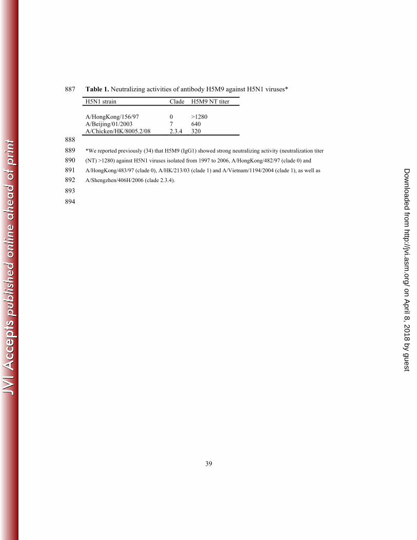
Table 1. Neutralizing activities of antibody H5M9 against H5N1 viruses* 887
H5N1 strain Clade H5M9 NT titer A/HongKong/156/97 0 >1280 A/Beijing/01/2003 7 640 A/Chicken/HK/8005.2/08 2.3.4 320
888
*We reported previously (34) that H5M9 (IgG1) showed strong neutralizing activity (neutralization titer 889
(NT) >1280) against H5N1 viruses isolated from 1997 to 2006, A/HongKong/482/97 (clade 0) and 890
A/HongKong/483/97 (clade 0), A/HK/213/03 (clade 1) and A/Vietnam/1194/2004 (clade 1), as well as 891
A/Shengzhen/406H/2006 (clade 2.3.4). 892
893
894
on April 8, 2018 by guest
http://jvi.asm.org/
Dow
nloaded from
40
Table 2. Data collection and refinement statistics of VN1203 HA and GD1 HA crystals. 895
Data set VN1203 HA –
H5M9 Fab´
GD1 HA GD1 HA –
H5M9 Fab´
Space group C2 P21 P3121
Unit cell (Å)
Unit cell (deg.)
a = 142.6,
b = 251.3,
c = 230.5
β = 107.1
a = 72.5,
b = 225.7,
c = 211.6
β = 99.0
a = b = 199.6,
c = 466.9
Resolution (Å) a 50.0-3.60
(3.83-3.60)
50.0-2.60
(2.69-2.60)
50.0-7.0
(7.44-7.0)
X-ray source APS 23ID-B SSRL 9-2 SSRL 11-1
Unique refs 85,101 196,526 16,538
Redundancy a 2.8 (2.2) 3.4 (2.2) 5.8 (6.2)
Average I/σ(I) a 11.5 (1.2) 22.8 (1.9) 12.9 (1.2)
Completeness a 94.9 (84.3) 95.3 (75.6) 93.3 (64.0)
Rsyma,b 0.10 (0.94) 0.10 (0.70) 0.14 (0.92)
Rpima,b 0.08 (0.72) 0.06 (0.49) 0.06 (0.39)
HA monomers in a.u. 3 9 6
Vm (Å3/Da) 6.1 3.4 4.3
Refs used in refinement
Refined residues
84,244
2,820
195,939
4,464
15,673
5,569
Refined waters 0 117 0
Rcrystc 0.201 0.189 0.377
Rfreed 0.238 0.243 0.387
B-values (Å2)
Protein
Waters
142
-
95
75
-
-
Wilson B-values (Å2) 100 79 -
Ramachandran plot (%)e 92.4, 0.7 97.1, 0.1 -
rmsd bond (Å) 0.012 0.009 -
rmsd angle (deg.) 1.6 1.3 -
896 a Parenthesis denote outer-shell statistics. 897 b Rsym = hkli |Ihkl,i - <Ihkl>| /hkli Ihkl,i and Rpim = hkl[1/(N-1)]1/2i |Ihkl,i - <Ihkl>| /hkli Ihkl,i, where Ihkl,i is 898 the scaled intensity of the ith measurement of reflection h, k, l, < Ihkl> is the average intensity for that 899 reflection, and N is the redundancy.. 900 c Rcryst = hkl |Fo - Fc| / hkl |Fo|, where Fo and Fc are the observed and calculated structure factors. 901 d Rfree was calculated as for Rcryst, but on 5% of data excluded before refinement. 902 e The values are percentage of residues in the favored and outliers regions analyzed by MolProbity (10). 903
on April 8, 2018 by guest
http://jvi.asm.org/
Dow
nloaded from

41
Table 3. Binding of H5 HAs by antibody H5M9 904
H5N1 strain Fab´ Kd (nM) IgG Kd (nM) A/Goose/Guangdong/1/96 1.0 0.008 A/Vietnam/1203/2004 0.9 0.04
905 906
on April 8, 2018 by guest
http://jvi.asm.org/
Dow
nloaded from
42
907 Table 4. Sequence comparison of HA1 domain of H5N1 HAs and H1, H3 HAsa. 908 _________________________________________________________ 909 H5 numbering: 1 10 20 30 40 910 H3 numbering: 10 20 30 34 911 A/Goose/Guangdong/1/96(H5N1) MEKIVLLLAIVSLVK----------SDQICIGYHANNSTEQVDTIMEKNV 912 A/Vietnam/1203/2004(H5N1) MEKIVLLFAIVSLVK----------SDQICIGYHANNSTEQVDTIMEKNV 913 A/Vietnam/1194/2004(H5N1) MEKIVLLFAIVSLVK----------SDQICIGYHANNSTEQVDTIMEKNV 914 A/New Caledonia/20/1999(H1N1) MKAKLLVLLCTFTATY---------ADTICIGYHANNSTDTVDTVLEKNV 915 A/Panama/2007/1999(H3N2) MKTIIALSYILCLVFAQKLPGNDNSTATLCLGHHAVSNGTLVKTITNDQI 916 917 H5 numbering: 41 50 60 70 80 90 918 H3 numbering: 35 40 50 60 70 80 83 919 A/Goose/Guangdong/1/96(H5N1) TVTHAQDILEKTHNGKLCDLNGVKPLILRDCSVAGWLLGNPMCDEFINVP 920 A/Vietnam/1203/2004(H5N1) TVTHAQDILEKKHNGKLCDLDGVKPLILRDCSVAGWLLGNPMCDEFINVP 921 A/Vietnam/1194/2004(H5N1) TVTHAQDILEKTHNGKLCDLDGVKPLILRDCSVAGWLLGNPMCDEFINVP 922 A/New Caledonia/20/1999(H1N1) TVTHSVNLLEDSHNGKLCLLKGIAPLQLGNCSVAGWILGNPECELLISKE 923 A/Panama/2007/1999(H3N2) EVTNATELVQSSSTGRICDSP-HQILDGENCTLIDALLGDPHCDGFQNKE 924 925 H5 numbering: 91 100 110 120 130 140 926 H3 numbering: 83A 90 100 110 120 129 927 A/Goose/Guangdong/1/96(H5N1) EWSYIVEKASPANDLCYPGDFNDYEELKHLLSRTNHFEKIQIIPKS-SWSN 928 A/Vietnam/1203/2004(H5N1) EWSYIVEKANPVNDLCYPGDFNDYEELKHLLSRINHFEKIQIIPKS-SWSS 929 A/Vietnam/1194/2004(H5N1) EWSYIVEKANPVNDLCYPGDFNDYEELKHLLSRINHFEKIQIIPKS-SWSS 930 A/New Caledonia/20/1999(H1N1) SWSYIVETPNPENGTCYPGYFADYEELREQLSSVSSFERFEIFPKESSWPN 931 A/Panama/2007/1999(H3N2) -WDLFVERSKAYSN-CYPYDVPDYASLRSLVASSGTLEFNNESF---NWTG 932 933 H5 numbering: 141 150 160 170 180 190 934 H3 numbering: 130 140 150 160 170 178 935 A/Goose/Guangdong/1/96(H5N1) HDASSGVSSACPYHGRSSFFRNVVWLIKKNSAYPTIKRSYNNTNQEDLLV 936 A/Vietnam/1203/2004(H5N1) HEASLGVSSACPYQGKSSFFRNVVWLIKKNSTYPTIKRSYNNTNQEDLLV 937 A/Vietnam/1194/2004(H5N1) HEASLGVSSACPYQGKSSFFRNVVWLIKKNSTYPTIKRSYNNTNQEDLLV 938 A/New Caledonia/20/1999(H1N1) HTVT-GVSASCSHNGKSSFYRNLLWLTGKNGLYPNLSKSYVNNKEKEVLV 939 A/Panama/2007/1999(H3N2) VAQN-GTSSACKRRSNKSFFSRLNWLHQLKYKYPALNVTMPNNEKFDKLY 940 941 H5 numbering: 191 200 210 220 230 240 942 H3 numbering: 180 190 200 210 220 228 943 A/Goose/Guangdong/1/96(H5N1) LWGIHHPNDAAEQTKLYQNPTTYISVGTSTLNQRLVPEIATRPKVNGQSG 944 A/Vietnam/1203/2004(H5N1) LWGIHHPNDAAEQTKLYQNPTTYISVGTSTLNQRLVPRIATRSKVNGQSG 945 A/Vietnam/1194/2004(H5N1) LWGIHHPNDAAEQTKLYQNPTTYISVGTSTLNQRLVPRIATRSKVNGQSG 946 A/New Caledonia/20/1999(H1N1) LWGVHHPPNIGDQRALYHTENAYVSVVSSHYSRRFTPEIAKRPKVRDQEG 947 A/Panama/2007/1999(H3N2) IWGVHHPSTDSDQISIYAQASGRVTVSTKRSQQTVIPNIGSSPWVRGVSS 948 949 H5 numbering: 241 250 260 270 280 290 950 H3 numbering: 230 240 250 260 270 277 951 A/Goose/Guangdong/1/96(H5N1) RMEFFWTILKPNDAINFESNGNFIAPEYAYKIVKKGDSAIMKSELEYGNC 952 A/Vietnam/1203/2004(H5N1) RMEFFWTILKPNDAINFESNGNFIAPEYAYKIVKKGDSTIMKSELEYGNC 953 A/Vietnam/1194/2004(H5N1) RMEFFWTILKPNDAINFESNGNFIAPEYAYKIVKKGDSTIMKSELEYGNC 954 A/New Caledonia/20/1999(H1N1) RINYYWTLLEPGDTIIFEANGNLIAPWYAFALSRGFGSGIITSNAPMDEC 955 A/Panama/2007/1999(H3N2) RISIYWTIVKPGDILLINSTGNLIAPRGYFKI-RSGKSSIMRSDAPIGKC 956 957 H5 numbering: 291 300 310 320 330 340 958 H3 numbering: 280 290 300 310 320 959 A/Goose/Guangdong/1/96(H5N1) NTKCQTPMGAINSSMPFHNIHPLTIGECPKYVKSNRLVLATGLRNTPQRE 960 A/Vietnam/1203/2004(H5N1) NTKCQTPMGAINSSMPFHNIHPLTIGECPKYVKSNRLVLATGLRNSPQRE 961 A/Vietnam/1194/2004(H5N1) NTKCQTPMGAINSSMPFHNIHPLTIGECPKYVKSNRLVLATGLRNSPQRE 962 A/New Caledonia/20/1999(H1N1) DAKCQTPQGAINSSLPFQNVHPVTIGECPKYVRSAKLRMVTGLRNIPS-- 963 A/Panama/2007/1999(H3N2) NSECITPNGSIPNDKPFQNVNRITYGACPRYVKQNTLKLATGMRNVPE-- 964 _________________________________________________________ 965 a Red indicates H5M9 epitope residues on A/Vietnam/1203/2004 (VN1203) HA. The only amino 966 acid difference between VN1203 HA and A/Vietnam/1194/2004 (VN1194) HAs is highlighted in 967 green. The amino acid difference between VN1203 and GD1 HAs are highlighted in cyan. The 968 H1 and H3 residues not conserved in H5M9 epitope are highlighted in yellow. H3 numbering 969 system is used throughout in this paper and H5 numbering is shown here for comparison. 970 971
on April 8, 2018 by guest
http://jvi.asm.org/
Dow
nloaded from
43
972 Table 5. Antibody H5M9 epitope residues in VN1203 HA and comparison with other 973
HAs from H5N1 viruses neutralized by H5M9 in this studya. 974
Amino acid position
Viral strain Clade 53 55 56 57 62 78 81 83 83a 117 119 273 274 276 278
A/Vietnam/1203/2004 1 D D V K R E N P E H E E Y N N
A/goose/Guangdong/1/96 0 · N · · · · · · · · · · · · ·
A/HongKong/156/97 0 · N · · · · · · · · · · · · ·
A/HongKong/483/97 0 · N · · · · · · · · · · · · ·
A/HongKong/213/03 1 · · · · · · · · · · · · · · ·
A/Vietnam/1194/2004 1 · · · · · · · · · · · · · · ·
A/Shenzhen/406H/2006 2.3.4 · · · · · · · · · · · · · · ·
A/Beijing/01/2003 7 · · · · · · · · · · · · · · ·
975 a The identified key epitope residues are shown in boldface type. 976 977
on April 8, 2018 by guest
http://jvi.asm.org/
Dow
nloaded from
44
Table 6. Conservation of H5M9 epitope residues in influenza H5N1 HAsa. 978 979
Amino Residue number
acid 53 55 56 57 62 78 81 83 83a 117 119 273 274 276 278
Ala 3 0 0 0 0 6 0 0 0 0 0 2 0 0 0
Cys 0 0 0 0 0 0 0 0 0 0 0 0 1 0 0
Asp 1,326 1,314 0 0 0 0 26 0 0 0 1 1 0 25 35
Glu 1 3 0 1 0 1,565 0 0 1,572 0 1,560 1,557 0 0 0
Phe 0 0 0 0 0 0 0 0 0 0 0 0 0 0 0
Gly 0 1 1 0 4 0 0 0 1 0 0 11 0 0 0
His 0 0 0 0 0 0 2 0 0 1,484 0 0 1 0 0
Ile 0 0 7 0 0 0 0 0 0 0 0 0 1 0 1
Lys 0 37 0 1,503 260 7 12 0 3 0 18 6 0 0 1
Leu 0 0 0 0 0 0 0 1 0 0 0 0 0 0 0
Met 0 0 2 0 0 0 0 0 0 0 0 0 0 0 0
Asn 197 221 0 0 0 0 1,537 0 0 0 0 0 0 1,545 1,480
Pro 0 0 0 0 0 0 0 1,443 0 0 0 0 0 0 0
Gln 0 0 0 0 0 0 0 7 0 0 0 0 0 0 1
Arg 0 0 0 75 1,315 0 0 0 0 95 0 0 0 0 0
Ser 52 0 0 0 0 0 1 127 0 0 0 0 0 5 56
Thr 0 0 0 0 0 0 0 0 0 0 0 0 0 0 2
Val 0 0 1,569 0 0 0 0 0 1 0 0 0 0 0 0
Trp 0 0 0 0 0 0 0 0 0 0 0 0 0 0 0
Tyr 0 3 0 0 0 0 0 0 0 0 0 0 1,574 2 1
980 a The incidence of an amino acid occurring at certain position is shown. A total of 1,579 full-length, non-981
redundant HA sequences from all influenza H5N1 viruses were available in the Influenza A Virus Resource 982
at the NCBI in January 22, 2013. 983
984
on April 8, 2018 by guest
http://jvi.asm.org/
Dow
nloaded from
45
Table 7. Conservation of H5M9 epitope residues of VN1203 H5 HA in different clades 985
and subclades of H5N1 virusesa. 986
Amino acid position
Viral strain Clade 53 55 56 57 62 78 81 83 83a 117 119 273 274 276 278
A/Vietnam/1203/2004 1 D D V K R E N P E H E E Y N N
A/Goose/Huadong/1/2000 0 · · · · · · · · · · · · · · ·
A/Cambodia/S1211394/2008 1.1 · · · R · · · · · · · · · · ·
A/Chicken/Indonesia/11/2003 2.1.1 · · · · · · · · · · · · · · ·
A/Indonesia/542H/2006 2.1.2 · · · · · · · · · · · · · · ·
A/Indonesia/5/2005 2.1.3.2 · · · · · · · · · · · · · · ·
A/Chicken/Medan/BPPVRI_15/2008 2.1.3.3 · · · · · · · · · · · · · · ·
A/bar-headed goose/Qinghai/12/05 2.2 · · · · · · · · · · · · · · ·
A/turkey/Turkey/1/2005 2.2.1 · · · · · · · · · · · · · · ·
A/Bangladesh/207095/2008 2.2.2 · · · · · · · · · · · · · · ·
A/Duck/Hunan/127/2005 2.3.1 · · · · · · · · · · · · · · ·
A/chicken/Guangdong/1/2005 2.3.2 · · · · · · · · · · · · · · ·
A/Hubei/1/2010 2.3.2.1 · N · · K · · · · · · · · · ·
A/chicken/Guiyang/3055/2005 2.3.3 · · · · · · · · · · · · · · ·
A/Anhui/1/2005 2.3.4 · · · · · · · · · · · · · · ·
A/chicken/Sichuan/81/2005 2.3.4.1 · · · · · · · · · · · · · · ·
A/Vietnam/HN36250/2010 2.3.4.2 · · · · · · · · · · · · · · · A/chicken/Vietnam/NCVD-188/2008 2.3.4.3 · · · · · · · · · · · · · · ·
A/chicken/jiyuan/1/03 2.4 · · · · · · · · · · · · · · ·
A/chicken/Korea/ES/03 2.5 · · · · · · · · · · · · · · ·
A/chicken/HongKong/YU562/01 3 · · · · · · · · · · · · · · ·
A/goose/Fujian/bb/2003 4 · · · · · · · · · · · · · · ·
A/goose/Guangxi/1097/2004 5 · · · · · · · · · · · · · · ·
A/swine/Anhui/ca/2004 6 · · · · · · · · · · · · · · ·
A/duck/Yunnan/5133/2005 7 · · · · · · · · · · · · · · ·
A/Ck/HK/YU777/02 8 · · · · · · · · · · · · · · ·
A/chicken/Fujian/1042/2005 9 · · · · · · · · · · · · · · ·
A/chicken/Egypt/1029/2010 2.2.1 N · · · · · · · · · · · · · ·
A/chicken/Shanxi/10/2006 7.1 N · · · K · · S · · K · · · ·
A/chicken/Henan/A-7/2006 7.2 N · · · K · · S · · K · · · ·
987 a The identified key epitope residues are shown in boldface type. 988 989
on April 8, 2018 by guest
http://jvi.asm.org/
Dow
nloaded from

46
Table 8. Conservation of H5M9 HA epitope in HAs of influenza virus subtypes H5N1 to 990 H5N9. 991 992
Residue
Percent Conservation from H5N1 to H5N9 Consensus a
H5N1
(1679)b H5N2 (379)
H5N3 (68)
H5N4 (4)
H5N5 (4)
H5N6 (2)
H5N7 (7)
H5N8 (5)
H5N9 (21)
53 D 85.9 c 1.8 (S)d 0.0 (S) 0.0 (S) 75.0 0.0 (S) 0.0 (S) 0.0 (S) 0.0 (S)
55 D 86.1 0.8 (K) 0.0 (N) 0.0 (K) 50.0 0.0 (N) 0.0 (K) 0.0 (N) 0.0 (N)
56 V 99.4 99.7 100.0 100.0 100.0 100.0 100.0 100.0 100.0
57 K 95.9 46.7 (R) 69.1 0.0 (R) 75.0 100.0 28.6 (R) 80.0 47.6 (R)
62 R 86.1 32.2 (K) 64.7 0.0 (K) 75.0 100.0 28.6 (K) 80.0 42.9 (K)
78 E 99.4 97.1 98.5 100.0 100.0 100.0 100.0 100.0 95.2
81 N 98.0 92.3 94.1 100.0 50.0 100.0 100.0 100.0 100.0
83 P 93.6 99.2 100.0 100.0 100.0 100.0 100.0 100.0 90.5
83a E 99.6 99.7 100.0 100.0 100.0 100.0 100.0 100.0 100.0
117 H 95.4 95.0 100.0 100.0 100.0 100.0 100.0 100.0 100.0
119 E 99.0 97.9 98.5 100.0 100.0 100.0 100.0 100.0 100.0
273 E 98.9 87.6 91.2 100.0 100.0 100.0 71.4 100.0 95.2
274 Y 99.7 100.0 100.0 100.0 100.0 100.0 100.0 100.0 100.0
276 N 98.4 98.1 100.0 100.0 100.0 100.0 100.0 100.0 100.0
278 N 95.2 40.6 (D) 72.1 0.0 (D) 75.0 100.0 28.6 (D) 80.0 47.6 (D)
993 a Most common residue at position by simple majority in H5N1 virus HA sequences (full-length, non-994
redundant) that were available in the Influenza A Virus Resource at the NCBI in January 22, 2013. 995 b Number of sequences available for this subtype in the Influenza A Virus Resource at the NCBI in January 996
22, 2013. 997 c Percent of all H5 sequences that are identical to the consensus. 998 d Most common residue in this subtype. 999
1000
on April 8, 2018 by guest
http://jvi.asm.org/
Dow
nloaded from
47
Figure Legends 1001 1002 FIG 1 Crystal structure of antibody H5M9 Fab´ in complex with VN1203 HA. (A) 1003
Overall structure of the H5M9 Fab´ - VN1203 HA complex. The H5M9 Fab´ binds in the 1004
vestigial esterase subdomain of the HA. One HA/ Fab´ protomer of the trimeric complex 1005
is colored with HA1 in pink and HA2 in cyan, with the corresponding bound Fab´ heavy 1006
chain in orange and Fab´ light chain in yellow. N-linked glycans are depicted in colored 1007
balls representing their atom types, and asparagines that code for potential N-1008
glycosylation sites are also shown in sticks. The other two protomers are in grey, but the 1009
third Fab molecule is hidden behind the HA trimer. (B) Surface presentation illustrating 1010
the H5M9 binding site (in green) on one VN1203 HA protomer, which is colored with 1011
HA1 in pink and HA2 in cyan. The H5M9 footprint on VN1203 HA is far from the 1012
receptor binding site (RBS, in red). (C) Closer view of the H5M9 epitope with selected 1013
epitope residues shown as sticks. The four identified key residues are labeled in red. (D) 1014
Footprint of HA on H5M9 combining site (i.e. paratope), highlighting contacting 1015
antibody residues from the heavy chain (colored with orange carbon atom) and light 1016
chain (with yellow carbon atoms). 1017
1018
FIG 2 Overall structures of GD1 H5 HA and its complex with antibody H5M9. (A) GD1 1019
H5 HA in apo form. One HA protomer of the trimeric complex is colored with HA1 in 1020
pink and HA2 in cyan, with the other two protomers in green. N-linked glycans are 1021
depicted in colored balls representing their atom types, and asparagines that code for 1022
potential N-glycosylation sites are also shown in sticks. (B) GD1 H5 HA in complex with 1023
H5M9 Fab´. The H5M9 Fab´ binds in the vestigial esterase subdomain of the HA, which 1024
on April 8, 2018 by guest
http://jvi.asm.org/
Dow
nloaded from

48
is the same epitope as that in VN1203 H5 HA (Fig. 1). One HA/ Fab´ protomer of the 1025
trimeric complex is colored with HA1 in pink and HA2 in cyan, with the corresponding 1026
Fab´ heavy chain in orange and Fab´ light chain in yellow. The other two protomers are 1027
in green and the third Fab´ molecule is hidden behind the HA trimer. 1028
1029
FIG 3 Structural comparison of GD1 HA apo-form (in yellow side chains and pink CĮ 1030
atoms) and VN1203 HA (PDB code 3GBM, in grey side chains and CĮ atoms). (A) 1031
Comparison of the receptor binding sites. The overall structures as well as key binding 1032
residues are virtually identical for except one amino acid substitution of residue 221 from 1033
Pro (GD1 HA) to Ser (VN1203 HA) near the receptor binding site. (B) Comparison of 1034
the H5M9 epitopes in GD1 and VN1203 HAs. The H5 epitope residues are 1035
superimposable with only one amino acid substitution of Asn55 (GD1 HA) to Asp55 1036
(VN1203 HA). 1037
1038
FIG 4 Receptor binding was investigated against printed glycans on a microarray (Fig. 1039
S2) for GD1 HA (A) and control VN1203 HA (B). Different categories of glycans on the 1040
array are highlighted in colors, and the bars denote the fluorescent signal intensity, and 1041
error bars indicate the standard errors. The normal starting concentration of 15 µg/ml was 1042
applied to both HAs. Both GD1 HA and VN1203 HA showed similar binding affinities to 1043
most Į2-3 linked glycans, but no obvious binding to any Į2-6 linked glycan. 1044
Interestingly, VN1203 HA also showed strong binding to fucosylated Į2-3 glycans (in 1045
yellow box), in contrast to GD1 HA, which did not bind these glycans. 1046
1047
on April 8, 2018 by guest
http://jvi.asm.org/
Dow
nloaded from
49
FIG 5 Escape mutant D53N of VN1994 HA to antibody H5M9. (A) Neutralization 1048
activity of H5M9 to H5N1 viruses with wild-type VN1194 HA and its escape mutant 1049
D53N. Neutralization activities were evaluated by plaque reduction neutralization assays 1050
using MDCK cells, which showed that D53N mutation confers resistance to H5M9. The 1051
data are representative of triple independent experiments. 1052
(B) Electrostatic potential surfaces around H5M9 paratope of antibody H5M9 1053
combining site with the interacting Asp53 from VN1203 HA labeled. Electrostatic 1054
potential surfaces around wild-type VN1203 HA with Asp53 (C) and VN1203 HA with 1055
the Asp53Asn mutant model (D). Electrostatic surface potentials were calculated using 1056
the APBS program (3). Negatively charged regions are red, positively charged regions are 1057
blue, and neutral regions are white (-10 to 10 KbT/ec potential range). The Asp53Asn 1058
mutation alters the electrostatic potential from weakly acidic (C) to near neutral (D) 1059
around the HA1 53 position, rendering it unfavorable to bind H5M9, which is weakly 1060
basic around the interaction site with HA1 Asp53 (B). 1061
1062
FIG 6 Flow cytometry histograms of antibody H5M9 binding to wild-type or mutant of 1063
VN1194 HA proteins from yeast surface display. Representative histograms are shown 1064
for anti-Xpress antibody, antibody H5M9, and antibody H5M11 with wild-type HA and 1065
each of the HA mutants. The H5 proteins expressed well on the yeast cell surface and can 1066
be detected by the reactivity of the Xpress epitope tag in the HA protein with the anti-1067
Xpress antibody, as well as the binding activity by a control antibody H5M11 (IgG2a) 1068
elicited from the same mouse immunization as H5M9 (IgG1), which appears to bind to a 1069
different HA epitope (34). Arrows indicate mutations that result in significant or almost 1070
on April 8, 2018 by guest
http://jvi.asm.org/
Dow
nloaded from
50
complete loss of antibody binding. Data shown are representative of at least three 1071
independent experiments. 1072
1073
FIG 7 Reducing SDS-PAGE of trypsin digestion of VN1203 HA in the absence (lanes 1 1074
and 2) and presence (lanes 3 and 4) of H5M9 Fab´. The HA or HA- Fab´ mixture was 1075
exposed to pH 4.9 (lanes 1 and 3) or pH 8.0 (lanes 2 and 4) at 37 ºC for one hour before 1076
trypsin digestion at pH 8.4. As expected, the HA1 could be completely digested at pH 4.9 1077
without H5M9 Fab´ (lane 1), while in the control at pH 8.0, the HA1 remains contact. 1078
After precomplexing the HA with H5M9 Fab´, the HA appeared to be resistant to trypsin 1079
digestion even at pH 4.9 in addition to pH 8.0. These results suggest H5M9 may prevent 1080
low pH-induced conformational changes of the HA. 1081
1082
FIG 8 Prophylactic and therapeutic efficacy of H5M9 against lethal H5N1 infection in 1083
mice. (A) Survival curves (left) and body weight loss (right) of BALB/c mice (n=6) that 1084
received 2 or 20 mg/kg of H5M9 on 24 hours before intranasal infection with 10 MLD50 1085
(50% mouse lethal dose) of VN1194 virus. (B) Survival curves (left) and body weight 1086
loss (right) of mice (n=6) that received 2 or 20 mg/kg of H5M9 on 24 hours post 1087
infection with 10 MLD50 of VN11904 virus. 1088
1089
FIG 9 Comparison of antibody epitopes mapped onto the surface of corresponding HAs. 1090
(A) VN1203 H5 HA with the H5M9 footprint shown in green. (B) A/Aichi/2/68 (H3N2) 1091
with the HC45 footprint shown in red (PDB ID code 1QFU). The H3 HA1 domain was 1092
superimposed on the HA1 domain of VN1203 H5 for this comparison. 1093
on April 8, 2018 by guest
http://jvi.asm.org/
Dow
nloaded from