1. a double sucrose-gap voltage-clamp t
TRANSCRIPT

J. Physiol. (1975), 250, pp. 175-202 175With 13 text-ftgurePrinted in Great Britain
EFFECTS OF STIMULATING THEACETYLCHOLINE RECEPTOR ON THE CURRENT-VOLTAGERELATIONSHIPS OF THE SMOOTH MUSCLE MEMBRANE
STUDIED BY VOLTAGE CLAMP OF POTENTIALRECORDED BY MICRO-ELECTRODE
BY T. B. BOLTONFrom the University Department of Pharmacology, Oxford, OX1 3QT
(Received 9 January 1975)
SUMMARY
1. A double sucrose-gap voltage-clamp technique is described for use onsmooth muscle strips longer than about 2 mm. It involves intracellularrecording by microelectrode of the membrane potential of a narrow regionof the strip ('node') sandwiched between two streams of deionized sucrosesolution. Current was passed into the node across one or both sucrosestreams.
2. Preliminary experiments in which potential was recorded intra-cellularly at two points during polarization of a 'short cable' preparation,formed by folding over a strip of smooth muscle, suggested that a nodewidth of less than 0O15 mm was needed to achieve uniform potentialduring inward current flow. However, when node width between sucrose-gaps was reduced to 05 mm, spontaneous electrical activity was lost, andbelow 05 mm spike threshold was raised and the regenerative spikebecame graded. The currents flowing during the application of rectangularvoltage-clamp command potentials were described.
3. Using taenia smooth muscle it was shown by recording with a second,independent micro-electrode that potential was not uniform for up to200 ms or more following a step change in potential under voltage-clampin nodes 004-0 5 mm wide where current was passed across both sucrosegaps. However, reasonably uniform nodal potentials were obtained usingramps with relatively slow rates of rise (25 mV/s).
4. Using such slow ramp commands under voltage clamp, the effects ofcarbachol on the current-voltage relationship of longitudinal muscle ofileum and taenia were studied in hypertonic solution.
5. In the presence of carbachol (10- to 10 g/ml.) additional inwardcurrent flowed across the membrane (in some experiments an equilibriumpotential was observed at which this current reversed direction). The

magnitude of this additional current was linearly related to potential atpotentials negative to the resting potential. At potentials positive to theresting membrane potential, this additional current increased with de-polarization over the range -40 to -10 mV; in ileum the effect of thisadditional inward current on the current-voltage relationship was toproduce a region of net inward current where before, in the absence ofcarbachol, a net outward current existed. In taenia the additional inwardcurrent flowing in the presence of carbachol was too small to produce aregion of net inward current; thus carbachol produced regenerative slowoscillations of potential (slow waves) in ileum but not in taenia.
6. These results support a previous suggestion that activation of theacetylcholine receptor of ileal smooth muscle produces an additionalinward current in the membrane which increases with depolarization andis responsible for the regenerative slow waves seen when muscarinicstimulants are applied. A similar effect apparently operates in taenia butthe additional inward current is too small to produce regenerative slowwaves.
INTRODUCTION
Stimulation of the acetylcholine receptor of smooth muscle of ileumproduces large, relatively slow (- 1 s) regenerative oscillations of themembrane potential. These may first appear as a prolonged delay ofrepolarization following a spike, and as the spike is reduced in size duringthe action of some agonist such as carbachol, they develop an almostsinusoidal form. The mechanism of these oscillations, or slow waves, isunknown, but they are sensitive to a reduction in the external sodiumconcentration and membrane conductance is increased at their peak. Insucrose hypertonic solution such waves do not occur in the absence ofstimulation of the acetylcholine receptor, depolarizing current elicitingonly repetitive spiking. However, if a small concentration of carbachol ispresent a similar depolarization (if suprathreshold) elicits slow waves ofabout 3 s duration upon which spikes are superimposed. Clearly thepresence of slow waves depends upon stimulation of the acetylcholinereceptor and their mechanism is a regenerative, voltage-dependent one(Bolton, 1971, 1972a).
Since these previous results were obtained using larger pieces of smoothmuscle, it is conceivable that slow waves are a form of spike modified bythe additional conductance introduced into the membrane by stimulatingthe acetylcholine receptor (Bolton, 1972b). Thus, a spike may arise in aremote or deep part of the muscle (Osa & Taga, 1973) but, upon pro-pagating to the region of recording its duration is greatly increased and itssize reduced by the increased conductance. An argument against this
176 T.B.BOLTON
VOLTAGE CLAMP OF SMOOTH MUSCLE
hypothesis is that normal spikes can sometimes be recorded as taking off atthe peak of the slow wave (fig. 7 of Bolton, 1971). Strong evidence againstthis hypothesis might be obtained if it was possible to record from piecesof smooth muscle which could be shown to be polarized sufficiently uni-formly for asynchronous firing of spikes to be unlikely. This paper de-scribes the results of experiments of this type.An alternative slow wave mechanism which was suggested (Bolton,
1971) was that stimulation of the acetylcholine receptor somehow alteredthe current-voltage relationship of an inward current channel whoseconductance increased with depolarization. Thus, without muscarinicstimulation this channel would carry insignificant current since slow wavesdo not occur, but, when the acetylcholine receptor is stimulated, thispostulated channel would carry a significant inward current producing aslow wave.
Evidence to support such a hypothesis must come from some form ofvoltage-clamp technique. Early experiments (Bolton, 1974a) using thedouble sucrose-gap voltage-clamp apparatus of Rougier, Vassort &Stampifli (1968) with extracellular potential recording, were somewhatdisappointing, since muscarinic stimulants did not produce the oscil-lations of potential typical of their action on larger pieces of ileal smoothmuscle. It was not, therefore, surprising that under voltage clamp, noimportant voltage-dependence of the additional conductance appearing inthe presence of carbachol could be detected. A significant factor in theexperiments was probably the small size of the region of active muscle('node') between sucrose streams, which has the effect of increasing therelative importance of leakage current (McGuigan, 1974). To reduce theimportance of leakage current, node width was increased in the presentexperiments, and an alternative voltage-clamp method developed (Bolton,1974b).There have been a number of voltage-clamp studies on smooth muscle
in which the potential has been recorded extracellularly and in which theassumption of uniform nodal potential under voltage-clamp conditions hasbeen made (e.g. Anderson, 1969; Kumamoto & Horn, 1970; Anderson,Ramon & Snyder, 1971; Kao, 1971; Mironneau & Lenfant, 1972; Miron-neau, 1973; Kao & McCullough, 1975). Voltage-clamp techniques on multi-fibre preparations have been severely criticized on the grounds of lack ofuniformity of nodal potential (Johnson & Lieberman, 1971). Experimentsdescribed in this paper substantially support the theoretical doubts whichwere raised. Because of this, and since the present voltage-clamp methodhas not been previously applied to smooth muscle, an important part ofthis paper is concerned with establishing conditions under which nodalpotential is uniform, or nearly so.
177
178 T. B. BOLTONThe results of the present work support the previously suggested hypo-
thesis that stimulation of the acetylcholine receptor of smooth muscleresults in additional inward membrane current at the resting membranepotential, and this current increases with depolarization, as reported inpreliminary communications (Bolton, 1975a, b).
METHODS
Double sucrose gapPreparation
Strips of longitudinal smooth muscle were cut from taenia or terminal ileum ofguinea-pigs weighing 200-400 g. Longitudinal ileal muscle was separated from thebulk of the circular muscle by a technique described previously (Bolton, 1972b). Incross-section taenia strips were about 0 1 mm thick and 0-3-0 5 mm wide when inposition in the apparatus. Ileal strips were slightly thinner and about 0-8 mm wide.
PSS
M. E. |M. E.
Kg-AgCI| 1 By Ag-AgCl v MEAgfAgCl KC
Ag-AgQ ~~Suc ma Suc TY3tjl
Current electrode Current electrode
Ag-AgCICurrent electrode
14 0 75 -1* 0 5 04140*75 1mm mm mm
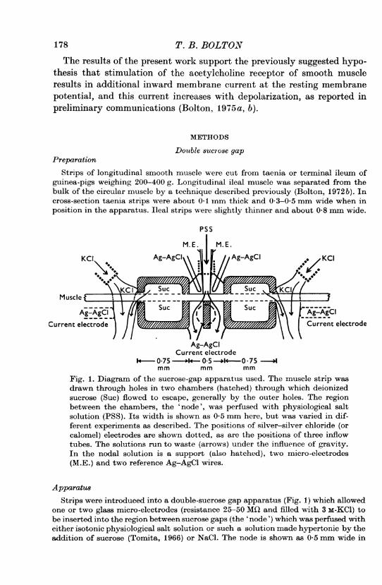
Fig. 1. Diagram of the sucrose-gap apparatus used. The muscle strip wasdrawn through holes in two chambers (hatched) through which deionizedsucrose (Suc) flowed to escape, generally by the outer holes. The regionbetween the chambers, the 'node', was perfused with physiological saltsolution (PSS). Its width is shown as 0 5 mm here, but was varied in dif-ferent experiments as described. The positions of silver-silver chloride (orcalomel) electrodes are shown dotted, as are the positions of three inflowtubes. The solutions run to waste (arrows) under the influence of gravity.In the nodal solution is a support (also hatched), two micro-electrodes(M.E.) and two reference Ag-AgCl wires.
ApparatusStrips were introduced into a double-sucrose gap apparatus (Fig. 1) which allowed
one or two glass micro-electrodes (resistance 25-50 MO and filled with 3 m-KCl) tobe inserted into the region between sucrose gaps (the 'node') which was perfused witheither isotonic physiological salt solution or such a solution made hypertonic by theaddition of sucrose (Tomita, 1966) or NaCl. The node is shown as 0 5 mm wide in

VOLTAGE CLAMP OF SMOOTH MUSCLE 179Fig. 1 but the effects of varying node width from 0-1 to 1 mm or more will bedescribed.The sucrose chambers were made of film base (Kodak 2495 RAR film) cemented
together with Araldite epoxy resin (Ciba). Their width was about 0-8 mm and musclestrips were drawn through holes 0 3 mm diameter in these chambers from the nodalside. Usually both ends of the muscle strip were depolarized in 154 mM-KCl solution.A 292 mm sucrose solution was made by dissolving Analar grade sucrose and de-ionized by passing through an ion exchange resin (Elgastat type B1102, Mk II). Inlater experiments 10-5 M-CaCl2 was added to this solution (New & Trautwein, 1972;Kleber, 1974).
Current was passed between silver-silver chloride or calomel electrodes (Ives &Janz, 1961) connected downstream to the central node or K streams by 3 M-KCl 4%agar bridges about 1 cm long. Two chlorided silver wires (100 jnm diam) dipped intothe nodal solution and were positioned touching, or almost touching, the muscle atone side.In the experiments where the effects of varying node width were examined, one
sucrose gap was replaced by a short tube (i.d. about 0 5 mm) through which sucrosesolution flowed and this side of the node was formed as an interface between physio-logical salt solution and sucrose solution without a physical partition. In this casethat end of the muscle was not depolarized in high K and current was passed acrossonly one sucrose gap. In all other experiments current was passed across both sucrosegaps.
Series resistance. This varied depending among other factors upon dimensions ofthe node. In the experiment illustrated in Fig. 8 the potential step observed uponwithdrawing the electrode from the cell was less than 1 mV when about 3-5 ,#A werepassed. Assuming all this current passes through r8 (Fig. 2) (i.e. re > r1, which isalmost certainly not true) then the series resistance is less than 300 Q. This current,passing through rm produced a steady-state hyperpolarizing electrotonic potentialof 50 mV. Since the current-voltage relationship is linear, node resistance at theresting membrane potential is about 15 ki and the ratio of rm/rs is 50. This ratio isunaffected if the assumption that re > ri is not true. In the experiment of Fig. 9where the node width was 0 5 mm, rm/r8 is about 25.
ElectronicsThe essentials of the circuit used are shown inFig. 2. The muscle strip in the node is
represented by a single battery, resistor, and capacitor in parallel but under someconditions this representation is insufficient and a second membrane unit is intro-duced connected by interrupted lines. The internal (core) resistance of the musclethrough both gaps (in parallel) is r1, and the external resistance across the gaps (inparallel) is re. The resistance in series with the membrane is rj(ohms).One chlorided silver wire was used as an independent electrode for the input
which was differential for both micro-electrodes. The other chlorided silver wire wasconnected to the negative input of an operational amplifier and nodal solution wasclamped at virtual ground potential by the appropriate position of switch C. Thecurrent required to do this during voltage-clamp, or when passing rectangular currentpulses was measured as the potential across a 1 kn resistor. This potential was fedinto the voltage-clamp amplifier, when rectangular current pulses were required, bythe appropriate position of switch B; RB was then reduced and RF increased, clamp-ing the current between nodal and KCl streams. Rectangular or ramp currents couldthen be applied from the wave-form generator. The membrane was always held atthe resting membrane potential (zero current) when in the current clamp mode.For voltage clamp the output from one electrode was chosen by the position of

switch A and fed into the voltage-clamp amplifier by means of switch B. Theresistance R8 was reduced and RF increased. The holding potential was chosen byadjusting the offset, and voltage-clamp commands were produced by the wave formgenerator. The resistance of both sucrose gaps in parallel (= (r, re)/(ri + re) wasmeasured as the potential across the 1 kn resistor when a 100 mV, 10 Hz, sine wavewas applied between the node and the KCl streams by the appropriate positions ofswitch C. Typical resistances lay in the range 40-200 kQ.
I C]CROP2SimpUlifSi KCIxa0 croa
OffsetRF
Votage-C Wae-form clamp
generatorRs
amplifier
Ground-clamp C 1amplifier
I K
Fig. 2. Simplified diagram of the circuit used. The double sucrose gap isrepresented by a single gap here (gaps were connected in parallel). Thusr1 (il) is the internal (core) resistance of the preparation across both gaps inparallel; re, (Qi) is the external resistance across both gaps in parallel.Operational amplifiers labelled 1 had high input impedance, those labelled2, low input impedance. CRO are the inputs to the oscilloscope. The hatchedboxes are Ag-AgCl chloride or calomel electrodes connected to the nodalsolution or KCI streams. By appropriate positions of switch B either thecurrent flowing between the node, the KC1 streams, or the potential recordedby one or other of the micro-electrodes, can be clamped. For further detailssee text.
To prevent oscillation during voltage or current clamp a suitable value of CF waschosen. As can be seen from the records, control of recorded potential after a steppotential change under voltage clamp could be achieved within a few millisecondswhich was adequate for present purposes. In some ramp experiments the currentoutput was filtered by a RC circuit with variable time constant immediately beforethe input to the oscilloscope. A routine check was made to ensure that this did notaffect the shape of the ramp obtained.
180 T.B.BOLTON

VOLTAGE CLAMP OF SMOOTH MUSCLE 181
Phyeiological 8alt 8olutioBn and drug
The isotonic Krebs solution used to bathe the node had the following composition(mM): NaCl 120; KCl 5 9, CaCJ2 2-5, NaHCO3 15, NaH2PO4 12, MgCl2 1P2, glucose 1.Hypertonic solution was made by adding 320 mM sucrose or 234 mx (extra) sodiumchloride to isotonic Krebs solution. All solutions were equilibrated with 3% CO2 and97 % 02 before use. The temperature of the nodal solution was 32-34' C. Carbacholchloride was introduced to the muscle by changing the physiological salt solution toone that contained it.
Short-cable folded preparations
A few experiments were done on ileal muscle strips about 2 mm wide. These werefolded over as shown diagrammatically in Fig. 3 to create a short cable in which thedecline of potential is expected to be less than in the usual 'infinite cable' prepara-tion. Current was injected close to the folded end of this preparation by means of asilver-silver chloride plate electrode (electrode 3) insulated on the side facing tworecording micro electrodes (electrodes 1 and 2) as depicted. To reduce short-circuitinga 2 mm sucrose gap was formed between the current passing electrode and itspartner (electrode 4) in a KCI pool. The folded end of the muscle was secured bymicropins (T. Gerrard & Co.) and bathed in sucrose hypertonic physiological saltsolution.
500 Ms
__U
0 025
025< 02 3o.6-4. 3 6 4_
Fig. 3. Decline of potential in a 'short-cable' preparation of ileal smoothmuscle. A closed end of the cable was formed by folding over the musclestrip as shown in the diagrams. Current was passed into the cable by meansof electrode 3 (an external Ag-AgCl electrode) and sucrose gap (Suc). Elec-trode 4 is the indifferent electrode in the KCl pool. Potential was recordedat the indicated points (dimensions in mm) in two preparations by meansof micro-electrodes 1 and 2. The records above are labelled accordingly.The inset shows the form of the current pulses applied. As shown by thedifferential records (D) when the preparation was short, X 0 5 (a-d, andleft-hand diagram) the decline in the electrotonic potential was very small,but, when the preparation was longer, X > A (e-h, and right-hand diagram)then there was an appreciable decline in the electrotonic potential.
T. B. BOLTON
h
.40
30 r-0
(4Ul -20 ja.
l l -101 101 l
_ 'I _ -- I 0
0 2 0.3 0 4 0-5 0-6 mm from closed.
MIE1 I T end of cable
M.E. 1 M.E. 2 Currentelectrode.
Fig. 4. For legend see facing page.
182
E0)An
I s
C
0-1

VOLTAGE CLAMP OF SMOOTH MUSCLE
RESULTS
Potential decline in 'short-cable' preparationsDespite their multifibre nature, the passive electrical properties of
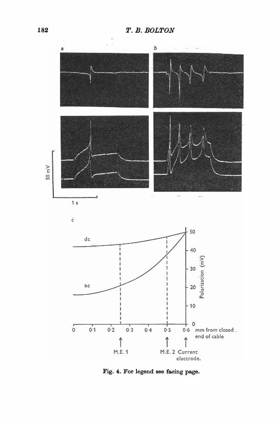
strips of intestinal smooth muscle are approximately those of an electricalcable, the decline of extrapolar potential with distance from a current-passing electrode being exponential (Shuba, 1961; Tomita, 1966; Abe &Tomita, 1968). Potential decline in a cable is reduced near a closed end(e.g. see Adrian, Chandler & Hodgkin, 1970). To test this, a 'short cable'preparation of smooth muscle was created by folding over strips of longi-tudinal ileal muscle, as indicated diagrammatically in the insets to Fig. 3.The folded ends of the strips were polarized as described by an externalcurrent passing electrode (and sucrose gap). In this arrangement the upperand lower parts of the folded strip can be considered to be two cables bothof which are 'closed' at the fold, since no axial current can flow at thispoint. (Alternatively the portion of the muscle between the externalelectrode and the fold can be considered to represent the end of a singlecable, terminating at the fold, with a longitudinal internal septum whichdoes not affect current distribution.) Thus the folded end of the smoothmuscle strip is theoretically analogous to the end of a skeletal musclefibre as used by Adrian et al. (1970). Two micro-electrodes were used torecord the potential at different points in hypertonic physiological saltsolution while hyperpolarizing or depolarizing rectangular current pulseswere passed. The difference in potential between these two points wa" alsodisplayed on the oscilloscope (Figs. 3b d, f-h and Fig. 4, upper line).When the length of muscle, x (mm), beyond the current electrode was
short compared to the space constant, A (mm), of this muscle (about1-3 mm according to Hidaka & Kuriyama, 1969), the decline in potentialobserved in response to constant hyperpolarizing currents was very small:
Fig. 4. Variation in potential in a 'short-cable' preparation during spikedischarge. The preparation is the same one as in Fig. 3a-d. The upper lines ina and b show the recorded potential difference between the two electrodes.Providing membrane resistance does not fall there is little decline of directcurrent polarization along this preparation, but considerable potentialvariation occurs during a spike. In c is shown the theoretical decline ofpotential in a short cable in which current injection is made 06 mm froma closed end. Line d.c. shows the decline of polarization produced by aconstant current when A = 1-0 mm. Line a.c. shows the expected decline ifmembrane resistance falls to a tenth of its normal value (A = 0.32), whichis equivalent to its capacitive reactance to a 50 Hz sine wave assuming themembrane capacitance is 1 ptF. cm-2. The positions of the two recordingmicro-electrodes (M.E.) are indicated. For further details see text.
183

for example, in Fig. 3b-d the largest electrotonic potential elicited was37 mV recorded at electrode 2 while the difference in potential recordedbetween the two electrodes (top line) was 1-2 mV. The short cable equa-tion, V = VOcosh (x/A), V (volts) being the potential recorded at a dis-tance x from the closed end of a cable (x = 0) where the potential is VO,predicts a 3 mV difference between electrodes in these positions for 50 mVpolarization (at x = 0-6 mm) of a cable of space constant 1 0 mm (Fig.4c, line d.c.). Thus, providing there is no appreciable decrease in the restingmembrane resistance, a closed cable length ofabout 'A achieves reasonableconstancy of potential. However, when the length of muscle beyond the
0O5E~t - tSEE0 -
1L52~~~~~~~~~
Fig. 5. Responses recorded from two cells of an 'infinite cable' preparationof taenia when 1*5 s rectangular current pulses were applied in isotonicphysiological salt solution. The sucrose chambers were about 1 mm apartand one sucrose stream was not switched on. At * the electrode was djs-lodged by the contraction.
current electrode exceeded one space constant, a significant decline inpotential was observed as recorded by two electrodes (Fig. 3f-h, top lines).These experiments suggest that, while the passive electrical properties of asmooth muscle strip can normally be described by equations for an infinitecable (Tomita, 1970) they can, under appropriate conditions, also be de-scribed by equations for a short cable.When spikes were elicited by a rectangular depolarizing current pulse
significant potential differences were recorded between the two electrodeseven at cable lengths < PA (Fig. 4a, b). The spike duration at half maximalsize was about 20 msec. Assuming the peak ofthe spike is roughly equivalentto a 50 Hz sine wave, the capacitative reactance, Z (= 1f(2irfC)) of the mem-brane would be about 3 kkQ. cm2, if a capacitance of 1 geF. cm-2 is assumed.A d.c. space constant of 1 0 mm gives a membrane resistance of about30kQ.cm2 if cable core specific resistivity is 3000.cm (Tomita, 1969,
184 T.B.BOLTON

VOLTAGE CLAMP OF SMOOTH MUSCLE
1970). Thus during the spike the impedance of the membrane is reducedto a tenth, and thus A to approximately a third, of its d.c. value. Thedecline in potential is greatly increased (Fig. 4c, line a.c.) and the cabculated potential difference between the two micro-electrodes expected inthis experiment would be 17 mV for a 50 mV polarization applied at thecurrent passing electrode, compared with a 3 mV difference during d.c.polarization. The actual difference observed was 14 mV for a 50 mV spike(Fig. 4b). These results suggested that, to obtain reasonably uniformpotential during inward current flow in this muscle in hypertonic solution,a short cable length of 0 15 Mm or less is needed.
Double-sucrose gap experimentsEffects of node width on electrophysiological properties of mmwcle
Constant currents. To obtain short cable preparations of smoothmuscle strips down to 0'1 mm in length, a double-sucrose gap apparatuswas used. Taenia strips showed normal electrophysiological activity inisotonic PSS providing the node width was 1 mm or more. Such stripswere often spontaneously active after a period in the apparatus. Themembrane potential was 55-60 mV and inward current elicited the usualelectrotonic potential with a time to 63% steady-state size of 70-110 msec.Depolarizing current elicited repetitive spiking, some of the spikes showingovershoot (Fig. 5).When node was reduced to 0S3-05 mm, spontaneous activity was
seldom seen. Depolarizing current usually produced only a single spikefollowed by a hump in the electrotonic potential (Fig. 6a-e). It can be seenthat the threshold for spiking was rather high. Further reduction of nodewidth, to 0 1-0 3 mm, produced a further increase in threshold and theregenerative spike seen in wider nodes was often converted to a gradedphenomenon (Fig. 7a-e). Thus, reducing node width had several effects onelectrophysiological activity, namely abolition of spontaneous activity,elevation of spike threshold, and conversion of all-or-none to graded spike.
Voltage clamp. When responses to rectangular current pulses (such as areshown in Figs. 6 and 7) had been obtained following a penetration, thenattempts were made to voltage-clamp the membrane potential at its rest-ing level and apply rectangular command potentials under voltage-clamp.Since these experiments were done in isotonic physiological salt solution,the contraction of the muscle sooner or later dislodged the micro-electrode.It was found easier to remain within a cell in narrow nodes (0 1-0.3 mm)than in wider nodes (> 0'3 mm) probably because the total amount ofmovement possible during contraction was less in the former. Fig. 6fshows the currents flowing in response to three rectangular depolarizing
185

command potentials, applied while recording from the same cell as thatfrom which records a-e were obtained, after clamping at the resting mem-brane potential. The three records obtained were stored on the oscilloscopeand then photographed.The smallest depolarization (by about 11 mV) elicited a roughly constant
outward current after the initial capacitative surge. Stronger depolarizationelicited a net inward current which reached a peak after about 45 ms(23 mV depolarization). The peak of this current occurred earlier in time
50 mV
40pAI S
Fig. 6. Responses recorded from a node 0-5 mm wide using taenia smoothmuscle in isotonic physiological salt solution. a-e, the responses to rect-angular current pulses. After clamping at the resting potential the recordsshown inf were obtained. This shows three responses (superimposed usinga storage oscilloscope) to rectangular depolarizing command potentials ofincreasing size (lower three lines). The upper three lines are the resultingcurrents which flowed. At ; the electrode was partially dislodged and therecord which follows (X) should be disregarded. Notice that in this and insubsequent records the actual potential recorded by micro-electrode undervoltage-clamp is shown and this attained its new value within a few milli-seconds after potential stepping.
(at about 30 ms) with stronger depolarization (by about 36 mV). Followingthis, current became outward and reached an early peak, declined, andreached a second more gradual peak at about 500-700 ms. The form ofthese current records is very similar to those described by Vassort (1974)using extracellular potential recording. However, the results describedearlier in this paper on short cable folded preparations make it very un-likely that all parts of the preparation at a node width of 0-5 mm (andusing current injection across only one sucrose gap) were held at the poten-tial recorded by the micro-electrode and displayed in Fig. 6f.A greater possibility of voltage homogeneity exists in narrow nodes
T. B. BOLTON186

VOLTAGE CLAMP OF SMOOTH MUSCLE
50
27 FA
I s
Fig. 7. Records from a node 0-15 mm wide of taenia in isotonic physiologicalsalt solution. a-e are the responses to rectangular current pulses. Noticethat the 'spike' is graded. Records f-j were obtained from the same cell,after clamping at the resting potential (-45 mV). They show the currentsflowing into the node in response to rectangular command potentials. Thenumbers are the potential (mV) existing during the clamp pulse.
187
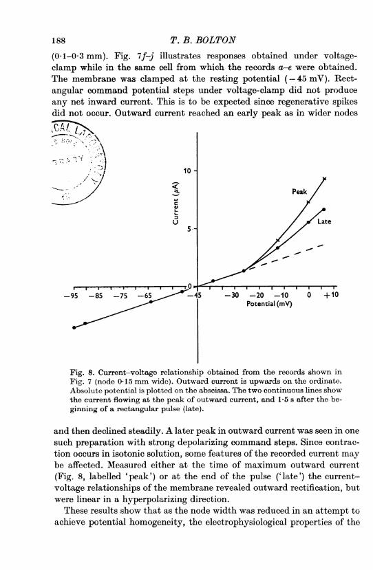
(0-1-0-3 mm). Fig. 7f-j illustrates responses obtained under voltage-clamp while in the same cell from which the records a-e were obtained.The membrane was clamped at the resting potential (-45 mV). Rect-angular command potential steps under voltage-clamp did not produceany net inward current. This is to be expected since regenerative spikesdid not occur. Outward current reached an early peak as in wider nodes
7\10-
- is X | Peak
U ate
5.
-95 -85 -75 -65 -45 -30 -20 -10 0 +10Potential (mV)
Fig. 8. Current-voltage relationship obtained from the records shown inFig. 7 (node 0-15 mm wide). Outward current is upwards on the ordinate.Absolute potential is plotted on the abscissa. The two continuous lines showthe current flowing at the peak of outward current, and 1-5 s after the be-ginning of a rectangular pulse (late).
and then declined steadily. A later peak in outward current was seen in onesuch preparation with strong depolarizing command steps. Since contrac-tion occurs in isotonic solution, some features of the recorded current maybe affected. Measured either at the time of maximum outward current(Fig. 8, labelled 'peak') or at the end of the pulse ('late') the current-voltage relationships of the membrane revealed outward rectification, butwere linear in a hyperpolarizing direction.
These results show that as the node width was reduced in an attempt toachieve potential homogeneity, the electrophysiological properties of the
T. B. BOTON188
VOLTAGE CLAMP OF SMOOTH MUSCLE
muscle were changed. It seems likely that most of the effects arose due tothe fact that a sucrose gap is an imperfect approximation to the 'shortcable' and that as the node width (x) becomes small compared to the d.c.space constant of the tissue, A, then leakage current becomes relativelyimportant. According to McGuigan & Tsein (McGuigan, 1974) whenX = x/A = 1.0 then the effects of leakage current are minimized. In theseexperiments, where X < 0 1, the leakage current would be expected to berelatively large.
Two micro-electrode experimentsConstancy of nodal potential. Since it was clear that the electrophysio-
logical properties of the muscle were changed as X 0-> 01, it was decided touse slightly wider nodes (04-0.5 mm) and to pass current into the nodeacross both sucrose gaps (both ends of the muscle depolarized in high K)instead of across only one sucrose gap as in the experiments described sofar. This ought to double the node width at which a given percentagepotential variation across the node occurs.
Additionally, the node was bathed in a hypertonic physiological saltsolution in order to prevent or minimize artifacts due to contraction. Thissolution also allowed penetrations to be maintained for a much longerperiod of time but in taenia converted the regenerative spike usually seenat this node width into a graded spike.The usual procedure in these experiments was to penetrate cells at two
different (usually well separated) points in the node. Responses to rect-angular current pulses were first obtained (Fig. 9a-b). Following this, ifthe form of these responses was similar, the potential recorded by onemicro-electrode was clamped at the resting membrane potential andrectangular voltage-clamp command pulses applied.Although the clamped potential could be efficiently stepped to a new
potential within a few milliseconds, the potential recorded by the second,independent, electrode always lagged. The difference between the potentialsrecorded by the two micro-electrodes was serious for 50-100 ms when thepotential stepping did not activate inward current (Fig. 9d-h). Wheninward current was activated a potential difference persisted for longerperiods and strong depolarization produced a spike-like response recordedat the monitoring electrode (Fig. 9i-k). However, in most experiments,after about 200 ms the potentials recorded at two points within a nodewere similar.
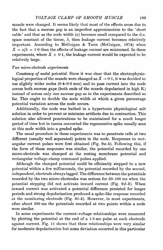
In some experiments the current-voltage relationships were measuredby plotting the potential at the end of a 1-5 sec pulse at each electrodeagainst current. Fig. 11 shows that these relationships were very similarfor moderate depolarization but some deviation occurred in this particular
189

experiment (part ofwhich is illustrated in Fig. 9) either with hyperpolariza-tion or strong depolarization. Presumably this situation may arise due tothe imperfect cable properties of the smooth muscle strip.A total of six experiments were done in which it was possible to monitor
U.-100 -UpA
50 mV
C MU
Node H~0 15mmP! 'I
*-0 4 mm-*
j 05mm-jFig. 9. Test for uniformity of nodal potential during rectangular voltageclamp commands. The membrane potential was recorded at two points ina taenia node at the positions indicated in the diagram. Node width was05 mm and current was passed across both sucrose gaps. Responses torectangular current pulses were first obtained (a-b). The potential recordedby electrode C was then clamped at the resting membrane potential andsubjected to hyperpolarizing (d-g) or depolarizing (h-k) rectangular com-mand potentials. Notice that when the inward current channel is notactivated, potential at the monitoring electrode (M) lags the clampedpotential (C) and differences exist for 50-100 ms following a step changein potential. When inward current channels are activated then differenceslast longer and even spiking may occur (j-k). Where ramp commands areused under voltage-clamp (c) then potential varied little between the twopoints of recording. I, the current record. Sucrose hypertonic PSS. Cali-bration 25 sA in a-i and 100 gA in j-k.
T. B. BOLTON190

VOLTAGE CLAMP OF SMOOTH MUSCLE19the potential at some other point in the node while voltage-clamping thepotential recorded by a second electrode. In some experiments, potentialvariations were greater than observed in Fig. 9, although the current tracegave no indication of this. Fig. l0a-g shows an experiment in which theresponses to rectangular current pulses recorded at two points were verysimilar, although not identical. However, when the potential recorded by
50 50 mV
100
PA4 500 msec
Fig. 10. Nodal potential variation under voltage clamp. Recording wasmade at two points within a 0-5 mm node of taenia at the positions shownin the diagram. The responses to rectangular current pulses were similar (a-g), but under voltage clamp, the potential recorded by electrode M differedconsiderably from clamped potential, and this difference persisted for upto 500 ins. Upon repolarization the potential recorded by electrode M washyperpolarized for a similar period. The monitoring electrode was dislodgedbefore recordj was obtained. Sucrose hypertonic physiological salt solution.Calibrations 10,sA (e, f), 251sA (a-d, g), 50 AA (h), 100 AsA (i-j).
191
7 PRY 250
electrode C was clamped at the resting membrane potential and thensubjected to rectangular command pulses, the potential recorded inde-pendently by electrode M showed considerable variation from the hoped-for rectangular form (Fig. lOh-j). Variation persisted for up to 500 msupon a depolarizing step and the early peak in outward current wasassociated with a varying potential at electrode M. Upon repolarizationto the resting membrane potential, potential recorded at electrode M wasconsiderably hyperpolarized while potential recorded at electrode C wasclamped at the resting (= holding) potential. Such experiments indicatethat under the conditions of these experiments, the assumption of con-stant nodal potential following a step depolarization or a step repolariza-tion would not be justified except possibly at times greater than 100-200 ms or more following such step changes. The assumption of constantnodal potentials, made by others using voltage-clamp of extracellularpotential ofsmooth muscle strip (e.g. Anderson, 1969; Kumamoto & Horn,1970; Anderson et al. 1971; Kao, 1971; Mironneau & Lenfant, 1972;Mironneau, 1973; Kao& McCullough, 1975)would, therefore, seem to requirere-examination, particularly since Tarr & Trank (1974) and McGuigan(1974) have obtained very similar results to these described here, on multi-fibre preparations of cardiac muscle.
Current-votage relationship. As a check on the voltage-clamp techniquea comparison was made between the current-voltage relationship obtainedusing rectangular current pulses and rectangular voltage-clamp commandpotentials, while the electrodes were within the same two cells. Currentwas measured at the end of a 1*5 s pulse in each case. The two methodsgave good agreement although current pulses were not tested over so widea range (Fig. 11).
Also tested were ramp command potentials under voltage-clamp, andramp currents. It was found that at a rate of ramp rise (voltage-clamp) of20-30 mV/s, then the potentials recorded at two different points showedinsignificant differences (Fig. 9c). The current-voltage relationship ob-tained was virtually identical to that found using rectangular pulses in adepolarizing direction (Fig. 11). However, in a hyperpolarizing directionthis relationship was similar for only moderate hyperpolarizations (50 mVin Fig. 11) and rectangular voltage-clamp pulses (0) activated morecurrent than ramps at more negative potentials. Hyperpolarizing rampcurrents also produced a similar current-voltage relationship for moderatehyperpolarizations (their rate of rise was sufficiently slow for capacitivecurrent to exert a negligible influence).
T. B. BOLTON192
VOLTAGE CLAMP OF SMOOTH MUSCLE 193
100
75.-
Monitor
0
2S ~~~~~Clamp
Potential hyperpolarization100 50 50s
o 25-
Potential hyperpolarization Potential depolarization1000 50 _>~50 100
o VC rectangular+ VC ramp*2 CC rectangularCCramp
EU
so0
Fig. It. Comparison of the current-voltage relationships obtained by twoelectrodes in the experiment of Fig. 9. The current-voltage relation wasobtained using rectangular voltage-clamp command potentials (0), rampvoltage clamp comans (+), rectangular current pulses (@) and rampcurrents (V). This relationship in the case of rectangular pulses (currentor potential) was measured at the end of a 1-5 s pulse. The rate of ramprise was about 30 mV/s. The potential shown is the deviation from theresting potential and has been corrected for the potential across the seriesresistance (r.).
7-2
194 P. B. BOLTON
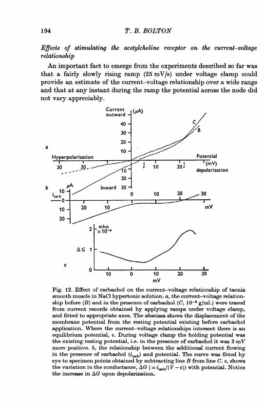
Effects of stimulating the acetylcholine receptor on the, current-voltagerelationshipAn important fact to emerge from the experiments described so far was
that a fairly slowly rising ramp (25 mV/s) under voltage clamp couldprovide an estimate of the current-voltage relationship over a wide rangeand that at any instant during the ramp the potential across the node didnot vary appreciably.
Current(,A
outward
40
30
20-a
10Hyperpolarization Potential
30 20 10 20 ~~~~~~~~~~depolarizationb #A Inward 30
10 -0 1 0 3icarbI-0
10 20 10 mY20
Lmho2 [X10-4
AG IF
C 01OI10 0 10 20 30
mY
Fig. 12. Effect of carbachol on the current-voltage relationship of taeniasmooth muscle in NaCl hypertonic solution. a, the current-voltage relation-ship before (B) and in the presence of carbachol (C, 10-6 g/ml.) were tracedfrom current records obtained by applying ramps under voltage clamp,and fitted to appropriate axes. The abscissa shows the displacement of themembrane potential from the resting potential existing before carbacholapplication. Where the current-voltage relationships intersect there is anequilibrium potential, e. During voltage clamp the holding potential wasthe existing resting potential, i.e. in the presence of carbachol it was 3 mVmore positive. b, the relationship between the additional current flowingin the presence of carbachol (ic.b) and potential. The curve was fitted byeye to specimen points obtained by subtracting line B from line C. C, showsthe variation in the conductance, AG (= i,:.b/( V - e)) with potential. Noticethe increase in AG upon depolarization.
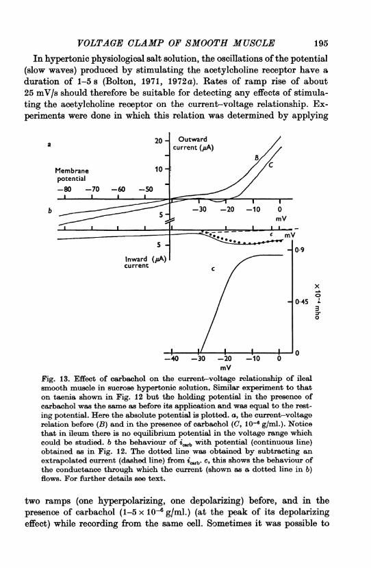
VOLTAGE CLAMP OF SMOOTH MUSCLE 195In hypertonic physiological salt solution, the oscillations of the potential
(slow waves) produced by stimulating the acetylcholine receptor have aduration of 1-5 s (Bolton, 1971, 1972a). Rates of ramp rise of about25 mV/s should therefore be suitable for detecting any effects of stimula-ting the acetylcholine receptor on the current-voltage relationship. Ex-periments were done in which this relation was determined by applying
20 - Outwardcurrent (#A)
Membrane 10potential
b
Inward (HA)current
mV
I 09
x
0-45 130
0-'tV -JV -AU -IU V
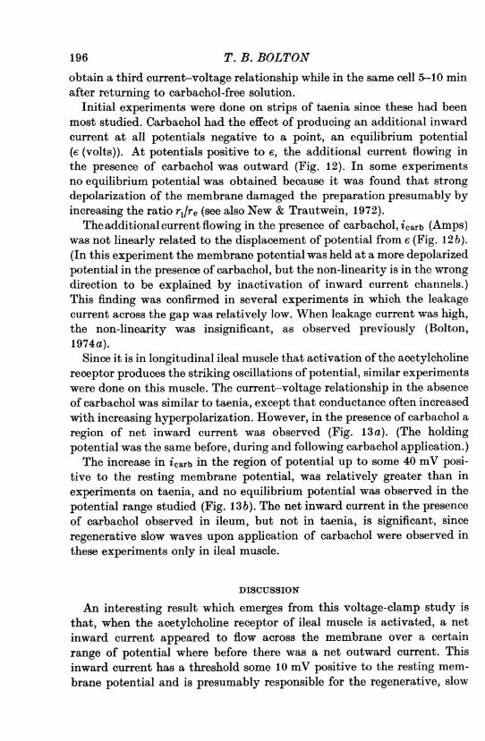
mVFig. 13. Effect of carbachol on the current-voltage relationship of ilealsmooth muscle in sucrose hypertonic solution, Similar experiment to thaton taenia shown in Fig. 12 but the holding potential in the presence ofcarbachol was the same as before its application and was equal to the rest-ing potential. Here the absolute potential is plotted. a, the current-voltagerelation before (B) and in the presence of carbachol (C, 10-6 g/ml.). Noticethat in ileum there is no equilibrium potential in the voltage range whichcould be studied. b the behaviour of i with potential (continuous line)obtained as in Fig. 12. The dotted line was obtained by subtracting anextrapolated current (dashed line) from iab. c, this shows the behaviour ofthe conductance through which the current (shown as a dotted line in b)flows. For further details see text.
two ramps (one hyperpolarizing, one depolarizing) before, and in thepresence of carbachol (1-5 x 10 g/ml.) (at the peak of its depolarizingeffect) while recording from the same cell. Sometimes it was possible to
Aen
obtain a third current-voltage relationship while in the same cell 5-10 minafter returning to carbachol-free solution.
Initial experiments were done on strips of taenia since these had beenmost studied. Carbachol had the effect of producing an additional inwardcurrent at all potentials negative to a point, an equilibrium potential(e (volts)). At potentials positive to e, the additional current flowing inthe presence of carbachol was outward (Fig. 12). In some experimentsno equilibrium potential was obtained because it was found that strongdepolarization of the membrane damaged the preparation presumably byincreasing the ratio ri/re (see also New & Trautwein, 1972).
The additional current flowing in the presence of carbachol, icarb (Amps)was not linearly related to the displacement of potential from e (Fig. 12 b).(In this experiment the membrane potential was held at a more depolarizedpotential in the presence of carbachol, but the non-linearity is in the wrongdirection to be explained by inactivation of inward current channels.)This finding was confirmed in several experiments in which the leakagecurrent across the gap was relatively low. When leakage current was high,the non-linearity was insignificant, as observed previously (Bolton,1974a).
Since it is in longitudinal ileal muscle that activation of the acetylcholinereceptor produces the striking oscillations of potential, similar experimentswere done on this muscle. The current-voltage relationship in the absenceof carbachol was similar to taenia, except that conductance often increasedwith increasing hyperpolarization. However, in the presence of carbachol aregion of net inward current was observed (Fig. 13 a). (The holdingpotential was the same before, during and following carbachol application.)The increase in icarb in the region of potential up to some 40 mV posi-
tive to the resting membrane potential, was relatively greater than inexperiments on taenia, and no equilibrium potential was observed in thepotential range studied (Fig. 13b). The net inward current in the presenceof carbachol observed in ileum, but not in taenia, is significant, sinceregenerative slow waves upon application of carbachol were observed inthese experiments only in ileal muscle.
DISCUSSION
An interesting result which emerges from this voltage-clamp study isthat, when the acetylcholine receptor of ileal muscle is activated, a netinward current appeared to flow across the membrane over a certainrange of potential where before there was a net outward current. Thisinward current has a threshold some 10 mV positive to the resting mem-brane potential and is presumably responsible for the regenerative, slow
T.B.BOLTON196
VOLTAGE CLAMP OF SMOOTH MUSCLE
oscillations of the membrane potential (slow waves) which occur in thismuscle if depolarization produced by stimulating the acetylcholine re-ceptor is sufficient (Bolton, 1971, 1972a).
It is a moot point to what extent voltage variations within the nodeaffect the interpretation of voltage-clamp results (Johnson & Lieberman,1971). It was possible in several experiments, using an independentmicro-electrode, to record from another region of the node during slowvoltage-clamp ramps and to show that the potential there was not greatlydifferent from the voltage-clamped one (step changes initially producedlarge variations in potential across the node which lasted for up to severalhundred milliseconds, as others have found in cardiac muscle (McGuigan,1974; Tarr & Trank, 1974)). Nevertheless there may be regions possiblyinaccessible to micro-electrode penetration, where the potential is con-siderably different from the voltage-clamped one. It seems unlikely thatthese are sufficient to affect the direction of the recorded currents, althoughtheir size may be altered. The increase in membrane conductance in thepresence of carbachol at potentials negative to the resting potential wasvery modest, and it seems most unlikely that an additional voltage-independent conductance alone could bring about sufficient undetectednodal voltage variation to explain the observed change in the current-voltage relationship. A likely and simple explanation for these results andothers (Bolton, 1971, 1972a) is that there is an opening of a population ofion channels whose conductance increases sharply with depolarizationbeyond about -35 mV.While regenerative slow waves occurred in ileum when carbachol was
applied in these experiments, they did not occur in taenia. Although icarbincreased slightly with depolarization in taenia, it is significant that itssize was insufficient to produce a region of inward current in the current-voltage relationship.The interpretation of these results in terms of the properties of ion
channels opened in the membrane during stimulation of the muscarinicreceptor by carbachol must be somewhat speculative. One interpretationmight be that there is a single set ofion channels with fixed ionic selectivitysuch that they have an equilibrium potential, e (V). Their conductancemight then be defined as AG = icarb/(V -e) where V (V) is the membranepotential at some instant. The way AG so defined varies with potential intaenia is shown in Fig. 12c. An alternative scheme would be to supposethat the ionic selectivity of these channels, e.g. for Na and K, varied withpotential.A more attractive hypothesis is suggested by the results on ileum (Fig.
13). Here no equilibrium potential was encountered in the potential rangethat could be studied. However, the linear relationship of icarb in a
197
hyperpolarizing direction might indicate a population of ion channels,operated by the activated acetylcholine receptor, whose conductance isindependent of potential and which have an equilibrium potential (obtainedby extrapolation) around -4 mV. Most of the additional current flowingin the presence of carbachol in the range between -32mVand zero potentialmight then be through a second population of channels with a differentionic selectivity and equilibrium potential. We may tentatively call this6Na, since this current is presumably sensitive to a reduction in the externalsodium concentration (Bolton, 1971, 1972a). Linear extrapolation of thecurrent-voltage relationships in Fig. 13a (and subtracting the part oficarb which is linearly related to potential) give eNa a value of + 55 mV.Using this value of eN. and defining the conductance of these channels asACNa = icarb, Na/( V- Na) the activation curve for these channels may beobtained and is shown in Fig. 13c. This ignores the possibility that at therate of ramp rise used in this experiment icarb, Na maynot be fully activated.The postulated presence of two populations of ion channels operated by
activated acetylcholine receptors in ileum (and possibly in taenia) shouldbe compared with the pharmacological evidence for the existence of twopopulations of acetylcholine receptors in this muscle (Burgen & Spero,1968, 1970; Burgen & Hiley, 1974).
It is necessary also to reconcile the present results with those of previousstudies (Bolton, 1972b) in which it was found that the maximum de-polarization produced by acetylcholine (or other strong agonists at themuscarinic receptor) in ileum was about -8 mV. This value was suggestedas being limiting upon depolarization because it was close to the equili-brium potential of the channels responsible for depolarization. Theseresults on ileum might suggest an equilibrium potential which ought to bemuch nearer to the Na equilibrium potential. However, recent experi-ments (unpublished) indicate that much Of carb activated over the potentialrange -40 to -10 mV can be inactivated by a few seconds depolarizationto around -10 mV. If this is the case, then the equilibrium potentialwould be determined by those channels remaining open when the potentialhad moved to around -10 mV under the influence of a fairly high con-centration of acetylcholine or carbachol, and no serious discrepancy wouldexist. Additionally, it is clear that the larger concentrations of cholinergicagonists which are needed to depolarize to this level, will also disturb theionic gradients across the membrane of these small cells, probably shiftingthe equilibrium potential (Bolton, 1973).Many of the features of the action of acetylcholine on ileal muscle are
seen when a variety of other stimulant substances act on the membranesof other smooth muscles. Noradrenaline has an action on the membranepotential of anococcygeus muscle which, superficially at least, closely
198 T.B.BOLTON
VOLTAGE CLAMP OF SMOOTH MUSCLE19resembles the action of acetylcholine on ileum in hypertonic solution(Gillespie, Creed & Muir, 1973). Without producing appreciable changesin the level of the resting potential, pentagastrin acts to increase slow wavefrequency in canine antrum muscle (Szurszewski, 1975), noradrenalineand histamine prolong the plateau of the guinea-pig ureter (Shuba, 1975;Shuba, Taranenko, Kochemasova, Gurkovskaya, & Klevetz, 1975) and anumber of stimulants (prostaglandin, oxytocin, acetylcholine) in low con-centrations increase the frequency of occurrence and duration of spikebursts in uterine muscle (Kuriyama, Gsa & Suzuki, 1975). It is conceivablethat an effect on a voltage-sensitive inward channel is a mechanism ofgeneral importance, underlying the actions of a variety of stimulant sub-stances on the membranes of rhythmically contracting visceral smoothmuscles.The action of acetylcholine on ileum resembles its action on cortical
neurones (Krnjevi6', Pumain & Renaud, 1971; Krnjevi6', 1974) and on
spinal motoneurones (Zieglginsberger & Reiter, 1974) where it producesdepolarization and a delay in repolarization following the spike. This canresult in a plateau-like component appearing. Krnjevi6' (1974) has sug-gested a unifying hypothesis, applicable to all these cells, of a reduction inoutward K current which has the dual effect of delaying repolarizationfollowing a spike and reducing the potential in the interspike interval.Certainly the actions of acetylcholine on ileum and on neurones are super-ficially very similar, but acetylcholine produces a large increase in con-ductance in smooth muscle (Bolton, 1972b; Magaribuchi, Ito & Kuriyama,1973; Osa & Taga, 1973; Biilbring & Szurszewski, 1974) while it reducesthe conductance of neuronal membrane (loc. cit.). The increase in con-ductance in smooth muscle is detectable when point injection of currentis made by micro-electrode (Hidaka & Kuriyama, 1969; Purves, 1974). Ithas been pointed out to me by Purves that this latter observation wouldseem to exclude any primary action of acetylcholine to uncouple smoothmuscle cells as suggested by Krnjevi6 (1974), since an increase in couplingresistance would be expected to increase, rather than decrease, the electro-tonic potential elicited by intracellular injection of current. Whilethe suggestion of a generalized reduction of smooth-muscle mem-
brane conductance by acetylcholine is clearly untenable, the idea of a
reduction by acetylcholine of the outward potassium current responsiblefor repolarizing following a spike, is worthy of consideration. The experi-ments described in this paper alone do not exclude a reduction in outwardcurrent rather than an increase in inward current, being responsible forthe effects of carbachol on the current-voltage relationshp. Howeversince a reduction in [Na+]0 abolished regenerative slow waves elicited bycarbachol (Bolton, 1971, 1972 a) while variations in [K+]0 had little effect
199

200 T. B. BOLTON(Bolton, 1972 a) it would seem that, at least in this muscle, activation of theacetylcholine receptor increases an inward Na current rather than de-creases an outward K current.
This work was done during the tenure of a Royal Society Locke Research Fellow.ship and was supported by a grant from the Medical Research Council. I am gratefulto Professor W. D. M. Paton for affording me the facilities of his department.
I am also indebted to Mr P. M. G. Bell and Mr T. Trinder for constructing some ofthe apparatus and to Miss P. M. Maxwell-Allen for painstaking technical assistance.
REFERENCES
ABE, Y. & ToMITA, T. (1968). Cable properties of smooth muscle. J. Physiol. 196,87-100.
ADRIAN, R. H., CHANDLER, W. K. & HODGKIN, A. L. (1970). Voltage-clamp experi-ments in striated muscle fibres. J. Physiol. 208, 607-644.
ANDERSON, N. C. (1969). Voltage-clamp studies on uterine smooth muscle. J. gen.Physiol. 54, 145-165.
ANDERSON, N. C., RAMON, F. & SNYDER, A. (1971). Studies on calcium and sodiumin uterine smooth muscle excitation under current-clamp and voltage-clampconditions. J. gen. Physiol. 58, 322-339.
BOLTON, T. B. (1971). On the nature of the oscillations of the membrane potential(slow waves) produced by acetylcholine or carbachol in intestinal smooth muscle.J. Physiol. 216, 403-418.
BOLTON, T. B. (1972a). The effects of varying the concentrations of ions in theexternal solution on the oscillations of the membrane potential (slow waves) pro-duced by carbachol in longitudinal ileal muscle. Pfliiger8 Arch. 335, 85-96.
BOLTON, T. B. (1972b). The depolarizing action of acetylcholine or carbachol inintestinal smooth muscle. J. Physiol. 220, 647-671.
BOLTON, T. B. (1973). The role of electrogenic sodium pumping in the response ofsmooth muscle to acetylcholine. J. Physiol. 228, 713-731.
BOLTON, T. B. (1974a). Voltage-clamp experiments on the potential-dependentbehaviour of membrane ion channels operated by the muscarinic receptor ofsmooth muscle. Br. J. Pharmac. 51, 129-130P.
BOLTON, T. B. (1974b). Voltage-clamp of potential recorded intracellularly withmicro-electrodes in smooth muscle. J. Physiol. 244, 25-26P.
BOLTON, T. B. (1975a). Voltage-clamp of taenia smooth muscle using intracellularrecording of membrane potential: Action of carbachol. Proceedings of 'PhysiologyofSmooth Muscles' Symposium held in Kiev 16th Int. Cong. Physiol. Sci. New York:Raven Press. (In the Press.)
BOLTON, T. B. (1975b). Evidence of a non-linear voltage-dependence of the addi-tional current flowing in the membrane of smooth muscle when the acetylcholinereceptor is stimulated. J. Physiol. 246, 63-64P.
BU;LBRING, E. & SZURSZEWSKI, J. H. (1974). The stimulant action of noradrenaline(x-action) on guinea-pig myometrium compared with that of acetylcholine. Proc. R.Soc. B 185, 225-262.
BLYRGEN, A. S. V. & HILEY, C. R. (1974). Two populations of acetylcholine receptorsin guinea-pig ileum. Br. J. Pharmac. 51, 127P.
BURGEN, A. S. V. & SPERO, L. (1968). The action of acetylcholine and other drugson the efflux of potassium and rubidium from smooth muscle of the guinea-pigintestine. Br. J. Pharmac. Chemother. 34, 99-115.

VOLTAGE CLAMP OF SMOOTH MUSCLE 201
BURGEN, A. S. V. & SPERO, L. (1970). The effects of calcium and magnesium on theresponse of intestinal smooth muscle to drugs. Br. J. Pharmac. 40, 492-500.
CoNNoR, J. A., PRaosER, C. L. & WEEms, W. A. (1974). A study of pace-makeractivity in intestinal smooth muscle. J. Physiol. 240, 671-701.
GTTwT.sprr, J. S., CREED, K. E. & Mum, T. C. (1973). Electrical changes underlyingexcitation and inhibition in intestinal and related smooth muscle. Phil. Trans. R.Soc. B 265, 95-106.nx1A-K, T. & KU IYAmA, H. (1969). Responses of the smooth muscle membrane ofguinea-pig jejunum elicited by field stimulation. J. gen. Physiol. 53, 471-486.
Ivirs, D. J. G. & JANz, G. T. (1961). Reference Electrodes, Theory anid Practice.New York: Academic Press.
JOHNSON, E. A. & LiEB3ERMAN, M. (1971). Heart: excitation and contraction. A. Rev.Physiol. 33, 479-532.
KAo, C. Y. (1971). Some new leads into the physiology of mammalian smoothmuscles. In Research in Physiology, ed. KAo, F. F., KoizumI, K. & VASSAL,, M.,pp. 365-372. Bologna: Aulo Gaggi.
KAo, E.&MCCULLOUGH, J. R. (1975). Ionic currents in the uterine muscle.J.Phys-iol.246, 1-36.
KL, iBER, A. G. (1974). Effects of sucrose solution on the longitudinal tissue re-sistivity of trabecular muscle from mammalian heart. Pfluigers Arch. 345, 195-205.
KRNJEvii6, K. (1974). Chemical nature of synaptic transmission in vertebrates.Physiol. Rev. 54, 418-540.
KRNJEvii6, K., Pum~iN, R. & RENAuiD, L. (1971). The mechanism of excitation byacetylcholine in the cerebral cortex. J. Physiol. 215, 247-268.
KuMAMOTO, M. & HORN, L. (1970). Voltage clamping of smooth muscle from taeia,coli. Microvasc. Res. 2, 188-201.
KuiIrYAmA, H., GSA, T. & Suzux, H. (1975). Ionic mechanism involved in theexcitatory action of prostaglandin E2 on the pregnant mouse myometrium. Pro-ceedings Kiev Symposium, ed. BtJBRING, E. & SHUBA, M. F. New York: RavenPress. (In the Press.)
KuRaIYAMA, H. & ToMITA, T. (1970). The action potential in the smooth muscle ofthe guinea pig tacnia coli and ureter studied by the double sucrose-gap method.J. gen. Physiol. 55, 147-162.
MAGARIBUCHI, T., ITO, Y. & KuRiYAMA, H. (1973). Desensitization of smooth musclecells in the guinea pig tacnia coli to prolonged application of carbachol.Jap. J. Physiol. 23, 447-464.
MOGUIGAN, J. A. S. (1974). Some limitations of the double sucrose gap, and itsuse in a study of the slow outward current in mammalian ventricular muscle.J. Physiol. 240, 775-806.
MIRoNNEAu, J. (1973). Excitation-contraction coupling in voltage clamped uterinesmooth muscle. J. Physiol. 233, 127-141.
MIRoNNEAu, J. & LENwANT, J. (1972). Activit6 eflectrique du faisceau musculairelisse de l'ut6rus. J. Physiol., Paris 64, 97-105.
NEw, W. & TRAuTwiEIN, W. (1972). Inward membrane currents in mammalianmyocardium. Pfluigers Arch. 334, 1-23.
OSA, T. & TAGA, F. (1973). Electrophysiological comparison of the action of oxytocinand earbachol on pregnant mouse myometrium. Jap. J. Physiol. 23, 81-96.
PuRviFs, R. D. (1974). Muscarinic excitation: a microelectrophoretic study oncultured smooth muscle cells. Br. J. Pharmac. 52, 77-86.
RouGI-ER, 0., VASSORT, G. & STAmi'FLi, R. (1968). Voltage clamp experiments onfrog atrial heart muscle fibres with the sucrose gap technique. Pflulgers Arch.301, 91-108.
SHUBA, M. (1961). Electrotonus in smooth muscle. Bioflzika 6, 56-64.SHUuBA, M. (1975). Mechanism of action of catecholamines and histamine on smooth
muscle cells of guinea-pig ureter. J. Physiol. 245, 88-89P.SHUBA, M. F., TARANENKO, V. M., Kocus~mAsOVA, N. G., GURKOVSKAYA, A. V. &KLEVETZ, M. (1975). Mechanism of excitatory and inhibitory action of catechol-amines on the membrane of smooth muscle. Proceedings Kiev Symposium, ed.BULBRING, E. & SHUBA, M. F. New York: Raven Press. (In the Press.)
SzuRszEwsxi, J. H. (1975). Effect of pentagastrin and acetylcholine on the electricaland mechanical activity of isolated longitudinal muscle of the canine autrum.Proceedings Kiev Symposium, ed. BtBLBrING, E. & SHU-BA, M. F. New York:Raven Press. (In the Press.)
TARR, M. & TRANK, J. W. (1974). An assessment of the double sucrose-gap voltageclamp technique as applied to frog atrial muscle. Biophys. J. 14, 627-643.
TOMITA, T. (1966). Electrical responses of smooth muscle to external stimulation inhypertonic solution. J. Physiol. 183, 450-468.
TOmiTA, T. (1969). The longitudinal tissue impedance of the smooth muscle of guinea-pig taenia coli. J. Physiol. 201, 145-159.
TOMITA, T. (1970). Electrical properties of mammalian smooth muscle. In SmoothMuscle, ed. BULBRING, E. et al., pp. 197-243. London: Edward Arnold.
VASSORT, G. (1974). Initial ionic currents in guinea-pig myometrium. J. Physiol. 237,50-51P.
ZIEGLGANSBERGER, W. & REITER, C. (1974). A cholinergic mechanism in the spinalcord of cats. Neuropharmacology 13, 519-527.
202 T. B.BOLTON