0dwhuldo (6, iru&khp&rpp 7klv - rsc.org · supplementary information in vitro selection of...
TRANSCRIPT

Supplementary Information
In vitro selection of electrochemical peptide probes using bioorthogonal tRNA for influenza virus detection
Tara Bahadur K. C.a,b[1], Seiichi Tadab[1], Liping Zhuc[1], Takanori Uzawaa,c, Noriko Minagawaa, Shyh-Chyang Luod,e, Haichao Zhaod, Hsiao-hua Yud,f, Toshiro Aigakib,c, and Yoshihiro Ito*a,b,c
aEmergent Bioengineering Materials Research Team, RIKEN Center for Emergent Matter Science, 2-1 Hirosawa, Wako, Saitama 351-0198, Japan.
bGraduate School of Biological Science, Tokyo Metropolitan University, 1-1 Minami-Osawa, Tokyo 192-0397, Japan.
cNano Medical Engineering Laboratory, RIKEN, 2-1 Hirosawa, Wako, Saitama 351-0198, Japan.
dResponsive Organic Materials Laboratory, RIKEN, 2-1 Hirosawa, Wako, Saitama 351-0198, Japan.
eDepartment of Material Science and Engineering, National Taiwan University, No1, Sec 4, Roosevelt Road, Taipei 10617, Taiwan.
fInstitute of Chemistry, Academia Sinica, 128 Academia Dr. Sec. 2, Nankang Taipei 11529, Taiwan.
Corresponding authors:Prof. Yoshihiro ItoEmergent Bioengineering Materials Research Team, RIKEN Center for Emergent Matter Science, 2-1 Hirosawa, Wako, Saitama 351-0198, Japan, Fax: +81(48)467-9300; Tel: +81(48)467-5809; Email: [email protected]
[1] These authors contributed equally to this work.
1
Electronic Supplementary Material (ESI) for ChemComm.This journal is © The Royal Society of Chemistry 2018

Material and Methods
Synthesis of 3,4-ethylenedioxythiophene (EDOT)-aminophenylalanyl-tRNA
Boc-ε-aminophenylalanine was coupled to 5′-O-phosphoryl-2′-deoxycytidylyl-(3′-5′)
adenosine (pdCpA) to give the corresponding aminophenylalanine-pdCpA, (AF-pdCpA).
Then, a DMSO solution of EDOT-succinimidyl ester was treated with a DMSO solution
of AF-pdCpA (5 mM, 40 µL) in aqueous pyridine-HCl (5M, pH 5.0, 80 µL), and the
resulting mixture was incubated at 37 °C for 3 h. The EDOT-AF-pdCpA product was
purified by reversed-phase HPLC using an XTerra C18 column (4.6 × 20 mm, 2.5 µm
particle size; Waters, Milford, MA, USA), which was eluted at a flow rate of 1.5 mL min-1
with a linear gradient of 0–100% acetonitrile in 0.1% trifluoroacetic acid over a period of
10 min. The identity of the product was confirmed by matrix-assisted laser
desorption/ionization time-of-flight mass spectrometry (Voyager, Applied Biosystems,
Foster City, CA, USA). The resulting EDOT-AF-pdCpA was ligated to an amber-
suppressor tRNA, which was derived from Mycoplasma capricolum Trp1 tRNA without
the 3′ dinucleotide, using a previously reported chemical ligation method.1 The purified
EDOT-AF-tRNA molecules were lyophilized and stored at –80 °C.
In vitro selection of an electrosensitive peptide ligand against the influenza virus
The selection protocol is shown schematically in Fig 2. A 13Trx plasmid was employed
in the current study carrying a promoter sequence for the T7 RNA polymerase, an
Escherichia coli ribosome-binding sequence, an SfiI restriction enzyme sequence, a
protein-linker sequence, and a ribosome-arrest sequence. Random double-stranded DNA
(dsDNA) was also prepared in parallel. The library sequences were obtained from
Eurofins Genomics (Tokyo, Japan). These sequences were based on the general sequence
5′-ATATGGCCATGCAGGCC(VVN)3TAG(VVN)7GGCCAGCTAGGCCAGTT-3′,
2

where V represents G, C, or A; and N represents G, C, T, or A. The VVN sequence covers
only 10 of the naturally-occurring amino acids, and was selected to exclude hydrophobic
amino acids, such as leucine, valine, and tryptophan, and stop codons (e.g. TAG, TAA,
and TGA). This design strategy was selected for peptides with high solubility in an
aqueous buffer, and to allow for the incorporation of one EDOT molecule at the same
position in each library sequence, whilst avoiding the unintentional incorporation of
additional EDOT molecules. To run selection, dsDNA was prepared by one cycle of a
polymerase chain reaction (PCR) using Takara Ex taq DNA polymerase (Takara Bio Inc.,
Otsu, Shiga, Japan), and the crude material was purified with a QIAquick PCR
Purification kit (Qiagen, Valencia, CA, USA). The resulting dsDNA and 13Trx plasmid
were digested with a restriction enzyme (SfiI; New England Biolabs, Ipswich, MA, USA)
and fused (DNA Ligation Kit; Takara Bio). Finally, a dsDNA library was prepared by
PCR with new primers (New-T7-fp-rec-1: 5′-
GTAATACGACTCACTATAGGCCGCGTCGACAATAA-3′ and New-rp-fp-M13-NS:
5′-GATTACGCCAAGCTGAGTGAGA-3′). Transcription was performed at 37 °C for 3
h, and the product was then treated with DNase. The mRNA was purified using a RNeasy
kit (Qiagen). In vitro translation was performed using a PURESYSTEM Classic II kit
(Wako Pure Chemical Industries Ltd., Osaka, Japan) with EDOT-AF-tRNAs. Given that
the mRNA did not contain any stop codons, it was not possible for the ribosome to release
the mRNA or the translated peptide, and the mRNA, ribosome, and peptide became
coupled as a ternary complex (the PRM complex, see Fig. 2). The reaction was stopped
by placing the mixture on ice for 10 min. The translated solution was then incubated with
inactivated influenza virus A/California/07/2009 (H1N1, kindly provided as a gift from
Denka Seiken Co., Ltd., Tokyo, Japan), which was immobilized on silica affinity beads
3

(Sumitomo Bakelite Co., Ltd., Tokyo, Japan) in the selection buffer (0.1% Tween 20, 50
mM Tris-acetate, 150 mM NaCl, 50 mM magnesium acetate, pH 7.4) at 4 °C for 1 h. The
virus-immobilized beads were collected by centrifugation at 10,000 × g for 5 min and
washed with buffer (50 mM Tris-acetate, 150 mM NaCl, 50 mM magnesium acetate, pH
7.4) at 4 °C to remove any free mRNA-ribosome-peptide complexes. The mRNA was
recovered from the bound mRNA-ribosome-peptide complex following a 30-min period
of incubation with ethylenediaminetetraacetatic acid at room temperature, which allowed
for the removal of the Mg2+ ions and resulted in the detachment of the ribosomes from
the mRNA. The isolated mRNA was purified using an RNeasy kit. Preparative PCR was
performed to amplify the reverse transcription products, and the DNA product was
purified using a QIAquick PCR purification kit (Qiagen). The isolated DNA was used as
the template for the next round of selection.
After six rounds, the sequences of the selected DNAs were analyzed by the next-
generation sequencing service provided by Takara-bio (Otsu, Japan) using a MiSeq
sequencer (Illumina, San Diego, CA, USA). The obtained sequences (approximately
200,000) were trimmed to use only the random library sequences and converted from
nucleotide sequences to amino acid sequences. The trimmed and translated reads were
aggregated, and those with 90% sequence identity were clustered using Cd-hit.2 The
clusters representing singletons (with a multiplicity of one) were discarded, and the
remaining clusters were ranked based on the sum of multiplicities within each cluster.
Representative sequences, which are defined as the reads with highest multiplicities, were
determined for the clusters. Representative sequences and multiplicities for each cluster
were tabulated, and the top 100 clusters were identified as the enriched sequences
mediated by the selection process (Table S1). The analysis indicates the enrichment of
4

selected peptide sequences against the influenza virus, and the three peptide sequences
with the highest enrichment (Sequence 1, Sequence 2, and Sequence 3) were selected for
interaction analysis (Table S2).
Synthesis and purification of peptide sequences.
Selected peptides sequences from the Next Generation Sequence Analysis (Sequence
1, Sequence 2, and Sequence 3) were synthesized using a conventional Fmoc-solid phase
peptide synthesis method on preloaded Wang resin (Watanabe Chemical Industries,
Hiroshima, Japan). Peptide coupling and deprotection reactions were performed with a
Discover microwave (CEM Corporation, Matthews, NC, USA) using standard CEM
protocols, with mild N2 bubbling, at a 0.1 mmol scale (at 70°C, 5 and 3min for coupling
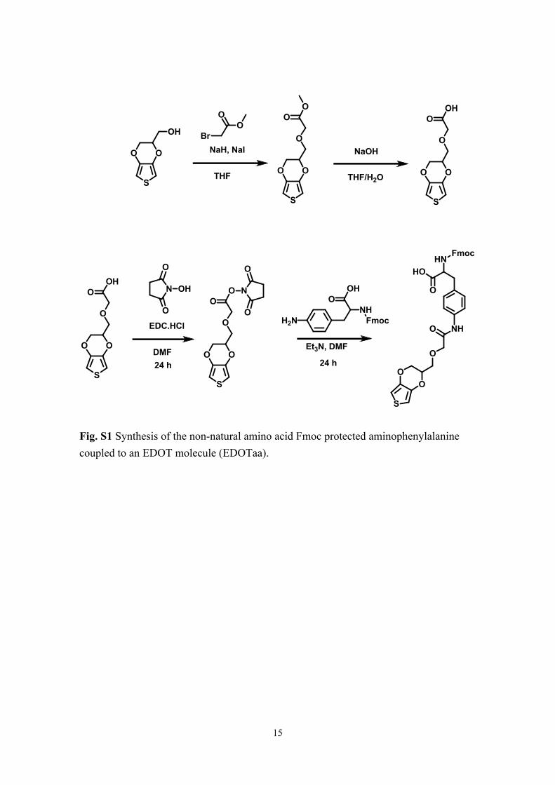
and deprotection, respectively). For the incorporation of EDOT into the selected peptide
sequences, a non-natural amino acid, Fmoc-aminophenylalanyl EDOT (EDOTaa), was
prepared according to the scheme shown in Fig S1. EDOTaa was attached using
optimized conditions for coupling (10 min at 50 °C and 25 min at 50 °C) and deprotection
(5 min at 50 °C). After coupling EDOTaa, elongation with other amino acids was also
carried out at reduced temperature but for prolonged times (at 50 °C, 7 and 5 min for
coupling and deprotection, respectively) to prevent the degradation of the non-natural
amino acid. The elongation of the peptide sequence was confirmed by checking the
MALDI MS at various steps, and after final cleavage from the resin. For cleavage, a ΔTIS
cocktail consisting of an 85:5:4:4:2 (v/v) mixture of trifluoroacetic acid: phenol:
thioanisole: ddH2O: ethane dithiol was used to avoid reducing the thiophene moiety. The
cleaved peptides were precipitated via chilled diethyl ether and centrifuged, and the
peptide pellet was washed and lyophilized for further purification. For the peptide-
5

binding assay, the N-terminal primary amine group of the peptide was modified with
fluorescein at the RIKEN Brain Science Institute, Japan. The fluorescein-labeled reaction
was carried out on a solid support using a fluorescein-NHS ester (Thermo Fisher
Scientific Inc., Rockford, IL, USA). After confirming the reaction, the resin was
thoroughly washed, and the peptide was cleaved as described above.
Peptide purification and analysis was performed using an Extrema HPLC (JASCO,
Tokyo, Japan). Purification was performed with a linear gradient using 0.1 %
trifluoroacetic acid in water, and acetonitrile in a COSMOSIL 5C18-AR-II column
(Nacalai Tesque, Kyoto, Japan). Finally, the peptide was characterized by HPLC, as well
as MALDI-TOF MS (Fig. S2a and S2b) prior to the interaction analysis.
Evaluation of peptide binding affinity and target selectivity
To determine the biding affinity using immobilized microbeads, first, the inactivated
influenza virus (1 µg) was immobilized on 20 mg of silica microbeads (Sumitomo
Bakelite, Tokyo, Japan) following the manufacturer’s protocol. Then, the peptides labeled
with fluorescein were incubated with 300 µg of the virus-immobilized silica microbeads
containing approximately 15 ng of inactivated influenza viral particles, maintaining the
volume at 200 µL in the selection buffer at 25 °C for 1 h. During the incubation process,
the beads and peptide solution were continuously mixed using a microtube mixer
(ThermoMixer C; Eppendorf, Hamburg, Germany) at 1000 rpm. After incubation, the
beads and peptide solution were centrifuged at 10,000 rpm for 5 min, and the solution
containing unbound peptide was discarded. The peptide-bound beads were washed three
times with 300 µL washing buffer (50 mM Tris-acetate, 150 mM NaCl, 50 mM
magnesium acetate, pH 7.4) and were protected from light during the experiment. For the
6

fluorescence measurements, the bead volume was adjusted to 100 µL, and the suspension
were transferred to a 96-well black microplate (PerkinElmer, Waltham, MA, USA), and
the fluorescence intensities were quantified at 530 nm using a microplate reader (Enspire
2300; PerkinElmer, Hamburg, Germany). All data are presented as the mean values ± SD
(n = 3).
Biological specificity
The biological specificity of the binding of the Sequence 2 peptide to the influenza
virus was studied using a dot blot analysis. A membrane (Immobilon-P Transfer
Membrane, pore size: 0.2 µm; Millipore, Bedford, MA, USA) was immersed in methanol
followed by blotting with a buffer solution [25 mM Tris, 192 mM glycine, 20% (v/v)
methanol]. The membrane was then placed on wet filter paper to avoid excessive drying.
A 2-µL portion of 1 mg mL−1 inactivated influenza virus in PBS buffer was then dropped
onto the membrane. Similar samples of Epstein–Barr virus, gelatine, and bovine serum
albumin (Sigma-Aldrich) were also dropped onto the membrane as negative controls.
After it had dried out, the membrane was immersed in methanol, followed by the blotting
buffer. The membrane was then immersed in a blocking buffer (5% w/v of ECL Advance
blocking agent) and a TBS-T buffer (50 mM Tris, 150 mM NaCl, 0.05% Tween 20, pH
7.4), and incubated for 1 h at room temperature to block any nonspecific binding. The
membrane was briefly washed with TBS-T buffer and put on parafilm to keep it wet.
Fluorescein-labeled Sequence 2 peptide (500 nM) was dropped on to the membrane to
cover the entire area. After being incubated for 1 h at room temperature in the dark, the
membrane was washed three times with TBS-T buffer for 10 min each time, and an image
of the surface of the membrane was recorded using a Molecular Imager FX system (Bio-
Rad Laboratories, Hercules, CA, USA) at the RIKEN Brain Science Institute, Japan. The
7

image was analyzed using the ImageJ64 software package to find the integrated densities
of the spots.
Electropolymerization of EDOT-conjugated peptide ligand
Electrochemical experiments were carried out using an Autolab potentiostat system
(ALS/CH, electrochemical analyzer, 700C, CH Instruments Inc. Austin, TX, USA). A
gold electrode (ALS Au 6 × 3.0 mm; ALS Co., Ltd., Tokyo, Japan) was used as the
working electrode, and a platinum wire and an Ag/AgCl electrode (RE-1S, ALS Co., Ltd.,
Tokyo, Japan) were used as the counter and reference electrodes, respectively. After
setup, the distance between the counter/reference electrodes and the working electrode
was kept at 1 mm. Twelve microliters of the Sequence 2 peptide solution were dropped
onto the working electrode, ensuring that the counter and reference electrodes were
submerged in the solution. Cyclic voltammetry (−0.6 to 0.95 V vs. Ag/AgCl, scanning at
0.1 V s−1) was conducted during the polymerization reaction in 0.1 M Tris-HCl buffer
(pH 7.0) supporting electrolyte. After the polymerization reaction, the gold electrode was
gently washed with Milli-Q water to remove unpolymerized peptide. For the optimization
of the electropolymerizing conditions, Sequence 2 peptide polymerization was performed
at various oxidative potentials (−0.6 and +0.70 to +1.4 V). At lower potentials (below
+0.75 V) the peptide did not polymerize, because no current inhibition was detected on
the gold electrode even after 15 cycles of polymerization. At higher potentials, the peptide
polymerized after fewer cycles and started to be destroyed, and this observed
phenomenon was similar to a reported EDOT polymerization.3 Thus, we selected the
parameters of –0.6 to 0.95 V at 0.1 V s−1 scanning speed for 12 cycles for further analysis.
For the FT-IR measurements, electropolymerization of the peptide was performed on
a gold-coated chip (5 × 10 mm) cut from a gold-coated silicon wafer (Sigma-Aldrich).
8

The experimental setup was similar to the peptide electropolymerization experiment
described above, but a gold-coated silicon chip was used instead of the gold working
electrode. A 20 µL portion of a 2 mg mL−1 solution of Sequence 2 peptide in 0.1 M Tris-
HCl buffer (pH 7.0) was dropped onto the gold-coated silicon chip, and cyclic
voltammetry (−0.6 to 0.95 V vs. Ag/AgCl, at a scanning speed of 0.1 V s−1 for 12 cycles)
was performed using a potentiostat system. The gold-coated chip was dipped in Milli-Q
water to remove any unpolymerized peptide. After drying, the sample was analyzed by
FT-IR spectroscopy using an FT-IR 4100 system (Jasco, Tokyo, Japan). The FT-IR
spectra were recorded in the range of 4500–600 cm−1 using an average of 80 scans to
increase the signal to noise ratio, and the spectral resolution was 4 cm−1. The FT-IR
spectrum of the Sequence 2 peptide monomer was also measured for comparison.
Electrochemical detection of influenza virus
Three electrodes were set up as described above for the peptide polymerization. First,
the effective concentration of the peptide was determined by running the polymerization
reaction on the gold electrode (−0.6 to 0.95 V vs. Ag/AgCl, at a scanning speed of 0.1 V
s−1 for 12 cycles) using various concentrations, and then the current was measured. The
cathodic current was effectively suppressed at 0.1 mg mL−1 of the peptide (Fig. S3), so
this concentration of peptide was used. For the detection system, 12 µL of Sequence 2
peptide was dropped on to the gold electrode, ensuring that the entire surface of the
electrode was covered. Cyclic voltammetry was performed for the polymerization
reaction, as described above. After cleaning, the working electrodes were set up in the
same manner as before, and 12 µL of 10 mM K3[Fe(CN)6] was dropped onto the surface
of the gold electrode. Then, cyclic voltammetry (−0.3 to 0.65 V vs. Ag/AgCl, at 0.1 V
s−1) was performed, and the current at the gold electrode was recorded. In the same way,
9

12 µL of the mixture of Sequence 2 peptide (final concentration, 0.1 mg mL−1) and
various concentrations of influenza virus were applied to the detection system, and the
current at the gold electrode was recorded as a function of the influenza virus
concentration. The spectrum of the second cycle is shown in the figures because the
spectra stabilized after the second cycle. To determine the limit of detection (LOD) of the
system, the anodic current obtained after the electrochemical analysis of inactivated
influenza virus at different concentrations in the presence of a fixed amount of Sequence
2 Peptide was analyzed statistically at the 95% confidence interval. The p-values of the
different concentrations were calculated by comparing the results with those recorded in
the absence of influenza virus using Student’s t-test.
Interference of microorganisms on the electrochemical detection system.
The possible interference of other microorganisms in the electrochemical detection
system was examined using bacteria commonly found in the oral cavity (Staphylococcus
aureus and Strepotococcus pneumonia). They were kindly provided by Dr. Suresh
Panthee at Teikyo University Institute of Medical Mycology, Japan, after heat treatment
at 121oC for 15 min by an autoclave. For this measurement, we used a much larger
amount of microorganisms (1 × 10⁶ CFU mL-1) than the amount of staphylococci species
usually found in saliva (102–104 CFU mL-1).4 Specifically, all three electrodes were set
up as described above for the peptide polymerization and then the current on a clean
working electrode was recorded by cyclic voltammetry (−0.3 to 0.65 V vs. Ag/AgCl, at
0.1 V s−1) using 12 µL of 10 mM K3[Fe(CN)6] in 0.1 M Tris-HCl buffer (pH 7.0). Cyclic
voltammetry was performed during the polymerization cycle (−0.6 to 0.95 V vs.
Ag/AgCl, at a scanning speed of 0.1 V s−1 for 12 cycles) using 12 µL of the Sequence 2
10

peptide (0.1 mg mL−1) in the presence of 1 × 10⁶ CFU mL-1 of microorganism, only the
Sequence 2 peptide (0.1 mg mL−1), and only the microorganism [1 × 10⁶ CFU mL-1 in 0.1
M Tris-HCl buffer (pH 7.0)]. Then, the current at the gold electrode was recorded again,
as described above, and the obtained cyclic voltammograms were compared (Fig S5). The
results showed that the microorganisms did not interfere with our influenza detection
system. Neither species of microorganism remarkably suppressed the CV current
measured after the polymerization cycle (blue and red lines in Fig S5) nor did their
presence affect the peptide polymerization (green and purple lines in Fig S5).
11

Table S1. Sequence list of the selected peptides from the Next Generation Sequencing analysis. “B” represents EDOT-aminophenylalanine.
12

Table S2: Nominated peptide sequences for the interaction analysis.Peptide Name Amino acid sequences[a] Ratio[b]
Sequence 1 AAPBKAGKGAP 2.65Sequence 2 ARRBGHRKPRR 2.52Sequence 3 AGRBRRGAHDT 1.93
Sequence 2 (∆EDOT) ARRFGHRKPRR –
[a] B stands for EDOT-coupled aminophenylalanine, which is a non-natural amino acid.
F indicates where phenylalanine was used in place of B to synthesize the Sequence 2
peptide without EDOT (∆EDOT).
[b] Sequence repetition percentage out of the total number of sequences.
13

Table S3: P-values calculated using the Student’s t-test for the detection limit of the influenza virus.
Influenza Virus 6.25 µg mL-1 12.5 µg mL-1 25 µg mL-1
P-Value 0.27 0.02 0.001
14
S
OO
OH
NaH, NaIBr
OO
THF
S
OO
O
OO
THF/H2O
S
OO
O
OHO
NaOH
S
OO
O
OHO
S
OO
O
OO
EDC.HCl
DMF
N
O
O
OH N
O
O
24 h
Et3N, DMF
24 h
S
OO
O
NHO
HNFmoc
HO
O
H2NNHFmoc
OHO
Fig. S1 Synthesis of the non-natural amino acid Fmoc protected aminophenylalanine coupled to an EDOT molecule (EDOTaa).
15

Fig. S2 Characterization of Sequence 2 peptide a) HPLC analysis showing the absorbance at 220 nm. b) MALDI MS analysis (Calculated M+H = 1663.99, experimental monoisotopic mass M+H = 1664.32)
16

Fig. S3 Averaged cathodic current after the polymerization of various concentrations of Sequence 2 peptide on a gold electrode. The cathodic current was effectively suppressed above a concentration of 0.1 mg mL−1 (effective peptide concentration).
17

Fig S4. Cyclic voltammogram changes caused by the influenza virus protein. Cyclic
voltammograms were measured on a clean electrode, after running the polymerization
cycle with 0.15 µg mL-1 of influenza virus.
18

Fig S5. Comparison of cyclic voltammogram (CV) current on a working electrode. CV current on a clean electrode (blue), after polymerization of Sequence 2 peptide in the presence of microorganism (green), only microorganism (red), and only Sequence 2 peptide (purple). (A) Staphylococcus aureus and (B) Strepotococcus pneumonia.
19

References
1. H. Taira, Y. Matsushita, K. Kojima, K. Shiraga and T. Hohsaka, Biochem. Biophys.
Res. Commun., 2008, 374, 304–308.
2. W. Li and A. Godzik, Bioinformatics, 2006, 22, 1658–1659.
3. V. S. Vasanth, R. Thangamuthu and S. M. Chen, Electroanalysis, 2008, 20, 1754–
1749.
4. Y. Ohara-Nemoto, H. Haraga, S. Kimura, T. K. Nemoto, J. Med. Microbiol., 2008,
57, 95-99
20