d12geb6i3t2qxg.cloudfront.net · web viewthey lead to inflammation and in extreme cases heavy...
TRANSCRIPT

Diarrhoea in Backyard Poultry
Many poultry text books cover in depth the subject of diarrhoea in poultry. However most of these books cover a very long list of differentials many of which are rarely seen in the UK.
Whilst there are indeed many pathogens that can be involved from a first opinion practice point of view there are really only three conditions to be aware of:
Worms Coccidiosis Dysbacteriosis
Worms:
There are several species of gastrointestinal worms that can infect poultry and all can lead to diarrhoea, weight loss, inappetence and poor egg production. It is important to note that worms can be fatal in extreme cases, especially for young goslings infected by Capillaria.
We often think of worms in poultry as requiring intermediate hosts, however all of the species of worms mentioned below require intermediate hosts and as such worms are not just a problem for free range birds.
There are three species to be aware of:
Capillaria worms are short hairworms and may just about be seen with the naked eye. There are many species of hairworms which vary in their predilection site from the crop right through to the oesophagus. They burrow into the mucosa causing inflammation, haemorrhage and necrosis. This leads to inappetence. Heavy infestations in goslings are often fatal.
Ascaridia galli are round worms that infest the jejunum. These worms are approximately 12cm in length and the thickness o a pencil lead. They lead to inflammation and in extreme cases heavy burdens may cause intestinal obstruction.
Heterakis gallinarum (the caecal worm) is a relatively non-pathogenic worm that is approximately 2cm in length inhabiting the caecae. The reason for concern is the fact that Heterakis may carry the harmful protozoal parasite Histomonas (blackhead). See below.
Diagnosis is often best carried out using faecal worm egg counts with counts of over 400 eggs/gram of faeces or the presence of any Capillaria eggs warranting worming.
For worming birds we recommend the use of Flubenvet in feed for 7 consecutive days- this can either be bought pre-mixed or in a small pot as a 1% powder.
Routine worming can either be carried out every three months or in response to faecal worm egg counts done every three months. Pasture rotation may help but remember worm eggs can survive for years on the pasture. Keeping the grass short will allow UV light to reach the soil killing the parasite and there is anecdotal evidence for using agricultural lime to help destroy pathogens on the pasture.

Note tapeworms are relatively non-pathogenic in poultry but many owners seem to be greatly upset by them. There are mixed reports as to the efficacy of Flubenvet against tapeworms. Should such infections fail to respond to Flubenvet the Praziquantel may be tried ‘off label’.
Coccidiosis:
Of all the potential causes for diarrhoea in poultry coccidiosis is one of the oldest and best known. Unfortunately it is also one of the most frequently over diagnosed. There are many species of coccidiosis/Eimeria each of which is both host specific and predilection site specific. From a first opinion veterinary point of view the species of coccidiosis involved isn’t of major importance and will not influence the treatment of the condition. The parasite tends to infect birds from three weeks up to three months of age with older birds having developed an immunity. It is worth noting that recently rehomed ex-bats will be naive to coccidiosis since they have spent their lives in cages and will not have been exposed to intestinal pathogens. Furthermore immuno-compromised birds such as those with Mareks disease may develop coccidiosis when older than three months of age. As in mammals the parasite replicates in the intestine destroying the enterocytes leading to the malabsorption of nutrients together with potential bleeding into the intestine due to capillary damage. This damaged intestine tries to protect itself by secreting mucus. Unfortunately Clostridia are mucolytic and coccidial damage is often closely followed by an overgrowth of clostridia leading to the formation of a necrotic diphtheritic membrane. The classical signs of coccidiosis are young birds huddling together with ruffled feathers together with a bloody diarrhoea. The anaemia may lead to them having a pale comb and wattles. However in very acute cases the birds may die of anaemia before getting the chance to pass blood. As such sudden death can be a common presenting sign. In other poultry species blood is not a common finding and as such its absence should not be used to rule out the condition. A clinical suspicion can be aided by faecal oocyst counts (anything over 50,000 oocysts per gram of faeces is too high) or by examining scrapings from the intestine microscopically at X 40 magnification.
Treatment involves three components:
1. TLC- Keeping the bird(s) hydrated with fluids. Electrolytes can be given in their drinking water. Keep affected flocks warm- it is amazing the difference heat can make to the demeanour of affected birds!
2. An anticoccidial medication can be given. Amprolium (Coccibal) can be given in drinking water for 5 days at 1ml/Litre of drinking water. This has a zero-egg withdrawal. Baycox is more commonly available in practice and can be given at 2ml/Litre of drinking water for 48 hours. Note Baycox has no license for use in laying birds.
3. The next step is to control secondary bacterial overgrowth through the use of antimicrobials. Amoxicillin is thought to be the best antimicrobial for the job but is not licensed in laying birds. As such we recommend the use of oral Tylan at 20mg/Kg in drinking water for 5 days. Tylan can safely be mixed with either Coccibal or Baycox.
Prevention of coccidiosis can take many forms. The most obvious one for breeders is to properly clean and disinfect their brooding pens between batches. This means removing the litter then washing the coop with a detergent to remove organic matter which may inactivate the disinfectant. Next the coop needs to dry so that the owner doesn’t dilute the disinfectant. Coccidial oocysts are very resistant to disinfectants and if a disinfectant doesn’t say that it destroys oocysts then we must

assume it doesn’t. CV recommends the use of Interkokask (an acidified phenol) as one of the best disinfectants for the job.
In order to sporulate oocysts need hot moist conditions. We cannot take heat away from chicks but we can reduce moisture through regular rebedding especially around the drinkers along with reducing the stocking density.
In feed anticoccidial agents can be given from two to ten weeks of age but such compounds can be highly toxic to turkeys and to birds being treated concurrently with Tiamulin (Denagard).
Vaccination is available but it is fraught with difficulties. The attenuated coccidiosis species in the vaccine need to cycle in the shed and if the shed to too clean and dry this will not happen effectively. Furthermore the oocysts in the vial of vaccine tend to sink to the bottom of the vial so some birds may be under-dosed.
Dysbacteriosis
The most common cause of diarrhoea that we see at CV is dysbacteriosis. This is a broad term to describe a disruption to the normal intestinal flora often leading to diarrhoea in slightly older birds.
There are several potential underlying causes:
Diet- mouldy feeds or inappropriate diets can disrupt the normal gut flora. Poultry need only their pellets or mash with few or no treats. Owners can often be very deceptive about what they feed their birds
Contaminated water- It can be shocking how few owners don’t change their birds drinking water daily. Furthermore in this wet weather dirty puddles can be a source of pathogens. I am always amazed that when presented with a drinker full of clean water and a dirty puddle the birds almost always choose the puddle. Owners should try to drain puddles or to cover them in bark to limit their birds access to them.
Worms and coccidiosis- as discussed above internal parasites can lead to a disruption of the normal intestinal flora
Viruses and bacterial species- there are a plethora of viruses and bacteria discussed in the text books which can contribute to diarrhoea but invariably they all lead to dysbacteriosis and it is rarely worth spending the clients money or your time isolating them through- culture, PCR or virus isolation as a specific diagnosis will not affect the treatment given
Underlying immuno-suppression thorough Mareks disease or Lymphoid Leukosis can lead to dysbacteriosis- this should be suspected where cases are either fully or partially unresponsive to therapy for dysbacteriosis.
When presented with a patient with diarrhoea always first rule out parasites, diet and dirty water as the cause. Such birds are rarely unwell in appearance but always check their hydration status and correct this if necessary.
Next there are two approaches:

1. Give probiotics such as Beryl’s to try and competitively exclude any pathogens by bombarding the gut with ‘friendly’ lactobacilli
2. Use Tylan for 5 days orally to control the overgrowth of pathogenic bacteria. Tylan should be given orally as we want it to be active locally in the intestine rather than systemically. Tylan can be followed 24 hours later by probiotics. Resist the temptation to use Baytril in such cases as Enrofloxacin will not only destroy pathogenic bacteria but it will also destroy the beneficial species too.
Finally always remember to rule out polyuria as a cause of ‘apparent diarrhoea’. Birds urinate and defecate in one motion. Diarrhoea tends to be homogenously liquid whereas polyuria tends to present as a solid mass of faeces with a puddle of water with it. Infectious Bronchitis (coronavirus) in chickens often causes renal damage leading to polyuria.
Respiratory Disease
One of the most common and frustrating health issues in backyard poultry is respiratory disease. There are a number of potential reasons for such a high prevalence of respiratory disease in backyard holdings:
Many holdings have multiple ages of birds Many holdings have several poultry species Birds are regularly bought in from multiple sources (such as markets) with little or no history Very few backyard keepers vaccinate for respiratory disease Many of these birds are free range and as such come into regular contact with wild birds
Before discussing the common respiratory diseases of backyard chickens it is worth discussing the unique physiology and anatomical features the avian respiratory system.
Anatomy
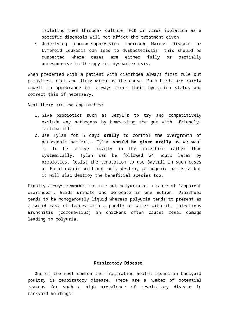
Like mammals when birds breathe air flows from their nares and into their sinuses where the air is warmed (many respiratory pathogens can infect the sinuses causing sinusitis).The air then enters the pharynx via the slit like opening in the hard palate (the choanae). Next the air enters the opened glottis into the trachea. Just as in mammals the upper respiratory tract is lined by ciliated epithelial cells which help move mucus and debris up out of the airway protecting the lower airway (lungs and air sacs) from pathogens.
Unlike mammals birds have fixed lungs which do not expand or reduce with breathing. Instead in order to move air through their lungs birds have expandable air sacs which fill with air
whilst inhaling and deflate when the bird exhales.
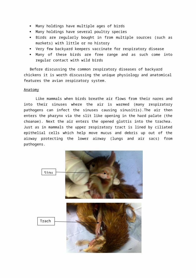
Birds have nine air sacs in total (a single intraclavicular air sac and paired cervical, cranial thoracic, caudal thoracic and abdominal air sacs). The intraclavicular air sac, the cervical air sacs and the cranial thoracic air sacs make up the cranial air sacs whilst the caudal thoracic and abdominal air sacs make up the caudal air sacs. These air sacs are extremely thin and are not vascularised. The air
sacs should be cling film-like (transparent with no thickening, mucus or vasculature).
Trachea
Sinus
Birds do not have diaphragms to move air into and out of the respiratory system. Instead the air sacs inflate and deflate due to the movement of the musculature of the abdominal wall.
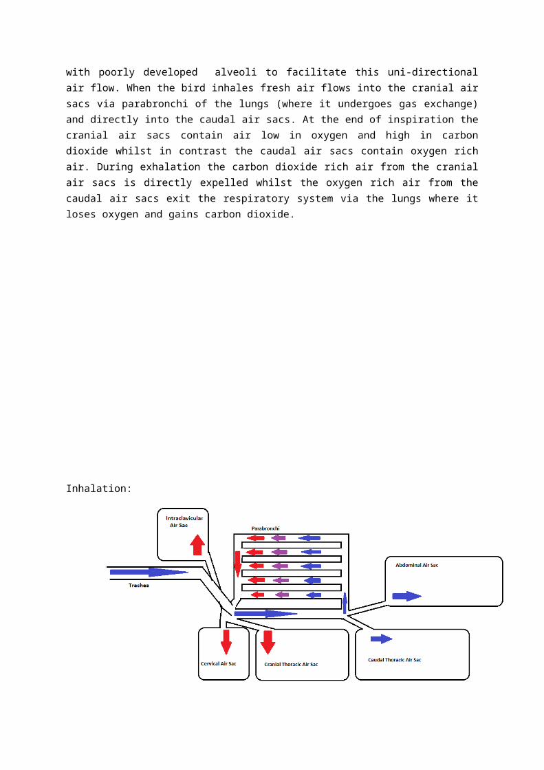
The fact that most birds fly means that they have a relatively high oxygen demand compared to mammals. Often flight occurs at high altitudes where the partial pressure of oxygen is low therefore their lungs must be capable of highly efficient gaseous exchange. To ensure this highly efficient exchange birds have arranged their air sacs to ensure unidirectional air flow through their lungs. Furthermore avian lungs have tube-like structures called parabronchi with poorly developed alveoli to facilitate this uni-directional air flow. When the bird inhales fresh air flows into the cranial air sacs via parabronchi of the lungs (where it undergoes gas exchange) and directly into the caudal air sacs. At the end of inspiration the cranial air sacs contain air low in oxygen and high in carbon dioxide whilst in contrast the caudal air sacs contain oxygen rich air. During exhalation the carbon dioxide rich air from the cranial air sacs is directly expelled whilst the oxygen rich air from the caudal air sacs exit the respiratory system via the lungs where it loses oxygen and gains carbon dioxide.
Air sac
Lungs (Should be a salmon-pink colour)

Inhalation:
Exhalation:
Pathogenesis
There are many potential pathogens that can cause respiratory disease most of which behave similarly and go on to produce similar clinical signs. (Each of these pathogens are discussed below in detail). These pathogens are often spread via the faeco-oral and aerosol routes. Fomites are also generally important in their spread.
The majority of these pathogens once inhaled attach to and damage the epithelial cells of the upper respiratory tract causing sinusitis and tracheitis. The damaged trachea and sinuses are then easy places for environmental organisms (such as E. coli) which are normally removed by the respiratory system to become established. This can result in a muco-purulent sinusitis and tracheitis.
As a result of the damage to the trachea the cilia are no longer able to waft mucus (often containing pathogens such as E. coli) up the trachea. This damage to the muco-cilliary escalator means that debris including many pathogens which are normally removed by this protective mechanism can reach the air sacs and lungs unimpeded.
Unfortunately the air sacs are very thin and have virtually no blood supply meaning once pathogens reach them they can establish themselves very easily causing air sacculitis. Subsequently this air sacculitis can then cause peritonitis by crossing the very thin poorly vascularised air sacs.
The ciliated epithelial cells of the trachea are similar to cells found in egg shell gland of the oviduct and the kidneys. This means that many respiratory pathogens can cause poor quality eggs shells, a loss of egg shell colour and nephritis.
Clinical signs
Many birds with respiratory disease may present with:
Dyspnoea Lethargy Weight loss Sneezing (correctly called snicking since sneezing requires a diaphragm) Naso-occular discharge Facial swelling due to swollen infraorbital sinuses hence the condition is commonly called
‘bulgy eye’ in game birds Pale eggs with poor shell quality Polyuria (appearing as diarrhoea)
Differential diagnosis:
Infectious Bronchitis Virus (IB) Mycoplasma gallisepticum (Mg) Mycoplasma synoviae (Ms) Mycoplasma meleagridis (Mm) Avian RhinoTracheitis virus (ART/TRT) Infectious LaryngioTracheitis (ILT) Syngamus trachea Aspergillus fumigatus Avian paramyxovirus including Newcastle’s Disease (NDV) Avian Influenza (AI)
Infectious Bronchitis :
IB is caused by a coronavirus. This virus is predominantly a pathogen of chickens. The virus is predominantly shed from the respiratory tract but faecal shedding is also common. The virus is very resistant and can survive in the environment for weeks. The virus will infect and damage the ciliated epithelial cells of the respiratory tract and after a short incubation period of 1-3 days the virus can cause snicking, sinusitis and a naso-occular discharge . The damage to the cilia allows environmental commensals such as E. coli to reach the air sacs where they may go on to cause air sacculitis and even peritonitis. Like many respiratory pathogens IB will infect the cells of the egg shell gland and will often lead to poor quality egg shells and a loss of shell colour. This damage to the egg shell gland can be temporary or permanent and there is no way of telling whether or not it will resolve.
In severe cases certain strains of IB (IB-QX) can cause such severe damage to the oviduct if the bird is infected pre-puberty that permanent adhesions form in the oviduct (often this results in huge fluid filled cysts) thus forever impeding the transport of the egg from the oviduct to the vent. In these cases the egg yolk ends up freely in the abdomen. These birds are called internal layers. The yolk will slowly be absorbed from the abdomen but if the bird lays internally every day then the yolks will be laid faster than they will be absorbed thus building up a mass of egg yolk in the abdomen. This mass has the potential to provide a rich nutrient medium for bacteria which can go on to cause egg peritonits. Due to the build up of yolks in the abdomen the bird adopts a penguin-like stance to assist in breathing.
IB can also infect the cells of the kidney leading to nephritis. In severe cases this renal damage can cause these birds to suffer dehydration, polyuria (manifesting itself as diarrhoea) and even visceral and articular gout.
Like all coronaviruses IB mutates frequently and immunity against one serotype doesn’t necessarily confer immunity against other strains, meaning birds can be infected with different strains throughout life.
The variability of IB strains presents a problem for vaccination and necessitates the designer of an IB vaccine program to be aware of the strains circulating locally in their area. Successful vaccination depends upon identifying the circulating IB strains in your area and ensuring that birds receive initially live vaccines for the relevant strains followed by a killed inactivated vaccine. Because this virus can infect young chicks it is often recommended to give the first vaccine before seven days of age.
A cockerel with an occular discharge

Above: Pale eggs (Note some breeds normally lay pale eggs)
Above: The distended abdomen of a pullet with a large fluid filled cyst caused by IB Qx
Mycoplasma gallisepticum (Mg)
Mg is a relatively common respiratory pathogen found in backyard chickens and turkeys. The bacteria is shed in the respiratory secretions of infected birds. This aerosolised bacteria is the inhaled by other birds. Like IB, Mg infects the cells of the respiratory tract leading to tracheitis and sinusitis which appears as peri-ocular swelling often containing caseous material (due to secondary bacteria). Similarly Mg also infects the cells of the egg shell gland leading to a reduction in the quality and colour of the egg shell.
Mg often infects birds concurrently with ART.
One of the biggest issues with Mg and other Mycoplasma species is that once infected with it, birds remain carriers for life. Often during periods of stress such as re-homing, these latent carriers recrudesce and develop clinical signs once again. This recrudescence can allow these carriers to infect other new flock mates.
It is possible to vaccinate against Mg using either two killed vaccines given at least four weeks apart or using a live attenuated vaccine to begin with followed by a single killed vaccine at least four weeks later.

Note: Mg can be transmitted vertically and via copulas. This is important as many people buy eggs on eBay for hatching and can bring in disease.
Above: A hen with peri-occular swelling caused by sinusitis caused by Mg
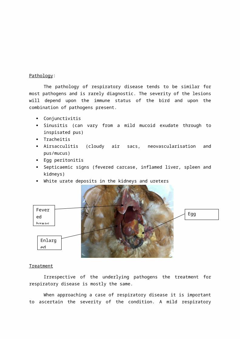
Pathology:
The pathology of respiratory disease tends to be similar for most pathogens and is rarely diagnostic. The severity of the lesions will depend upon the immune status of the bird and upon the combination of pathogens present.
Conjunctivitis Sinusitis (can vary from a mild mucoid exudate through to inspisated pus) Tracheitis Airsacculitis (cloudy air sacs, neovascularisation and pus/mucus) Egg peritonitis Septicaemic signs (fevered carcase, inflamed liver, spleen and kidneys) White urate deposits in the kidneys and ureters
Fevered breast muscle
Enlarged liver
Egg peritonitis

Treatment
Irrespective of the underlying pathogens the treatment for respiratory disease is mostly the same.
When approaching a case of respiratory disease it is important to ascertain the severity of the condition. A mild respiratory disease case where the bird is snicking(sneezing) but is otherwise fine will likely recover without the need for antibiotics. Mintamix is a plant extract based decongestant which will help birds breathe. If the bird is unwell, the clinical signs severe or the signs are not resolving then antimicrobials may be indicated. Even if the cause is viral you will want to protect against secondary bacterial infection.
There are a number of antimicrobials available but it is important to use those which have activity against Mycoplasma and secondary pathogens if you are unsure about the diagnosis.
For the majority of conditions many vets will use Fluroquinolones (Enrofloxacin) which is a broad spectrum powerful antimicrobial (which will kill Mycoplasma) however there is concern about overuse leading to resistance and its harmful effects on the intestinal flora.
Doxycycline and Tetracyclines are broad spectrum antimicrobials which have activity against Mycoplasma and secondary pathogens such as E. coli. Generally either antimicrobial is given for five days at 20mg/Kg in drinking water. It is worth noting that there is considerable resistance to Tetracyclines.
Macrolides such as Tylosin (Tylan- Zero egg withdrawal) are commonly prescribed as they are effective against Mycoplasma but they have a narrow spectrum of activity against other secondary pathogens. Tylan can be given up to 200mg/Kg for five days.
Tiamulin (Denagard- zero egg withdrawal) is licensed against Mycoplasma but like Tylosin has poor activity against secondary pathogens. A typical course of Denagard is 2ml/Litre of drinking water for five days.
Aminoglycosides such as Lincospectin can be used effectively (for up to seven days at 50mg/Kg) as they have activity against both Mycoplasma and E. coli.
When treating respiratory disease it is often advised to treat the entire group of birds as many of them will likely have subclinical disease.
Many antimicrobials destroy the gut flora and it is recommended about a week after the end of treatment to give a probiotic such as Beryl’s to repopulate the intestine with ‘friendly bacteria’.
Prevention:
Prevention of respiratory disease like other diseases is based upon biosecurity. Always obtain birds from a reliable source (local markets and eBay are bad sources). Ideally quarantine new birds for three weeks before introducing them to an existing flock. If your client has high value birds then testing them before admitting them to the flock may be a good idea.

Trying to keep wild birds and vermin away by keeping feed stored in metallic containers and not placing feed on the ground or in the sight of wild birds.
Minimising stress will reduce the chances of respiratory disease taking hold and should help reduce the chances of recrudescence in latent carriers. This means avoiding stresses such as poor diet, over stocking, extremes of temperature, draughts and poor ventilation. Note poor ventilation can lead up to high ammonia levels which can damage the cilia leaving the respiratory tract more vulnerable to infection.
Vaccination is possible for respiratory disease but it is not straight forward.
Vaccination:
It is firstly important to decide whether or not a client needs to vaccinate against respiratory disease. A client with a few backyard birds probably doesn’t need to vaccinate as the stocking density and hopefully stress levels are low. For clients keeping and breeding several hundred birds it is definitely worthy vaccinating. The problem clients are those with say 30-100 birds and you will have to use information about their holding’s disease history to ascertain the disease risk.
Once you have decided to vaccinate a holding you next need to decide which respiratory diseases you need to vaccinate for (Ideally include at least IB). Before instigating a vaccination program it is probably worth carrying out serology on the holding to determine which pathogens are present. For most respiratory diseases a vaccination with a killed vaccine repeated one month later followed by annual boosters is the most practical option.
Many of these vaccines come in 1,000 dose packs so there will be considerable waste however these 1,000 dose packs are relatively cheap costing approximately £30-40 per pack but if you have to give several vaccines the costs will mount.
There is a single killed vaccine available containing: IB, ART and NDV.
Vaccinated birds should be boosted with killed vaccines annually.
Key points to remember
Warn owners that many of these diseases can become latent and can recrudesce at a later date especially when their birds are stressed
If there is egg shell gland damage it may not resolve fully Mycoplasma can be transmitted vertically IB immunity is serotype specific Vaccination is neither straight forward nor will it give 100% protection
Egg peritonitis
Egg peritonitis is caused by a bacterial infection of the abdomen usually involving infected egg yolk. In most cases E.coli is the main pathogen involved. The routes of infection are not always clear but known causes include:
- respiratory disease (through cilliary damage of the upper air way allowing bacteria to reach the thin poorly vascularised air sacs, which the bacteria then crosses into the abdomen)
- ascending infection from the vent
Affected birds often show a severe loss of condition leading to a razor sharp breast bone and an enlarged abdomen. These birds are often pyrexic on clinical examination. Confirmation can be made through abdomenocentesis.
Treatment options include:
A broad spectrum antibiotic, fluid therapy and NSAIDs- but ultimately this only provides temporary respite
Euthanasia- this sounds harsh but is a valid course of action as the prognosis is very poor Speying- possible but the survival rate is poor (30%) Suprelorin implant, antibiotics and NSAIDs
Egg Bound Hens
The bird is unable to pass an egg (dystocia). The exact cause is unknown but predisposing factors include:
Obesity large eggs- diet is the main cause of large eggs through linoleic acid levels calcium imbalances stress
Presenting signs include straining and occasionally a vent discharge.
Diagnosis is based upon clinical signs, digital probing of the vent and occasionally radiography.
Conservative treatment is best, with apparently otherwise healthy, birds having their vents lubricated and being sent home to a dark quiet area in which to lay.
If conservative treatment fails then breaking the egg internally may be considered however remember sharp pieces of shell may damage an already inflamed reproductive tract.
Alternatively surgery with concurrent speying may be carried out under GA.
Birds that repeatedly become egg bound may require suprelorin implants- off license!!!!!.

Birds have no Oxytocin receptors in their reproductive tracts and as such it is unhelpful.
Calcium supplementation may be given if hypocalcaemia is suspected.
Red Mites
Red Mite (Dermanyssus gallinae) are ectoparasites that feed on the blood of poultry at night. Their numbers increase rapidly during the warmer summer months. These mites have a knack for hiding in the smallest of crevices and can survive for long periods without feeding (years). It is advised that owners keep a tight vigilance on their poultry houses. Due to the nocturnal nature of red mites they cannot be seen by day. One traditional way to look for them is to go out by night with a torch. For those that their sleep, then attaching a drinking straw to the perches and blow out the arachnid contents in the morning works well. If all that sounds too disgusting, then new mite traps are now available (these are plastic boxes the size of a match box that contain sticky paper to trap the mites). Owners should regularly clean out and check in all the corners and cracks to avoid a build-up of these parasites without noticing.
Low numbers of mites can cause irritation and causing the bird restlessness, however large numbers of mites can cause symptoms such as anaemia (Pale comb and wattles), lethargy, reduced egg production, owner complaints of itching and presence of grey/white/red mites around the birds vent area.
Treatment of red mites can be tackled in two ways: firstly by treating the environment by removing all the litter, then washing the shed with a detergent to remove dirt and grease(washing up liquid will suffice), next let the shed dry before applying a suitable disinfectant- Poultry Shield is a cheap, safe and highly recommended red mite disinfectant. After cleaning out mite numbers can be kept down by applying mite powder (Diamataceous earth) to the nest boxes and dust baths to abrade the waxy cuticle of the mites allowing them to dehydrate and die. Secondly Ivermectin 1% spot on can be used at 1ml/500g of bodyweight, however this is off label use and an appropriate egg withdrawal period should be set by the prescribing veterinary surgeon- this should be at least 14 days. The eggs from such birds should not ever be sold for human consumption. Similarly some owners use Fipronil.
Remember treating red mites is a war and not a single battle.

Scaly leg Mites
Scaly leg mites and depluming mites are two closely related mites which both belong to the genus Knemidocoptes.
The scaly leg mite (Knemidocoptes mutans) is commonly found on older backyard chickens.
This mite burrows beneath the scales of the leg causing damage which causes the damaged tissue of the leg to ooze with tissue fluid on which the mites feed.
This burrowing causes irritation, raised scales and crusting.
In the early stages of infection the affected bird will be no more than mildly irritated by the mites but as the scales become thickened and the mites cause more extensive damage the affected legs will become very painful.
It is important to note that it takes several months for the scales of the legs to become raised and consequently to heal again after treatment.
The mites are transmitted by direct contact with infected birds
Treatment of scaly leg involves killing the mites and softening the roughened scales.
Killing the mites can be done using Ivermec drops but it is important to note that Ivermec isn’t licensed for chickens and as an appropriate egg withdrawal should be set (minimum of 14 days).
Dunking the legs into a jar of surgical spirit or methylated spirits twice weekly for three weeks will kill the mites.
Vaseline can be used to soften the scales on the leg and to suffocate the mites.
Washing the legs using baby shampoo and a soft toothbrush can be used to soften the scales on the legs.